Article

Ni(II) and Cu(II) Ion Coordination by the Novel (2E,2′E)-N,N′-(2-Hydroxypropane-1,3-diyl)bis[(2-hydroxyimino)propanamide] Ligand in the Solid State and in Aqueous Medium

by
Igor Vasyl Nikolayenko
1,*,†,
Carla Bazzicalupi
2,*,
Thomas-John Theron
1,‡ and
Craig Grimmer
1
1
School of Chemistry and Physics, University of KwaZulu-Natal, Private Bag X01, Scottsville, Pietermaritzburg 3209, South Africa
2
Department of Chemistry “Ugo Schiff”, University of Florence, Via della Lastruccia 3, 50019 Sesto Fiorentino, Italy
*
Authors to whom correspondence should be addressed.
Retired.
Current Address: Intiki Raw Materials, Richmond 3780, South Africa.
Inorganics 2024, 12(12), 330; https://doi.org/10.3390/inorganics12120330
Submission received: 20 November 2024 / Revised: 9 December 2024 / Accepted: 11 December 2024 / Published: 17 December 2024
(This article belongs to the Special Issue Metal Complexes with N-donor Ligands, 2nd Edition)

Abstract

In the process of a systematic study of the bis-chelate oxime-amide ligands with different polymethylene spacers and their transition metal complexes, two new Ni(II) and Cu(II) complexes of the mhiea2poh ligand have been isolated, and their molecular and crystal structures determined by single crystal X-ray diffraction. Potentiometric titrations were performed on a range of aqueous solutions in both systems, which allowed the identification of various complex species and afforded their stability constants. ESR spectra of samples with optimised concentrations of complexes in Cu(II)–L–H system were recorded. A distinguishing feature of both systems is the dimerization of anionic pseudo-macrocyclic complexes. The latter is caused either by centrosymmetric hydrogen bonding of the hydroxy group on the propane spacer to the oximato oxygen of the opposing unit or by back-to-back π-stacking of planar complex units. ESR measurements indicated likely coupling of Cu-Cu paramagnetic centres in the dimers.

1. Introduction

Complexes of transition metals with polydentate ligands remain the focus of scientific research in view of their versatile applications [1,2,3]. In particular, the complexes of bis-chelate ligands with an oxime-amide moiety demonstrate a range of interesting properties [4]. They have been shown to exhibit biological activity, namely, as catalysts in the decomposition of acetyl phosphate [5,6]. They are also capable of stabilising higher oxidation states of such metals [7,8,9,10]. Homo- and hetero-polymetallic oximato-bridged complexes of paramagnetic transition metal ions attracted attention in view of their molecular magnetism, particularly in relation to efficient exchange interactions (mostly antiferromagnetic) between non-zero spin 3d-metal centres [11,12,13,14,15,16]. Another area of interest is the study of structural motifs, which are made particularly versatile by the rotation around the –(HON=)C–C(=O)–NH– bond in the oxime-amide unit, leading to the chelation of metal ions in a variety of coordination modes, namely, (NoxNad), (NoxOad), and (NoxOox′). Which particular mode is realised in each case is determined by a range of factors: the softness of the metal and its affinity towards oxygen donor, medium pH, metal-to-ligand ratio, etc. The ambivalent nature of the oxime group is responsible for numerous structural types of its metal complexes: mono- and poly-nuclear, homo- and hetero-nuclear, oligomeric, and metal clusters. Particularly interesting are various stacking arrangements of planar complexes in view of the opportunities for supramolecular self-assembly, formation of synthetic analogues of enzymes, and preparation of materials with unusual magnetic properties [17,18,19,20,21]. For instance, stacking of novel Ni(II)-complexes in the solid state affords infinite parallel columns with direct short contacts between Ni(II) centres, resulting in the material made up of 1-D metal ion chains in ligand insulation [22].
Previously some of us have reported the synthesis, characterisation, and protonation parameters for the new ligand mhiea2poh, Scheme 1 [4]. The feature that distinguishes this ligand from all previously reported ones is the presence of a hydroxy group on the flexible polymethylene link that joins two chelating moieties. It is able to influence the ligand protonation behaviour by altering the nucleophilicity of the oxime and amide donor centres and, consequently, their acidity. Next, we wanted to investigate the effect an additional contact point for hydrogen bonding and steric crowding in the bridge area might have on the metal coordination and packing behaviour in the solid state in comparison to previously reported compounds (see [22] and the references therein for a comprehensive account of all such compounds). Keeping in mind that all reported metal complexes of this kind demonstrated a rich hydrogen bond environment, we expected the above-mentioned hydroxy group to have an impact on the complex conformations and crystal packing, possibly with new types of H-bonding arrangements.
In the current paper, we present the results of an investigation of metal complexes formed by the mhiea2poh ligand with Ni(II) and Cu(II) ions, both in the solid state and in aqueous medium. In particular, two solid-state structures of these systems were determined by single-crystal X-ray diffraction. The composition and stability constants of complexes formed in aqueous solution were determined by potentiometric titrations. Thermodynamic results are presented in conjunction with the hypothetical structures of the metal complexes in solution. We have also recorded the ESR spectra for a selected series of Cu complexes in solution. In addition, we have simulated the complex 1H-NMR spectrum of the ligand in DMSO-d6 solution in close resemblance to the experimental one.

2. Results and Discussion

2.1. X-Ray Diffraction

The preparation of the samples for crystal growth is described in Section 3.5. Despite more than one type of yellow crystal being observed in some Ni samples, only the tea-yellow prisms turned out to be of the quality suitable for the XRD; they happened to represent complex [NiLH]·½[Ni(H2O)6]2+·H2O (1). Numerous coloured solids also precipitated out of the target Cu-solutions; however, only the pale-red prisms that crystallised out of sample 111 (see Section 3.5) were of the XRD quality and turned out to represent complex [CuLH]· [PPh4]+·4H2O (2).
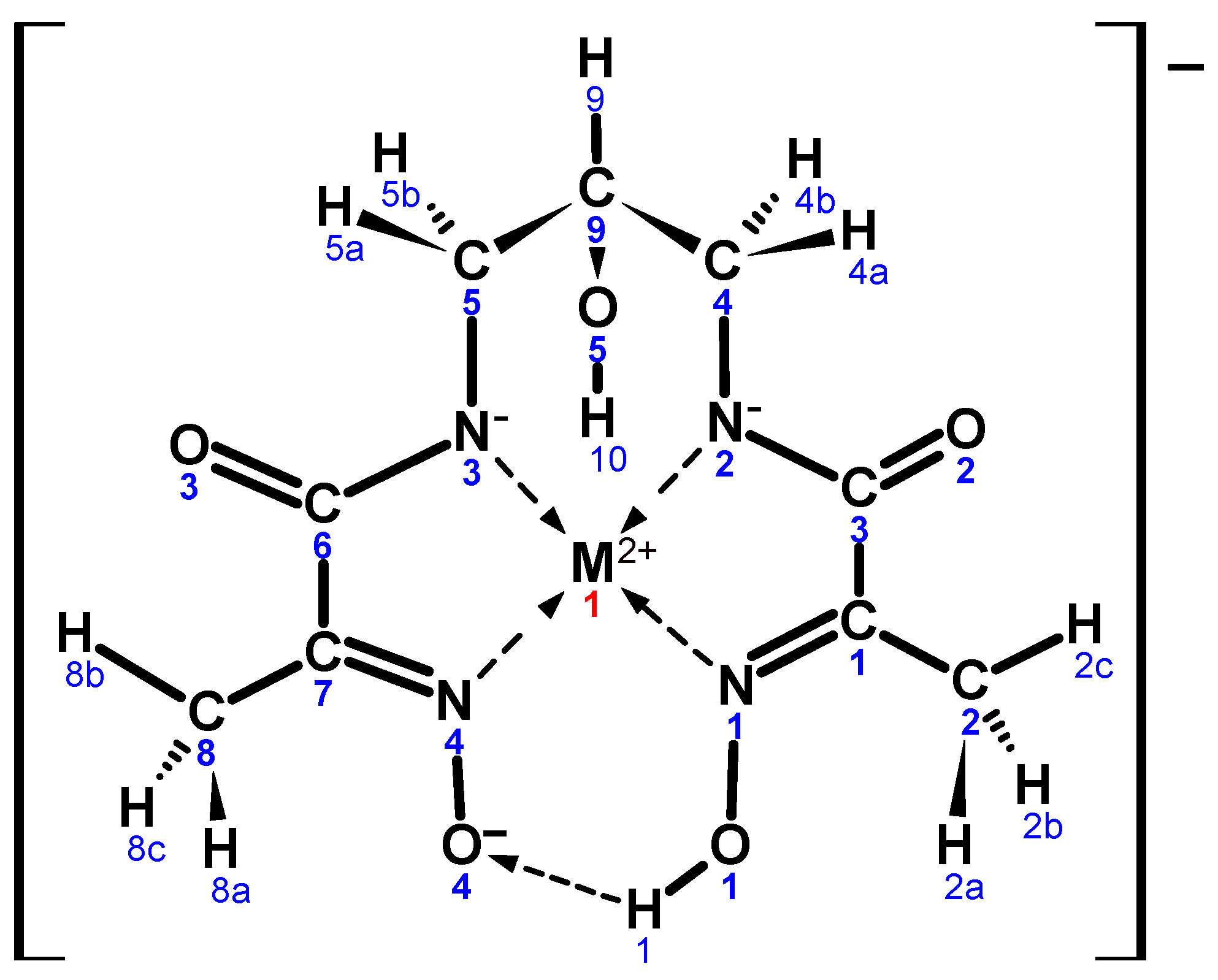
The summary of data and parameters for the single crystal diffraction experiments, which led to successful refinement of crystal structures, is presented in Table 1. Both complexes contain similar pseudo-macrocyclic anionic core, also similar to the one reported previously for the ligands with a polymethylene bridge between two chelating oxime-amide units [22]. In present work, we have adopted a numbering scheme for this core, as shown in Scheme 2.
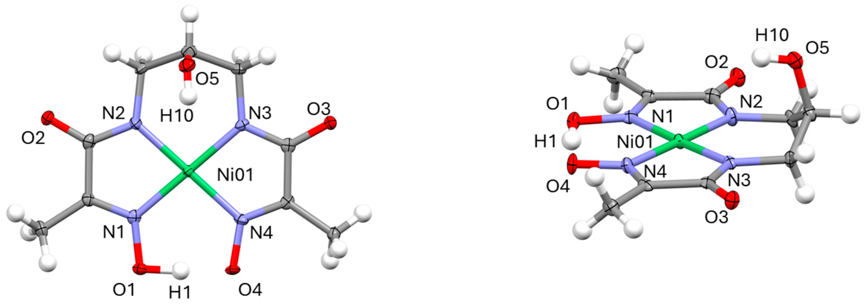
The molecular structures of Ni(II) and Cu(II) complexes determined in this work are shown in Figure 1 and Figure 2. Selected values of bond lengths and angles for these compounds are given in Table 2.
A new feature not seen previously in the reported structures of anionic complexes is the presence of an aliphatic hydroxy group and its involvement in hydrogen bonding in the solid state. A series of images illustrating various aspects of crystal packing in structures (1) and (2) can be found in Supplementary Materials (SM) Figures S1–S9.
Overall, this work reports the isolation in monocrystalline form of one new Ni(II) and one new Cu(II) complex. The 2 (NoxNad) binding mode observed in these structures has already been reported for the Ni(II) and Cu(II) complexes with three bis-chelate oxime-amide ligands with the unsubstituted polymethylene bridge of variable length; see [22] and the references therein, CSD [23] codes: NOXBIL, XANBET, BICGEA, XAPHUS, XEQBIF, DUCNIZ. Geometric parameters of (1)–(2), Table 2, are representative of such systems.
In the case of the Ni(II)-complex (1), three of the coordination Ni–N bonds are nearly identical in length at 1.866 (7) Å to 1.868 (6) Å, and only the fourth Ni–Nox bond adjacent to the deprotonated oxime terminal is somewhat longer at 1.878 (6) Å. Such proximity of the M–N bonds is uncommon for these complexes, where distinct differences are observed for the M–Nad and M–Nox bonds. In this sense, Cu(II)–complex (2) fits the expected pattern with the Cu–Nox bonds being about 0.032 Å longer than the Cu–Nad ones. The latter is clearly related to the anionic state of amide nitrogens and their higher donor ability. It is also interesting to note that the marked asymmetry associated with the oximato-oxime terminals in (1) is practically absent in (2). In general, the M:–N bonds are longer for the Cu(II)-complex (2) by about 0.050 Å to 0.087 Å. This is somewhat counterintuitive. On one hand, according to [24], the Cu(II) ion radius in the square-planar coordination environment is only marginally (by 0.020 Å) larger than that of Ni(II) ion. On the other hand, from the Irving-Williams series [25], one may expect stronger metal binding in (2) and, consequently, shorter bond length. However, if one takes into account the ionic radii for the square-planar coordination environment reported in [26], the difference in size increases to 0.080 Å in favour of Cu(II) ion. Perhaps this accounts for our observations. As expected, with the Cu(II) ion being larger in size than the Ni(II) ion, which is also reflected in noticeably longer Cu:–N bonds in comparison to the Ni:–N ones, the wider opening of the bis-amide mouth is observed in (2) vs. (1) (Nad:–M(II):–Nad angle is 98.8 (2)° vs. 97.0 (3)°). However, this does not lead to a wider bis-oxime mouth opening for the Cu(II)-complex. In fact, the opposite is the case (Nox:–M(II):–Nox angle is 95.2 (2)° for (2) vs. 97.2 (3)° for (1)). At the level of standard deviations, the O1—H1 and O4⋯H1 bonds in the two complexes are indistinguishable (1.1 (2) Å and 1.4 (2) Å for (1) versus 1.16 (6) Å and 1.31 (6) Å for (2)). Both structures are characterised by high degree of planarity with the exception of methyl group hydrogens and the hydroxypropane bridge, which has predictable “flap of the envelope” conformation. Thus, the deviation of atoms that constitute the complex circumference from their average planes drawn through 12 ligand atoms ranges from 0.000 (6) Å to 0.039 (7) Å in (1) and from 0.004 (3) Å to 0.071 (4) Å in (2), the C9 atom being out of this plane by 0.672 (8) Å in (1) and by 0.742 (4) Å in (2), respectively. The metal ions also conform to such planarity with their distance from the basal plane drawn through four donor atoms being 0.023 (1) Å for Ni and 0.0413 (6) Å for Cu.
Both crystal structures (1) and (2) exhibit extensive arrays of hydrogen bonds, which to a large degree define the overall structure. An interesting similarity in the crystal packings of these two compounds is the presence of dimers, where two anionic cores face each other in (1) at an interplanar distance of about 3.43 Å and an intermetallic distance of 3.526 (2) Å, Figure 3 (left), and in (2) at about 3.26 Å and 3.990 (8) Å, respectively, Figure 4 (left). Formation of such dimers is attributable to the back-to-back π-stacking interactions of planar cores—a phenomenon well established, CSD codes: NOXBIL, BICGEA, XAPHUS, XEQBIF. The hydroxypropane bridges stretch away from the dimer and provide inter-dimer contact points in both structures, with actual contacts being affected by the counterion size and nature.
In the nickel structure, these dimers are joined together by a pair of centrosymmetric H-bonds between hydroxy groups and oximato oxygens from adjacent dimers with the interplanar distance of 3.49 Å and Ni-Ni distance of 4.398 (2) Å, Figure 3 (left), thus forming columns of stacked dimers. Obviously, this dimerization is the consequence of the hydroxy group riding the propane spacer—a unique feature for the current complex that has not been observed previously. Under such arrangements, the carbonyl oxygens are exposed and available for H-bonding to Ni(II) hexa-aqua cations, which fill the voids between neighbouring columns, Figure 3 (right). Each Ni(II) hexa-aqua counter cation is also H-bonded through its axial water molecules and four more water molecules to two neighbouring aqua-ions in a diamond-shaped pattern, resulting in infinite cationic chains, Supplementary Materials Figure S2. Furthermore, each Ni(II) hexa-aqua cation is H-bonded through a pair of adjacent water molecules to a pair Ni(II) complex anions, totalling six such bonds, Supplementary Materials Figure S3. Finally, a free water molecule in the crystal structure of (1) is H-bonded to the amide oxygens of two complex anions and the axial water molecules of two Ni(II) hexa-aqua cations in tetrahedral configuration, Supplementary Materials Figure S4. The combination of the above factors afforded the crystal structure that consists of parallel columns of complex anions and cations, Figure 3 (right).
In the copper structure, the need to accommodate bulky tetraphenylphosphonium cations requires more space and prevents participation of hydroxy groups in direct dimer formation. Instead, two such groups of two neighbouring anionic complexes link the latter via a diamond-shaped double water molecule bridge, Figure 4 (left). Both amide oxygens of the complex anion are similarly bonded to two neighbouring complexes through pairs of water molecules, Supplementary Materials Figure S8. The combination of the above-mentioned factors accounts for the formation of layers of anionic complexes alternating with layers of bulky TPP+ cations, thus forming the “layered cake” structure, Supplementary Materials Figure S9.

2.2. Potentiometric Titrations and Solution Equilibria

The speciation and stability constants of the Ni(II) and Cu(II) complexes in 0.100 M NMe4Cl(aq) medium have been determined in a series of potentiometric titrations as described in Section 3.2. In brief, all titrations started from a solution of total volume 25.000 mL acidified to a pH of about 3, which also contained metal ion and ligand in various metal-to-ligand molar ratios, from ligand-rich to metal-rich. At this level of acidity, the ligand is fully protonated [4], and no coordination of metal ions takes place. The titrations were carried out with ca 0.1 M NMe4OH(aq) of accurately determined concentration until the pH was about 11.5. To test the possibility of oligomerisation in solution, and, in particular, that of dimerization, a few runs with the same metal-to-ligand ratio but of different overall concentrations were performed.
The refinement of the titration results was performed with the aid of Hyperquad2008 (ver. 5.2.19) software [27,28]. We started with a model where the species of all conceivable stoichiometric compositions, about 30 in total, were tested sequentially. They were gradually weeded out until the best-fit model with the minimal number of species remained. Two additional criteria were employed before the final model was accepted: (a) allowing adjustment of “dangerous parameters”, namely of the ligand and total hydrogen concentrations, led to an even better fit with only minor changes (of a few percent) to their initial values, and (b) close values of stability constants were obtained in different titrations for similar metal-to-ligand ratios.
The cumulative protonation constants for mhiea2poh, βqr, which represent the following equilibria
q L a q + r H a q L q H r ( a q )
have been determined by us earlier [4] and are given in Table 3 together with the stepwise protonation constants Kqr. As it turned out, this ligand is tetraprotic in aqueous medium and should be denoted as LH4. In the above equation, the charges on species are omitted for clarity, as is the convention in the Hyperquad set of programmes. The former and latter constants are related as follows
β q r = K q 1 K q 2 K q r
The cumulative stability constants, βpqr, for the M(II)-mhiea2poh complexes represent the following equilibria
p M a q + q L a q + r H a q   M p L q H r ( a q )
One example each of the titration outcome in the Ni(II)-L-H and Cu(II)-L-H systems, together with the refined solution model, is shown in Figure 5 and Figure 6. A complete set of individual experimental parameters, titration outcomes, and refined models is given in Supplementary Materials Figures S10–S18, Files S1–S9, SHq1–SHq9, and Tables S1 and S2.
Acceptable speciation models for Ni(II)-L-H and Cu(II)-L-H systems in aqueous medium are presented in Table 4 and Table 5 together with the cumulative stability constants. The latter were calculated as a weighted average of such constants from individual titrations.
The titration experiments revealed a rich variety of equilibrium complexes present in solution in both systems. Processing the results for solutions with an equimolar metal-to-ligand ratio unequivocally confirmed the formation of dimeric species. Ligand-rich solutions did not reveal any new species in comparison to the equimolar ones and, in fact, had fewer species, with less stable ones suppressed. In contrast, the metal-rich solutions demonstrated the whole plethora of new species, both monomeric and dimeric, with the molar ratio of M:L = 2:1. In the copper system the number of such species detected was less as the progress of titrations was hampered by the onset of precipitation, even at very low concentrations. The precipitates did not resemble copper hydroxide but rather appeared as black globular aggregates.
Stability constants for the accepted species are given in Table 4 and Table 5. Simulations on the basis of the above data speciation diagrams for the representative [M] to [L] ratios are shown in Figure 7 and Figure 8.
Conceivable structures for Ni(II)- and Cu(II)-mhiea2poh complexes in aqueous medium are shown in Scheme 3. In drawing these structures, we were guided by empirical knowledge of both crystal structures and solution spectra for the analogous Ni(II)- and Cu(II)-mhiea2p systems. In particular, it was established that due to the low affinity of Ni(II) ion to oxygen donors no (NoxOad) coordination mode is observed with this metal. In contrast, for Cu(II) and analogous mhiea2e, mhiea2p, and mhiea2b ligands in the process of titration with hydroxide, the formation of green species is observed first, identified as cationic dimers [ C u I I L H 3 ] 2 2 + [22,29,30,31], followed by the formation of anionic pseudo-macrocyclic monomers [ C u I I L H ] [9,22,29,32,33]. Ni(II) forms yellow complexes of the same structure with these ligands. Visual observation in the process of titration in both Ni(II)- and Cu(II)-mhiea2poh systems revealed a similar change of colours. Consequently, we have rejected all structures with the (NoxOad) chelation mode for Ni(II). For the 226 Cu(II)-complex, on the grounds of both solid-state data and entropy considerations, we favour the E structure over the F one, Scheme 3. The A structure is the only one conceivable for the anionic pseudo-macrocyclic complexes 111, 110, 11-1, and 11-2. The presence of dimeric species 223, 222, 221, and 220 is a signature feature of the new ligand and on the grounds of hydroxy group involvement in H-bonding favours the B structure over D and G. The C dimer, which was observed in the solid state of both systems, can certainly play a role in solution. On one hand, a pair of centrosymmetric hydrogen bonds may appear advantageous over short-contact interactions from the energy perspective. On the other hand, π-stacking in the solid state affords dimers with a shorter interplanar distance, which might be energetically competitive. As our unpublished results of potentiometric titrations in Ni(II)- and Cu(II)-mhiea2p systems also indicate the presence of dimeric species, the C structure should be given serious consideration. The choice between H and I structures for 21-1, 21-2, and 21-4 complexes we made in favour of the former, based on the prominent presence of 21-4 species in the copper system. It seems likely that the structure with more distributed negative charges would be more stable than the left-hand side Cu(II) ion in the coordination environment of four anionic oxygen donors in I. The riding-the-bridge hydroxy group may stabilise both structures via H-bonds. The presence of dimeric tetranuclear species 423, 422, 421, and 420 is yet another feature of the mhiea2poh ligand. In the Ni-L-H system, they are only observed in the metal-rich mixtures. These complexes are clearly more stable in the Cu-L-H system, where they are detected even in the ligand-rich solutions. The choice between the J and K structures is not easy to make. On the grounds of structural rigidity, perhaps, J should be favoured. On the other hand, the K-dimer is held together by six hydrogen bonds versus three in J. The K structure also affords shorter intermetal Cu-Cu distances, which is more in tune with our ESR results that indicate direct Cu-Cu coupling.
On the balance of said above, our preference is for the K structure over J, however counterintuitive it might be. Finally, the tetracopper L structure, which is a structural analogue of the C-dimer, cannot be entirely discounted. What weighs against it is the presence of the 420 species in the Ni(II)-L-H system. As we have discussed previously, the coordination of the Ni(II)-ion to the oximato terminals is without a precedent. The opposite is true for the Cu(II)-ion. The complex stabilisation due to intermolecular hydrogen bonding and metal–metal interaction was known to lead towards much increased acidity of the Nox and Nad donors, which might account for the relatively low pH onset of both 422 and 420 species formation.
Comparing the stability constants of Ni(II) and Cu(II) complexes with the same stoichiometry, namely, 221, 11-2, 21-2, 422, and 420, one may notice much higher values for the latter (from 9.67 to 16.75 orders of magnitude). This is not uncommon for such complexes, as the difference of 9.5 orders of magnitude was reported previously for the 111 species with the mhiea2p ligand [29,34,35]. The higher stability of copper complexes is in accord with expectations based on the Irving–Williams series [25]. Direct comparison of our stability constants to the literature values is obviously not possible as the mhiea2poh ligand is a new one. The closest comparison to be made is to the data for the structurally similar, but for the riding hydroxy group, mhiea2p ligand. Such data are available [29,34,35,36]. The problem lies in the fact that quoted authors accepted the ligand as diprotic, with the formula L′H2, a fact refuted by us in [4]. The values of stability constants are meaningfully comparable only when they refer to reactions with: (a) the same number of similar bonds broken and formed and (b) the same number of particles involved. Thus, rather than trying to compare the cumulative stability constants, Equation (3), one should compare the equilibrium constants for the reactions that meet the above criteria. For example, with respect to the 111 species in our notation, the comparison should be made for the constants of reactions (4) and (5)
Cu a q + L H 2 a q + 3 O H a q     C u L H 1 a q + 3 H 2 O ( l )
Cu a q + L H 4 a q + 3 O H a q     C u L H a q + 3 H 2 O O ( l )
where L′ = mhiea2p and L = mhiea2poh, and the charges on species are omitted for clarity. These constants can be easily calculated from the stability constants, protonation constants, and the constant of self-ionisation of water. The only common species in the nickel and copper systems detected by us and reported in the literature for L′ are those of 111 and 110 in our notation. The respective values of stability constants are given in Table 6. Calculated equilibrium constants for comparable reactions are presented in Table 7.
As can be seen from Table 7, the Ni(II)-coordination reaction constants agree within experimental errors. For Cu(II)-coordination reactions our value is slightly less, but the difference is not significant to merit discussion. In all the reported literature, the data were collected on solutions with the ligand excess, from [M]:[L] = 1:5 to 1:2. As we have seen in the current work, such a concentration ratio suppresses formation of minor species. A remarkable fact is that the most stable monomeric species in M(II)-L′-H systems, where M(II) is Ni(II) or Cu(II), are of limited importance in M(II)-L-H systems, where various dimeric species are prominent. Given the structural difference between L′ and L is exclusively related to the presence of the hydroxy group riding the polymethylene bridge, a conclusion of the crucial role played by this group in the stabilisation of complex dimers via intramolecular H-bonding in solution could not be avoided. This conclusion is supported by direct evidence from the XRD studies of solid complexes.

2.3. ESR Spectra of Selected Cu(II)-Mhiea2poh Solutions

Based on the titration diagrams for equimolar and Cu-rich solutions, Supplementary Materials Figure S28, simulated from the above data by means of HySS (ver. 4.0.31) [28], we have prepared a series of mixtures optimised with respect to the expected amount of individual Cu(II)-complexes, Section 3.5. Thereafter, ESR spectra of these solutions were recorded and are shown in Figure 9 and Figure 10.
The spectra presented in Figure 9 are representative of the Cu(II) complexes in the coordination environment of four nitrogen atoms [37,38]. As the nuclear spin of both 63Cu and 65Cu isotopes is 3⁄₂, the ESR spectrum is expected to show a hyperfine splitting into four lines of equal intensity. In our square-planar complexes, Cu(II) ion is ligated to two pairs of almost equivalent nitrogen nuclei. For 14N the nuclear spin is 1, and super-hyperfine splitting of various features, arising from the magnetic interaction of the unpaired electron spin with the nuclear magnetic moments of nitrogen donors, should produce a pattern of 25 lines. As no such pattern is observed, two types of nitrogen donors (Nad and Nox) must be nearly equivalent in terms of magnetic interaction, and, consequently, nine lines of super-hyperfine structure with intensities of about 1:4:10:16:19:16:10:4:1 are observed, Figure 10. This confirms coordination of the Cu(II) centre in the complex to four nitrogen atoms, with expected maximum coupling along either the x or y (the perpendicular) direction and minimum coupling along the z (the parallel) direction.
All samples exhibited an ESR signal indicative of a Cu(II) ion, with a variable degree of signal suppression. The spectra clearly fall into two groups, namely, 1:1_73, 2:1_138 and 1:1_88, 1:1_112, 1:1_170. According to our potentiometric results, the major complex species expected to be present in these samples are 73: 422 at 52% and 420 at 27%, 138: 420 at 91%, 112: 111 at 48%, 21-2 at 33% and 221 at 12%, and 170: 21-4 at 76% and 11-2 at 23%. Thus, according to Scheme 3, it appears the difference is that between dimeric and monomeric species. As the excitation amplitude and receiver gain were the same for all samples, a progressively weakening partial coupling was observed for 73, 88, 112, and 170 samples. This is understandable in view of the decreasing concentration of the dimeric species in this direction. Though full suppression of the ESR signal was not observed, the change in the ESR spectra supports the conclusion of dimerization in solution, in agreement with our findings from the crystallographic and potentiometric studies. The average values of the magnetogyric ratio and the coupling constant, giso and Aiso, are determined from the ESR spectra for Cu(II)-complexes in free tumbling motion at room temperature. The ESR spectra presented in Figure 9 showed four copper hyperfine lines with Aiso = 93 G and its centre at giso = 2.083 for the first group and Aiso = 92 G and its centre at giso = 2.080 for the second group. The super-hyperfine splitting by 14N nuclei is observed, which indicates the presence of four almost equivalent nitrogen nuclei (Aiso = 15.7 G in both cases). The lack of evidence that two distinct (in the ESR sense) species are present in the samples rules out the possibility of triplet-state dimeric species. In contrast, the consistent decline in the spectrum intensity across the titration series strongly points towards direct Cu-Cu coupling in the dimeric species.
From the electronic structure point of view, in the square-planar crystal field, the degeneracy of five d-orbitals is removed with the energy levels splitting as follows: d x z = d y z < d x y < d z 2 d x 2 y 2 . For Ni(II), which has an electronic configuration of 3d84s0, the d x 2 y 2 orbital remains empty, as the pairing energy is usually much less than Δ. Thus, square-planar Ni(II) complexes are almost always low spin (S = 0). Formation of the σ-molecular orbitals in square-planar complexes of d-metals proceeds via an overlap of four 4 s 4 p x 4 p y 3 d x 2 y 2 hybridised orbitals of the metal and s- or p-orbitals of ligands, resulting in Ni(II) complexes in a HOMO of A1g and a LUMO of B 1 g * symmetry. For Cu(II), which has an electronic configuration of 3d94s0, an unpaired electron will populate the orbital of B 1 g * symmetry, essentially 4pz in character. As two mononuclear complexes approach each other along the z-direction (perpendicular to the complex plane), an opportunity for the direct Cu-Cu coupling arises, with the creation of σ-type bonding and antibonding orbitals in the z-direction. Two previously unpaired electrons from two Cu-centres will populate the lower energy orbital, resulting in the decrease of the number of unpaired electrons and lower intensity of absorption bands in the ESR spectrum.

2.4. The Ligand 1H NMR Spectrum

The 1H NMR spectrum of the mhiea2poh ligand in DMSO-d6 solution turned out to feature a series of multiplets, Figure 11. Interpretation of one of them, namely, the one in the range 3.09 ppm to 3.22 ppm, was not immediately obvious. We had to turn to the simulation, using SpinWorks (ver. 4.2.8.0) software [39] with the NUMMRIT algorithm [40], in order to reproduce key features of these multiplets. In the above simulation, slightly different chemical shifts had to be adopted for two protons of the methylene groups of the bridge, as well as allowing the coupling between axial and equatorial positions. The diagram of the ligand fragment used in the simulation is shown in Supplementary Materials Scheme S1, while the parameters of the simulated spectrum are given in Supplementary Materials Table S3. The experimental and simulated multiplets are shown in Figure 12.

3. Materials and Methods

3.1. Materials

Organic solvents, reagents, and other materials were purchased from commercial suppliers (Aldrich, St. Luis MO, USA; Sigma-Aldrich, Burlington VT, USA; Fluka Analytical, Radnor CA, USA; Merck, Modderfontein, South Africa) and were of analytical or reagent grade. They were used without further purification.

3.2. Instrumental

Melting point temperatures: Melting points were recorded on MPA 100 Optimelt (Stanford Research Systems, San Jose, CA, USA). The melting point range is reported from the onset to the clear point. It was determined at a heating rate of 1 °C min−1 with the apparatus calibrated against melting points of vanillin, phenacetin, and caffeine SRS melting point standards, traceable to the WHO standards.
IR: FTIR spectra were recorded in KBr discs on Spectrum 100 spectrometer (Perkin Elmer, Shelton, CT, USA) in the range 450 to 4000 cm−1 with a resolution of 1 cm−1.
CHN: Elemental analyses were performed in the Laboratorio di Microanalisi, University of Florence (Italy).
NMR:1H, 13C, 15N, and 31P NMR spectra were recorded on either an Avance-III 400 or an Avance-III 500 spectrometer (Bruker, Ettlingen, Germany) at frequencies of 400/500 MHz (1H) and 100/125 MHz (13C) using either a 5 mm BBOZ-[31P-109Ag]-{1H} probe or a 5 mm TBIZ-[1H]-{31P}-{31P-109Ag} probe. All proton and carbon chemical shifts are quoted relative to the relevant solvent signal (e.g., for DMSO-d6, 1H: 2.50 ppm, 13C: 39.50 ppm). Coupling constants are reported in Hertz (Hz). All experiments were conducted at 30 °C.
MS-ToF: High-resolution mass spectra were recorded on UPLC Acquity—Micromass LCT Premier ToF/MS spectrometer (Waters Limited, Wilmslow, UK). Samples were dissolved in DMSO to a concentration of approximately 2 mg L−1. For low-resolution measurements, the instrument was internally calibrated with either reserpine (positive ionisation mode) or raffinose (negative ionisation mode). High-resolution measurements were performed using DMSO as the lock mass standard. Pure samples were injected directly into the MS port, i.e., bypassing the UPLC system.
XRD: Reflection data were acquired on the single-crystal diffractometers XcaliburPX Ultra or Xcalibur3 (Oxford Diffraction Ltd., London, UK). Once collected, the integrated intensities were corrected for Lorentzian and polarisation effects, and an empirical absorption correction was applied, SCALE3 ABSPACK [41]. Crystal structures were solved by direct methods with SIR97 [42], and refinements were performed by means of full-matrix least-squares using SHELXL (ver. 2019/2) [43]. Non-hydrogen atoms were refined anisotropically, while riding models were used for all hydrogen atoms, with the exception of oxygen-bound ones. Mercury 2024.2.0 (Build 415171) [44,45], enCIFer 2024.2.0 (Build 415171) [46], and ORTEP-3 (ver. 1.076, 2020) [47] were used for processing, visualisation, and presentation of structural data. New structures have been deposited into the CCDC [28] with reference code numbers: 2402136-2402137.
Potentiometric Titrations: In a typical experiment, the required amount of ligand (between 2 mg and 10 mg) was weighed on a 6-decimal place balance (Hewlett-Packard AD-4 Autobalance) and transferred to the titration cell, followed by calculated volumes of stock solutions: 0.125 M NMe4Cl(aq), 0.1436 M NiCl2 (aq, in 1.000 mM HCl) or 0.1996 M CuCl2 (aq, in 1.000 mM HCl) and 0.1987 M HCl(aq), with the balance of 25.000 mL made up with water. The solution was stirred under a continuous flow of humidified argon gas until all the ligand had dissolved. Actual parameters of the solutions prepared are given in Supplementary Materials Files SHq1–SHq9. Titrations were performed using the Metrohm automatic titration system (Titrando-808 and Dosimat-805 with 1 mL, 5 mL, and 50 mL burette exchange units) (Metrohm, Schweiz AG, Zofingen, Switzerland) [48]. They took place in a 50 mL water-jacketed Metrohm cell under continuous flow of argon bubbled through support electrolyte solution to reduce the water evaporation from the solution in the cell. The temperature inside the cell was continuously recorded with Metrohm Pt 1000 temperature sensor and remained constant at the level of ± 0.05 °C over 24 h period. We have employed two types of Metrohm combination glass sensors in our studies: Micro-electrode (“T”-type glass, hemisphere membrane, ceramic pin diaphragm) and Unitrode (”U”-type glass, spherical membrane, ceramic pin diaphragm). Each sensor was calibrated daily before the titration in a Gran procedure [49,50,51] that afforded electrode parameters, as well as corrected concentration of the alkali solution and the level of protolytic impurity (carbonate and silicate). The following 2-parameter equation was used to convert the sensor e.m.f. readings into pH values:
E = E° + f·s·log10[H]
where E° is the standard e.m.f., s and f are the Nernstian slope and slope correction factor, respectively, and the quantity in brackets represents the concentration of H+ ions. As the values of pH readings in our experiments remained within the 2.5 to 11.3 range, no corrections for the glass-electrode acid or alkali errors were applied. Starting solutions contained metal ions and ligands in ligand-rich, equimolar, or metal-rich molar ratio and were acidified to a pH of about 3. The titrations were carried out with ca 0.1 M NMe4OH(aq) of accurately determined concentration in 10 μL, 15 μL, or 20 μL steps (burette drive on Ttrando-808 unit has 20,000 digitally controlled steps, which affords a resolution of 0.05 μL on a 1 mL burette) until the pH of about 11.5 with typically about 100 titration points recorded in one experiment. The equilibration time after each addition varied in different titrations from 15 min to 30 min. To test the possibility of oligomerisation in solution, and, in particular, that of dimerization, a few runs with the same metal-to-ligand ratio but of different overall concentrations were carried out. Primary data were collected and processed under Metrohm tiamo (ver. 1.0) software [52]. Further processing of the information from potentiometric titrations was carried out with a suite of software applications from the Protonic Software [24]. In particular, GLEE (ver. 3.0.21) was used for the glass-electrode evaluation, HySS 2009 (ver. 4.0.31) was used for the titration planning and speciation simulation, and the module Hyperquad 2008 (ver. 5.2.19) was used for the refinement of the potentiometric data. The value of the water self-ionisation constant at 25.00 °C in 0.1 NMe4Cl(aq), p K W = 13.81 , [53] was used in all calculations. The Ni(II) and Cu(II) hydrolysis constants [54] were used in all models.
ESR: The spectra were acquired on EMX-Plus X-band spectrometer (Bruker, Seoul, Korea) at room temperature. They were recorded in a flat-bed cell with the same excitation amplitude and receiver gain for all samples. Generic acquisition parameters used are shown in Supplementary Materials Figure S29. The intensity derivative for all ESR spectra can be found in Supplementary Materials File SEx1.

3.3. Synthesis and Characterisation

mhiea2poh: The new ligand was synthesised as follows. 1,3-Diaminopropane-2-ol (0.522 g, 5.5 mmol) was dissolved in dry methanol (10 mL) under an argon atmosphere, acid-washed glass beads added, followed by ethyl pyruvate oxime (1.311 g, 10 mmol). The mixture was stirred for 24 h at room temperature, glass beads filtered off, and the solvent removed by rotary evaporation. A nearly colourless oil was washed with diethyl ether (20 mL) and recrystallized from hot water (15 mL) as white powder. Further recrystallization from acetonitrile: water = 2:1 produced an analytical sample. Yield 0.42 g (32.4%).
Mp: 180.3–181.4 °C.
Microanalysis: Calcd. for C9H18N4O6 (L·H2O): C, 38.85; H, 6.52; N, 20.13. Found: C, 38.65; H, 7.09; N, 20.63.
According to microanalysis, the solid phase represented monohydrate, i.e., L·H2O.
FTIR (KBr, ν ¯ /cm−1): 3383 (s, N–H), 2949, 2925, 1654 (s, C=O), 1626 (s, C=O′), 1536 (s, –C(=O)–CN), 1441, 1419, 1369, 1314, 1259, 1224, 1189, 1121, 1100, 1078, 1024 (s, N–O), 1003 (s, N–O′), 916, 803, 720.
1H NMR (DMSO-d6, δ/ppm): 1.88 (s, 6H, –CH3), 3.09–3.22 (m, 4H, –CH2–), 3.62 (hx, 1H, J = 5.4 Hz, –CH(OH)–), 5.06 (d, 1H, J = 4.8 Hz, –CH(OH)–), 7.73 (t, 2H, J = 6.2 Hz, 2H, –NH–), 11.69 (s-br, 2H, =N–OH).
13C NMR (DMSO-d6, δ/ppm): 9.4 (q, –CH3), 42.3 (t, –CH2–), 68.0 (d, –CH(OH)–), 150.0 (s, –C(=NOH)–), 163.7 (s, –C(=O)–).
HRMS [ES+] m/z(%): Calculated for [C9H16N4O5Na]+ 283.1018; found 283.1014 (100); δ −1.4 ppm.
Original spectra for this ligand can be found in Supplementary Materials Figures S19–S27.

3.4. Solution Preparation

All solutions were prepared in deionised type-I ultrapure water (18.2 MΩ cm, ELGA PURELAB Ultra Inorganic), which was boiled and cooled under argon in a quartz flask to remove dissolved CO2. The 0.100 M NMe4Cl support electrolyte medium was employed in all cases, with the value of ionic strength altered by no more than 4 percent in an average titration. 0.1 M HCl solution was prepared from ultra-pure (99.999%) concentrated acid and standardised against tris(hydroxymethyl)aminomethane (THAM) in a seven-sample titration procedure. The 0.1 M NMe4OH solution was prepared from a 25 wt. % concentrate, kept permanently under argon, and standardised daily against 0.1 M HCl in a Gran procedure [50,51,52]. The solution was discarded when the level of protolytic impurity (mainly carbonate) exceeded 1.5%.

3.5. Sample Preparation

The samples for crystal growth were made on the basis of speciation diagrams derived from the potentiometric titrations, with the aim of isolating complexes present in solution. The ratio of ingredients was chosen to yield the samples with the concentration of the desired species close to maximum. In particular, the following aqueous (unless stated otherwise) stock solutions were prepared: [L]0 = 5.012 mM in EtOH, [HCl]0 = 0.1987 M, [NiCl2]0 = 0.1436 M in 1.00 mM HCl(aq), [CuCl2]0 = 0.1942 M in 1.00 mM HCl(aq), [TPPB]0 = 49.94 mM, [STPB]0 = 50.07 mM, [PHFA]0 = 50.00 mM. (The above abbreviations stand for: TPPB—tetraphenylphosponium bromide, STPB—sodium tetraphenylborate, PHFA—potassium hexafluoroantimonate) The target values of the ingredients in the sample of 5.000 mL total volume were chosen as follows: [L] = 1.00 mM, [M] = 1.00 mM or 2.00 mM, [H] = 1.00 mM, [CI] = 1.00 mM or 2.00 mM, where the meaning of the labels is as follows: M stands for Ni(II) or Cu(II), H represents a proton, and CI stands for the counterion: TPP+, TPB, or HFA.
The samples of Ni(II) complexes were made by adding sequentially the volumes of stock solutions listed in Table 8. Thereafter the mixtures were stirred for 24 h and left to evaporate on the bench in open vials. From our previous experience, see [55], we expected the formation of only neutral or anionic complexes with the Ni(II) ion. Consequently, only the TPP+ cation was employed in this case.
The samples of Cu(II) complexes were prepared by adding the volumes of stock solutions sequentially as listed in Table 9. In this case, the formation of cationic, neutral, and anionic complexes was anticipated. Therefore, both the TPP+ cation and TPB, HFA anions were employed.
The samples for ESR measurements were also prepared on the basis of simulated titration diagrams, Supplementary Materials Figure S28. The samples of total volume 5.000 mL were made from the following aqueous stock solutions: [CuCl2] = 0.1942 M, [L] = 0.9823 mM, [HCl] = 0.1987 M, and [NMe4OH] = 0.1250 M according to Table 10, to meet the figures of species concentrations shown on the top of the above diagrams. The volumes of base solution added were expected to yield samples with a substantial concentration of the following species: 422, 420, 111, 21-4, 422, and 420.

4. Conclusions

Two new Ni(II) and Cu(II) complexes with the bis-chelate oxime-amide mhiea2poh ligand, [NiLH]·½[Ni(H2O)6]2+·H2O (1) and [CuLH] [PPh4]+·4H2O (2), have been isolated and their molecular and crystal structures determined by single crystal X-ray diffraction. In the solid state, both coordination compounds are dimerised due to π-stacking of their planar anionic pseudo-macrocyclic cores. In addition, in (2) such dimers are joined together by pairs of centrosymmetric H-bonds between the hydroxy group of the bridge and the oximato oxygen of the opposite unit—a unique feature for the complexes of this kind.
Potentiometric titrations performed on a series of solutions in both Ni-L-H and Cu-L-H systems revealed a variety of complex species present across the pH range, both monomeric and dimeric, with their compositions and stability constants determined. Hypothetical structures of complex species have been suggested. In contrast to previously reported results for similar complexes, in both systems the dimeric species 221 and 220 dominate monomeric species 111, 110, and 11-2, where the pqr numbers represent the species MpLqHr stoichiometry. In addition, in the Cu-system tetranuclear species 422 and 420 are present alongside dimeric species.
ESR spectra for a selected series of Cu-L-H solutions revealed partial coupling of Cu(II) paramagnetic centres, which was progressively weakening with pH as one moves away from the regions of 422 and 420 existence. This is in agreement with the hypothesis of dimer formation and indirectly supports the assumption of Cu-Cu interaction in the dimeric tetra-copper species.
In our opinion, the stability of polynuclear metal complexes is enhanced by the possible formation of dimeric structures, the latter being a direct consequence of the presence of the hydroxy group on the polymethylene spacer and its involvement in intermolecular hydrogen bonding. As a matter of fact, both the increased hydration and species solubility, attributable to this additional hydroxy group, made an important contribution to the overall stabilisation of metal complexes in aqueous medium.

Supplementary Materials

The following supporting information can be downloaded at: https://www.mdpi.com/article/10.3390/inorganics12120330/s1, Schemes: Scheme S1. A fragment of the ligand with the labelling scheme used for the simulation of 1H NMR spectrum; Tables: Table S1. Common logarithms of stability constants, log10βpqr, with standard deviations, σ, for the MpLqHr complexes in the Ni(II)-L-H system refined from individual potentiometric titrations. Table S2. Common logarithms of stability constants, log10βpqr, with standard deviations, σ, for the MpLqHr complexes in the Cu(II)-L-H system refined from individual potentiometric titrations. Table S3. Optimised chemical shifts (δ) and coupling constants (J) in the simulated 1H NMR spectrum of mhiea2poh ligand; Figures: Figure S1. Molecular structure of the anionic Ni(II) complex in crystal structure (1). Figure S2. Hydrogen-bonded chains of Ni(II) hexa-aqua counter-ions in crystal structure (1). Figure S3. Hydrogen-bonding interactions of Ni(II) hexa-aqua counterion through the equatorial water molecules in crystal structure (1). Figure S4. Hydrogen-bonding interactions of a free water molecule in crystal structure (1). Figure S5. View down c-axis in crystal structure (1). Figure S6. Molecular structure of the anionic Cu(II) complex in crystal structure (2). Figure S7. Hydrogen-bonding interaction of the hydroxy group in crystal structure (2). Figure S8. Hydrogen-bonding interaction of amide oxygens in crystal structure (2). Figure S9. View down c-axis in crystal structure (2). Figure S10. Potentiometric titration data and converged model for experiment 20090502. Figure S11. Potentiometric titration data and converged model for experiment 20090413. Figure S12. Potentiometric titration data and converged model for experiment 20090503. Figure S13. Potentiometric titration data and converged model for experiment 20090506. Figure S14. Potentiometric titration data and converged model for experiment 20090505. Figure S15. Potentiometric titration data and converged model for experiment 20090818. Figure S16. Potentiometric titration data and converged model for experiment 20090819. Figure S17. Potentiometric titration data and converged model for experiment 20090922. Figure S18. Potentiometric titration data and converged model for experiment 20090923. Figure S19. FTIR spectrum of mhiea2poh ligand. Figure S20. 1H NMR spectrum of mhiea2poh ligand. Figure S21. 13C NMR spectrum of mhiea2poh ligand. Figure S22. g-HSQC NMR spectrum of mhiea2poh ligand. Figure S23. g-HMBC NMR spectrum of mhiea2poh ligand. Figure S24. COSY NMR spectrum of mhiea2poh ligand. Figure S25. NOESY NMR spectrum of mhiea2poh ligand. Figure S26. High resolution mass spectrum of mhiea2poh ligand. Figure S27. High resolution mass spectrum of mhiea2poh ligand with the best formulae analysis. Figure S28. Simulated titration diagrams in Cu(II)-L system. Figure S29. ESR spectra acquisition parameters; Files: S1_Ni_mhiea2poh-H_20090413_fin.docx, S2_Ni_mhiea2poh-H_20090502_fin.docx, S3_Ni_mhiea2poh-H_20090503_fin.docx, S4_Ni_mhiea2poh-H_20090505_fin.docx, S5_Ni_mhiea2poh-H_20090506_fin.docx, S6_Cu_mhiea2poh-H_20100818_fin.docx, S7_Cu_mhiea2poh-H_20100819_fin.docx, S8_Cu_mhiea2poh-H_20100922_fin.docx, S9_Cu_mhiea2poh-H_20100923_fin.docx, SHq1_Ni_mhiea2poh_H_20090413_fin.HQD, SHq2_Ni_mhiea2poh_H_20090502_finHQD, SHq3_Ni_mhiea2poh_H_20090503_fin.HQD, SHq4_Ni_mhiea2poh_H_20090505_fin.HQD, SHq5_Ni_mhiea2poh_H_20090506_fin.HQD, SHq6_Cu_mhiea2poh-H_20100818_fin.HQD, SHq7_Cu_mhiea2poh-H_20100819_fin.HQD, SHq8_Cu_mhiea2poh-H_20100922_fin.HQD, SHq9_Cu_mhiea2poh-H_20100923_fin.HQD, SH10_model_Ni_L4-H_2024_fin.HQD, SH11_model_Cu-L4-H_2024_fin.HQD, SEx1_Cu-L-H_ESR_combined.xlsx.

Author Contributions

Conceptualization, I.V.N.; methodology, I.V.N. and C.B.; software, C.B, I.V.N. and C.G.; formal analysis, I.V.N. and C.B.; investigation, I.V.N., C.B. and T.-J.T.; resources, I.V.N., C.B and C.G.; data curation, I.V.N., C.B., T.-J.T. and C.G.; writing—original draft preparation, I.V.N. and C.B.; writing—review and editing, I.V.N., C.B. and C.G.; visualisation, I.V.N., C.B. and C.G.; supervision, I.V.N.; project administration, I.V.N.; funding acquisition, I.V.N. and C.B. All authors have read and agreed to the published version of the manuscript.

Funding

This research received no external funding.

Data Availability Statement

The data for new structures have been deposited into the CSD [28] with reference code numbers: 2402136-2402137.

Acknowledgments

The authors gratefully acknowledge the use of research equipment and instrumental services of the School of Chemistry and Physics, University of KwaZulu-Natal Pietermaritzburg and the Department of Chemistry, University of Florence.

Conflicts of Interest

Author Thomas-John Theron was employed by the company Intiki Raw Materials. The remaining authors declare that the research was conducted in the absence of any commercial or financial relationships that could be construed as a potential conflict of interest.

References

  1. Abu-Dief, A.M.; Mohamed, I.M.A. A review on versatile applications of transition metal complexes incorporating Schiff bases. Beni-Suef Univer. J. Basic Appl. Sci. 2015, 4, 119–133. [Google Scholar] [CrossRef] [PubMed]
  2. Lesnikov, V.K.; Golovanov, I.S.; Nelyubina, Y.V.; Aksenova, S.A.; Sukhorukov, A.Y. Crown-hydroxylamines are pH-dependent chelating N,O-ligands with a potential for aerobic oxidation catalysis. Nat. Commun. 2023, 14, 7673. [Google Scholar] [CrossRef] [PubMed]
  3. Mezgebe, K.; Mulugeta, E. Synthesis and pharmacological activities of Schiff bases with some transition metal complexes: A review. Med. Chem. Res. 2024, 33, 439–463. [Google Scholar] [CrossRef]
  4. Nikolayenko, I.V.; Barry, J.R.; Manival, A.; Theron, T.-J.; Grimmer, C. Peculiar sequence of the thermodynamic protonation parameters for bis-chelate ligands with methylhydroxyiminoethanamide moieties. Curr. Inorg. Chem. 2015, 5, 83–97. [Google Scholar] [CrossRef]
  5. Gutsche, C.D.; Mei, G.C. Polyfunctional catalysis of acetyl phosphate decomposition. J. Am. Chem. Soc. 1978, 100, 1857–1865. [Google Scholar] [CrossRef]
  6. Lau, H.-P.; Gutsche, C.D. Mixed chelate and comicellar catalysis of acetyl phosphate “olysis” reactions. J. Am. Chem. Soc. 1985, 107, 7964–7967. [Google Scholar] [CrossRef]
  7. Fritsky, I.O.; Kozlowski, H.; Sadler, P.J.; Yefetova, O.P.; Swatek-Kozlowska, J.; Kalibabchuk, V.A.; Glowiak, T. Template synthesis of square-planar nickel(II) and copper(III) complexes based on hydrazide ligands. Dalton Trans. 1998, 19, 3269–3274. [Google Scholar] [CrossRef]
  8. Hanss, J.; Beckmann, A.; Krueger, H.-J. Stabilization of copper(III) ions with deprotonated hydroxyiminoamide ligands. Syntheses, structures, and electronic properties of copper(II) and copper(III) complexes. Eur. J. Inorg. Chem. 1999, 1, 163–172. [Google Scholar] [CrossRef]
  9. Kanderal, O.M.; Kozlowski, H.; Dobosz, A.; Swiatek-Kozlowska, J.; Meyer, F.; Fritsky, I.O. Effect of metal ionic radius and chelate ring alternation motif on stabilization of trivalent nickel and copper in binuclear complexes with double cis-oximato bridges. Dalton Trans. 2005, 1428–1437. [Google Scholar] [CrossRef]
  10. Fritsky, I.O.; Kozlowski, H.; Kanderal, O.M.; Haukka, M.; Swiatek-Kozlowska, J.; Gumienna-Kontecka, E.; Meyer, F. Efficient stabilization of copper(III) in tetraaza pseudo-macrocyclic oxime-and-hydrazide ligands with adjustable cavity size. Chem. Commun. 2006, 39, 4125–4127. [Google Scholar] [CrossRef]
  11. Cervera, B.; Ruiz, R.; Lloret, F.; Julve, M.; Cano, J.; Faus, J.; Bois, C.; Mrozinski, J. Tuning the nature of the exchange interaction in out-of-plane oximato-bridged dinuclear copper(II) complexes. J. Chem. Soc. Dalton Trans. 1997, 395–401. [Google Scholar] [CrossRef]
  12. Colacio, E.; Dominguez-Vera, J.M.; Escuer, A.; Kivekas, R.; Klinga, M.; Moreno, J.-M.; Romerosa, A. Bis(didentate) open-chain and tetradentate pseudo-macrocyclic bridging co-ordination modes of N,N′-bis(1,3-dimethyl-5-nitroso-2,4-dioxopyrimidin-6-yl)butane-1,4-diamine. Dalton Trans. 1997, 1685–1689. [Google Scholar] [CrossRef]
  13. Costes, J.-P.; Dahan, F.; Dupuis, A.; Laurent, J.-P. Bridging ability of a novel polydentate ligand (H2L) comprising an oxime function. Structures of a mononuclear precursor [NiL] and a dinuclear CuII2 complex. Magnetic properties of mononuclear (NiII and CuII), dinuclear (CuII2, NiII2, NiIICuII and CuIICrIII) and trinuclear (CuII3, CuIIMnIICuII and CuIIZnIICuII) complexes. Dalton Trans. 1998, 1307–1314. [Google Scholar] [CrossRef]
  14. Chaudhuri, P. Homo- and hetero-polymetallic exchange coupled metal-oximates. Coord. Chem. Rev. 2003, 243, 143–190. [Google Scholar] [CrossRef]
  15. Chesman, A.S.R.; Turner, D.R.; Moubaraki, B.; Murray, K.S.; Deacon, G.B.; Batten, S.R. Nucleophilic addition of water and alcohols to dicyanonitrosomethanide: Ligands with diverse bonding modes in magnetically coupled d-block complexes. Eur. J. Inorg. Chem. 2010, 2010, 59–73. [Google Scholar] [CrossRef]
  16. Razali, M.R.; Urbatsch, A.; Langley, S.K.; MacLellan, J.G.; Deacon, G.B.; Moubaraki, B.; Murray, K.S.; Batten, S.R. Linear trinuclear copper(II) complexes derived from the nucleophilic addition products of dicyanonitrosomethanide [C(CN)2(NO)]: Syntheses, structures, and magnetic properties. Aust. J. Chem. 2012, 65, 918–925. [Google Scholar] [CrossRef]
  17. Giri, S.; Maity, D.; Godsell, J.F.; Roy, S.; Drew, M.G.; Ghosh, A.; Mukhopadhyay, G.; Saha, S.K. A new tetranuclear copper(II) complex with oximate bridges: Structure, magnetic properties and DFT study. Inorg. Chim. Acta 2011, 377, 99–104. [Google Scholar] [CrossRef]
  18. Pagonda, H.; Yogesh, P.P.; Katreddi, H.R.; Munirathinam, N. Novel tetranuclear distorted open-cubane copper complex contain-ing oximate bridges: Synthesis, crystal structure, DNA binding and cleavage activity. Inorg. Chim. Acta 2012, 392, 478–482. [Google Scholar] [CrossRef]
  19. Dass, L.K.; Biswas, A.; Kinyon, J.S.; Dalal, N.S.; Zhou, H.; Ghosh, A. Di-, tri-, and tetranuclear nickel(II) complexes with oximato bridges: Magnetism and catecholase-like activity of two tetranuclear complexes possessing rhombic topology. Inorg. Chem. 2013, 52, 11744–11757. [Google Scholar] [CrossRef]
  20. Afati, T.; Zaleski, C.M.; Dendrinou-Samara, C.; Mezei, G.; Kampf, J.W.; Pecoraro, V.; Kessissoglou, D.P. Di-2-pyridyl ketone oxime in copper chemistry: Di-, tri-, penta- and hexanuclear complexes. Dalton Trans. 2007, 2658–2668. [Google Scholar] [CrossRef]
  21. Alcazar, L.; Cordero, B.; Esteban, J.; Tangoulis, V.; Font-Bardia, M.; Calvet, T.; Escuer, A. Manganese clusters derived from 2-pyridylcyanoxime: New topologies and a large spin ground state in pyridyloximate chemistry. Dalton Trans. 2013, 12334–12345. [Google Scholar] [CrossRef] [PubMed]
  22. Bazzicalupi, C.; Grimmer, C.; Nikolayenko, I.V. Old acquaintances and novel complex structures for the Ni(II) and Cu(II) complexes of bis-chelate oxime–amide ligands. Molecules 2024, 29, 522. [Google Scholar] [CrossRef] [PubMed]
  23. Groom, C.R.; Bruno, I.J.; Lightfoot, M.P.; Ward, S.C. The Cambridge Structural Database. Acta Crystallogr. 2016, B72, 171–179. [Google Scholar] [CrossRef] [PubMed]
  24. Shannon, R.D. Revised effective ionic radii and systematic studies of interatomie distances in halides and chaleogenides. Acta Crystallogr. 1976, A32, 751–767. [Google Scholar] [CrossRef]
  25. Irving, H.; Williams, R.J.P. The stability of transition-metal complexes. J. Chem. Soc. 1953, 637, 3192–3210. [Google Scholar] [CrossRef]
  26. Banks, C.V.; Barnum, D.W. Intermolecular Metal-Metal Bonds and Absorption Spectra of Some Nickel(II) and Palladium(II) Complexes of vic-Dioximes1. J. Am. Chem. Soc. 1958, 80, 4767–4772. [Google Scholar] [CrossRef]
  27. Gans, P.; Sabatini, A.; Vacca, A. Investigation of equilibria in solution. Determination of equilibrium constants with the HYPERQUAD suite of programs. Talanta 1996, 43, 1739–1753. [Google Scholar] [CrossRef]
  28. Available online: http://www.hyperquad.co.uk/ (accessed on 1 November 2024).
  29. Duda, A.M.; Karaczyn, A.; Kozłowski, H.; Fritsky, I.O.; Głowiak, T.; Prisyazhnaya, E.V.; Sliva, T.Y.; Świątek-Kozłowska, J. Co-ordination of copper(II) and nickel(II) ions by a novel open chain oxime ligand. Dalton Trans. 1997, 3853–3859. [Google Scholar] [CrossRef]
  30. Fritsky, I.O.; Kozłowski, H.; Prisyazhnaya, E.V.; Karaczyn, A.; Kalibabchuk, V.A.; Głowiak, T. A short intramolecular hydrogen bond is a key factor in the self-assembly of a dimeric complex with a 22-membered metallamacrocyclic cavity. J. Chem. Soc. Dalton Trans. 1998, 10, 1535–1536. [Google Scholar] [CrossRef]
  31. Fritsky, I.O.; Kozłowski, H.; Prisyazhnaya, E.V.; Rzączyńska, Z.; Karaczyn, A.; Sliva, T.Y.; Głowiak, T. Co-ordination ability of novel tetradentate amide-and-oxime ligands: Differential binding to CuII and NiII. J. Chem. Soc. Dalton Trans. 1998, 21, 3629–3633. [Google Scholar] [CrossRef]
  32. Fritsky, I.O.; Swiatek-Kozlowska, J.; Kapshuk, A.A.; Kozlowski, H.; Sliva, T.Y.; Gumienna-Kontecka, E.; Prisyazhnaya, E.V.; Iskenderov, T.S. Preparation and crystal structure of a mixed metal assembly [Ni(phen)3][Cu(H-1pap)]2(NO3) · 8 H2O featuring octahedral cationic and square-planar anionic modules. Z. Naturforsch. 2000, 55, 966–970. [Google Scholar] [CrossRef]
  33. Fritsky, I.O.; Swiatek-Kozlowska, J.; Dobosz, A.; Sliva, T.Y.; Dudarenko, N.M. Hydrogen bonded supramolecular structures of cationic and anionic module assemblies containing square-planar oximate complex anions. Inorg. Chim. Acta 2004, 357, 3746–3752. [Google Scholar] [CrossRef]
  34. Nomkoko, E.T.; Jackson, G.E.; Nakani, B.S. In vitro and in vivo stability investigations of Cu(II), Zn(II), Ca(II) and Gd(III) complexes with N,N′-bis(2-hydroxyiminopropionyl) propane-1,3-diamine. Dalton Trans. 2004, 9, 1432–1440. [Google Scholar] [CrossRef] [PubMed]
  35. Nomkoko, E.T.; Jackson, G.E.; Nakani, B.S.; Bourne, S.A. Computer simulation of nickel in blood-plasma following the in vitro investigations of complex formation chemistry with polyamine(amide) ligands. Dalton Trans. 2004, 12, 1789–1796. [Google Scholar] [CrossRef]
  36. Kufelnicki, A.; Tomyn, S.V.; Nedelkov, R.V.; Haukka, M.; Jaciubek-Rosińska, J.; Pająk, M.; Jaszczak, J.; Świątek, M.; Fritsky, I.O. Synthesis of cobalt(III) complexes with novel open chain oxime ligands and metal–ligand coordination in aqueous solution. Inorg. Chim. Acta 2010, 363, 2996–3003. [Google Scholar] [CrossRef]
  37. Sawada, T.; Fukumaru, K.; Sakurai, H. Coordination-Dependent ESR Spectra of Copper(II) Complexes with a CuN4 Type Coordination Mode: Relationship between ESR Parameters and Stability Constants or Redox Potentials of the Complexes. Chem. Pharm. Bull. 1996, 44, 1009–1016. [Google Scholar] [CrossRef]
  38. El-Tabl, A.S.; Shakdofa, M.M.E.; El-Seidy, A.M.A. Synthesis, characterisation and ESR studies of new copper(II) complexes of vicinal oxime ligands. J. Korean Chem. Soc. 2011, 55, 603–611. [Google Scholar] [CrossRef]
  39. Marat, K. SpinWorks 4.2.8.0. University of Manitoba: Winnipeg, MB, Canada, 2017. [Google Scholar]
  40. Quirt, A.R.; Martin, J.S. NMR spectra of symmetric molecules. I. The spin Hamiltonian for twofold symmetry. J. Magn. Reson. 1971, 5, 318–327. [Google Scholar] [CrossRef]
  41. CrysAlisPro, Version 1.171.34.44, Oxford Diffraction Ltd.: Abingdon, UK, 2010.
  42. Altomare, A.; Burla, M.C.; Camalli, M.; Cascarano, G.L.; Giacovazzo, C.; Guagliardi, A.; Moliterni, A.G.; Polidori, G.; Spagna, R. SIR97: A new tool for crystal structure determination and refinement. J. Appl. Crystallogr. 1999, 32, 115–119. [Google Scholar] [CrossRef]
  43. Lübben, J.; Wandtke, C.M.; Hübschle, C.B.; Ruf, M.; Sheldrick, G.M.; Dittrich, B. Aspherical scattering factors for SHELXL—Model, implementation and application. Acta Crystallogr. Sect. A Found. Adv. 2019, A75, 50–62. [Google Scholar] [CrossRef]
  44. Mercury 2024.2.0 (Build 415171). Copyright © CCDC, 2001–2023. Available online: https://www.ccdc.cam.ac.uk/solutions/software/mercury/ (accessed on 1 November 2024).
  45. Macrae, C.F.; Sovago, I.; Cottrell, S.J.; Galek, P.T.A.; McCabe, P.; Pidcock, E.; Platings, M.; Shields, G.P.; Stevens, J.S.; Towler, M.; et al. Mercury 4.0: From visualization to analysis, design and prediction. J. Appl. Crystallogr. 2020, 53, 226–235. [Google Scholar] [CrossRef] [PubMed]
  46. enCIFer 2024.2.0 (Build 415171). Copyright © CCDC, 2001–2024. Available online: https://www.ccdc.cam.ac.uk/solutions/software/encifer/ (accessed on 1 November 2024).
  47. Farrugia, L.J. ORTEP-3 for windows—A version of ORTEP-III with a graphical user interface (GUI). J. Appl. Cryst. 1997, 30, 565. [Google Scholar] [CrossRef]
  48. Available online: http://www.metrohm.com/com/Produkte2/Titration/PotentiometricTitration/ (accessed on 1 November 2024).
  49. Gran, G. Determination of the equivalence point in potentiometric titrations. Analyst 1952, 77, 661–671. [Google Scholar] [CrossRef]
  50. Rossotti, F.J.C.; Rossotti, H. Potentiometric titrations using Gran plots: A textbook omission. J. Chem. Educ. 1965, 42, 375–378. [Google Scholar] [CrossRef]
  51. Gans, P.; O’Sullivan, B. GLEE, a new computer program for glass electrode calibration. Talanta 2000, 51, 33–37. [Google Scholar] [CrossRef]
  52. Available online: http://www.metrohm.com/com/Produkte2/Titration/PotentiometricTitration/tiamo.html (accessed on 1 November 2024).
  53. Lebruška, V.; Dobrovolná, T.; Gemperle, T.; Kubíček, V.; Susanne Kossatz, S.; Hermann, P. A UV-Vis method for investigation of gallium(iii) complexation kinetics with NOTA and TRAP chelators: Advantages, limitations and comparison with radiolabelling. Dalton Trans. 2024, 53, 17554–17564. [Google Scholar] [CrossRef]
  54. NIST Standard Reference Database 46. NIST Critically Selected Stability Constants of Metal Complexes Database, Version 8.0 for Windows; Users’ Guide Data collected and selected by: R. M. Smith and A. E. Martell, Program developed by: R. J. Motekaitis, Texas A&M University, College Station, TX; U.S. Department of Commerce, Technology Administration, National Institute of Standards and Technology, Standard Reference Data Program: Gaithersburg, MD, USA, 2004.
  55. Nikolayenko, I.; Barry, J.; Chalmers, C. The nature and solution thermodynamics of Ni(II) and Cu(II) complexes with tetradentate oxime-and-amide ligands. In Proceedings of the 37th International Conference on Coordination Chemistry, Cape Town, South Africa, 13–18 August 2006; p. 245. [Google Scholar]
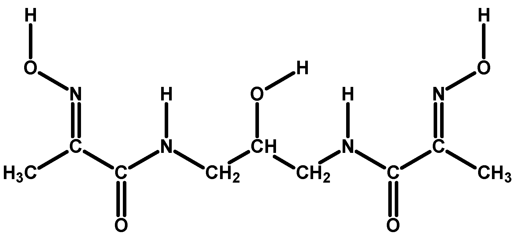
Scheme 1. L = mhiea2poh: (2E,2′E)-N,N′-(2-Hydroxypropane-1,3-diyl)bis[(2-hydroxyimino)propana-mide].
Inorganics 12 00330 sch001
Scheme 2. Anionic pseudo-macrocyclic complex core, M ( I I ) L H , with the numbering scheme.
Inorganics 12 00330 sch002
Figure 1. Molecular structure of the anionic Ni(II) complex in crystal structure (1) with selected labels. (Left): view of the anionic core from above. (Right): side view of the anionic core.
Inorganics 12 00330 g001
Figure 2. Molecular structure of the anionic Cu(II) complex in crystal structure (2) with selected labels. (Left): view of the anionic core from above. (Right): side view of the anionic core.
Inorganics 12 00330 g002
Figure 3. (Left): π-Stacked pseudo-macrocyclic Ni(II)-complex dimers joined into vertical columns by pairs of centrosymmetric H-bond contacts with plane-to-plane and metal-to-metal distances shown. Hydrogen bonds connecting adjacent dimers are also shown. (Right): View down a-axis in crystal structure (1) on the developing columns of H-bonded Ni(II)-complex dimers and the columns of H-bonded Ni(II) hexa-aqua cations; the latter fill the voids in the lattice. Co-crystallised water molecules are omitted for clarity. The unit cell box is also shown.
Inorganics 12 00330 g003
Figure 4. (Left): π-Stacked pseudo-macrocyclic Cu(II)-complex dimers with plane-to-plane and metal-to-metal distances. Water-bridged hydrogen bonds connecting adjacent dimers are also shown. (Right): View down c-axis in crystal structure (2) on the developing columns of water-bridged H-bonded Cu(II)-complex dimers and tetraphenylphosphonium cations filling the voids in the lattice. The unit cell box is also shown (a axis = red, b axis = green).
Inorganics 12 00330 g004
Figure 5. An example of the titration experiment output in the Ni(II)-L-H system with the refined solution model. The species detected are 421, 223, 221, and 11–2 (from left to right), where the numbers represent the units of Ni, L and H in the formula, respectively. The diamonds represent experimental titration points (pH values), while the solid lines represent individual species abundance.
Inorganics 12 00330 g005
Figure 6. An example of the titration experiment output in the Cu(II)-L-H system with the refined solution model. The species detected are 226 (minor), 422, 420, 111, 221, 21–4, and 11–2 (from left to right), where the numbers represent the units of Cu, L and H in the formula, respectively. The meaning of diamonds and solid lines is the same as in Figure 5.
Inorganics 12 00330 g006
Figure 7. Simulated speciation diagrams in the Ni(II)–L–H system for the solutions with [M]:[L] = 1:2, 1:1, and 2:1 (from top to bottom).
Inorganics 12 00330 g007aInorganics 12 00330 g007b
Figure 8. Simulated speciation diagrams in the Cu(II)–L–H system for the solutions with [M]:[L] = 1:2, 1:1, and 2:1 (from top to bottom).
Inorganics 12 00330 g008aInorganics 12 00330 g008b
Scheme 3. Hypothetical molecular structures for Ni(II)- and Cu(II)-mhiea2poh complexes in aqueous medium and their stoichiometric compositions pqr (for MpLqHr species). The structure actually shown for each type is the one whose composition is highlighted in bold.
Inorganics 12 00330 sch003aInorganics 12 00330 sch003bInorganics 12 00330 sch003c
Figure 9. Full-range ESR spectra of the selected aqueous solutions in the Cu–L–H system. The labels refer to [Cu]:[L] molar ratio and the volume (in μL) of NMe4OH(aq) added.
Inorganics 12 00330 g009
Figure 10. Zoomed–in part of the ESR spectra with super–hyperfine structure clearly visible. The labels have the same meaning as in Figure 9.
Inorganics 12 00330 g010
Figure 11. Experimental 1H spectrum of the mhiea2poh ligand in DMSO-d6. The inserts show multiplets in more detail. Labels at the top represent chemical shifts (ppm); labels at the bottom represent peak integrals; red lines are common geometrical representation of the peak integrals.
Inorganics 12 00330 g011
Figure 12. Simulated (top) and experimental (bottom) multiplets in the 1H spectrum of the mhiea2poh ligand in DMSO-d6. The labels of spectral lines refer to the experimental spectrum. Chemical shifts are given in ppm.
Inorganics 12 00330 g012
Table 1. Crystal data and structure refinement parameters for complexes (1)–(2).
Complex(1)(2)
Empirical formulaC18H42N8O18Ni3C33H41N4O9PCu
M834.72732.21
T/K293 (2)293 (2)
Crystal systemmonoclinictriclinic
Space groupP 21/cP 1 ¯
a7.4351(2)11.3575(7)
b12.8172(4)12.880(1)
c16.0660(7)12.900(1)
α9077.163(7)
β94.436(3)68.123(7)
γ9082.197(6)
V−31526.46(9)1704.5(2)
Z22
dc/g cm−31.8161.427
μ/mm−12.9851.851
F(0 0 0)868766
Crystal size/mm0.25 × 0.2 × 0.20.15 × 0.15 × 0.1
Colourtea yellowpale red
λ/Å1.541801.54180
θrange/°4.418 to 72.4614.538 to 71.309
Index range−9 ≤ h ≤ 7,−11 ≤ h ≤ 13,
−15 ≤ k ≤ 15,−14 ≤ k ≤ 15,
−19 ≤ l ≤ 15−15 ≤ l ≤ 14
Reflections collected11,08513,080
Independent Reflections/Rint2956/0.03716339/0.0585
Data/25534769
restraints/12
parameters254463
Goodness of fit on F21.1611.063
R indices
[I > 2σ(I)]
R10.07200.0603
wR20.19140.1582
R indices
(all data)
R10.08250.0849
wR20.19620.1783
Max/min electron density/e Å−31.2/−0.81.7/−0.9
Table 2. Selected values of bond lengths (Å) and angles (°) for the anionic Ni(II) and Cu(II) complexes in crystal structures (1)–(2).
(1)(2)
Ni01 N11.866(7)N3 C51.466(9)Cu01 N11.953(4)N3 C51.463(7)
Ni01 N21.868(6)C4 C91.520(10)Cu01 N21.923(4)C4 C91.537(5)
Ni01 N31.867(7)C5 C91.520(10)Cu01 N31.917(3)C5 C91.527(7)
Ni01 N41.878(6)C9 O51.414(9)Cu01 N41.951(4)C9 O51.383(5)
C3 O21.259(2)O1 O42.436C3 O21.265(6)O1 O42.472
C6 O31.261(8)BP 1–Ni010.023(1)C6 O31.267(4)BP 1–Cu010.0413(6)
N1 C11.290(10)N1 Ni01 N282.7(3)N1 C11.287(6)N1 Cu01 N282.7(2)
N4 C71.297(9)N2 Ni01 N397.0(3)N4 C71.278(5)N2 Cu01 N398.8(2)
O1 N11.366(8)N3 Ni01 N483.1(3)O1 N11.363(5)N3 Cu01 N483.2(2)
O4 N41.347(8)N4 Ni01 N197.2(3)O4 N41.354(5)N4 Cu01 N195.2(2)
C3 N21.319(9)N1 Ni01 N3177.8(3)C3 N21.304(5)N1 Cu01 N3176.2(2)
C6 N31.326(9)N2 Ni01 N4179.4(3)C6 N31.319(6)N2 Cu01 N4177.4(2)
N2 C41.458(9)C9 O5 H10112(5)N2 C41.464(6)C9 O5 H10109.5(4)
1 BP stands for the basal plane—an average plane drawn through four donor atoms N1-N2-N3-N4 of the chelating ligand.
Table 3. Common logarithms of the cumulative, βqr, and stepwise, Kqr, protonation constants with standard deviations, σ, for the mhiea2poh ligand at 25.00 °C in 0.100 M NMe4Cl(aq) medium [4].
qrlog10βqr (σ)log10Kqr (σ)
1111.303 (10)11.303 (10)
1221.75 (2)10.44 (3)
1331.49 (2)9.74 (4)
1440.70 (3)9.48 (5)
Table 4. Common logarithms of cumulative stability constants, log10βpqr, with standard deviations, σ, for Ni(II)-mhiea2poh complexes at 25.00 °C in 0.100 M NMe4Cl(aq) medium.
pqrlog10βpqr (σ)
11013.8 (6)
11−1 12.59 (1)
11−2−8.5 (6)
22361.3 (8)
22254 (2)
22141.3 (5)
21−110.4 (9)
21−2−0.28 (15)
42366.55 (8)
42260.4 (9)
42153.86 (5)
42045.72 (8)
1 A negative value for the number of protons means that more than 4 of them have been removed from a single ligand unit. Alternatively, this may mean the ligation of hydroxide groups by the complex. As the former is impossible for our ligand in aqueous medium, the latter is certainty. In the Hyperquad convention [OH] is proportional to [H]−1, which stems from the self-ionisation of water equilibrium: O H = K w [ H ] = K w [ H ] 1 or in logarithmic terms: l o g 10 O H = l o g 10 K w l o g 10 [ H ] .
Table 5. Common logarithms of cumulative stability constants, log10βpqr, with standard deviations, σ, for Cu(II)-mhiea2poh complexes at 25.00 °C in 0.100 M NMe4Cl(aq) medium.
pqrlog10βqr (σ)
11128.85 (10)
11−21.2 (7)
22681.89 (9)
22153.5 (4)
21−216.14 (13)
21−40.5 (6)
42272.1 (3)
42062.5 (3)
Table 6. Logarithms of protonation constants for mhiea2p ligand, L′, and stability constants for its Ni(II) and Cu(II) complexes (MpLqHr) with standard deviations (σ) according to the literature.
Reference[29][34][35][36]
Mp q rlog10β (σ)Mediumlog10β (σ)Mediumlog10β (σ)Mediumlog10β (σ)Medium
H+0 1 1
0 1 2
10.383 (9)
19.933 (7)
0.1 M KNO310.408 (1)
20.072 (1)
0.15 M
KCl
10.54
(1)
20.31
(1)
0.1 M KNO3
Ni2+1 1 1

1 1 −1

1 1 −2


1.14
(9)
−7.05 (11)
0.1 M KNO3 15.460 (35)
2.842 (10)
−8.368 (35)
0.15 M
KCl
Cu2+1 1 1

1 1 0

1 1 −1

1 1 −2
19.61
(4)


9.95
(4)
2.08
(8)
0.1 M KNO3

14.076 (30)
9.903 (30)
1.625 (53)
0.15 M
KCl
Table 7. Comparable equilibrium constants for the reactions of Ni(II) and Cu(II) coordination with mhiea2poh ligand, L, (this work) and mhiea2p ligand, L′, (literature).
This WorkLiterature
Reactionlog10KReactionlog10K′Reference
Ni + LH4 + 4OH ⇌ NiL + 4H2O28.3 (9)Ni + L′H2 + 4OH ⇌ NiL′H−2 + 4H2O28.2 (2)[29]
Ni + L′H2 + 4OH ⇌ NiL′H−2 + 4H2O26.80 (9)[35]
Cu + LH4 + 3OH ⇌ CuLH + 3H2O29.3 (4)Cu + L′H2 + 3OH ⇌ CuL′H−1 + 3H2O31.39 (8)[29]
Cu + L′H2 + 3OH ⇌ CuL′H−1 + 3H2O31.26 (7)[34]
Table 8. Target solutions for the growth of Ni(II)-complex crystals and the observed outcomes.
Target Complex Stoichiometry, pqr a224223222111
[L]0/mL0.9980.9980.9980.998
H2O/mL3.9423.8923.8423.842
[HCl]0/μL25.225.225.225.2
[NiCl2]0/μL34.834.834.834.8
[KOH]0/μL117153185500
[TPPB]0/μL050.1100.1100.1
Target pH6.216.9610.0011.46
Observed solution colouryellowyellowyellowyellow
Observed solidsclustered honey-yellow prismsbright-yellow needles and honey-yellow prismstea-yellow slabs and prismstea-yellow prisms and branched colourless crystals
a These are the stoichiometric coefficients of M, L, and H in the complex.
Table 9. Target solutions for the growth of Cu(II)-complex crystals and the observed outcomes.
Target Complex Stoichiometry, pqr420, 224111, 22111-242021-4
[L]0/mL0.9980.9980.9980.9980.998
H2O/mL3.9513.8513.7513.7253.925
[HCl]0/μL25.225.225.225.225.2
[CuCl2]0/μL25.725.725.725.725.7
[KOH]0/μL135180300178226
[TPPB]0/μL0100.1200.200
[STPB]0/μL000199.7 (a)0
[PHFA]0/μL000200.0 (b)0
Target pH6.257.8511.026.578.73
Observed solution colourpale green yellowlight
terracotta
light
terracotta
(a) sea green + white ppt
(b) sea green
brown green
Observed
solids
small fused pink, yellow, green, and colourless crystalsbranched pink-red poorly shaped crystalspale red prisms and colourless very long thin needles(a) colourless prisms + pink and green powder
(b) green rosettes + white fine powder
colourless prisms + brown ppt and green glass
(a) and (b) refer to the same solution but for the counter-anion, the TPB in the first case and the HFA in the second case.
Table 10. ESR sample preparation guide.
Target
Species
Desired RatioConcentrations of Stock Solutions
0.9823 mM00.1987 M0.1942 M0.1250 M
pqr[L]:[M]L/μLH2O/μLH/μLCu/μLNMe4OH/μL
4221:13054183225.215.573
4201:13054181725.215.588
1111:13054179325.215.5112
21−41:13054173525.215.5170
4221:23054177925.230.9111
4201:23054175225.230.9138
Disclaimer/Publisher’s Note: The statements, opinions and data contained in all publications are solely those of the individual author(s) and contributor(s) and not of MDPI and/or the editor(s). MDPI and/or the editor(s) disclaim responsibility for any injury to people or property resulting from any ideas, methods, instructions or products referred to in the content.