Article

N-Based Polydentate Ligands and Corresponding Zn(II) Complexes: A Structural and Spectroscopic Study

Department of Chemistry, University of Turin, Via P. Giuria, 7, 10125 Turin, Italy
*
Author to whom correspondence should be addressed.
Inorganics 2023, 11(11), 435; https://doi.org/10.3390/inorganics11110435
Submission received: 28 August 2023 / Revised: 27 October 2023 / Accepted: 7 November 2023 / Published: 10 November 2023
(This article belongs to the Section Coordination Chemistry)

Abstract

:
Herein, the structural and photophysical features of two N-based polydentate ligands and the corresponding Zn(II) complexes are investigated. The obtained compounds were characterized using different spectroscopic techniques and their optical properties are discussed in relation to their chemical structure, defined by single-crystal X-ray diffraction and mass spectrometry. The spontaneous and quantitative complexation, investigated by UV-Vis, fluorescence, NMR, IR spectroscopies and mass spectrometry, makes these N-based polydentate ligands interesting candidates for possible applications in chelation therapy and in Zn(II) sensors.

1. Introduction

The human body contains up to several grams of Zn(II), making this element the second most abundant transition metal cation. Biological functions of Zn(II) have been reported for different purposes: it is required for the turnover of more than 300 catalytically active Zn(II) metal-proteins, as well as more than 2000 Zn(II)-dependent transcription factors [1]. Zn(II) is found at high concentrations, especially in the brain, pancreas and spermatozoa, and can be visualized with fluorescent dyes [2,3]. However, the biological functions of free or complexed Zn(II) are less certain. Recently, the exceptionally high intracellular Zn(II) concentration of β-cells (cells that produce insulin) has been widely investigated; indeed, β-cell insulin granules contain up to 20 mM Zn(II) compared to 2–10 μM in the Zn(II)-rich compartments of most other cell types. In addition, labile Zn(II) in the brain is reported to have a key role in the vesicles of presynaptic neurons, modulating the functions of fundamental ion channels and receptors [4,5,6].
A specific range of concentrations of metal ions in the body is required to ensure normal metabolic activities and physiological functions, as well as a healthy life. Contrariwise, the accumulation of metal ions (i.e., iron, lead, copper and zinc) in any organ, tissue, cellular or sub-cellular compartment represents a dangerous prognostic factor for many disorders and illness. The use of chelating drugs can re-equilibrate the metal imbalance and treat the associated diseases. For example, deferoxamine, deferiprone, deferasirox and their combinations are commonly used drugs for the medical treatment of Fe(III) overload in thalassaemia [7] and the chelating drugs penicillamine and triethylenetetramine are often used for Cu(II) overload in Wilson’s disease [8,9,10]. Similarly, many other organic ligands are employed for the detoxification of different transition metal ions [11].
In this regard, polydentate ligands fill a unique area, due to their chemical and structural behaviours. They have been extensively studied because they integrate the properties of large-sized molecules, such as structural tunability and high selectivity, moderate manufacturing costs, positive pharmaco-kinetic behaviour and availability. Well-documented cases, such as triethylenetetramine, ethylenediaminetetraacetate (EDTA), cyclodextrins, substituted porphyrins and crown ethers, represent useful scaffolds between aromatic ligands and functional structures [12,13]. Over the past few decades, polydentate ligand chemistry has been widely investigated and innovative synthetic approaches have been reported to collect new molecular skeletons with different binding properties, geometries, sizes and solubility [14,15,16,17,18,19]. New polydentate ligands with specific properties persist as a sought-after item for many innovative technological applications [20,21]. In general, polydentate ligands and their corresponding complexes can be utilized in various biomedical and pharmaceutical contexts, such as in drug delivery, biomaterials and diagnostic agents, ensuring minimal toxicity and compatibility with biological systems.
Initially, polydentate ligand derivatives captured escalating focus for their intriguing skeleton, and then for their use in supramolecular architectures [22,23,24], catalysis [25,26], high selectivity for ions and small-molecules recognition, supramolecular chemistry [27,28] and as chelating drugs for medical treatment [29,30,31,32,33,34,35]. In 2002, Zhao and Moore highlighted the structural relevance of the imine bond as a compositional unit in previously reported N-polydentate ligands such as macrocycles [36]. In particular, imine-containing phenylene portions are a significant subclass of shape-persistent products, because of their hetero-polydentate ligand capacity [37,38]. Phenylene-imine bonds have a few possible conformations, and the structural rigidity turns them into perfect systems and building blocks for larger structures, starting from macrocycles to open-chain polydentate ligands. Furthermore, the incorporation of substituent groups and N-heteroatoms into the backbone allows for powerful transition metal ion chelation along with the tuning of the optical and physical behaviour of the entire system [39].
Herein, we report the use of two N-based polydentate ligands (see Figure 1) obtained from common diamine and di-ketone precursors in a condensation process involving two consecutive imine-bond formations and a successive reduction. Recently, D. Carbajo et al. employed similar di-aldehyde (instead of ketone), obtaining innovative N-based polydentate ligands and macrocycles, as reported by the authors: “these systems open the door to their use in stimuli-responsive materials with appealing properties” [40]. These two ligands (L1 and L2) represent the N-based polydentate nucleus for the spontaneous formation of the corresponding new Zn(II) complexes ([ZnL1]2+ and [ZnL2]2+). The complexation reaction has been investigated for possible therapeutic applications as chelating drugs or sensitive fluorescence ‘turn-on’ chemo-dosimeters [41,42,43,44,45].

2. Results and Discussion

2.1. Complexation Behaviour

The employed ligands contain six N-atoms which can potentially act as coordinating sites for metal–ligand chelation. Each ligand has four pyridinic nitrogen atoms plus two N-atoms from imine (L1) or amine (L2) groups, making these compounds interesting candidates for removing heavy metals from solutions, as well as for possible therapeutic applications as chelating drugs. To investigate the coordinating abilities of L1 and L2, we prepared methanolic solutions of the ligands and added increasing equivalents of Zn(NO3)2, while monitoring the evolution of the 1H NMR signals (see Figure 2) and the UV-Vis absorption features (see Figure 3).
Compounds L1 and L2 show a similar structure, with the important difference of the imino/amino bonds (see Figure S1 for the 1H NMR spectrum for L1 and Figure 2 for L2) [46]. In the 1H NMR spectrum, L1 exhibits eight multiplets corresponding to the sixteen pyridine protons (range 6–9 ppm) and two multiplets corresponding to the eight methylene protons of the alkyl chain (range 1–4 ppm). Similarly, L2 exhibits four multiplets corresponding to the sixteen pyridine protons (range 6–9 ppm), with one singlet associated with the two methine proton (at about 5 ppm) confirming the reduction of the imino group, and two multiplets corresponding to the eight methylene protons of the alkyl chain (in the range 1–4 ppm). In the case of L2, the high symmetry of the skeleton greatly simplifies the spectrum with respect to the complexity of L1, in which the imine bonds imply the asymmetry of the pyridine rings. These considerations are further confirmed by the 13C NMR spectra where, in particular, the imine (quaternary carbon signal at 166.5 ppm for L1) and the amine (68.4 ppm signal for L2) signals are clearly recognisable.
In the 1H and 13C NMR spectra of the corresponding Zn(II) complexes, [ZnL1]2+ and [ZnL2]2+, all the signals are shifted compared to those of the employed ligands, providing clear evidence of the complexation that occurred with the Zn(II) ion. [ZnL1]2+ and [ZnL2]2+ show a very similar resonance pattern that highlights the asymmetrical nature of the coordinated pyridine rings on the Zn(II) centre (see Figure S1 for the 1H NMR spectrum of [ZnL1]2+ and Figure 2 for [ZnL2]2+). The complexation reaction is spontaneous and immediate, as shown in Figure 2 and Figure 3. Indeed, the addition of sub-stoichiometric amounts of Zn(NO3)2 to a methanolic solution of L2 results in the progressive formation of [ZnL2]2+ and the concomitant disappearance of the free L2. The octahedral coordination of L2 on the metal centre (confirmed by single crystal XRD data, see Section 2.4) implies symmetry breaking, with the splitting of the 1H NMR signals of the coordinate pyridine rings and the aliphatic chain (four signals for the diastereotopic methylene protons, see Figure 2). Conversely, in the case of [ZnL1]2+, the protons of the aliphatic chain remain isochronous (2 methylene signals, see Figure S1), proving a tetrahedral coordination. This inference is further supported by the DFT optimised geometries of [ZnL1]2+ and [ZnL2]2+ computed in methanol solution (see Figure S2).
Additionally, the complexation monitored by UV-Vis absorption spectroscopy (10–5 M methanol solutions at 25 °C) confirms the complete conversion of each ligand into a corresponding single product (presence of isosbestic points, see Figure 3 for L1 and [ZnL1]2+ and Figure S3 for L2 and [ZnL2]2+).

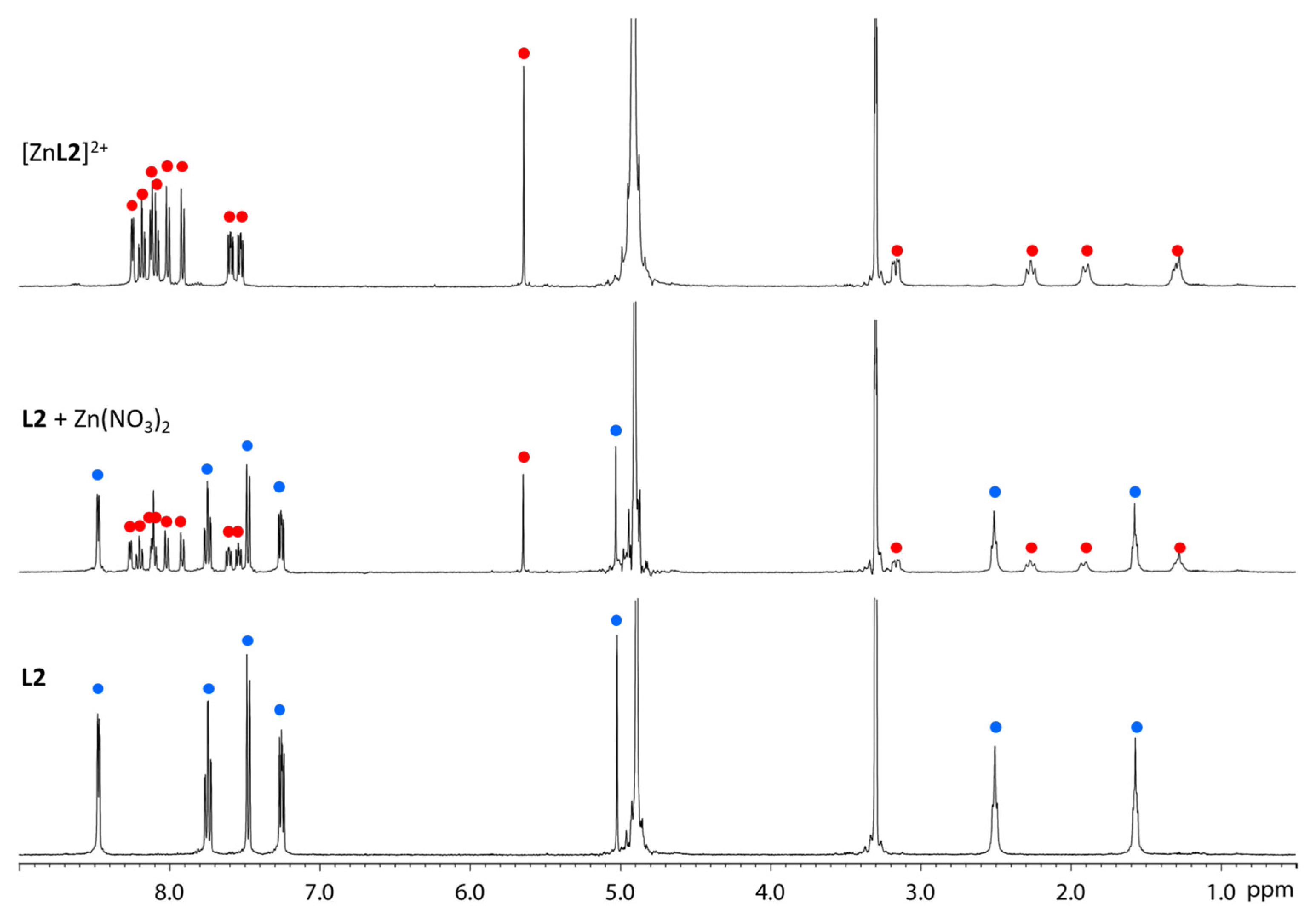
2.2. Spectroscopic Characterization

The absorption and emission spectra of L1, L2, [ZnL1]2+ and [ZnL2]2+ in methanol solutions are reported in Figure 3 and in Figure S3, whereas selected optical data are collected in Table 1.
L1 shows two principal bands, at 238 nm and 267 nm, with almost no absorption beyond 330 nm. The absorption profile of L2, which represents the corresponding reduced form of L1 (from imine to amine), conversely shows a single band centred at 262 nm with two defined shoulders at 257 and 272 nm.
The complex [ZnL1]2+ shows a noticeable change in the absorption profile, compared to the corresponding free ligand: the first band at 238 nm (well defined for L1) decreases in [ZnL1]2+, while the second band is red shifted at 278 nm. The compound [ZnL2]2+ displays a more structured band centred at 266 nm (surrounded by two shoulders more evident than for the free ligand), corresponding to n→π* and π→π* transitions.
All the studied compounds showed an appreciable emission between 350 and 550 nm, with emission maxima at about 410 nm (excitation wavelength at 300 nm). The modifications of the ligand emission after the addition of Zn(NO3)2 are reported in Figure 3 and Figure S13). The emission intensity increased after complexation with Zn(II), showing a little red-shift (11 nm) in the case of [ZnL1]2+ in comparison to the free L1 (see Table 1).
Solid state ATR-FTIR spectra of L1 and L2 and the corresponding [ZnL1]2+ and [ZnL2]2+ complexes are reported in the Supplementary Materials (see Figures S4 and S5). A DFT modelling of L1, L2, [ZnL1]2+ and [ZnL2]2+ was performed to check the vibrational assignments. In order to evaluate the effect of the Zn(II) coordination on the vibrational modes of the ligands, we focused on a few vibrational modes, in particular the symmetric νCC and νCN of the pyridyl ring and the νC=N and νC-N of imine and the corresponding amine of L2. The coordination of pyridine to Zn(II) induces a little shortening of the C-C bonds with a shift of νCC and νCN and brring to higher wavenumbers, respectively, 1580, 1482 and 992 cm−1 in py [47] and 1609, 1491 and 1016 cm−1 in [ZnCl2(py)2] [48]. In L1, the doublet at 1572 and 1565 cm−1, attributable to νCC, shifted to 1583 and 1572 cm−1 in [ZnL1]2+, and the doublet at 1001 and 994 cm−1, attributable to brring, shifted to 1019 and 1018 cm−1. The imide νC=N stretching, attributable to 1634 cm−1 in L1, remained almost unchanged in [ZnL1]2+, with a modest shift to 1683 cm−1 in [ZnL1]2+, supporting the involvement of only pyridyl nitrogen in Zn coordination and the tetrahedral coordination of L1 in [ZnL1]2+.
In L2, the νNH at 3289 and 3261 cm−1, because of the Zn(II) coordination, underwent a shift to lower wavenumbers and a broadening of the bands due to the hydrogen bond with NO3 groups. Similar to L1, the symmetric νCC may be assigned to 1586 and 1568 cm−1 in L2, shifted to 1601 and 1586 cm−1 in [ZnL2]2+; brring, assignable in L2 to the mode at 994 cm−1, shifted to 1018 cm−1 in [ZnL2]2+. The infrared spectra of both [ZnL1]2+ and [ZnL2]2+ showed the characteristic bands of the nitrate anion, with a medium-strong band at 1376 cm−1 due to asymmetric N-O stretching modes and a medium band at 828 cm−1 assignable to symmetric νNO. These nitrate modes do not show variation with respect to ionic nitrate groups of common salts, such as ZnNO3 or NaNO3.

2.3. Mass Spectrometry Characterization

The ESI-MS spectra of L1, L2 and their corresponding Zn(II) complexes are reported in the Supplementary Materials (see Figures S6–S13). Collected spectra show the molecular peaks of the protonated form [L+H]+ for L1 and L2 (see Figures S6 and S8, respectively) in methanol solutions (with 2% of formic acid). MS/MS experiments (see Figures S7 and S9) highlight similar fragmentation patterns for L1 and L2 species. Protonation due to formic acid likely occurs on the imine and amine N atoms, respectively, and subsequent heterolytic C-N cleavage yields the neutral fragments coming from the di-pyridine-ketone precursor, i.e., C11H9N3 for L1 and C11H11N3 for L2.
The mass spectra of the corresponding complex solutions exhibited abundant peaks ascribed to [ZnL1]2+ = C26H24N6Zn (calculated average mass m/z = 242; found m/z = 242) and to [ZnL2]2+ = C26H28N6Zn (calculated average mass m/z = 244; found m/z = 244) (see Figures S10 and S12). The other signals in the spectra may be assigned either to ligand fragmentation or to clustering reactions taking place in the gas phase. Furthermore, the experimental isotopic distributions for all the observed ions fit nicely with the calculated isotopic simulation, thus representing additional proofs for product assignment; as can be observed, Figures S11 and S13 report as examples the cases of [ZnL1]2+ and of [ZnL2]2+, respectively.

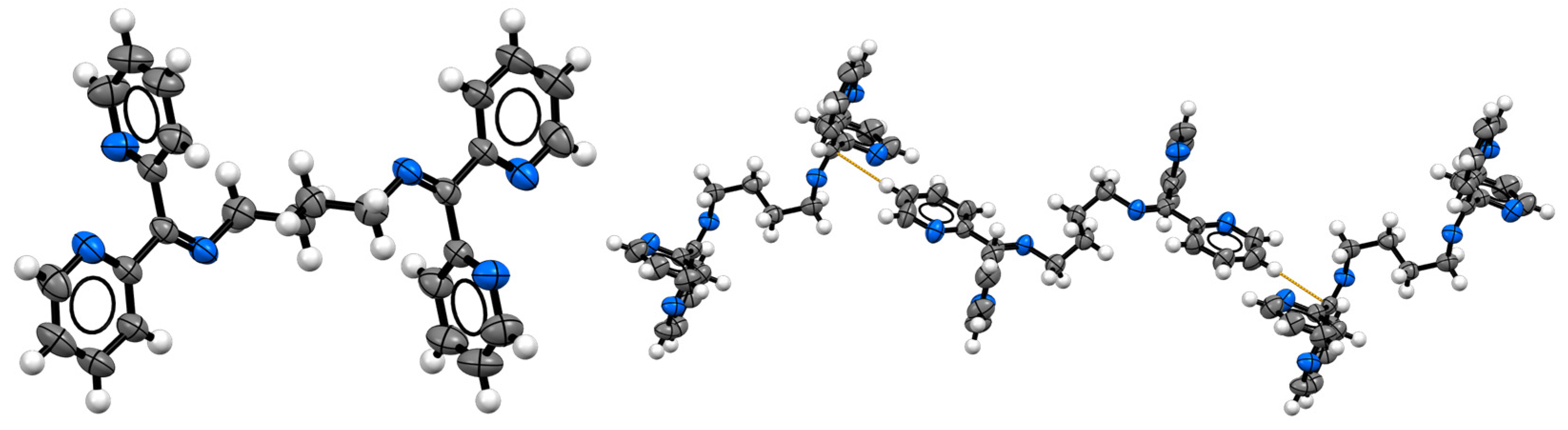
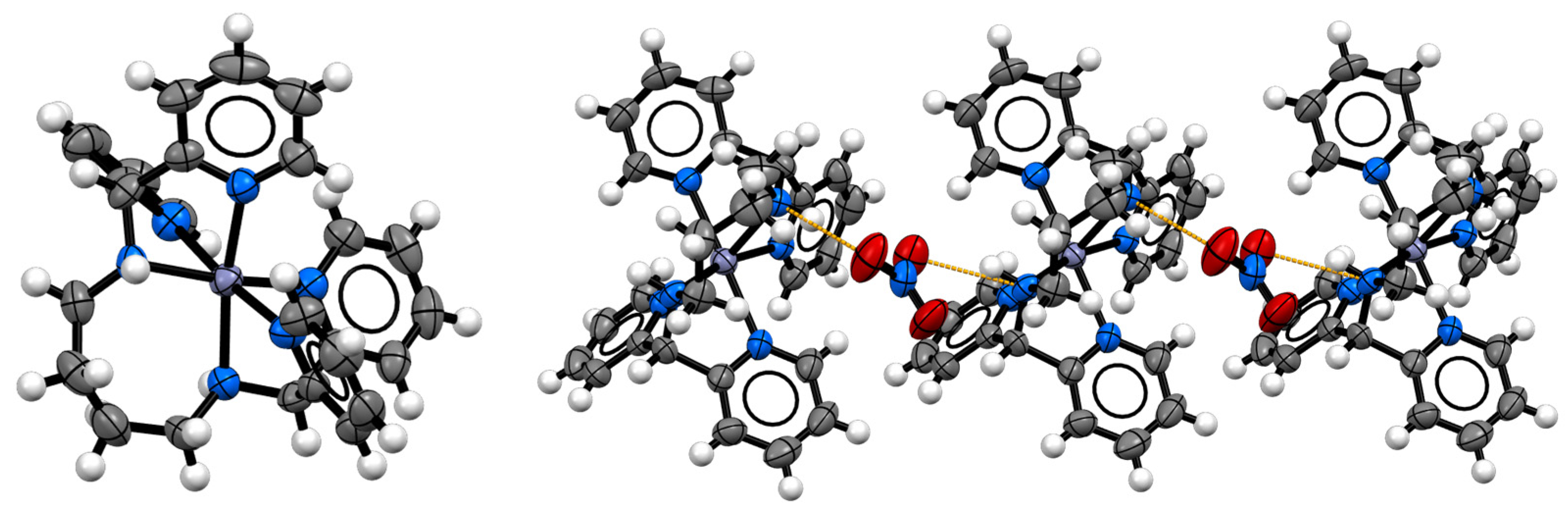
2.4. Structural Characterization

The ligand L1 crystallized from ethanol by slow evaporation in yellow prisms. The space group type is monoclinic P21/c, with half a molecule in the asymmetric unit and the entire molecule generated by an inversion centre. The imide nature is clear by the distance between C3 and N1 (1.276(5) Å, typical of imides, with respect to 1.493(6) Å found for L2 in the complex [ZnL2]2+) and for the planarity of the nearby fragments. The two pyridine groups are staggered to each other, to minimize the steric hindrance (see Figure 4). Moreover, in the crystal packing, the molecules are disposed orthogonally, interacting with dispersion interactions and C-H···π contacts (d(C-H···π)= 3.662 Å, see Figure 4, right).
On the other hand, crystals of [ZnL2](NO3)2·DMF have been obtained from DMF. The complex crystallizes as DMF solvate in the monoclinic non-centrosymmetric Cc space group type. Neither the nitrate counterion nor the DMF are bonded directly to the metal centre, while the Zn(II) is completely surrounded by the six coordination sites of L2 to form a nearly regular octahedron (see Figure 5, left, and Table 2).
The Zn-N distances are irregular, as are the angles, specifically the ones regarding the bonds with the most distant pyridyl group, probably due to the steric hindrance internal of L2 (see Table 2 and Table S3). In the crystal packing, it is worth noting the presence of hydrogen-bond-forming chains between one of the nitrate and the complexes through the amine hydrogens (d(N4-H···O2) = 2.983(8) Å, see Figure 5, right).

3. Materials and Methods

3.1. Experimental Details

All solvents and raw materials were used as received from commercial suppliers (Sigma-Aldrich and Alfa Aesar, Milano, Italy) without further purification. TLC was performed on Fluka silica gel TLC-PET foils GF 254, particle size 25 nm, medium pore diameter 60 Å. Column chromatography was performed on Sigma-Aldrich silica gel 60 (70–230 mesh ASTM).
1H and 13C NMR spectra were recorded on a JEOL ECP 400 FT-NMR spectrometer (1H NMR operating frequency 400 MHz). Chemical shifts are reported relative to TMS (δ = 0) and referenced against solvent residual peaks. The following abbreviations are used: s (singlet), d (doublet), t (triplet), dd (doublet of doublets), m (multiplet). Mass spectra were recorded on a Thermo-Finnigan Advantage Max Ion Trap Spectrometer equipped with an electrospray ion source (ESI) in positive and negative ion acquiring mode and with a Sciex Triple Quad 5500 equipped with a Turbo V™ source in positive electrospray mode. UV-Vis absorption spectra were recorded on a Cary60 spectrometer; infrared spectra were recorded on solid samples with a PerkinElmer Spectrum Two FT-IR Spectrometer equipped with a Universal Attenuated Total Reflectance accessory. DFT calculations were performed with the Gaussian 16 program package [49], employing the density functional theory (DFT) method with the Becke three-parameter hybrid functional [50] and the Lee–Yang–Parr gradient-corrected correlation functional (B3LYP) [51]. The solvent effect was included by using the conductor-like polarizable continuum model (CPCM) with methanol as the solvent [52,53]. The 6–31g(d,p) basis set was used for all atoms [54]. Geometries were optimized without any symmetry constraints and harmonic frequencies were computed. The nature of the optimized structures was verified by using harmonic vibrational frequency calculations. No imaginary frequencies were found, thus indicating we had located the minima on the potential energy surfaces. Molecular-graphic images were produced by using the UCSF Chimerax package from the Resource for Biocomputing, Visualization, and Informatics at the University of California, San Francisco (supported by NIH P41 RR001081) [55].
Single crystal data were collected on a Gemini R Ultra diffractometer (Agilent Technologies UK Ltd., Oxford, UK) using graphite-monochromatic Mo Kα radiation (λ = 0.7563 Å) with the ω-scan method. Copper-derived radiation was preferred for the cases of very weakly diffracting crystals. CrysAlisPro v42 software was used for retrieving cell parameters, for performing data reduction and for absorption correction (with multi-scan technique). All structures were solved with direct methods using ShelXS-14 and refined with full-matrix least-squares on F2 using the SHELXL-14 using the Olex2 program. All non-hydrogen atoms were anisotropically refined. Hydrogen atoms were calculated and were riding on the corresponding atom. Structure images were obtained using Mercury 2023.1.0 software. Crystal data and refinement, selected bond lengths and angle amplitudes and asymmetric units of compounds are reported in ESI. The crystallographic data for the crystallized compounds are deposited within the Cambridge Crystallographic Data Centre as supplementary publications under the CCDC numbers 2289875-2289876. This information can be obtained free of charge from the Cambridge Crystallographic Data Centre.

3.2. Synthesis

The synthetic approach has been previously employed by us to successfully produce different ligands from the versatile di-pyridyl ketone reagent; the obtained derivatives show excellent coordination skills towards different metal ions [56,57]. The N-based polydentate ligands L1 and L2 were obtained as previously reported [56].

3.2.1. L1: N,N’-(Butane-1,4-diyl)bis(1,1-di(pyridin-2-yl)methanimine)

In 30 mL of methanol were mixed di-2-pyridyl ketone (839 mg, 4.56 mmol), butane-1,4-diamine (200 mg, 2.28 mmol, 0.5 eq) and 5 drops of acetic acid. After 4 h refluxing, the solution was cooled. The solvent evaporated under vacuum. The final product was obtained after column chromatography on silica gel, using CH2Cl2/CH3OH (98:2) as eluent. Reaction yield: 906 mg, 2.16 mmol, 95%. 1H NMR (methanol-d4, 400 MHz) δ: 8.59 (d, J = 5.0 Hz, 2H), 8.40 (d, J = 5.0 Hz, 2H), 8.07 (d, J = 8.0 Hz, 2H), 7.91 (t, J = 7.7 Hz, 2H), 7.87 (t, J = 7.7 Hz, 2H), 7.45 (dd, J = 7.7 Hz, J = 5.0 Hz, 2H), 7.39 (dd, J = 7.5 Hz, J = 4.9 Hz, 2H), 7.31 (d, J = 7.7 Hz, 2H), 3.37 (m, 4H), 1.74 (m, 4H) ppm. 13C NMR (methanol-d4, 100 MHz) δ: 166.5, 156.2, 154.6, 148.9, 148.2, 137.1, 136.9, 124.6, 124.1, 123.7, 122.3, 53.1, 28.2 ppm. MS (ESI+) m/z calculated for [L1+H]+ = C26H25N6 m/z = 421.21; found m/z = 421.07.

3.2.2. L2: N1,N4-Bis(di(pyridin-2-yl)methyl)butane-1,4-diamine

The precursor L1 (500 mg, 1.19 mmol) was reduced with an excess of NaBH4 in methanol at 0 °C for 1 h. The solvent was removed under vacuum. Then, the obtained crude product was dissolved in CH2Cl2, and the solution extracted with water and brine and filtered and dried over Na2SO4. The solvent evaporated under vacuum. The oily product was obtained in quantitative yield: 504 mg, 1.19 mmol. 1H NMR (methanol-d4, 400 MHz) δ: 8.46 (d, J = 5.0 Hz, 4H), 7.73 (t, J = 7.7 Hz, 4H), 7.46 (t, J = 7.9 Hz, 4H), 7.24 (dd, J = 7.5 Hz, J = 5.0 Hz, 4H), 5.01 (s, 2H), 2.49 (m, 4H), 1.56 (m, 4H) ppm. 13C NMR (methanol-d4, 100 MHz) δ: 160.5, 148.6, 137.3, 122.6, 122.5, 68.4, 47.0, 27.0 ppm. MS (ESI+) m/z calculated for [L2+H]+ = C26H29N6 m/z = 425.24; found m/z = 425.10.
The Zn(II) complexes [ZnL1]2+ and [ZnL2]2+ were obtained in quantitative yield by the reaction of Zn(NO3)2 with a stoichiometric amount of the corresponding ligand in methanol at room temperature. All the complexes are uncoloured crystalline powders.

3.2.3. [ZnL1]2+

1H NMR (methanol-d4, 400 MHz) δ: 8.85 (m, 4H), 8.28 (t, J = 7.8 Hz, 2H), 8.16 (t, J = 7.8 Hz, 2H), 7.98 (dd, J = 7.5 Hz, J = 5.0 Hz, 2H), 7.78 (d, J = 7.8 Hz, 2H), 7.71 (dd, J = 7.8 Hz, J = 4.9 Hz, 2H), 7.50 (d, J = 7.9 Hz, 2H), 3.76 (m, 4H), 2.08 (m, 4H) ppm. 13C NMR (methanol-d4, 100 MHz) δ: 168.0, 150.6, 149.7, 149.6, 148.0, 142.2, 138.2, 129.3, 128.2, 125.9, 124.5, 52.2, 27.0 ppm. MS (ESI+) m/z calculated for [ZnL1]2+ = C26H24N6Zn2+ m/z = 242.07; found m/z = 242).

3.2.4. [ZnL2]2+

1H NMR (methanol-d4, 400 MHz) δ: 8.25 (d, J = 5.0 Hz, 2H), 8.19 (t, J = 7.8 Hz, 2H), 8.12 (d, J = 5.0 Hz, 2H), 8.10 (t, J = 7.8 Hz, 2H), 8.01 (d, J = 7.8 Hz, 2H), 7.92 (d, J = 7.8 Hz, 2H), 7.60 (dd, J = 7.7 Hz, J = 5.1 Hz, 2H), 7.53 (dd, J = 7.7 Hz, J = 5.1 Hz, 2H), 5.64 (s, 2H), 3.16 (m, 2H), 2.27 (m, 2H), 1.90 (m, 2H), 1.23 (m, 2H) ppm. 13C NMR (methanol-d4, 100 MHz) δ: 157.0, 155.4, 148.6, 147.9, 141.5, 141.0, 125.7, 125.5, 124.8, 124.1, 65.5, 49.1, 28.2 ppm. MS (ESI+) m/z calculated for [ZnL2]2+ = C26H28N6Zn2+ (calculated average mass: m/z= 244.08; found m/z = 244).

4. Conclusions

Two interesting N-based polydentate ligands have been synthesized and characterized in comparison with the corresponding Zn(II) complexes. The structure of the obtained compounds was determined using 1H and 13C NMR, UV-Vis absorption, emission and FT-IR spectroscopies, Electrospray Ionization Mass Spectrometry and X-Ray Diffraction. Obtained from a mild, straightforward and efficient preparation, the studied polydentate skeletons could inspire the design of a possible wide range of new ligands with different metal ion affinities.
The comparison between the structures of the free ligands L1 and L2 and corresponding Zn(II) complexes [ZnL1]2+ and [ZnL2]2+ shows an interesting modification in the spectroscopic properties. In particular, a noticeable change in the absorption profile (comparing the free ligand and the corresponding Zn(II) complex), a strong quenching in the emission along with appreciable shift and the splitting and shift of the NMR signals highlight the asymmetrical nature of the coordinated pyridine rings on the Zn(II) centre.
The proposed study shows that the investigated complexation reaction is spontaneous, instantaneous and complete, all key aspects for possible future applications.
Overall, these results encourage further studies on these N-based polydentate skeletons, aiming to introduce new functional groups, varying the chelating geometry and the electronic behaviours to further investigate the coordinating properties of these ligands.
In general, polypyridine compounds have shown promise in various medical applications due to their ability to form stable metal complexes. Furthermore, designing polypyridine-based materials for medical applications is a multidisciplinary endeavour that combines knowledge of chemistry, biology and materials science. In this context, it is essential to consider the specific requirements and challenges of each possible application to develop safe and effective solutions such as biocompatibility, stability, regulatory compliance and many others.
In conclusion, the synthetic accessibility and the demonstrated complete interactions with Zn(II) ions make these promising polydentate ligands interesting candidates for chelation therapy, Zn(II) sensors and possibly in metal ion removal, as recently reported for similar innovative structures [58,59,60,61,62,63,64].

Supplementary Materials

The following supporting information can be downloaded at: https://www.mdpi.com/article/10.3390/inorganics11110435/s1, Figure S1. 1H NMR spectrum of L1 and [ZnL1]2+ in CD3OD; Figure S2. DFT optimized structures of [ZnL1]2+ and [ZnL2]2+ in CD3OD solution; Figure S3. UV-vis absorption and emission spectra of L2 and the corresponding Zn(II) complex [ZnL2]2+ (methanol, 25 °C, 10–5M); Figure S4. Vibrational comparisons between L1 and [ZnL1]2+; Figure S5. Vibrational comparisons between ligand L2 and macrocycle [ZnL2]2+; Figure S6. ESI-MS mass spectrum of ligand L1; Figure S7. ESI-MS/MS mass spectrum of [L1+H]+; Figure S8. ESI-MS mass spectrum of ligand L2; Figure S9. ESI-MS/MS mass spectrum of [L2+H]+; Figure S10. ESI-MS mass spectrum of complex [ZnL1]2+; Figure S11. Experimental and theoretical isotopic composition of the [ZnL1]2+ ion; Figure S12. ESI-MS mass spectrum of complex [ZnL2]2+; Figure S13. Experimental and theoretical isotopic composition of the [ZnL2]2+ ion; Figure S14. ORTEP plot of asymmetric unit of L1; Figure S15. ORTEP plot of asymmetric unit of [ZnL2](NO3)2·DMF; Table S1. Crystal data and structure refinement for L1; Table S2. Bond Lengths for L1; Table S3. Bond Angles for L1; Table S4. Crystal data and structure refinement for [ZnL2](NO3)2·DMF; Table S5. Crystal data and structure refinement for [ZnL2](NO3)2·DMF; Table S6. Bond Angles for [ZnL2](NO3)2·DMF.

Author Contributions

Conceptualization, G.V. and C.G.; formal analysis, S.Z. and E.P.; investigation, G.V. and R.R.; resources, R.G. and E.D.; writing—original draft preparation, G.V. and R.R.; writing—review and editing, R.G. and C.G.; visualization, C.G.; supervision, G.V. All authors have read and agreed to the published version of the manuscript.

Funding

This research received no external funding.

Data Availability Statement

The data presented in this study are available on request from the corresponding author.

Acknowledgments

Authors acknowledge technical support from the Project CH4.0 under the MUR program “Dipartimenti di Eccellenza 2023–2027” (CUP: D13C22003520001).

Conflicts of Interest

The authors declare no conflict of interest.

References

  1. Vahidi Ferdowsi, P.; Ng, R.; Adulcikas, J.; Sohal, S.S.; Myers, S. Zinc Modulates Several Transcription-Factor Regulated Pathways in Mouse Skeletal Muscle Cells. Molecules 2020, 25, 5098. [Google Scholar] [CrossRef] [PubMed]
  2. Tang, J.; Yin, H.-Y.; Zhang, J.-L. Chapter One—Luminescent Zinc Complexes as Bioprobes for Imaging Molecular Events in Live Cells. In Inorganic and Organometallic Transition Metal Complexes with Biological Molecules and Living Cells; Lo, K.K.-W., Ed.; Academic Press: Cambridge, MA, USA, 2017; pp. 1–53. ISBN 978-0-12-803814-7. [Google Scholar]
  3. Endo, I.; Nagamune, T. Nano/Micro Biotechnology; Springer Science & Business Media: Berlin/Heidelberg, Germany, 2010; ISBN 978-3-642-14946-7. [Google Scholar]
  4. Horton, T.M.; Allegretti, P.A.; Lee, S.; Moeller, H.P.; Smith, M.; Annes, J.P. Zinc-Chelating Small Molecules Preferentially Accumulate and Function within Pancreatic β Cells. Cell Chem. Biol. 2019, 26, 213–222.e6. [Google Scholar] [CrossRef] [PubMed]
  5. Das, R.K.; Pachapur, V.L.; Lonappan, L.; Naghdi, M.; Pulicharla, R.; Maiti, S.; Cledon, M.; Dalila, L.M.A.; Sarma, S.J.; Brar, S.K. Biological Synthesis of Metallic Nanoparticles: Plants, Animals and Microbial Aspects. Nanotechnol. Environ. Eng. 2017, 2, 18. [Google Scholar] [CrossRef]
  6. Kikuchi, K.; Komatsu, K.; Nagano, T. Zinc Sensing for Cellular Application. Curr. Opin. Chem. Biol. 2004, 8, 182–191. [Google Scholar] [CrossRef]
  7. Neufeld, E.J. Oral Chelators Deferasirox and Deferiprone for Transfusional Iron Overload in Thalassemia Major: New Data, New Questions. Blood 2006, 107, 3436–3441. [Google Scholar] [CrossRef]
  8. Purchase, R. The Treatment of Wilson’s Disease, a Rare Genetic Disorder of Copper Metabolism. Sci. Prog. 2013, 96, 19–32. [Google Scholar] [CrossRef]
  9. Delangle, P.; Mintz, E. Chelation Therapy in Wilson’s Disease: From d-Penicillamine to the Design of Selective Bioinspired Intracellular Cu(i) Chelators. Dalton Trans. 2012, 41, 6359–6370. [Google Scholar] [CrossRef]
  10. Siegemund, R.; Lößner, J.; Günther, K.; Kühn, H.-J.; Bachmann, H. Mode of Action of Triethylenetetramine Dihydrochloride on Copper Metabolism in Wilson’s Disease. Acta Neurol. Scand. 1991, 83, 364–366. [Google Scholar] [CrossRef]
  11. Kontoghiorghes, G.J. Advances on Chelation and Chelator Metal Complexes in Medicine. Int. J. Mol. Sci. 2020, 21, 2499. [Google Scholar] [CrossRef]
  12. Kim, K.; Murray, J.; Selvapalam, N.; Ko, Y.H.; Hwang, I. Cucurbiturils: Chemistry, Supramolecular Chemistry and Applications; WORLD SCIENTIFIC (EUROPE): London, UK, 2018; ISBN 978-1-84816-408-6. [Google Scholar]
  13. Andreo, L.; Volpi, G.; Rossi, F.; Benzi, P.; Diana, E. Two-Step Synthesis of a New Twenty-Membered Macrocycle: Spectroscopic Characterization and Theoretical Calculations. ChemistrySelect 2022, 7, e202202564. [Google Scholar] [CrossRef]
  14. Knipe, A.C. Crown Ethers. A New Aid to Synthesis and to Elucidation of Reaction Mechanisms. J. Chem. Educ. 1976, 53, 618. [Google Scholar] [CrossRef]
  15. Evans, N.H. Chiral Catenanes and Rotaxanes: Fundamentals and Emerging Applications. Chem. Eur. J. 2018, 24, 3101–3112. [Google Scholar] [CrossRef] [PubMed]
  16. Ji, X.; Ahmed, M.; Long, L.; Khashab, N.M.; Huang, F.; Sessler, J.L. Adhesive Supramolecular Polymeric Materials Constructed from Macrocycle-Based Host–Guest Interactions. Chem. Soc. Rev. 2019, 48, 2682–2697. [Google Scholar] [CrossRef] [PubMed]
  17. Volpi, G.; Garino, C.; Priola, E.; Diana, E.; Gobetto, R.; Buscaino, R.; Viscardi, G.; Barolo, C. Facile Synthesis of Novel Blue Light and Large Stoke Shift Emitting Tetradentate Polyazines Based on Imidazo[1,5-a]Pyridine—Part 2. Dye. Pigment. 2017, 143, 284–290. [Google Scholar] [CrossRef]
  18. Nelson, S.M. Developments in the Synthesis and Coordination Chemistry of Macrocyclic Schiff Base Ligands. Pure Appl. Chem. 1980, 52, 2461–2476. [Google Scholar] [CrossRef]
  19. Wu, J.-R.; Yang, Y.-W. New Opportunities in Synthetic Macrocyclic Arenes. Chem. Commun. 2019, 55, 1533–1543. [Google Scholar] [CrossRef]
  20. Ball, M.; Zhang, B.; Zhong, Y.; Fowler, B.; Xiao, S.; Ng, F.; Steigerwald, M.; Nuckolls, C. Conjugated Macrocycles in Organic Electronics. Acc. Chem. Res. 2019, 52, 1068–1078. [Google Scholar] [CrossRef]
  21. Saha, S.K.; Dutta, A.; Ghosh, P.; Sukul, D.; Banerjee, P. Novel Schiff-Base Molecules as Efficient Corrosion Inhibitors for Mild Steel Surface in 1 M HCl Medium: Experimental and Theoretical Approach. Phys. Chem. Chem. Phys. 2016, 18, 17898–17911. [Google Scholar] [CrossRef]
  22. Buston, J.E.H.; Young, J.R.; Anderson, H.L. Rotaxane-Encapsulated Cyanine Dyes: Enhanced Fluorescence Efficiency and Photostability. Chem. Commun. 2000, 905–906. [Google Scholar] [CrossRef]
  23. Fujiwara, T.; Muranaka, A.; Nishinaga, T.; Aoyagi, S.; Kobayashi, N.; Uchiyama, M.; Otani, H.; Iyoda, M. Preparation, Spectroscopic Characterization and Theoretical Study of a Three-Dimensional Conjugated 70 π-Electron Thiophene 6-Mer Radical Cation π-Dimer. J. Am. Chem. Soc. 2020, 142, 5933–5937. [Google Scholar] [CrossRef]
  24. Mako, T.L.; Racicot, J.M.; Levine, M. Supramolecular Luminescent Sensors. Chem. Rev. 2019, 119, 322–477. [Google Scholar] [CrossRef] [PubMed]
  25. Schettini, R.; Sicignano, M.; De Riccardis, F.; Izzo, I.; Della Sala, G. Macrocyclic Hosts in Asymmetric Phase-Transfer Catalyzed Reactions. Synthesis 2018, 50, 4777–4795. [Google Scholar] [CrossRef]
  26. Aucagne, V.; Berná, J.; Crowley, J.D.; Goldup, S.M.; Hänni, K.D.; Leigh, D.A.; Lusby, P.J.; Ronaldson, V.E.; Slawin, A.M.Z.; Viterisi, A.; et al. Catalytic “Active-Metal” Template Synthesis of [2]Rotaxanes, [3]Rotaxanes, and Molecular Shuttles, and Some Observations on the Mechanism of the Cu(I)-Catalyzed Azide−Alkyne 1,3-Cycloaddition. J. Am. Chem. Soc. 2007, 129, 11950–11963. [Google Scholar] [CrossRef]
  27. Peng, R.; Xu, Y.; Cao, Q. Recent Advances in Click-Derived Macrocycles for Ions Recognition. Chin. Chem. Lett. 2018, 29, 1465–1474. [Google Scholar] [CrossRef]
  28. Mantooth, S.M.; Munoz-Robles, B.G.; Webber, M.J. Dynamic Hydrogels from Host-Guest Supramolecular Interactions. Macromol. Biosci. 2019, 19, 1800281. [Google Scholar] [CrossRef]
  29. Zhou, T.; Ma, Y.; Kong, X.; Hider, R.C. Design of Iron Chelators with Therapeutic Application. Dalton Trans. 2012, 41, 6371–6389. [Google Scholar] [CrossRef]
  30. Kim, J.-J.; Kim, Y.-S.; Kumar, V. Heavy Metal Toxicity: An Update of Chelating Therapeutic Strategies. J. Trace Elem. Med. Biol. 2019, 54, 226–231. [Google Scholar] [CrossRef]
  31. Liu, Z.D.; Hider, R.C. Design of Clinically Useful Iron(III)-Selective Chelators. Med. Res. Rev. 2002, 22, 26–64. [Google Scholar] [CrossRef]
  32. Roy, M.; Chakravarthi, B.V.S.K.; Jayabaskaran, C.; Karande, A.A.; Chakravarty, A.R. Impact of Metal Binding on the Antitumor Activity and Cellular Imaging of a Metal Chelator Cationic Imidazopyridine Derivative. Dalton Trans. 2011, 40, 4855–4864. [Google Scholar] [CrossRef]
  33. Liu, Z.D.; Hider, R.C. Design of Iron Chelators with Therapeutic Application. Coord. Chem. Rev. 2002, 232, 151–171. [Google Scholar] [CrossRef]
  34. Chaudhari, V.; Bagwe-Parab, S.; Buttar, H.S.; Gupta, S.; Vora, A.; Kaur, G. Challenges and Opportunities of Metal Chelation Therapy in Trace Metals Overload-Induced Alzheimer’s Disease. Neurotox. Res. 2023, 41, 270–287. [Google Scholar] [CrossRef] [PubMed]
  35. Bissani Gasparin, C.; Pilger, D.A. 8-Hydroxyquinoline, Derivatives and Metal-Complexes: A Review of Antileukemia Activities. ChemistrySelect 2023, 8, e202204219. [Google Scholar] [CrossRef]
  36. Zhao, D.; Moore, J.S. Synthesis and Self-Association of an Imine-Containing m-Phenylene Ethynylene Macrocycle. J. Org. Chem. 2002, 67, 3548–3554. [Google Scholar] [CrossRef] [PubMed]
  37. Akine, S.; Taniguchi, T.; Nabeshima, T. Synthesis and Crystal Structure of a Novel Triangular Macrocyclic Molecule, Tris(H2saloph), H2saloph=N,N′-Disalicylidene-o-Phenylenediamine. and Its Water Complex. Tetrahedron Lett. 2001, 42, 8861–8864. [Google Scholar] [CrossRef]
  38. Curtis, N.F. Compounds of Amine-Imine Macrocycles: Syntheses and Structures of Compounds of Aza-Macrocycles with Amine-β-Imine Ring Segments. Inorganica Chim. Acta 2021, 527, 120164. [Google Scholar] [CrossRef]
  39. Ma, J.; Vannam, R.; Terwilliger, D.W.; Peczuh, M.W. Synthesis, Structure and Reactivity of a Macrocyclic Imine: Aza-[13]-Macrodiolides. Tetrahedron Lett. 2014, 55, 4255–4259. [Google Scholar] [CrossRef]
  40. Carbajo, D.; Ruiz-Sánchez, A.J.; Nájera, F.; Pérez-Inestrosa, E.; Alfonso, I. Spontaneous Macrocyclization through Multiple Dynamic Cyclic Aminal Formation. Chem. Commun. 2021, 57, 1190–1193. [Google Scholar] [CrossRef]
  41. Mukhopadhyay, S.; Biswas, A.; Pandey, R.; Gupta, R.K.; Pandey, D.S. A Highly Selective and Femto-Molar Sensitive Fluorescence ‘Turn-on’ Chemodosimeter for Hg2+. Tetrahedron Lett. 2014, 55, 1437–1440. [Google Scholar] [CrossRef]
  42. Heng, S.; Reineck, P.; Vidanapathirana, A.K.; Pullen, B.J.; Drumm, D.W.; Ritter, L.J.; Schwarz, N.; Bonder, C.S.; Psaltis, P.J.; Thompson, J.G.; et al. Rationally Designed Probe for Reversible Sensing of Zinc and Application in Cells. ACS Omega 2017, 2, 6201–6210. [Google Scholar] [CrossRef]
  43. Sun, F.; Zhang, G.; Zhang, D.; Xue, L.; Jiang, H. Aqueous Fluorescence Turn-on Sensor for Zn2+ with a Tetraphenylethylene Compound. Org. Lett. 2011, 13, 6378–6381. [Google Scholar] [CrossRef]
  44. Shellaiah, M.; Wu, Y.-H.; Lin, H.-C. Simple Pyridyl-Salicylimine-Based Fluorescence “Turn-on” Sensors for Distinct Detections of Zn2+, Al3+ and OH Ions in Mixed Aqueous Media. Analyst 2013, 138, 2931–2942. [Google Scholar] [CrossRef] [PubMed]
  45. Xu, J.; Xiong, J.; Qin, Y.; Li, Z.; Pan, C.; Huo, Y.; Zhang, H. A Novel Quinolinyl-Tetraphenylethene-Based Fluorescence “Turn-on” Sensor for Zn2+ with a Large Stokes Shift and Its Applications for Portable Test Strips and Biological Imaging. Mater. Chem. Front. 2020, 4, 3338–3348. [Google Scholar] [CrossRef]
  46. Pandey, R.; Kumar, P.; Singh, A.K.; Shahid, M.; Li, P.; Singh, S.K.; Xu, Q.; Misra, A.; Pandey, D.S. Fluorescent Zinc(II) Complex Exhibiting “On-Off-On” Switching Toward Cu2+ and Ag+ Ions. Inorg. Chem. 2011, 50, 3189–3197. [Google Scholar] [CrossRef] [PubMed]
  47. Corrsin, L.; Fax, B.J.; Lord, R.C. The Vibrational Spectra of Pyridine and Pyridine-d5. J. Chem. Phys. 2004, 21, 1170–1176. [Google Scholar] [CrossRef]
  48. Wong, P.T.T.; Brewer, D.G. Nature of the Coordination Bond in Metal Complexes of Substituted Pyridine Derivatives. IV. Skeletal Vibration Spectra and the Substituent Effect upon the Coordination Bond of the Pyridine Complex of Zinc(II) Ion. Can. J. Chem. 1969, 47, 4589–4597. [Google Scholar] [CrossRef]
  49. Frisch, M.J.; Trucks, G.W.; Schlegel, H.B.; Scuseria, G.E.; Robb, M.A.; Cheeseman, J.R.; Scalmani, G.; Barone, V.; Petersson, G.A.; Nakatsuji, H.; et al. Gaussian 16; Revision B.01; Gaussian, Inc.: Wallingford, CT, USA, 2016. [Google Scholar]
  50. Becke, A.D. Density-functional Thermochemistry. III. The Role of Exact Exchange. J. Chem. Phys. 1993, 98, 5648–5652. [Google Scholar] [CrossRef]
  51. Lee, C.T.; Yang, W.T.; Parr, R.G. Development of the Colle-Salvetti Correlation-Energy Formula into a Functional of the Electron-Density. Phys. Rev. B 1988, 37, 785–789. [Google Scholar] [CrossRef]
  52. Cossi, M.; Scalmani, G.; Rega, N.; Barone, V. New Developments in the Polarizable Continuum Model for Quantum Mechanical and Classical Calculations on Molecules in Solution. J. Chem. Phys. 2002, 117, 43–54. [Google Scholar] [CrossRef]
  53. Miertuš, S.; Scrocco, E.; Tomasi, J. Electrostatic Interaction of a Solute with a Continuum. A Direct Utilizaion of AB Initio Molecular Potentials for the Prevision of Solvent Effects. Chem. Phys. 1981, 55, 117–129. [Google Scholar] [CrossRef]
  54. McLean, A.D.; Chandler, G.S. Contracted Gaussian Basis Sets for Molecular Calculations. I. Second Row Atoms, Z = 11–18. J. Chem. Phys. 1980, 72, 5639–5648. [Google Scholar] [CrossRef]
  55. Pettersen, E.F.; Goddard, T.D.; Huang, C.C.; Meng, E.C.; Couch, G.S.; Croll, T.I.; Morris, J.H.; Ferrin, T.E. UCSF ChimeraX: Structure Visualization for Researchers, Educators, and Developers. Protein Sci. 2021, 30, 70–82. [Google Scholar] [CrossRef] [PubMed]
  56. Volpi, G.; Garino, C.; Nervi, C. Exploring Synthetic Pathways to Cationic Heteroleptic Cyclometalated Iridium Complexes Derived from Dipyridylketone. Dalton Trans. 2012, 41, 7098–7108. [Google Scholar] [CrossRef] [PubMed]
  57. Volpi, G.; Garino, C.; Gobetto, R.; Nervi, C. Dipyridylmethane Ethers as Ligands for Luminescent Ir Complexes. Molecules 2021, 26, 7161. [Google Scholar] [CrossRef] [PubMed]
  58. Hasan, M.M.; Kubra, K.T.; Hasan, M.N.; Awual, M.E.; Salman, M.S.; Sheikh, M.C.; Rehan, A.I.; Rasee, A.I.; Waliullah, R.M.; Islam, M.S.; et al. Sustainable Ligand-Modified Based Composite Material for the Selective and Effective Cadmium(II) Capturing from Wastewater. J. Mol. Liq. 2023, 371, 121125. [Google Scholar] [CrossRef]
  59. Montaño-Medina, C.U.; Lopéz-Martínez, L.M.; Ochoa-Terán, A.; López-Maldonado, E.A.; Salazar-Gastelum, M.I.; Trujillo-Navarrete, B.; Pérez-Sicairos, S.; Cornejo-Bravo, J.M. New Pyridyl and Aniline-Functionalized Carbamoylcarboxylic Acids for Removal of Metal Ions from Water by Coagulation-Flocculation Process. Chem. Eng. J. 2023, 451, 138396. [Google Scholar] [CrossRef]
  60. Hozien, Z.A.; EL-Mahdy, A.F.M.; Ali, L.S.A.; Markeb, A.A.; El-Sherief, H.A.H. One-Pot Synthesis of Some New s-Triazole Derivatives and Their Potential Application for Water Decontamination. ACS Omega 2021, 6, 25574–25584. [Google Scholar] [CrossRef]
  61. Volpi, G.; Ginepro, M.; Tafur-Marinos, J.; Zelano, V. Pollution Abatement of Heavy Metals in Different Conditions by Water Kefir Grains as a Protective Tool against Toxicity. J. Chem. 2019, 2019, 8763902. [Google Scholar] [CrossRef]
  62. Costamagna, G.; Volpi, G.; Ghibaudi, E.; Ginepro, M. Quantitative Insights on the Interaction between Metal Ions and Water Kefir Grains: Kinetics Studies and EPR Investigations. Nat. Prod. Res. 2022, 36, 3440–3444. [Google Scholar] [CrossRef]
  63. Bianco Prevot, A.; Ginepro, M.; Peracaciolo, E.; Zelano, V.; De Luca, D.A. Chemical vs Bio-Mediated Reduction of Hexavalent Chromium. An In-Vitro Study for Soil and Deep Waters Remediation. Geoderma 2018, 312, 17–23. [Google Scholar] [CrossRef]
  64. Gul, Z.; Salman, M.; Khan, S.; Shehzad, A.; Ullah, H.; Irshad, M.; Zeeshan, M.; Batool, S.; Ahmed, M.; Altaf, A.A. Single Organic Ligands Act as a Bifunctional Sensor for Subsequent Detection of Metal and Cyanide Ions, a Statistical Approach toward Coordination and Sensitivity. Crit. Rev. Anal. Chem. 2023, 1–17. [Google Scholar] [CrossRef]
Figure 1. Chemical structures of the N-based polydentate ligands L1 and L2.
Inorganics 11 00435 g001
Figure 2. 1H NMR of a deuterated methanolic solution of L2 (blue dots) with increasing quantities (0.5 eq) of Zn(NO3)2, forming [ZnL2]2+ (red dots).
Inorganics 11 00435 g002
Figure 3. UV-Vis absorption and emission spectra of L1 and the corresponding Zn(II) complex [ZnL1]2+ (methanol, 25 °C, 10–5 M). Grey lines represent absorption after different additions of Zn(NO3)2 (0.2 eq each) to the L1 solution.
Inorganics 11 00435 g003
Figure 4. ORTEP plot of the molecule L1 (left) and the corresponding weak contacts in the crystal packing (right) (50% probability) (colour code: grey, carbon; blue, nitrogen; white, hydrogen).
Inorganics 11 00435 g004
Figure 5. ORTEP plot of the molecule [ZnL2](NO3)2·DMF (left) and the corresponding weak contacts in the crystal packing (right) (50% probability) (colour code: violet, zinc; grey, carbon; blue, nitrogen; red, oxygen; white, hydrogen).
Inorganics 11 00435 g005
Table 1. Optical characterization of L1, L2 and the corresponding Zn(II) complexes [ZnL1]2+ and [ZnL2]2+ (methanol at 25 °C, concentrations 10–5 M).
Compoundλabs (nm)log ε (L mol−1 cm−1)λem (nm)Stokes Shift (cm−1)
L1238
267
4.17
4.20
40712,883
[ZnL1]2+2784.1541812,048
L2257 (sh)
262
272 (sh)
4.10
4.12
4.03
40613,538
[ZnL2]2+257
266
272
4.10
4.10
4.08
40612,134
Table 2. Distances of bonds of ligand sites around the Zn(II) ion.
d(Zn-N)Distance (Å)
Zn-N12.231(4)
Zn-N22.105(4)
Zn-N32.160(5)
Zn-N42.126(4)
Zn-N52.143(4)
Zn-N62.240(4)
Disclaimer/Publisher’s Note: The statements, opinions and data contained in all publications are solely those of the individual author(s) and contributor(s) and not of MDPI and/or the editor(s). MDPI and/or the editor(s) disclaim responsibility for any injury to people or property resulting from any ideas, methods, instructions or products referred to in the content.

