
Formation of Ketimines from Aldimines in Schiff Base Condensation of Amino Acids and Imidazole-2-Carboxaldehydes: Tautomerization of Schiff Bases of Amino Acids Resulting in the Loss of Stereogenic Center


Abstract
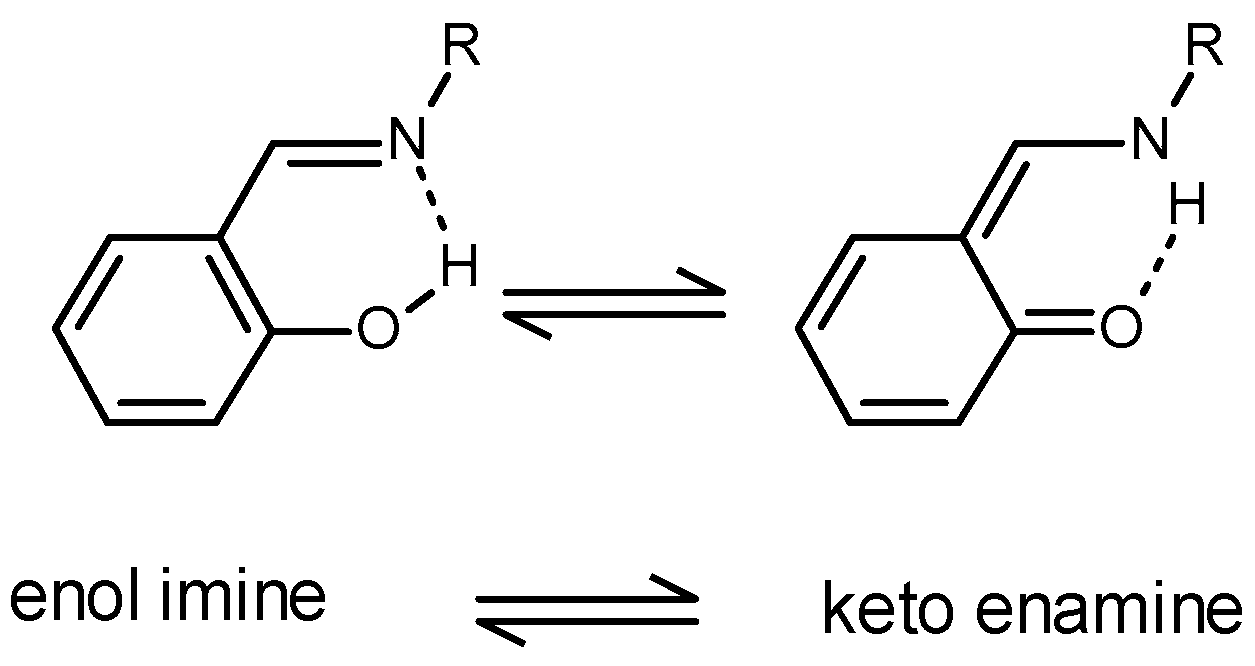
:1. Introduction
2. Results and Discussion
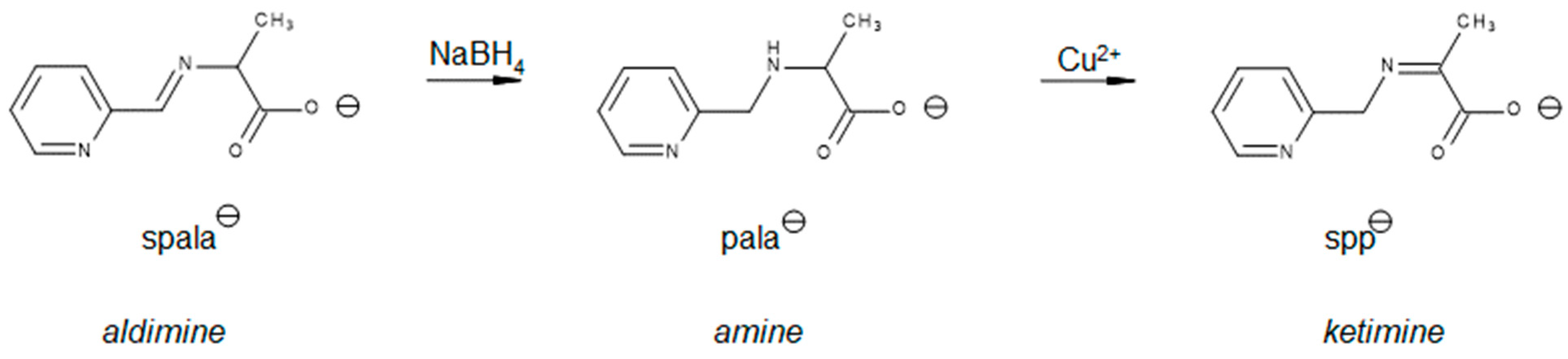
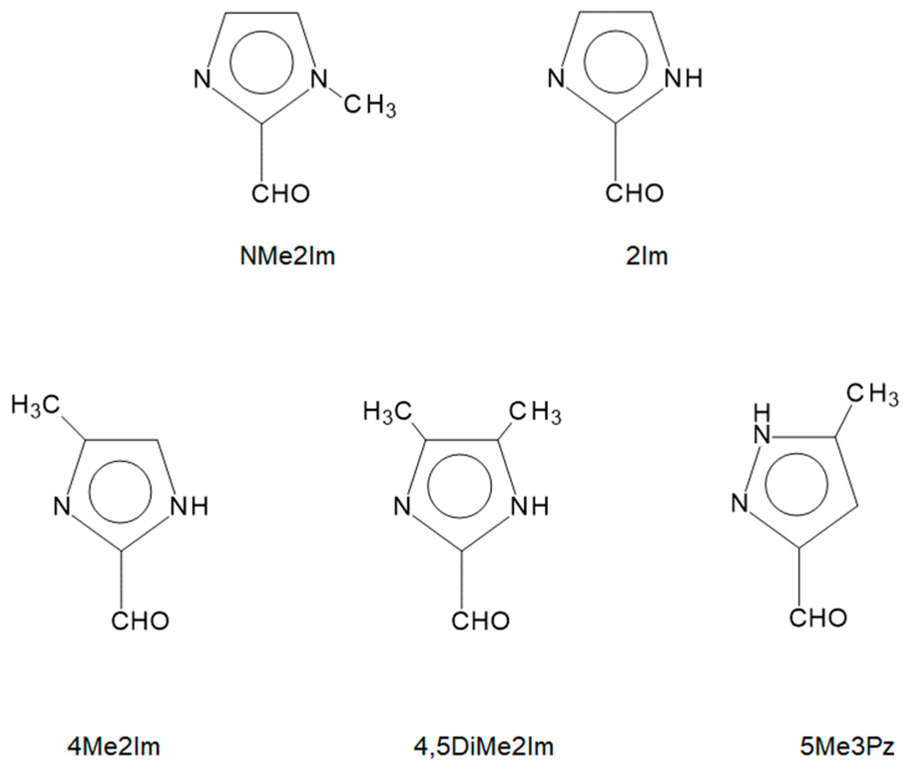
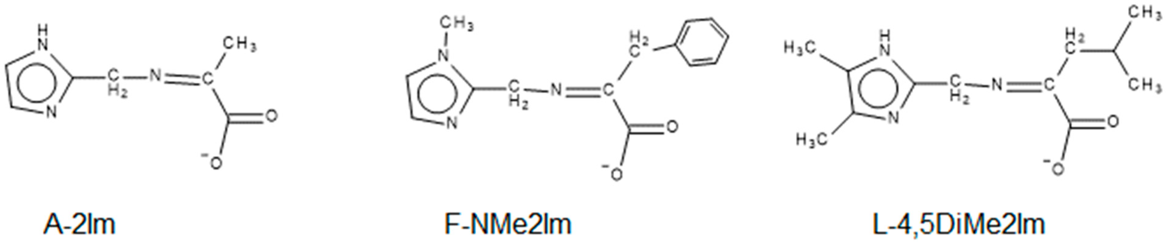
2.1. Choice of Amino Acids and Imidazole Carboxaldehydes
2.2. Synthesis and Preliminary Characterization of Complexes
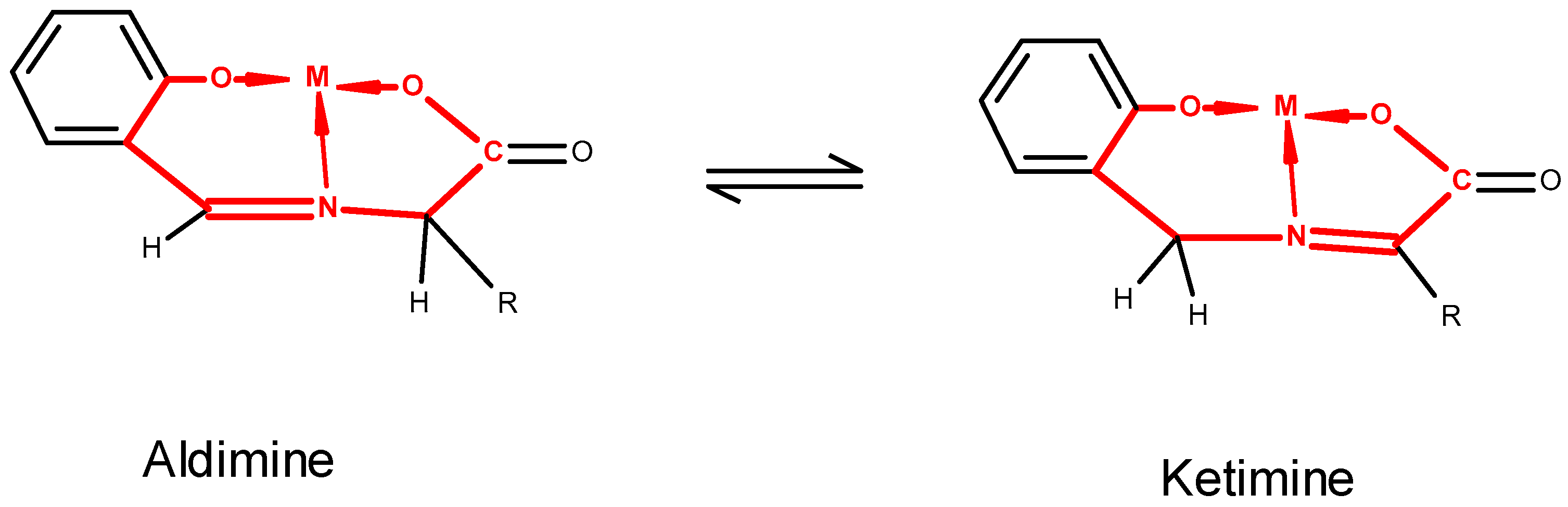
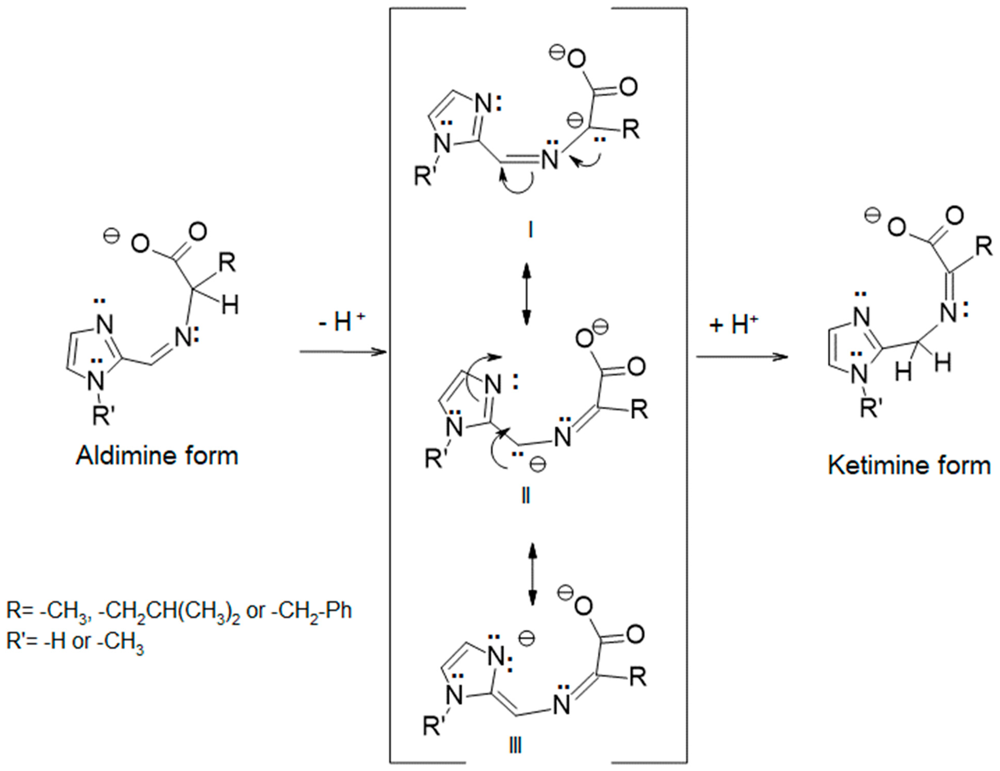
2.3. Background Information on Structure of Tridentate Schiff Bases of AA
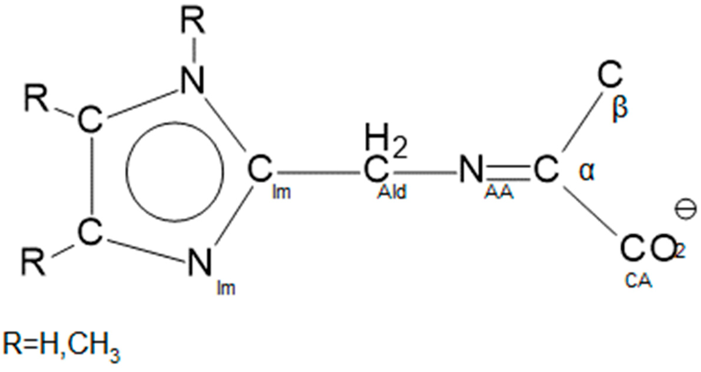
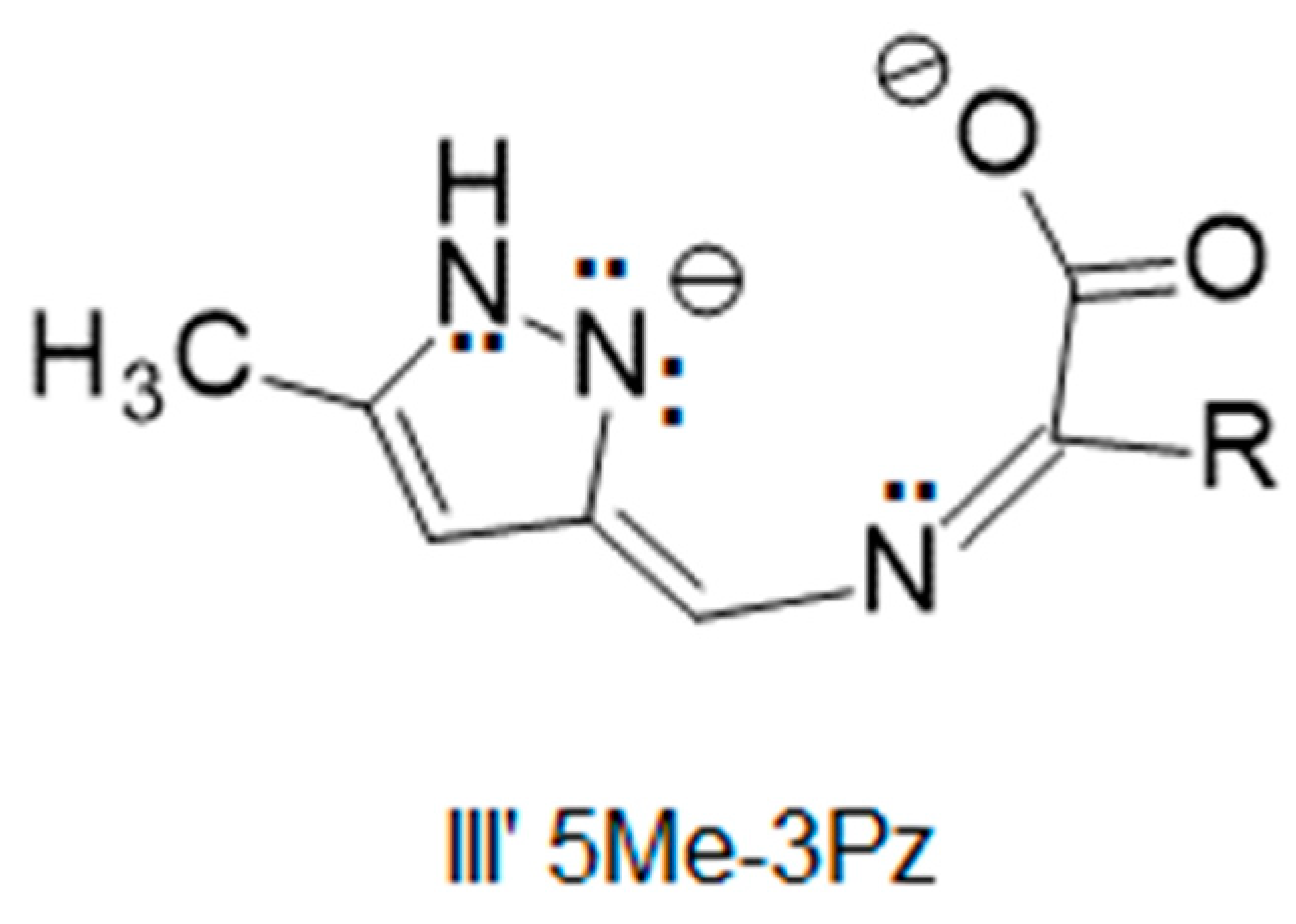
2.4. Organization of Structural Data
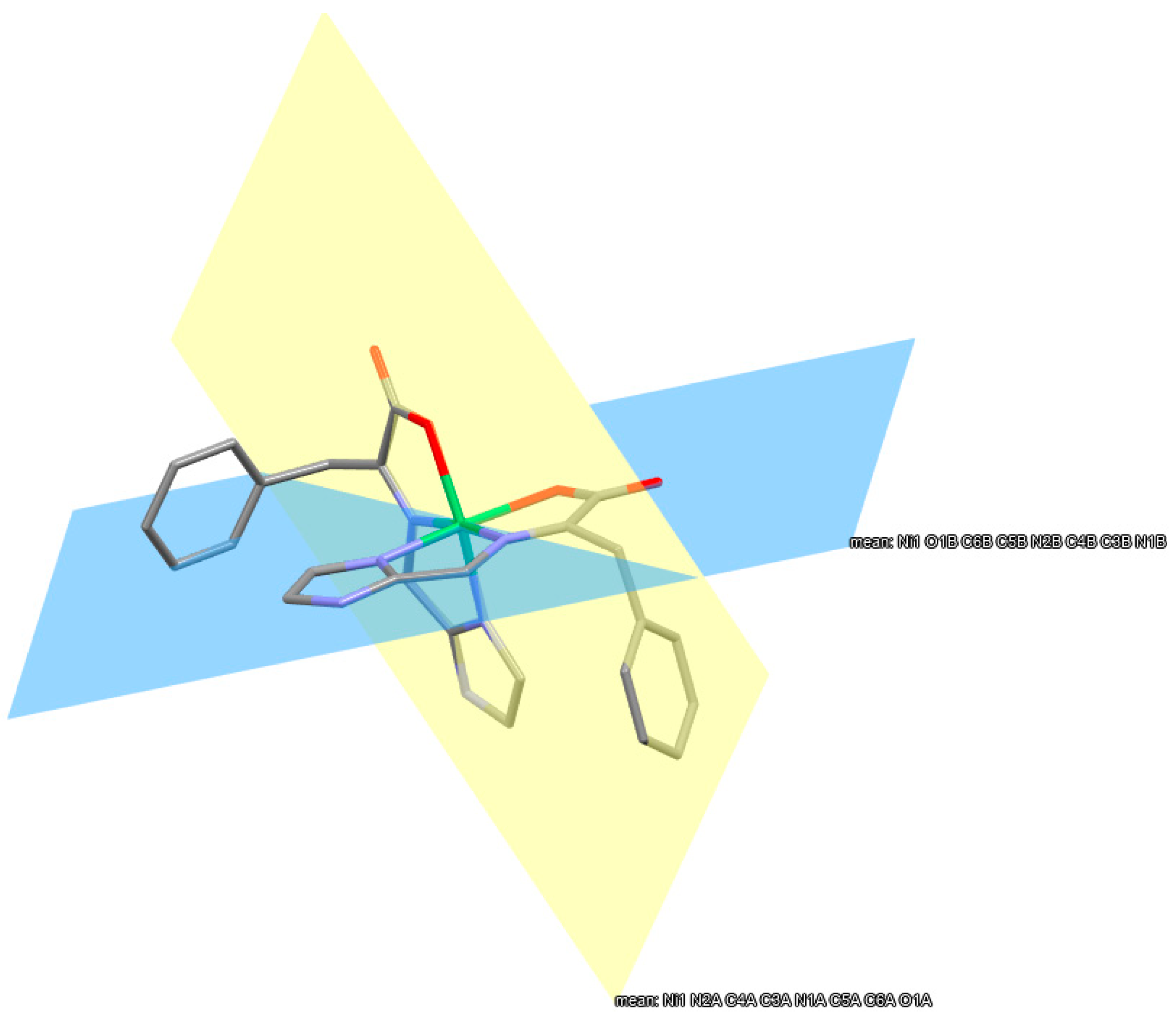
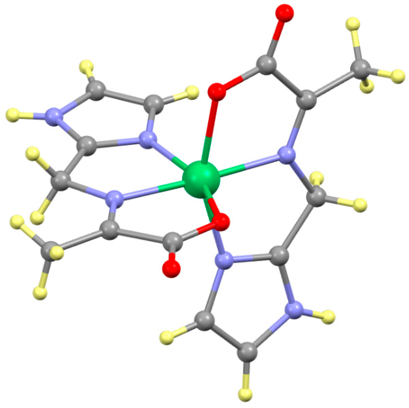
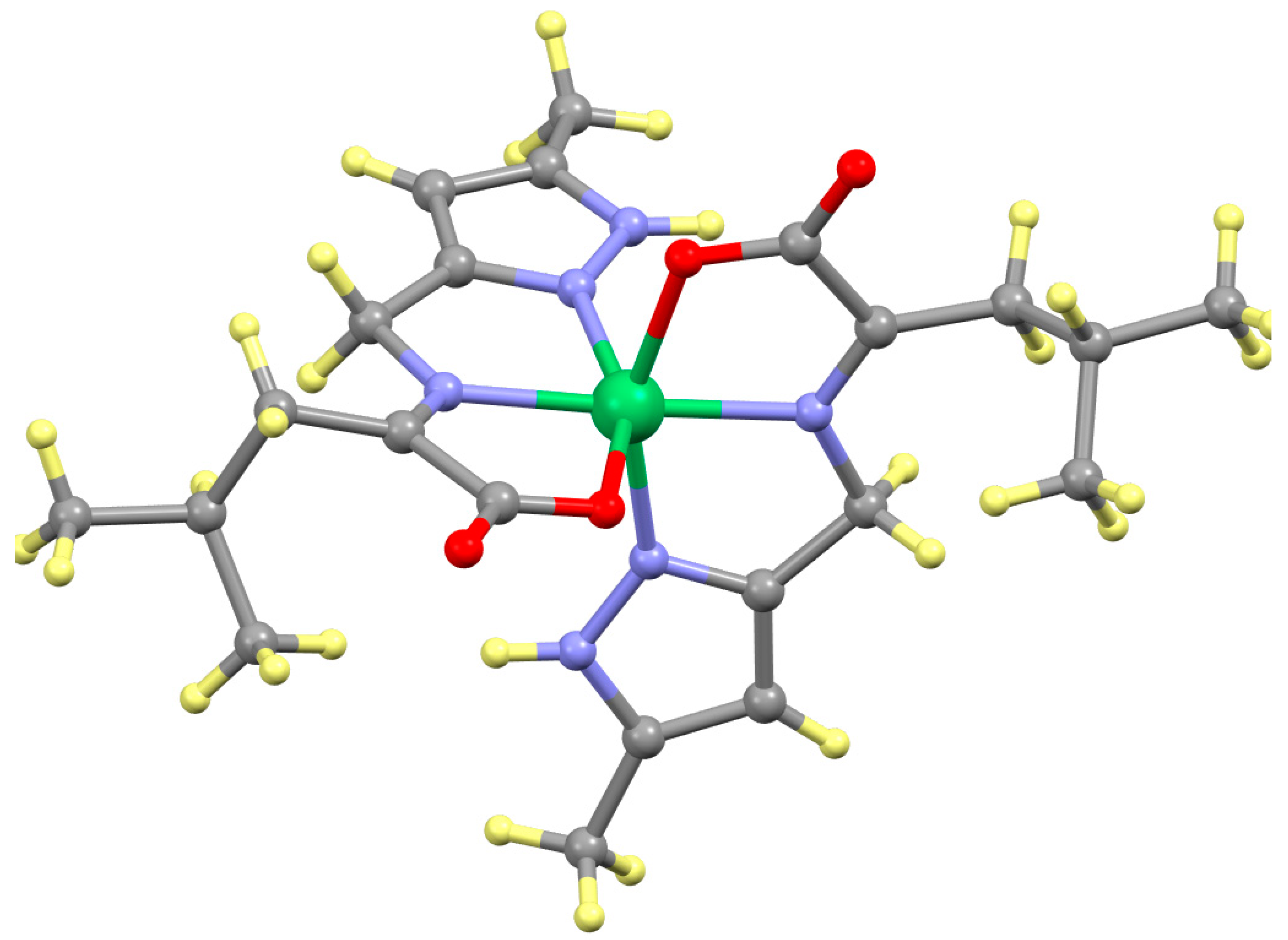
2.5. Structure of the Ni(ketimine)2 Complexes, Table 1

| Compound | Distance (Å) | Value 1 | Value 2 | Angle (°) | Value 1 | Value 2 |
|---|---|---|---|---|---|---|
| Ni-Nim | 2.0782(9) | 2.0870(9) | NAA-Ni-NAA | 177.68(3) | ||
| Ni(A-NMe2Im)2 | Ni-NAA | 2.0259(9) | 2.0212(9) | NIm-Ni-O | 157.05(3) | 157.19(3) |
| Ni-O | 2.0901(8) | 2.1175(8) | ||||
| Ni-Nim | 2.0768(15) | 2.0531(14) | NAA-Ni-NAA | 179.34(6) | ||
| Ni(L-NMe2Im)2 | Ni-NAA | 2.0335(13) | 2.0388(13) | NIm-Ni-O | 157.59(5) | 157.56(5) |
| monoclinic | Ni-O | 2.0977(12) | 2.0838(12) | |||
| Ni-Nim | 2.07(2) | 2.09(2) | NAA-Ni-NAA | 176.3(7) | ||
| Ni(F-NMe2Im)2 | Ni-NAA | 2.085(18) | 1.957(15) | NIm-Ni-O | 155.5(7) | 157.2(7) |
| Ni-O | 2.029(17) | 2.065(18) | ||||
| Ni-Nim | 2.065(2) | 2.069(2) | NAA-Ni-NAA | 177.12(10) | ||
| Ni(A-2Im)2 | Ni-NAA | 2.025(2) | 2.031(2) | NIm-Ni-O | 157.70(8) | 156.57(7) |
| Ni-O | 2.100(2) | 2.1146(19) | ||||
| Ni-Nim | 2.0968(19) | 2.0592(19) | NAA-Ni-NAA | 170.24(8) | ||
| Ni(L-2Im)2 | Ni-NAA | 2.0347(19) | 2.0258(19) | NIm-Ni-O | 157.60(7) | 158.13(7) |
| Ni-O | 2.1011(16) | 2.1153(16) | ||||
| Ni-Nim | 2.093(4) | 2.072(4) | NAA-Ni-NAA | 174.44(16) | ||
| Ni(F-2Im)2 | Ni-NAA | 2.028(4) | 2.028(4) | NIm-Ni-O | 157.05(15) | 158.38(18) |
| Ni-O | 2.107(3) | 2.073(4) | ||||
| Ni-Nim | 2.094(4) | 2.099(5) | NAA-Ni-NAA | 174.8(2) | ||
| Ni(L-4Me2Im)2 | Ni-NAA | 2.043(5) | 2.091(6) | NIm-Ni-O | 157.12(18) | 156.74(19) |
| Ni-O | 2.094(4) | 2.099(5) | ||||
| Ni-Nim | 2.077(13) | 2.085(14) | NAA-Ni-NAA | 178.43(5) | ||
| Ni(A-4,5DiMe2Im)2 | Ni-NAA | 2.0210(13) | 2.0248(13) | NIm-Ni-O | 157.63(5) | 156.75(5) |
| Ni-O | 2.1301(12) | 2.1193(12) | ||||
| Ni-Nim | 2.0947(19) | 2.0822(19) | NAA-Ni-NAA | 174.80(8) | ||
| Ni(L-4,5DiMe2Im)2 | Ni-NAA | 2.043(2) | 2.0251(18) | NIm-Ni-O | 154.92(2) | 156.54(8) |
| Ni-O | 2.097(9) | 2.1207(16) | ||||
| Ni-Nim | 2.082(2) | 2.060(2) | NAA-Ni-NAA | 176.41(8) | ||
| Ni(L-5Me3Pz)2 | Ni-NAA | 2.029(2) | 2.028(2) | NIm-Ni-O | 156.58(9) | 156.53(8) |
| Ni-O | 2.076(2) | 2.1000(18) | ||||
2.6. Structure of the Ketimine Ligand, Table 2
| Compound | Distance (Å) | Value 1 | Value 2 | Angle (°) | Value 1 | Value 2 |
|---|---|---|---|---|---|---|
| Cα-NAA | 1.2805(13) | 1.2796(14) | NAA-Cα-Cβ | 127.16(10) | 126.27(9) | |
| Ni(A-NMe2Im)2 | Cα-Cβ | 1.4857(15) | 1.4824(15) | NAA-Cα-CCA | 113.00(9) | 112.86(9) |
| Cα-CCA | 1.5357(14) | 1.5390(14) | Cβ -Cα-CCA | 119.84(9) | 120.86(9) | |
| Cα-NAA | 1.281(2) | 1.280(8) | NAA-Cα-Cβ | 126.61(15) | 126.7(10) | |
| Ni(L-NMe2Im)2 | Cα-Cβ | 1.494(2) | 1.511(7) | NAA-Cα-CCA | 112.91(14) | 112.4(5) |
| monoclinic | Cα-CCA | 1.546(2) | 1.549(3) | Cβ -Cα-CCA | 120.48(14) | 119.9(11) |
| Cα-NAA | 1.255(16) | 1.259(16) | NAA-Cα-Cβ | 127.1(18) | 124.6(17) | |
| Ni(F-NMe2Im)2 | Cα-Cβ | 1.50(3) | 1.54(3) | NAA-Cα-CCA | 113.0(15) | 113.8(14) |
| Cα-CCA | 1.537(19) | 1.535(19) | Cβ -Cα-CCA | 119.8(17) | 121.4(16) | |
| Cα-NAA | 1.269(4) | 1.283(4) | NAA-Cα-Cβ | 126.9(3) | 126.4(2) | |
| Ni(A-2Im)2 | Cα-Cβ | 1.486(4) | 1.476(4) | NAA-Cα-CCA | 113.0(2) | 113.2(2) |
| Cα-CCA | 1.534(5) | 1.541(3) | Cβ -Cα-CCA | 120.0(3) | 120.5(2) | |
| Cα-NAA | 1.272(3) | 1.278(3) | NAA-Cα-Cβ | 126.9(2) | 125.7(4) | |
| Ni(L-2Im)2 | Cα-Cβ | 1.490(3) | 1.485(9) | NAA-Cα-CCA | 114.5(2) | 113.8(2) |
| Cα-CCA | 1.546(3) | 1.540(3) | Cβ -Cα-CCA | 118.6(2) | 120.4(4) | |
| Cα-NAA | 1.304(7) | 1.285(8) | NAA-Cα-Cβ | 121.0(10) | 129.7(10) | |
| Ni(F-2Im)2 | Cα-Cβ | 1.43(4) | 1.48(2) | NAA-Cα-CCA | 112.4(5) | 113.4(5) |
| Cα-CCA | 1.537(7) | 1.50(2) | Cβ -Cα-CCA | 125.7(9) | 114.9(9) | |
| Cα-NAA | 1.251(8) | 1.267(8) | NAA-Cα-Cβ | 126.1(10) | 125.9(1) | |
| Ni(L-4Me2Im)2 | Cα-Cβ | 1.530(10) | 1.48(2) | NAA-Cα-CCA | 113.9(6) | 112.5(6) |
| Cα-CCA | 1.537(9) | 1.551(9) | Cβ -Cα-CCA | 119.5(10) | 121.6(12) | |
| Cα-NAA | 1.277(2) | 1.272(2) | NAA-Cα-Cβ | 125.76(15) | 126.35(16) | |
| Ni(A-4,5DiMe2Im)2 | Cα-Cβ | 1.492(2) | 1.492(2) | NAA-Cα-CCA | 113.35(14) | 113.99(14) |
| Cα-CCA | 1.535(2) | 1.535(2) | Cβ -Cα-CCA | 120.88(14) | 119.95(15) | |
| Cα-NAA | 1.176(9) | 1.294(3) | NAA-Cα-Cβ | 128.6(7) | 125.1(2) | |
| Ni(L-4,5DiMe2Im)2 | Cα-Cβ | 1.520(9) | 1.487(4) | NAA-Cα-CCA | 112.4(6) | 112.6(2) |
| Cα-CCA | 1.542(8) | 1.534(3) | Cβ -Cα-CCA | 118.8(7) | 122.3(2) | |
| Cα-NAA | 1.261(4) | 1.279(3) | NAA-Cα-Cβ | 126.0(3) | 127.0(2) | |
| Ni(L-5Me3Pz)2 | Cα-Cβ | 1.505(4) | 1.494(4) | NAA-Cα-CCA | 113.9(3) | 112.9(2) |
| Cα-CCA | 1.534(4) | 1.530(4) | Cβ -Cα-CCA | 120.1(3) | 120.1(2) |
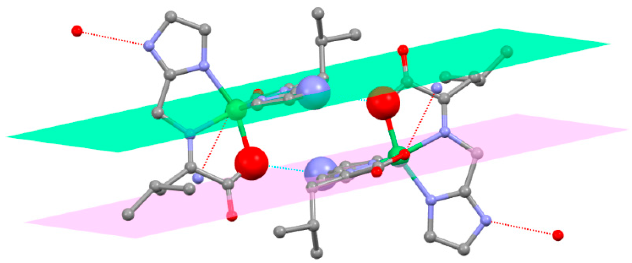
2.7. Structure of Triclinic Ni(L-NMe2Im)2, Table 3
| Distance | Aldimine | Ketimine | Angle | Aldimine | Ketimine |
|---|---|---|---|---|---|
| CAld-CIm | 1.478(19) | 1.491(7) | CIm CAld-NAA | 114.6(12) | 104.3(4) |
| CAld-NAA | 1.34(2) | 1.465(8) | CIm CAld-H | 122.2 | 110.9 |
| CAld-H | 0.9500 | 0.9900 | CIm CAld-H′ | xxxxxx | 110.9 |
| CAld-H | xxxxxx | 0.9900 | NAA-CAld-H | 122.2 | 110.9 |
| NAA-CAld-H′ | xxxxxx | 110.9 | |||
| H-CAld-H′ | xxxxxx | 108.9 | |||
| NAA-CAld | 1.34(2) | 1.465(8) | Ni-NAA-CAld | 116.7(10) | 116.1(3) |
| NAA-Cα | 1.375(13) | 1.280(5) | Ni-NAA-Cα | 123.6(9) | 115.8(3) |
| NAA-Ni | 1.921(11) | 2.077(4) | Cα-NAA-CAld | 114.1(11) | 127.1(3) |
| Cα-NAA | 1.375(13) | 1.280(5) | NAA-Cα-Cβ | 118.8(6) | 121.5(3) |
| Cα-Cβ | 1.4905(13) | 1.4905(13) | NAA-Cα-CCA | 104.3(7) | 114.4(2) |
| Cα-CCA | 1.690(13) | 1.5341(13) | NAA-Cα-H | 108.7 | xxxxxxx |
| Cα-H | 1.000 | xxxxxx | Cβ-Cα-CCA | 106.2(5) | 122.8(3) |
| Cβ-Cα-H | 108.7 | xxxxxx | |||
| CCA-Cα-H | 108.7 | xxxxxx |
2.8. Isomeric Justification of Outcome of Aldimine to Ketimine Tautomerism
2.9. Structure of Ni(L-5Me3Pz)2
2.10. Reactivity of Valine and Isoleucine
3. Experimental Methods
3.1. General
3.2. ESI-MS Were Obtained by Axis Pharm Laboratory, San Diego, CA
3.3. X-ray Crystallography
3.4. Synthesis
3.5. Available Data
4. Conclusions
Supplementary Materials
Author Contributions
Funding
Data Availability Statement
Conflicts of Interest
References
- Sunatsuki, Y.; Ikuta, Y.; Matsumoto, N.; Ohta, H.; Kojima, M.; Iijima, S.; Hayami, S.; Maeda, Y.; Kaizaki, S.; Dahan, F.; et al. An Unprecedented Homochiral Mixed-Valence Spin-Crossover Compound. Angew. Chem. Int. Ed. 2003, 42, 1614–1618. [Google Scholar] [CrossRef] [PubMed]
- Ikuta, Y.; Ooidemizu, M.; Yamahata, Y.; Yamada, M.; Osa, S.; Matsumoto, N.; Iijima, S.; Sunatsuki, Y.; Kojima, M.; Dahan, F.; et al. A New Family of Spin Crossover Complexes with a Tripod Ligand Containing Three Imidazoles: Synthesis, Characterization, and Magnetic Properties of [FeIIH3LMe](NO3)2·1.5H2O, [FeIIILMe]·3.5H2O, [FeIIH3LMe][FeIILMe]NO3, and [FeIIH3LMe][FeIIILMe](NO3)2 (H3LMe = Tris [2-(((2-methylimidazol-4-yll)methylidene)amino)ethyl]amine). Inorg. Chem. 2003, 42, 7001–7017. [Google Scholar] [PubMed]
- Yamada, M.; Ooidemizu, M.; Ikuta, Y.; Osa, S.; Matsumoto, N.; Iijima, S.; Kojima, M.; Dahan, F.; Tuchagues, J.P. Interlayer Interaction of Two-Dimensional Layered Spin Crossover Complexes [FeIIH3LMe][FeIILMe]X (X− = ClO4−, BF4−, PF6−, AsF6−, and SbF6−; H3LMe = Tris [2-(((2-methylimidazol-4-yl)methylidene)amino)ethyl]amine). Inorg. Chem. 2003, 42, 8406–8416. [Google Scholar] [CrossRef] [PubMed]
- Sunatsuki, Y.; Ohta, H.; Kojima, M.; Ikuta, Y.; Goto, Y.; Matsumoto, N.; Iijima, S.; Akashi, H.; Kaizaki, S.; Dahan, F.; et al. Supramolecular Spin-Crossover Iron Complexes Based on Imidazole−Imidazolate Hydrogen Bond. Inorg. Chem. 2004, 43, 4154–4171. [Google Scholar] [CrossRef]
- Brewer, G. Structural Evidence of Spin State Selection and Spin Crossover Behavior of Tripodal Schiff Base Complexes of tris(2-aminoethyl)amine and Related Tripodal Amones. Magnetochemistry 2020, 6, 28. [Google Scholar] [CrossRef]
- Shahraki, S. Schiff base compounds as artificial metalloenzymes. Colloids Surf. B Biointerfaces 2022, 218, 112727–112743. [Google Scholar] [CrossRef]
- Bos, J.; Roelfes, G. Artificial metalloenzymes for enantioselective catalysis. Curr. Opin. Chem. Biol. 2014, 19, 135–143. [Google Scholar] [CrossRef]
- Erxleben, A. Transition metal salen complexes in bioinorganic and medicinal chemistry. Inorg. Chim. Acta 2018, 472, 40–57. [Google Scholar] [CrossRef]
- Yoshida, N.; Oshio, H.; Ito, T. Supramolecular motifs in metal complexes of Schiff bases. Part 6. Topology of two types of self-assembly of bis-N,O-bidentate Schiff baseligands by copper(II) ions. J. Chem. Soc. Perkin Trans. 2 2001, 1674–1678. [Google Scholar] [CrossRef]
- Alcazar, J.J.; Geue, N.; Valladares, V.; Canete, A.; Perez, E.G.; Garcia-Rio, L.; Santos, J.G.; Aliaga, M.E. Supramolecular Control of Reactivity toward Hydrolysis of 7-Diethylaminocoumarin Schiff Bases by Cucurbit [7]uril Encapsulation. ACS Omega 2021, 6, 10333–10342. [Google Scholar] [CrossRef]
- Hulushe, S.T.; Malan, F.P.; Hosten, E.C.; Lobb, K.A.; Khanye, S.D.; Watkins, G.M. Photo-and thermoresponsive N-salicylideneaniline derivatives: Solid-state studies and structural aspect. New J. Chem. 2022, 46, 20940–20950. [Google Scholar] [CrossRef]
- Martinez, R.F.; Avalos, M.; Babiano, R.; Cintas, P.; Jimenez, J.L.; Light, M.E.; Palacios, J.C. Tautomerism in Schiff bases. The cases of 2-hydroxy-1-naphthaldehyde and 1-hydroxy-2-naphthaldehyde investigated in solution and the solid state. J. Org. Biomol. Chem. 2011, 9, 8268–8275. [Google Scholar] [CrossRef] [PubMed]
- Gallant, A.J.; Yun, M.; Sauer, M.; Yeung, C.S.; MacLachlan, M.J. Tautomerization in naphthalenediimines: A keto-enamine Schiff base macrocycle. Org. Lett. 2005, 7, 4827–4830. [Google Scholar] [CrossRef] [PubMed]
- Perona, A.; Sanz, D.; Claramunt, R.M.; Elguero, J. NMR studies of novel Schiff bases derived from L-α-amino methyl esters and 3-hydroxypyridin-4-carboxaldehyde. J. Magn. Reson. Chem. 2008, 46, 930–938. [Google Scholar] [CrossRef] [PubMed]
- Dudek, G.O.; Dudek, E.P. Spectroscopic Studies of Keto Enol Equilibria. IX. N15 Substituded Anilides. J. Am. Chem. Soc. 1966, 88, 2407. [Google Scholar] [CrossRef]
- Herscovitch, R.; Charette, J.J.; De Hoffmann, E. Physicochemical properties of Schiff Bases. II. Tautomeric equilibrium of substituted N-Salicylidene-2-aminopropanes and p-hydroxy-N-benzylidene-2-aminopropane. J. Am. Chem. Soc. 1973, 95, 5135–5140. [Google Scholar] [CrossRef]
- Metzler, C.M.; Cahill, A.; Metzler, D.E. Equilibriums and absorption spectra of Schiff bases. J. Am. Chem. Soc. 1980, 102, 6075–6082. [Google Scholar] [CrossRef]
- Heinert, D.D.; Martell, A.E. Pyridoxine and Pyridoxal Analogs. VI. Electronic Absorption Spectra of Schiff Bases. J. Am. Chem. Soc. 1963, 85, 183–188. [Google Scholar] [CrossRef]
- Blagus, A.; Cincic, D.; Friscic, T.; Kaitner, B.; Stilinovic, V. Schiff bases derived from hydroxyaryl aldehydes: Molecular and crystal structure, tautomerism, quinoid effect, coordination compounds. Maced. J. Chem. Chem. Eng. 2010, 29, 117–138. [Google Scholar] [CrossRef]
- Gasnow, O.A.; Holm, R.H. A proton resonance investigation of equilibria, solute structures, and transamination in the aqueous systems pyridoxamine-pyruvate-zinc(II) and –aluminum(III). J. Am. Chem. Soc. 1969, 91, 5984–5993. [Google Scholar]
- Viedma, C.; Ortiz, J.E.; de Torres, T.; Izumi, T.; Blackmond, D.G. Evolution of solid phase homochirality for a proteinogenic amino acid. J. Am. Chem. Soc. 2008, 130, 15274–15275. [Google Scholar] [CrossRef]
- Klussman, M.; Iwamura, H.; Mathew, S.P.; Wells, D.H., Jr.; Pandya, U.; Armstrong, A.; Blackmond, D.G. Thermodynamic control, of asymmetric amplification in amino acid catalysis. Nature 2006, 441, 621–623. [Google Scholar] [CrossRef]
- Hein, J.E.; Blackmond, D.G. On the origin of single chirality of amino acids and sugars in biogenesis. Acc. Chem. Res. 2012, 45, 2045–2054. [Google Scholar] [CrossRef] [PubMed]
- Weinstein, G.N.; Holm, R.H. Synthesis of Tautomeric Schiff Base Complexes and Ketimine Aldimine Conversion Rates of Copper(II) Complexes. Inorg. Chem. 1972, 11, 2553–2556. [Google Scholar] [CrossRef]
- Weinstein, G.N.; O’Connor, M.J.; Holm, R.H. Preparation, Properties, and Racemization Kinetics of Copper(II)-Schiff Base-Amino Acid Complexes Related to Vitamin B6 Catalysis. Inorg. Chem. 1970, 9, 2104–2112. [Google Scholar] [CrossRef]
- Yamada, S.; Hongo, C.; Yoshioka, R.; Chibata, I. Method for the Racemization of Optically Active Amino Acids. J. Org. Chem. 1983, 48, 843–846. [Google Scholar] [CrossRef]
- Metzler, D.; Snell, E. Some Transamination Reactions Involving Vitamin B6. J. Am. Chem. Soc. 1952, 74, 979–983. [Google Scholar] [CrossRef]
- Longenecker, J.; Snell, E. The Comparative Activities of Metal Ions in Promoting Pyridoxal-catalyzed Reactions of Amino Acids1. J. Am. Chem. Soc. 1957, 79, 142–145. [Google Scholar] [CrossRef]
- Jung, M.J. Enzyme Inhibition by Amino Acids and their Derivatives. In Chemistry and Biochemistry of the Amino Acids; Barrett, G.C., Ed.; Chapman and Hall Ltd.: London, UK, 1985; pp. 228–234. [Google Scholar]
- Voet, D.; Voet, J.G.; Pratt, C.W. Amino Acid Metabolism. In Fundamentals of Biochemistry; John Wiley and Sons: New York, NY, USA, 2002; pp. 616–619. [Google Scholar]
- Eichhorn, G.; Dawes, J. The Metal Complexes of Vitamin B6 and Schiff’s Base Derivatives. J. Am. Chem. Soc. 1954, 76, 5663–5667. [Google Scholar] [CrossRef]
- Matsushima, Y.; Martell, A. Pyridoxal Analogs. X. Zinc(II)-Chelate Catalysis of Transamination in Methanol Solution. J. Am. Chem. Soc. 1967, 89, 1331–1335. [Google Scholar] [CrossRef]
- Zabinski, R.F.; Toney, M.D. Metal Ion Inhibition of Nonenzymatic Pyridoxal Phosphate Catalyzed Decarboxylation and Transamination. J. Am. Chem. Soc. 2001, 123, 193–198. [Google Scholar] [CrossRef] [PubMed]
- Li, X.; Liu, T.; Hu, B.; Li, G.; Zhang, H.; Cao, R. Homochiral Supramolecular Compounds Constructed from Amino Acid Derivatives: Syntheses, Structures, Chiroptical, and Photoluminescence Properties. Cryst. Growth Des. 2010, 10, 3051–3059. [Google Scholar] [CrossRef]
- Brewer, G.; Brewer, C.; Butcher, R.J.; Zemba, M. Structural evidence of a ketimine as the product of an amino acid with an aldehyde. An intermediate in the racemization and transamination of amino acids. Inorg. Chem. Commun. 2016, 64, 35–38. [Google Scholar] [CrossRef]
- Iihoshi, T.; Sato, T.; Towatari, M.; Matsumoto, N.; Kojima, M. Enantioselective Assembly Structures of Copper(II) and Zinc(II) Complexes with the 1:1 Condensation Products of Imidazole-4-carbaldehyde Derivatives and DL-Phenylalanine. Bull. Chem. Soc. Jpn. 2009, 82, 458–466. [Google Scholar] [CrossRef]
- Cruz, C.; Gonzalez, C.; Rubio, F.; Erices, J.; Wrighton-Araneda, K.; Cortés-Arriagada, D.; Venegas-Yazigi, D.; Audebrand, N.; Paredes-García, V. Chiral 1D Metal–Organic Materials Based on Cu(II) and Amino Acid Schiff Bases. Cryst. Growth Des. 2022, 22, 237–250. [Google Scholar] [CrossRef]
- Shintoya, S.; Yamauchi, S.; Oishi, T.; Matsumoto, N.; Fujiami, T. Assembly structures of 1:2 nickel(II) complex of 5-methylimidazol-4-yl-methylidene-l-phenylalanine and 1:2 nickel(II) complex of its racemic ligand. Inorg. Chim. Acta 2013, 406, 59–64. [Google Scholar] [CrossRef]
- Brewer, G.; Butcher, R.J.; Zemba, M. Experimental Crystal Structure Determination CCDC 961032, HOVRAM01; CSD Communication: Cambridge, UK, 2018. [Google Scholar] [CrossRef]
- Yamouchi, S.; Fujinami, T.; Matsumoto, N.; Mochida, N.; Ishida, T.; Sunatski, Y.; Watanabe, M.; Tsuchimoto, M.; Coletti, C.; Re, N. Synthesis, Structure, Luminescence, and Magnetic Properties of a Single-Ion Magnet“mer”-[Tris(N-[(imidazol-4-yl)-methylidene]-DL-phenylalaninato)terbium(III) and Related“fac”-DL-Alaninato Derivative. Inorg. Chem. 2014, 53, 5961–5971. [Google Scholar] [CrossRef]
- Hamada, D.; Fujinami, T.; Yamauchi, S.; Matsumoto, N.; Mochida, N.; Ishida, T.; Sunatski, Y.; Watanabe, M.; Tsuchimoto, M.; Coletti, C.; et al. Luminescent DyIII single ion magnets with same N6O3 donor atoms but different donor atom arrangements, ‘fac’-[DyIII(HLDL-ala)3]·8H2O and ‘mer’-[DyIII(HLDL-phe)3]·7H2O. Polyhedron 2016, 109, 120–128. [Google Scholar] [CrossRef]
- Sakar, S.; Biswas, S.; Liao, M.S.; Kar, T.; Aydogu, Y.; Dagdelen, F.; Mostafa, G.; Chattopadhyay, A.P.; Yap, G.P.A.; Xie, R.H.; et al. An attempt towards coordination supramolecularity from Mn(II), Ni(II) and Cd(II) with a new hexadentate [N4O2] symmetrical Schiff base ligand: Syntheses, crystal structures, electrical conductivity and optical properties. Polyhedron 2008, 27, 3359–3370. [Google Scholar] [CrossRef]
- Felber, T.; Schafer, T.; Herrmann, H. Five-Membered Heterocycles as Potential Photosensitizers in the Tropospheric Aqueous Phase: Photophysical Properties of Imidazole-2-carboxaldehyde, 2-Furaldehyde, and 2-acetylfuran. J. Phys. Chem. A 2020, 124, 10029–10039. [Google Scholar] [CrossRef]
- Lin, X.; Huang, M.; Lu, T.; Zhao, W.; Hu, C.; Gu, X.; Zhang, W. Characterization of Imidazole Compounds in Aqueous Secondary Organic Aerosol Generated from Evaporation of Droplets Containing Pyruvaldehyde and Inorganic Ammonium. Atmosphere 2022, 12, 970. [Google Scholar] [CrossRef]
- Brewer, G.; Brewer, C.; Butcher, R.J.; Robichaux, G.T.; Viragh, C. Correlation of nitrogen chirality, R or S, with metal chelate chirality, Δ or Λ, in a series of reduced tripodal Schiff base complexes. A route to total spontaneous resolution. Inorg. Chim. Acta 2014, 410, 171–177. [Google Scholar] [CrossRef]
- Yang, Y.; Hansen, L.; Baldi, A. Suppression of simultaneous Fmoc-His-(Trt)-OH racemization and Nα-DIC endcapping in solid phase peptide synthesis through design of experiments and its implication for an amino acid activation strategy in peptide synthesis. Org. Process Res. Dev. 2022, 26, 2464–2474. [Google Scholar] [CrossRef]
- Zhou, Y.; Li, H.; Huang, Y.; Li, J.; Deng, G.; Chen, G.; Xi, Z.; Zhou, C. Suppression of alpha-carbon racemization in peptide synthesis based on a thiol-labile amino protecting group. Nat. Commun. 2023, 14, 5324–5334. [Google Scholar] [CrossRef] [PubMed]
- Ohishi, T.; Hashibe, T.; Takahashi, S.; Hagiwara, H.; Matsumoto, N.; Sunatsuki, Y. Enantioselective assembling into tetra- and octanuclear structures by deprotonation of copper(II) complexes of N-[(5-methylimidazol-4-yl)methylidene]-dl-phenylalanine and its l-form ligand. Polyhedron 2012, 33, 209–217. [Google Scholar] [CrossRef]
- Schwarzer, S.; Bohme, U.; Fels, S.; Gunther, B.; Brendler, E. (S)-N-[(2-hydroxynaphthalen-1-yl)methylidene]valine—A valuable ligand for the preparation of chiral complexes. Inorg. Chim. Acta 2018, 483, 136–147. [Google Scholar] [CrossRef]
- Capasso, S.; Giordano, F.; Mattia, C.; Mazzarella, L.; Ripamonti, A. Stereochemistry of model compounds for pyridoxal-catalysed reactions. Crystal structures of the hydrated complexes bis(pyridoxylidene-DL-valinato)nickel(II) and bis(pyridoxylidene-L-valinato)zinc(II). J. Chem. Soc. Dalton Trans. 1974, 2228–2233. [Google Scholar] [CrossRef]
- Zhang, Z.; Li, F.R.; Sun, C.; Huang, M.; Zhang, H. Synthesis, crystal structures, antibacterial activities, and fluorescence properties of Schiff base ligand and Its nickel(II) complex. Russ. J. Coord. Chem. 2014, 40, 432–438. [Google Scholar] [CrossRef]
- Brewer, G.; Brewer, C.; Butcher, R.J. Catholic University of America: Washington, DC, USA, 2023; manuscript in preparation.
- Wanders, R.J.A.; Duran, M.; Loupatty, F.J. Enzymology of the branched-chain amino acid oxidation disorders: The valine pathway. J. Inherit. Metab. Dis. 2012, 35, 5–12. [Google Scholar] [CrossRef]
- Sheldrick, G.M. Phase annealing in SHELXL—90: Direct methods for larger molecules. Acta Crystallogr. 1990, 46, 467–473. [Google Scholar] [CrossRef]
- Sheldrick, G.M.; Sheldrick, G.M.; Sheldrick, G.M. SHELXL-97: FORTRAN Program for Crystal Structure Refinement; ScienceOpen, Inc.: Burlington, MA, USA, 1997. [Google Scholar]
- Sheldrick, G.M. SHELXL-97: FORTRAN Program for Crystal Structure Analysis; Göttingen University, Instit. Fr Anorganisheche Chemie der Universitat, Tammanstrasse 4, D-3400: Göttingen, Germany, 1998. [Google Scholar]


Disclaimer/Publisher’s Note: The statements, opinions and data contained in all publications are solely those of the individual author(s) and contributor(s) and not of MDPI and/or the editor(s). MDPI and/or the editor(s) disclaim responsibility for any injury to people or property resulting from any ideas, methods, instructions or products referred to in the content. |
© 2023 by the authors. Licensee MDPI, Basel, Switzerland. This article is an open access article distributed under the terms and conditions of the Creative Commons Attribution (CC BY) license (https://creativecommons.org/licenses/by/4.0/).
Share and Cite
Brewer, G.; Brewer, C.; Butcher, R.J.; Zavalij, P. Formation of Ketimines from Aldimines in Schiff Base Condensation of Amino Acids and Imidazole-2-Carboxaldehydes: Tautomerization of Schiff Bases of Amino Acids Resulting in the Loss of Stereogenic Center. Inorganics 2023, 11, 381. https://doi.org/10.3390/inorganics11100381
Brewer G, Brewer C, Butcher RJ, Zavalij P. Formation of Ketimines from Aldimines in Schiff Base Condensation of Amino Acids and Imidazole-2-Carboxaldehydes: Tautomerization of Schiff Bases of Amino Acids Resulting in the Loss of Stereogenic Center. Inorganics. 2023; 11(10):381. https://doi.org/10.3390/inorganics11100381
Chicago/Turabian StyleBrewer, Greg, Cynthia Brewer, Raymond J. Butcher, and Peter Zavalij. 2023. "Formation of Ketimines from Aldimines in Schiff Base Condensation of Amino Acids and Imidazole-2-Carboxaldehydes: Tautomerization of Schiff Bases of Amino Acids Resulting in the Loss of Stereogenic Center" Inorganics 11, no. 10: 381. https://doi.org/10.3390/inorganics11100381
APA StyleBrewer, G., Brewer, C., Butcher, R. J., & Zavalij, P. (2023). Formation of Ketimines from Aldimines in Schiff Base Condensation of Amino Acids and Imidazole-2-Carboxaldehydes: Tautomerization of Schiff Bases of Amino Acids Resulting in the Loss of Stereogenic Center. Inorganics, 11(10), 381. https://doi.org/10.3390/inorganics11100381