Abstract
The spin-coating technique was used to produce composite films consisting of PMMA polymer doped with anisometric complexes of Eu(III) and Tb(III). It was found that an increase in the content of Tb3+ complexes intensifies emission of both ions due to the intermolecular energy transfer from the Tb(III) complex to the Eu(III) complex, which results in the increase in the relative luminescence quantum yield of Eu(III) ion by 36%. The temperature sensitivity of the film luminescence intensity and lifetime in the range of 296–363 K was investigated. The maximum relative sensitivity of the films reaches 5.44% × K−1 and exceeds that of all known lanthanide-containing thermal sensors designed for measuring physiological temperatures. In combination with changing luminescence color, such a sensitivity makes these films promising colorimetric thermal sensors for in situ temperature measurements.
1. Introduction
Luminescent thermometry is an emerging technology that can be used in microelectronics, photonics, microfluidics, medicine and biology [1,2,3,4,5]. Compared to other electronic thermal sensor techniques, the advantages of luminescent thermometry are its ability to monitor surface temperatures with micro- and nanometer-scale spatial resolution (<10 μm), high sensitivity (>1% × K−1), fast response (<1 ms) and measurement accuracy, small size of sensitive elements and resistance to electromagnetic interference [2,6]. The working principle of luminescent thermometers is monitoring the effect of temperature on the luminescence parameters: quenching time, intensity, emission peak area or position and the ratio of intensities of emission bands [7]. Several types of luminophores are already used as temperature sensors, including organic compounds, quantum dots, metal clusters, dye-doped nanoparticles, metal-ligand complexes and lanthanide-containing materials [8,9]. Narrow emission bands of lanthanides make it possible to measure temperature changes with high accuracy. In addition to these properties, lanthanides have a long luminescence lifetime and allow for using temporal methods for increasing signal-to-noise ratios [10]. Materials with two different lanthanide ions are attractive components of ratiometric thermometers [11,12]. Such materials are characterized by high accuracy, photostability and temporal resolution of signals. The accuracy of temperature measurements by an absolute emission intensity is sensitive to external factors such as an excitation power and errors in opto-electronic systems. On the other hand, the luminescence intensity ratio of two luminophores is almost constant, so no additional calibration of a thermal probe is needed for such a double system [13,14].
Among lanthanide coordination compounds, β-diketonate complexes are the most promising materials due to their luminescence with high quantum yield (up to 85% [15,16]), large Stokes shifts, decay time of luminescence in the range of tens to hundreds of microseconds and high temperature sensitivity of luminescence intensity (>1% × K−1) [17]. They also possess a set of attractive chemical properties: a relatively easy synthesis, a good solubility in a wide range of common solvents and an ability to be incorporated into various matrices [18,19,20].
In recent years, the focus of studies in this area is shifting more and more from synthesis of coordination lanthanide compounds to their inclusion into various types of matrices (inorganic, organic or organic-inorganic). A matrix is noticed to enhance not only thermal and chemical stability of complexes and their mechanical strength [21,22] but luminescence efficiency as well [23,24]. Two principal approaches are considered in this aspect: encapsulation of a lanthanide complex into polymer [25,26,27] and synthesis of lanthanide-containing polymers [28,29]. Polymers are ideal candidates for matrices as they possess attractive technological properties such as flexibility, processability, mechanical strength and low cost.
However, to control the distribution of temperature over a surface, we need film materials with intensive luminescence that are transparent in the visible range. The majority of currently known lanthanide(III) β-diketonate complexes have a crystalline structure, so it is difficult to use them for producing homogeneous films. It is necessary to use hard UV radiation for their excitation [30], which makes the resulting thermal sensors expensive and reduces their photostability.
In this work, we examined the possibility of making thermosensitive films using PMMA polymer and anisometric Tb(III) and Eu(III) complexes. The presence of long hydrocarbon substituents at the ends of molecules makes such compounds amorphous, which allows them to form transparent media with no crystalline defects. Good solubility of such complexes in organic solvents and miscibility with polymers [31,32] ensure their uniform dispersal in the film material. Additionally, they absorb light effectively in a wide range from 250 to 400 nm, which eliminates the need to use hard UV radiation for their excitation [33,34,35]. In addition to the advantages listed above, the use of multi-ion lanthanide complexes should enhance an intermolecular energy transfer, according to known theoretical prerequisites and experimental data, thus increasing the efficiency of a thermal sensor [36,37].
2. Results and Discussion
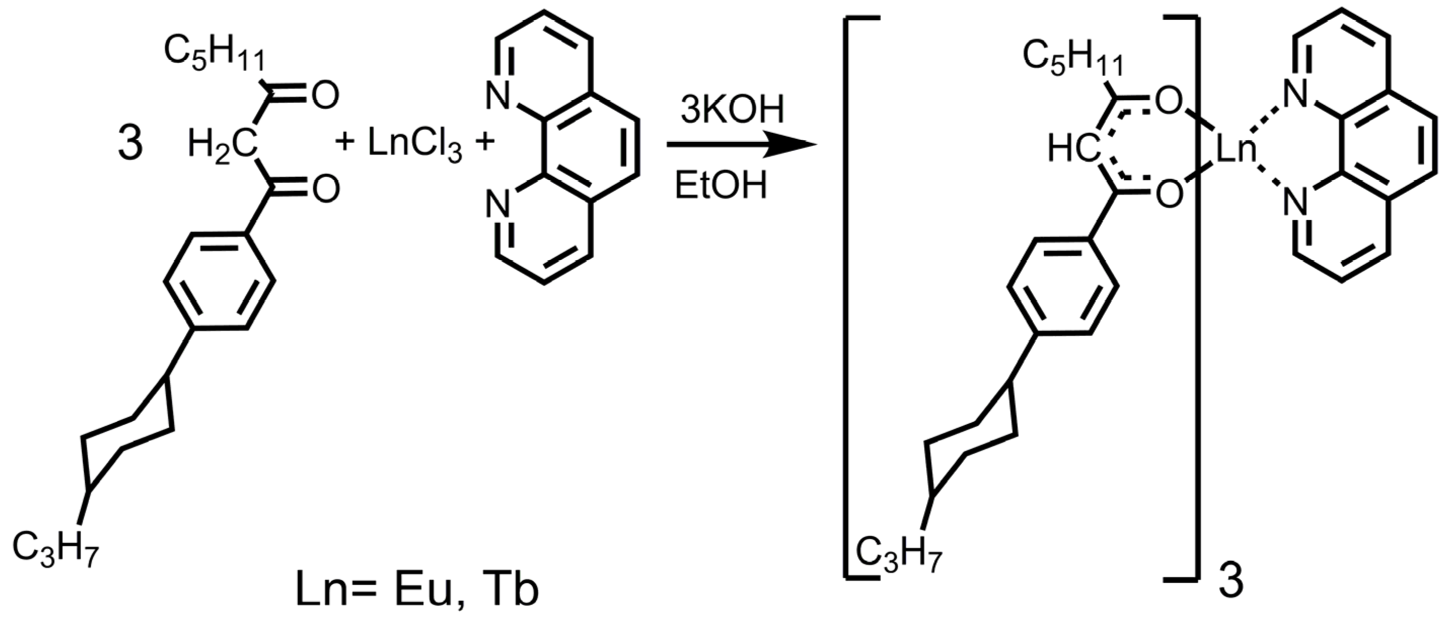
The original 1,10-phenanthroline and β-diketone were used as the ligands for the synthesis of the complexes. The triplet levels of these compounds provide an effective energy transfer to the emitting levels of Eu3+ and Tb3+ ions (Figure 1). Their composition and structure were confirmed by mass spectrometry and elemental analysis.
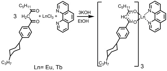
Figure 1.
Synthesis of Ln(III) compounds.
These complexes are amorphous powders that are soluble in organic non-polar and weakly polar solvents. They are mutually miscible as well as miscible with PMMA polymer [31,38].
A spin-coating process was used to produce composite films of PMMA polymer doped with 3 w.% of the Eu(III) compound and 1–20% w.% of the Tb(III) compound (3%EuX%Tb) [39]. The films are 300 nm thick (±10%). The films are almost transparent in the infrared and visible ranges (transmittance over 90%), whereas they absorb light strongly in the UV range (Figure S1).
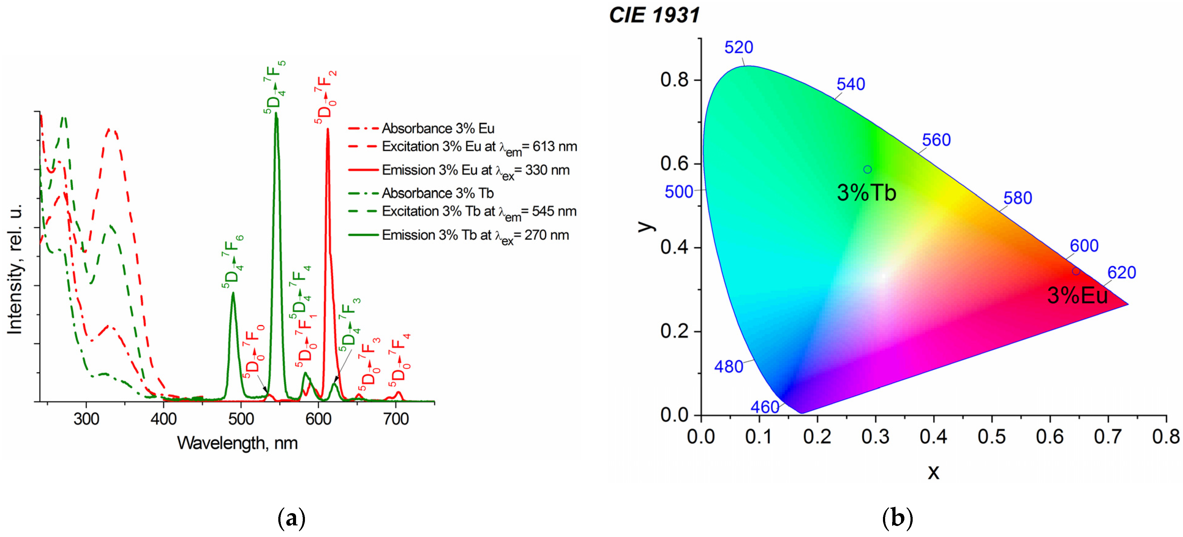
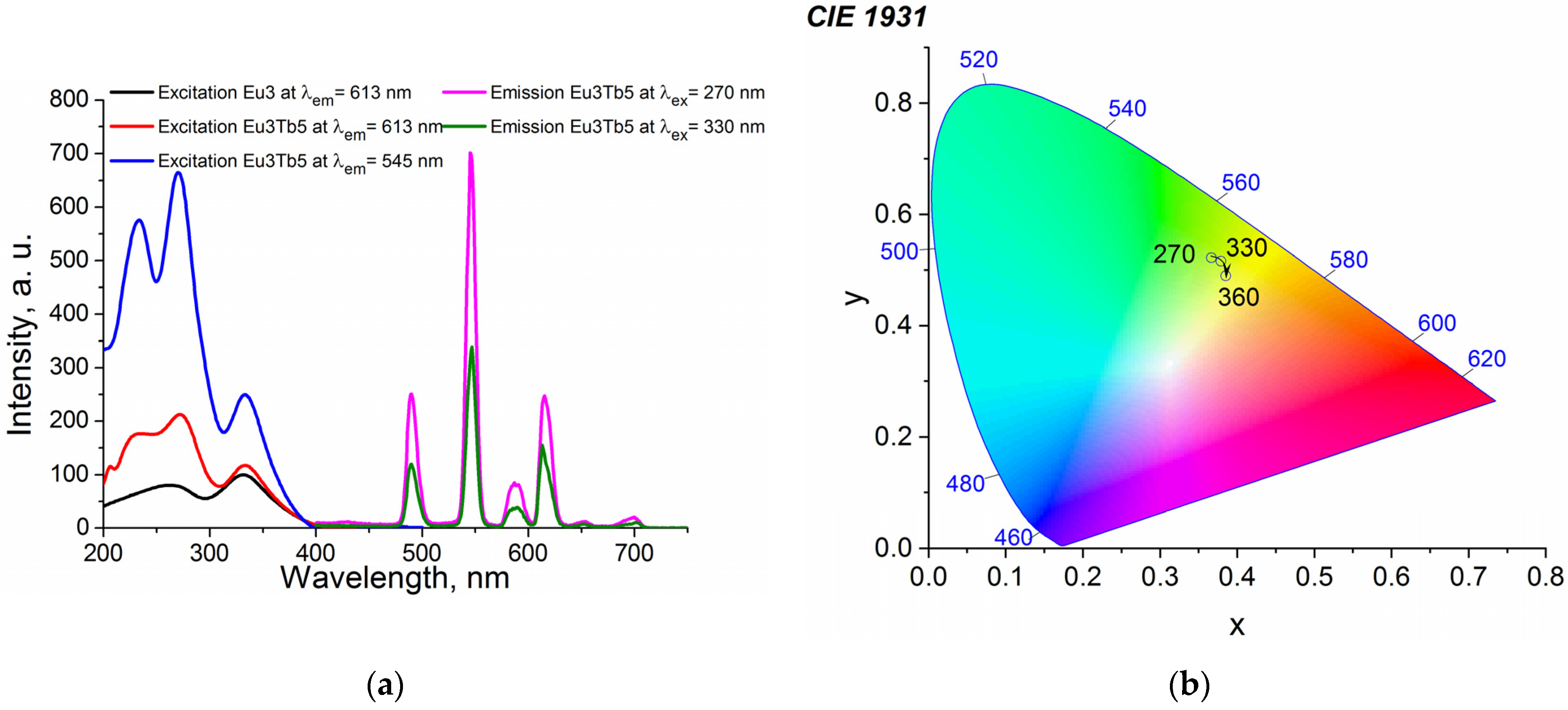
UV irradiation of the PMMA films doped with the individual Tb(III) and Eu(III) ions results in luminescence that is typical for the respective Ln(III) ions (Figure 2).
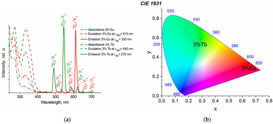
Figure 2.
Luminescence and absorption spectra (a) and the CIE diagram (b) of PMMA films with 3 w.% of Tb(III) and Eu(III) complexes.
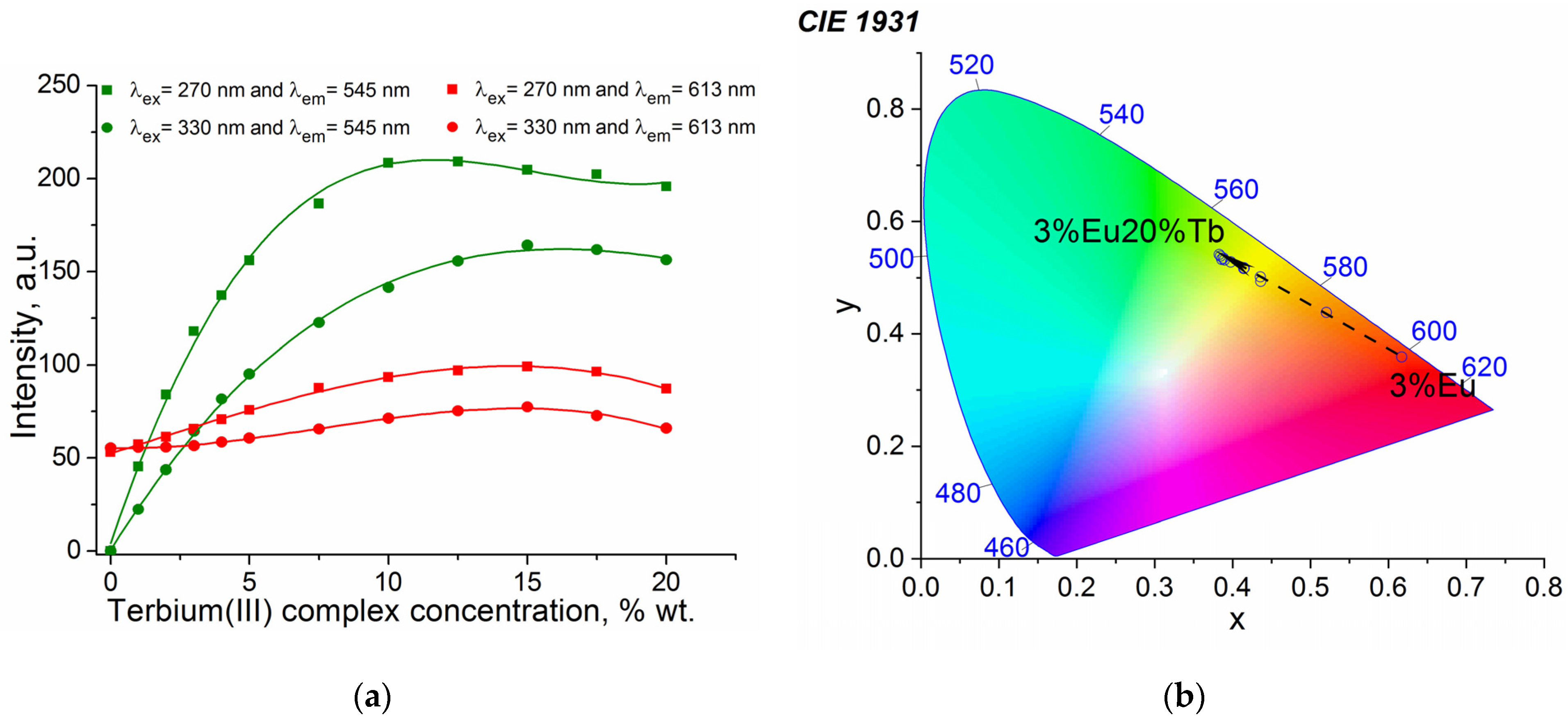
An increase in the content of the Tb(III) complexes in the 3%EuX%Tb films intensifies emission of both the Tb3+ and Eu3+ ions due to occurring intermolecular energy transfer (Figure 3). Because of large distances between ions, the most probable energy transfer mechanism is a dipole–dipole interaction (Förster) rather than an exchange energy transfer (Dexter).

Figure 3.
Concentration dependences of luminescence (a) obtained for the 3%EuX%Tb at λex = 270 and 330 nm and λem = 545 and 613 nm; the CIE diagram (λex = 330 nm) (b).
It was demonstrated that the emission intensity at λem = 613 nm of the 3%Eu15%Tb film increases by 2 times at λex = 270 nm and by 1.4 times at λex = 330 nm, respectively, as compared with the film of the individual Eu(III) compound in PMMA (3%Eu).
The overall luminescence quantum yield of the complex upon excitation of the chromophore is determined by the efficiency of the sensitization (ηsens) and by the emission quantum efficiency (φLn) of the lanthanide luminescence (Equation (1)).
Due to the energy transfer from the Tb(III) complex, the relative quantum yield of the Eu(III) ion luminescence increases by 36% at λex = 270 nm and 26% at λex = 330 nm (Equations (2)–(5)) (Tables S1 and S2). A stronger luminescence of the Eu(III) complex is mostly related to the energy transfer from the ligands (ηsens), whereas the contribution of the quantum efficiency of the Eu3+ ion luminescence itself (φLn) does not show considerable growth. The Tb(III) complex represents an additional “antenna” that provides a direct energy transfer from the Tb(III) coordination compound to the Eu3+ ion. Concentration changes of the Tb(III) complex also lead to changes in the emission color and provide an opportunity to control the color of the films (Figure 3b).
The 3%Eu5%Tb sample was selected to characterize luminescence of the composites at different temperatures. We chose this ratio of luminophores because it provides the optimal color visualization of temperature and almost zero concentration quenching. A relatively low content of luminophores ensures low cost of a resulting thermal sensor. It is known from the literature that luminescence quenching of Tb3+ ions is more strongly dependent on temperature than that of Eu3+ ions [3,40,41]. A simultaneous presence of Eu(III) and Tb(III) complexes in the composite films allows for a more accurate determination of temperature by evaluating the ratio of their luminescence peaks, which is not sensitive to measurement conditions.
At room temperature, the luminescence spectra of the PMMA films doped with both complexes contain the transition bands of the Eu3+ and Tb3+ ions (Figure 4). Due to the energy transfer from the Tb(III) complex to the Eu(III) complex, the excitation spectrum of the 3%Eu5%Tb film contains a more intensive excitation peak at 270 nm (Figure 4) as compared with the spectrum of the PMMA film doped with the individual Eu(III) complex. According to the CIE diagram, changing the wavelength does not lead to a considerably different perception of color (Figure 4b).
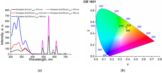
Figure 4.
Luminescence spectra (a) and the CIE diagram (b) of 3%Eu5%Tb at various λex.
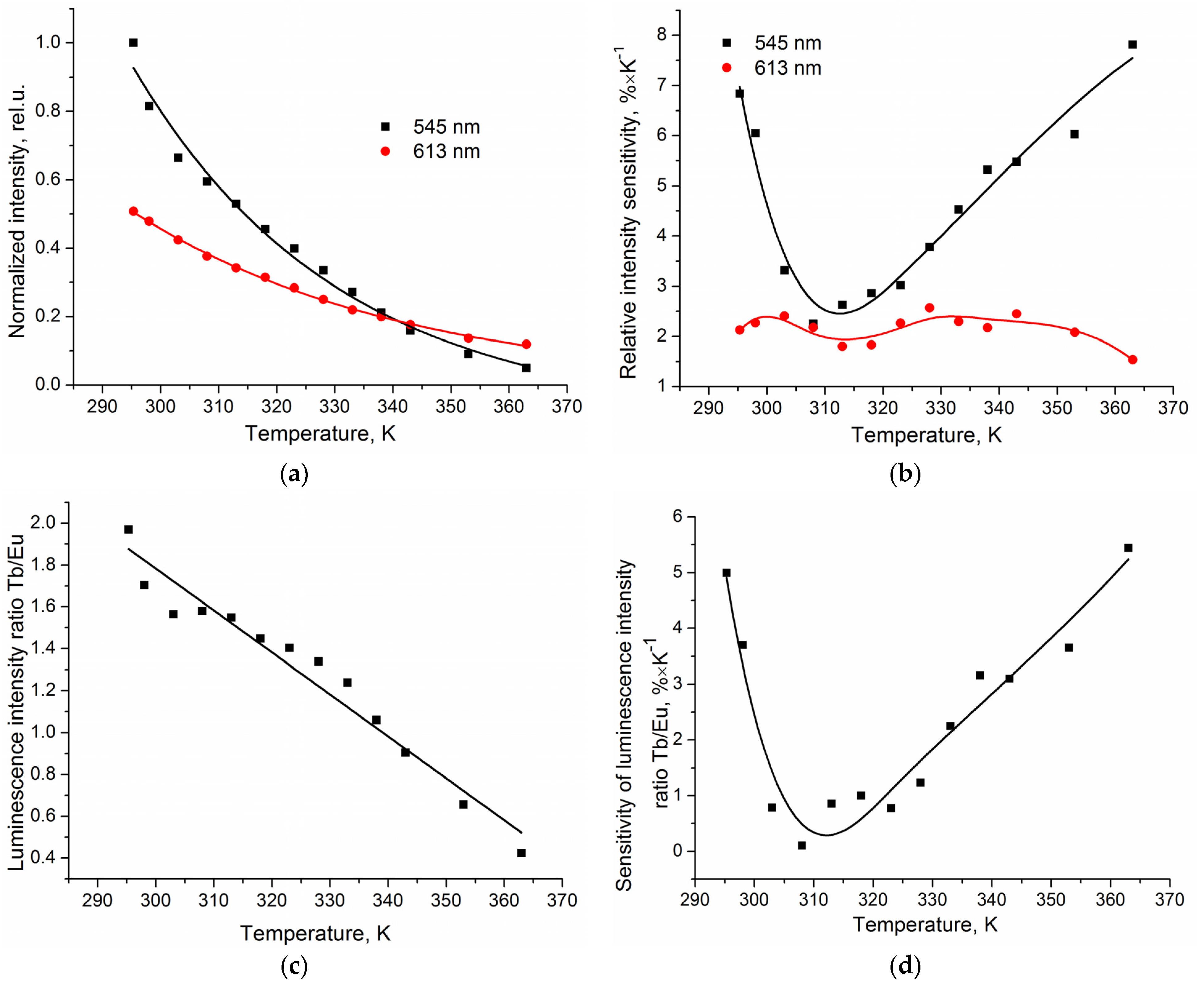
Figure 5a illustrates the influence of temperature on the luminescence intensity of the analyzed film in the range of 296–363 K at λex = 330 nm and λem = 545 and 613 nm (Figure S2 at λex = 270 nm and λem = 545 and 613 nm). Decreasing dependences of emissions from the 5D0 → 7F2 (Eu(III), 613 nm) and 5D4 → 7F5 (Tb(III), 545 nm) transitions are adequately described by the exponential functions with the correlation coefficients R2 > 0.98.
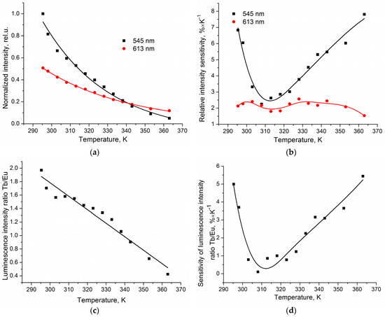
Figure 5.
Temperature dependences of the normalized luminescence intensity (a), relative sensitivity of intensity (b), the ratio of luminescence intensities of the transitions 5D4 → 7F5 (Tb(III), 545 nm) and 5D0 → 7F2 (Eu(III), 613 nm) (c), and the sensitivity of the luminescence intensity ratio of the 3%Eu5%Tb composite film (d) at λex = 330 nm.
The maximum value of the relative sensitivity of intensity (SI) varies from 2.6% × K−1 for the 5D0 → 7F2 transition (Eu(III), 613 nm) to 7.8% × K−1 for the 5D4 → 7F5 transition (Tb(III), 545 nm) (Equation (7)).
Since luminescence of both the Tb3+ and Eu3+ ions has a specific temperature behavior, it is possible to measure temperature by the ratiometric method from the intensity ratio of the 5D4 → 7F5 (Tb(III), 545 nm) and 5D0 → 7F2 (Eu(III), 613 nm) transitions. The ratio of the transition intensities is almost linear (R2 > 0.96) (Figure 5b and Figure S2). Therefore, the ratio of the peaks can be used to determine the surface temperature with high accuracy. The value of the maximum relative sensitivity (Sm) reaches 5.44% × K−1 and exceeds that of all known lanthanide-containing thermal sensors designed for measuring physiological temperatures (Table 1) [1,17].

Table 1.
Comparative temperature relative sensitivity of various Ln3+-based luminescent thermometers.
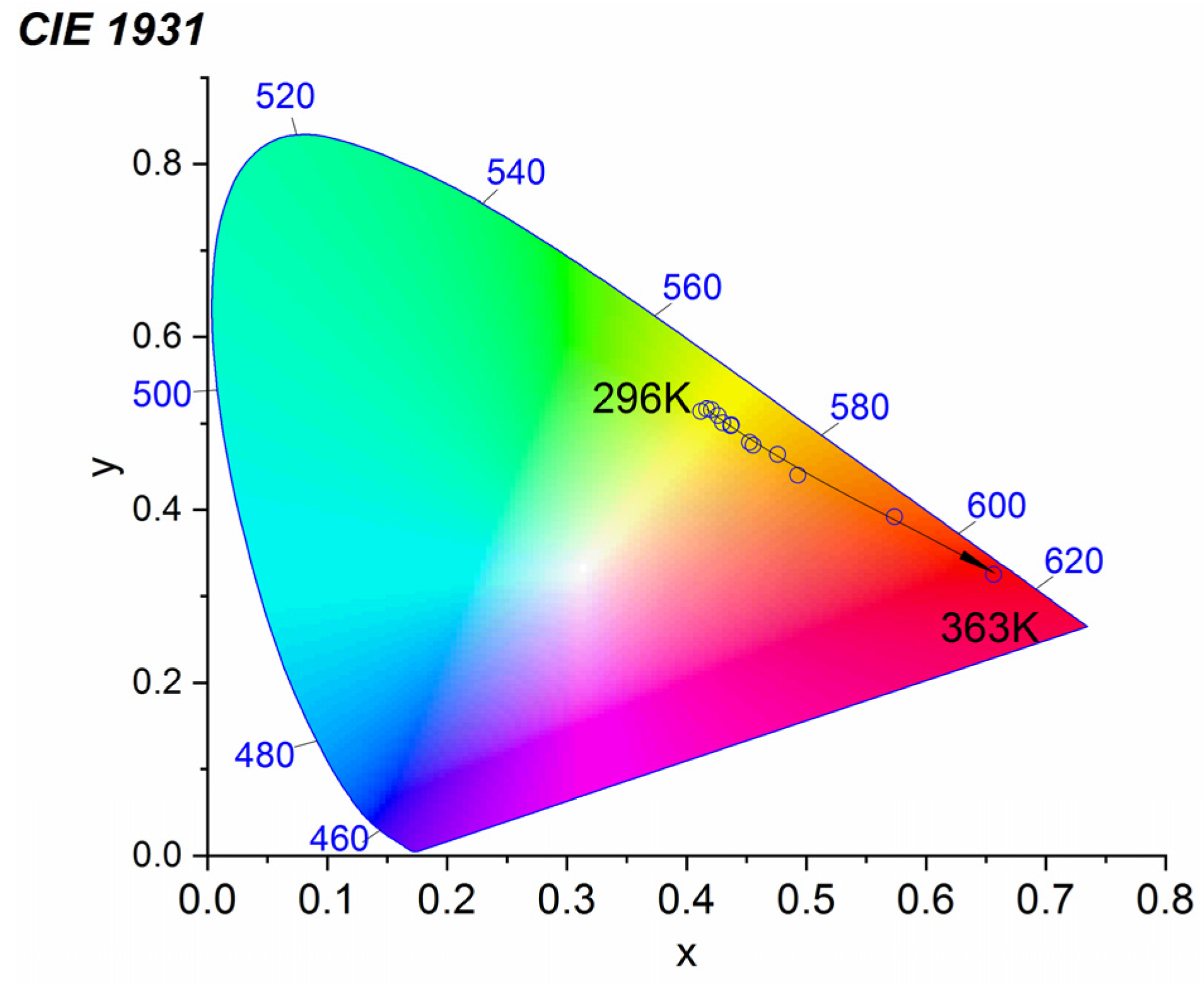
The films demonstrate luminescence color changes in the temperature range of 296–363 K. Therefore, they are promising colorimetric thermometers for in situ temperature measurements (Figure 6 and Figure S3). The coordinates at the CIE diagrams were calculated using the respective emission spectra from 296 K to 363 K. The luminescence color of the film changes from yellow (X = 0.411, Y = 0.514) at 296 K to red (X = 0.656, Y = 0.325) at 363 K (Figure 7 and Figure S3).

Figure 6.
CIE luminescence diagram of the PMMA films doped with 3 w.% of the Eu(III) complex and 5 w.% of the Tb(III) complex (3%Eu5%Tb) at various temperatures, the excitation wavelength λex = 330 nm.
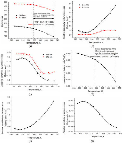
Figure 7.
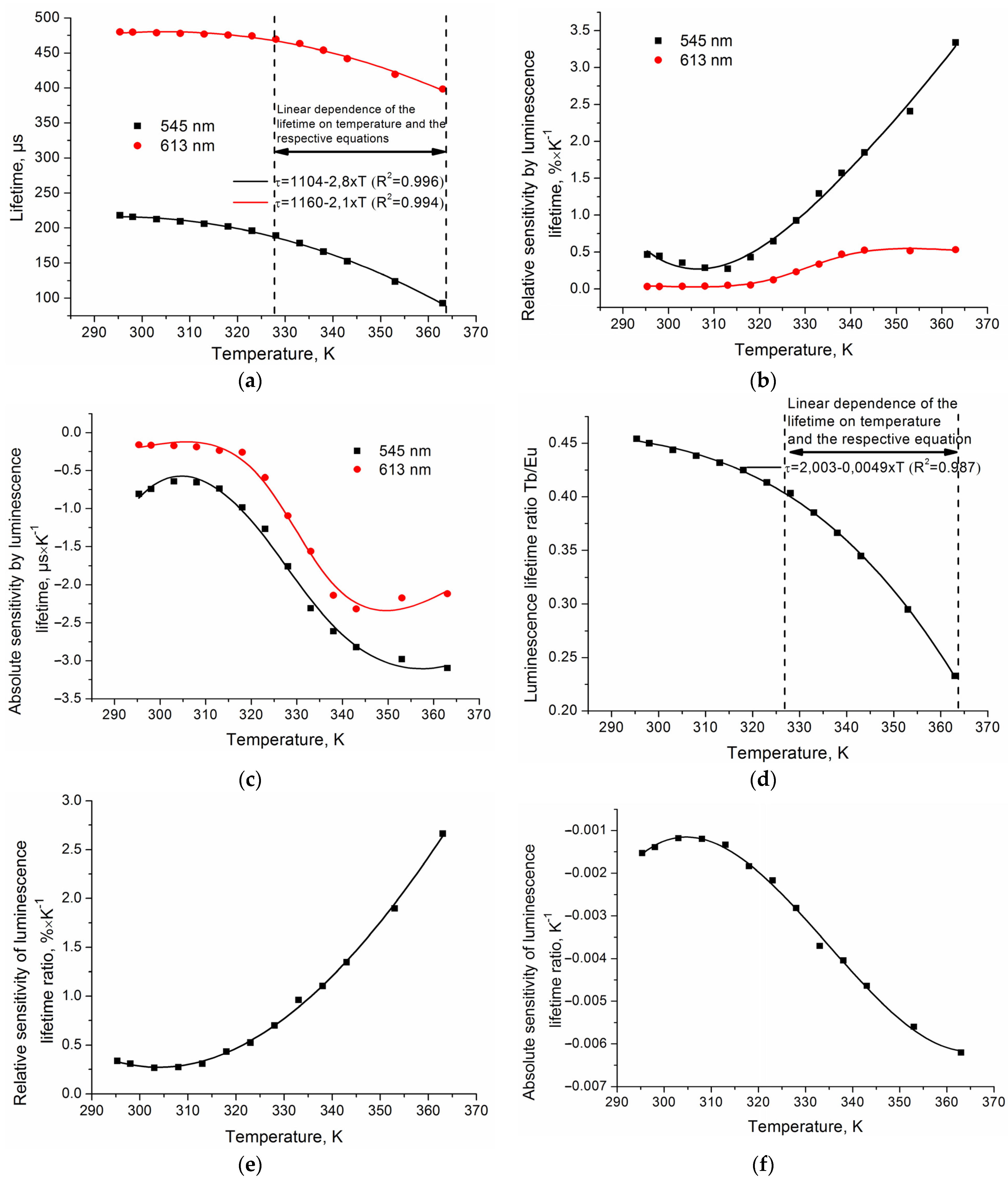
Temperature dependences of the luminescence lifetime (a), relative sensitivity of lifetime (b), absolute sensitivity of lifetime (c), the ratio of lifetimes of the 5D4 → 7F5 (Tb(III), 545 nm) and 5D0 → 7F2 (Eu(III), 613 nm) transitions (d), the relative sensitivity of the lifetime ratio (e), and the absolute sensitivity of the lifetime ratio (f) of the 3%Eu5%Tb composite film at λex = 330 nm.
The luminescence intensity of thermally sensitive films is well-known to depend largely on the characteristics of a sample and measurement conditions [1,2]. Moreover, it is rather difficult to consider the factor of film degradation under UV radiation to avoid a significant error in temperature measurements [55,56,57]. Thermal sensors can also experience changes in their emission intensity when the refractive index of the medium changes (for instance, when water or other liquid is absorbed by the sensor’s surface) or when chemical or biological substances are present. The luminescence quenching time, however, does not depend on the factors mentioned above [1,2]. Therefore, this parameter is often used for more reliable and accurate temperature measurements. Figure 7a illustrates the temperature dependence of the luminescence lifetime demonstrated by the composite film samples.
In the range of 303–363 K, the luminescence lifetime of the samples decreases almost linearly as temperature increases. The slope of the temperature line between 328 K and 363 K is 2.1–2.8 μs/K (Figure 7b). The maximum relative lifetime sensitivity (Equation (7)) reaches 3.3% × K−1. The curves representing the absolute sensitivity of the luminescence lifetime (Equation (6)) have similar profiles and relatively close values of 2.3–3.1 μs × K−1 (Figure 7).
In turn, the ratio of the luminescence lifetimes found for the 5D4 → 7F5 (Tb(III), 545 nm) and 5D0 → 7F2 (Eu(III), 613 nm) transitions is adequately described by a linear function (R2>0.987) in the range of 328-363 K (Figure 7 and Figure S4). Thus, the ratio of the lifetimes of the 5D4 → 7F5 (Tb(III), 545 nm) and 5D0 → 7F2 (Eu(III), 613 nm) transitions allows for a high accuracy determination of a surface temperature by the ratiometric method. The slope of the temperature line representing the ratios in the range of 328–363 K is 0.0049 K−1. The maximum relative lifetime sensitivity reaches 2.67% × K−1. The curves representing the absolute sensitivity of the luminescence lifetime ratio have similar profiles and relatively close values of 0.0062 K−1.
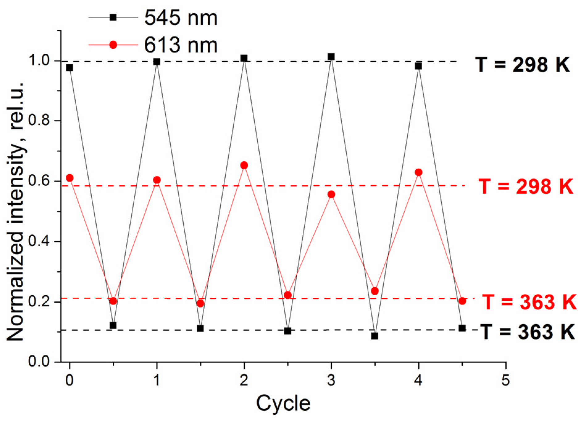
Stability and reversibility of thermosensor readings during heating are critical for its smooth operation. Therefore, the reversibility of intensity readings during cyclic temperature changes was examined. The luminescence intensity of the film is shown to change reversibly in the temperature range of 296–363 K (Figure 8 and Figure S5).

Figure 8.
Reversible changes in the luminescence intensity of the 3%Eu5%Tb composite film at λex = 330 nm during cycles of heating and cooling between 296 and 363 K.
The samples withstand five cycles of heating and cooling under irradiation at 330 nm and demonstrate a relatively small reduction in intensity (less than 2%).
Thus, these composite materials are promising components of luminescent thermometers capable of measuring temperatures in the range of 296–363 K, including the temperature of the human body.
3. Materials and Methods
3.1. Materials
EuCl3*6H2O (99.9%), TbCl3*6H2O (99.9%), 1,10-Phenanthroline (99%), poly(methyl methacrylate) (PMMA) (Mw~120,000 by GPC) and solvents were purchased from Sigma-Aldrich.
3.2. Characterization Techniques
CHN analysis was performed with an isotope mass spectrometer Delta V Plus Thermo Fisher Scientific. X-ray Fluorescence analysis was performed with a M4 «Tornado» Bruker spectrometer. Absorption and transmission spectra were measured with a UV/Vis spectrophotometer Lambda-35 Perkin–Elmer. Luminescence properties were measured with a Cary Eclipse Varian spectrofluorimeter.
3.3. Synthesis of Complexes
The β-diketone (1-[4-(4-propylcyclohexyl)phenyl]-octane-1,3-dione) was prepared according to the method described in [39,58,59,60].
General procedure used for synthesis of the Ln(III) complexes: EtOH solution (2 mL) of LnCl3 × 6H2O (Ln = Tb or Eu) (0.04 mmol) was added dropwise to a stirred hot EtOH solution (10 mL) containing 1,10-Phenanthroline (0.04 mmol), β-diketone (0.12 mmol), and KOH (0.125 mmol). The resulting yellow precipitate was isolated by hot filtration, washed with hot EtOH (25 mL), and dried under vacuum. Further, the product was dissolved in toluene, filtered and the solvent was evaporated. The structural characterization results are shown in Table S3.
3.4. Preparation of PMMA–Ln(III) Films
Spin-coating of films was performed in a spin-coater (WS-650 MZ-23NPP Laurel) by deposition of 0.3 mL solution in toluene on a quartz plate [61].
3.5. Calculation of the Quantum Efficiency and Quantum Yield
To calculate the quantum efficiency of the films doped with the Eu(III) complexes, the following formula was used for Equation (2):
where Arad and Anrad are the radiative and non-radiative rates, respectively.
The contributions to the Anrad include the reverse energy transfer to the ligand [62,63,64], electron transfer quenching (mostly for Eu3+) [64,65], and, most importantly, quenching by matrix oscillations. Luminescence of lanthanides is effectively quenched by O–H oscillations [66,67,68,69]. In addition, other oscillations of organic molecules can also contribute to Anrad [69]. Non-radiative processes influence the observed luminescence lifetime (τobs = (Arad + Anrad)−1). In turn, the lifetime of radiative processes can be defined as (τrad = Arad−1).
Thus, we can calculate ΦLn according to the value of the observed luminescence lifetime τobs (Equation (3)).
In turn, τrad can be calculated by assuming that the energy of the 5D0 → 7F1 transition and its dipole strength are constant. The resulting Equation (4) connects the form of the Eu3+ emission spectrum and its radiation lifetime:
where n is the refraction index of the medium (for a first approximation, we can use the PMMA index value)), AMD,0 is the probability of a spontaneous emission for the 5D0 → 7F1 transition in vacuum and Itot/IMD is the ratio of the total area of the Eu3+ emission to the area of the 5D0 → 7F1 band. For the theoretical value of the dipole strength, it was found that AMD,0 = 14.65 s−1.
The quantum yields of the PMMA–Ln(III) complexes hybrid films were calculated with Equation (5) [70]:
The subscripts st and u indicate the standard and unknown sample, A—corresponds to the absorbance of the films at the excitation wavelength λ and S is the integrated luminescence spectrum. The standard fluorophore for measurements was Eu(tta)3phen with φstd = 53% [70].
3.6. Calculation of the Thermal Sensitivity
The absolute thermal sensitivity (Sa) is calculated as (Equation (6)) [71]:
where Δ is luminescence lifetime or intensity.
The relative thermal sensitivity (Sr) is calculated as (Equation (7)) [71]:
4. Conclusions
A spin-coating method was used to produce composite films of PMMA polymer doped with 3 w.% of the Eu(III) compound and 1–20 w.% of the Tb(III) compound. Addition of the Tb3+ complex increases the emission intensity of both ions due to the intermolecular energy transfer from the Tb(III) compound to the Eu(III) compound, which leads to the 36% growth of the relative luminescence quantum yield of the Eu(III) ion. We investigated the temperature sensitivity of the luminescence intensity and lifetime of the 3%Eu5%Tb films in the 296–363 K temperature range. The maximum relative sensitivity of intensity (SI) varies from 2.6% × K−1 for the 5D0 → 7F2 transition (613 nm) to 7.8% × K−1 for the 5D4 → 7F5 transition (545 nm). The maximum relative sensitivity of these films reaches 5.44% × K−1 and exceeds that of all known lanthanide-containing thermal sensors designed for measuring physiological temperatures. Considering their luminescence color changes, the films demonstrate a significant potential as colorimetric thermal sensors for in situ temperature measurements.
Supplementary Materials
The following supporting information can be downloaded at: https://www.mdpi.com/article/10.3390/inorganics10070094/s1.
Author Contributions
This work is the collaborative development of all the authors. Conceptualization, A.A.K. and Y.G.G.; Methodology, A.A.K. and Y.G.G.; Software, A.S.K.; Validation, A.A.K., A.S.K. and Y.G.G.; Formal Analysis, A.S.K.; Investigation, A.A.K., A.S.K. and Y.G.G.; Resources, Y.G.G.; Data Curation, A.A.K. and Y.G.G.; Writing—Original Draft Preparation, A.A.K. and A.S.K.; Writing—Review and Editing, Y.G.G.; Supervision, A.A.K. and Y.G.G. All authors have read and agreed to the published version of the manuscript.
Funding
This research was funded by the Russian Science Foundation, grant number 18-13-00112.
Acknowledgments
This study was carried out using the equipment of the Center for Collective Use “Nanomaterials and Nanotechnology” of the Kazan National Research Technological University with the financial support of the Ministry of Science and Higher Education of the Russian Federation under agreement No. 075-15-2021- 699.
Conflicts of Interest
The authors declare no conflict of interests.
References
- Wang, X.D.; Wolfbeis, O.S.; Meier, R.J. Luminescent probes and sensors for temperature. Chem. Soc. Rev. 2013, 42, 7834–7869. [Google Scholar] [CrossRef] [PubMed]
- Brites, C.D.S.; Millán, A.; Carlos, L.D. Lanthanides in Luminescent Thermometry. In Handbook on the Physics and Chemistry of Rare Earths; Elsevier: Amsterdam, The Netherlands, 2016; Volume 49, pp. 339–427. [Google Scholar]
- Brites, C.D.S.; Balabhadra, S.; Carlos, L.D. Lanthanide-Based Thermometers: At the Cutting-Edge of Luminescence Thermometry. Adv. Opt. Mater. 2019, 7, 1801239. [Google Scholar] [CrossRef] [Green Version]
- Moßhammer, M.; Brodersen, K.E.; Kühl, M.; Koren, K. Nanoparticle- and microparticle-based luminescence imaging of chemical species and temperature in aquatic systems: A review. Microchim. Acta 2019, 186, 126. [Google Scholar] [CrossRef] [PubMed]
- Hemmer, E.; Acosta-Mora, P.; Méndez-Ramos, J.; Fischer, S. Optical nanoprobes for biomedical applications: Shining a light on upconverting and near-infrared emitting nanoparticles for imaging, thermal sensing, and photodynamic therapy. J. Mater. Chem. B 2017, 5, 4365–4392. [Google Scholar] [CrossRef]
- Brites, C.D.S.; Lima, P.P.; Silva, N.J.O.; Millán, A.; Amaral, V.S.; Palacio, F.; Carlos, L.D. Thermometry at the nanoscale. Nanoscale 2012, 4, 4799–4829. [Google Scholar] [CrossRef] [Green Version]
- Dramićanin, M.D. Sensing temperature via downshifting emissions of lanthanide-doped metal oxides and salts. A review. Methods Appl. Fluoresc. 2016, 4, 042001. [Google Scholar] [CrossRef] [Green Version]
- Qin, T.; Liu, B.; Zhu, K.; Luo, Z.; Huang, Y.; Pan, C.; Wang, L. Organic fluorescent thermometers: Highlights from 2013 to 2017. TrAC—Trends Anal. Chem. 2018, 102, 259–271. [Google Scholar] [CrossRef]
- Hasegawa, Y.; Kitagawa, Y. Thermo-sensitive luminescence of lanthanide complexes, clusters, coordination polymers and metal-organic frameworks with organic photosensitizers. J. Mater. Chem. C 2019, 7, 7494–7511. [Google Scholar] [CrossRef]
- Dai, Z.; Tian, L.; Song, B.; Ye, Z.; Liu, X.; Yuan, J. Ratiometric Time-Gated Luminescence Probe for Hydrogen Sulfide Based on Lanthanide Complexes. Anal. Chem. 2014, 86, 11883–11889. [Google Scholar] [CrossRef]
- Zhou, J.; Xia, Z.; Bettinelli, M.; Liu, Q. Photoluminescence tuning via energy transfer in Eu-doped Ba2(Gd,Tb)2Si4O13 solid-solution phosphors. RSC Adv. 2015, 6, 2046–2054. [Google Scholar] [CrossRef]
- Bao, G.; Wong, K.-L.; Jin, D.; Tanner, P.A. A stoichiometric terbium-europium dyad molecular thermometer: Energy transfer properties. Light Sci. Appl. 2018, 7, 1–10. [Google Scholar] [CrossRef]
- Wang, H.; Zhao, D.; Cui, Y.; Yang, Y.; Qian, G. A Eu/Tb-mixed MOF for luminescent high-temperature sensing. J. Solid State Chem. 2017, 246, 341–345. [Google Scholar] [CrossRef]
- Lu, H.; Meng, R.; Hao, H.; Bai, Y.; Gao, Y.; Song, Y.; Wang, Y.; Zhang, X. Stark sublevels of Er3+–Yb3+ codoped Gd2(WO4)3 phosphor for enhancing the sensitivity of a luminescent thermometer. RSC Adv. 2016, 6, 57667–57671. [Google Scholar] [CrossRef]
- Lima, N.B.D.; Gonçalves, S.M.C.; Júnior, S.A.; Simas, A.M. A Comprehensive Strategy to Boost the Quantum Yield of Luminescence of Europium Complexes. Sci. Rep. 2013, 3, srep02395. [Google Scholar] [CrossRef] [PubMed]
- Malta, O.L.; Brito, H.F.; Menezes, J.F.S.; Gonçalves e Silva, F.R.; De Mello Donegá, C.; Alves, S. Experimental and theoretical emission quantum yield in the compound Eu(thenoyltrifluoroacetonate)3.2(dibenzyl sulfoxide). Chem. Phys. Lett. 1998, 282, 233–238. [Google Scholar] [CrossRef]
- Brites, C.D.S.; Lima, P.P.; Silva, N.J.O.; Millán, A.; Amaral, V.S.; Palacio, F.; Carlos, L.D. Lanthanide-based luminescent mo-lecular thermometers. New J. Chem. 2011, 35, 1177–1183. [Google Scholar] [CrossRef] [Green Version]
- Binnemans, K. Rare-earth beta-diketonates. In Handbook on the Physics and Chemistry of Rare Earths; Elsevier: Amsterdam, The Netherlands, 2005; Volume 35, pp. 107–272. ISBN 9780444520289. [Google Scholar]
- Binnemans, K. Lanthanide-Based Luminescent Hybrid Materials. Chem. Rev. 2009, 109, 4283–4374. [Google Scholar] [CrossRef] [Green Version]
- Felinto Brito, H.; Manoel Loureiro Malta, O.; Claudia França Cunha Felinto, M.; Epaminondas de Sousa Teotonio, E. Lumi-nescence Phenomena Involving Metal Enolates. In PATAI’S Chemistry of Functional Groups; Wiley: Hoboken, NJ, USA, 2010. [Google Scholar]
- De Bettencourt-Dias, A. Lanthanide-based emitting materials in light-emitting diodes. Chem. Rev. 2007, 22, 2229–2241. [Google Scholar] [CrossRef]
- Li, Y.; Bian, Y.; Yan, M.; Thapaliya, P.S.; Johns, D.; Yan, X.; Galipeau, D.; Jiang, J. Mixed (porphyrinato)(phthalocyaninato) rare-earth(III) double-decker complexes for broadband light harvesting organic solar cells. J. Mater. Chem. 2011, 21, 11131–11141. [Google Scholar] [CrossRef]
- Lenaerts, P.; Storms, A.; Mullens, J.; D’Haen, J.; Görller-Walrand, C.; Binnemans, K.; Driesen, K. Thin Films of Highly Luminescent Lanthanide Complexes Covalently Linked to an Organic−Inorganic Hybrid Material via 2-Substituted Imidazo[4,5-f]-1,10-phenanthroline Groups. Chem. Mater. 2005, 17, 5194–5201. [Google Scholar] [CrossRef]
- Li, H.R.; Lin, J.; Zhang, H.J.; Li, H.C.; Fu, L.S.; Meng, Q.G. Novel, covalently bonded hybrid materials of europium (terbium) complexes with silica. Chem. Commun. 2001, 1, 1212–1213. [Google Scholar] [CrossRef]
- He, Y.; Liu, L.; Zhang, Z.; Fu, G.; Lü, X.; Wong, W.-K.; Jones, R.A. A tris-diketonate-Eu(III) complex with the brominated 2,2′-bpy ancillary ligand doped in PMMA for high color-purity red luminescence. Inorg. Chem. Commun. 2016, 64, 13–15. [Google Scholar] [CrossRef]
- George, T.M.; Sajan, M.J.; Gopakumar, N.; Reddy, M.L.P. Bright red luminescence and triboluminescence from PMMA-doped polymer film materials supported by Eu 3+ -triphenylphosphine based β-diketonate and 4,5-bis(diphenylphosphino)-9,9-dimethylxanthene oxide. J. Photochem. Photobiol. A Chem. 2016, 317, 88–99. [Google Scholar] [CrossRef]
- Li, H.; Qi, W.; Sun, H.; Li, P.; Yang, Y.; Wu, L. A novel polymerizable pigment based on surfactant-encapsulated polyoxometalates and their application in polymer coloration. Dye Pigment. 2008, 79, 105–110. [Google Scholar] [CrossRef]
- Zhang, X.; Zhang, Z.; Liu, L.; Yu, C.; Lü, X.; Zhu, X.; Wong, W.-K.; Jones, R.A. Dual-nodal PMMA-supported Eu 3+ -containing metallopolymer with high color-purity red luminescence. Inorg. Chem. Commun. 2015, 60, 51–53. [Google Scholar] [CrossRef]
- Xu, C.-J.; Wan, J.-T.; Li, B.-G. Monochromatic light-emitting copolymer of methyl methacrylate and Eu-complexed 5-acrylamido-1,10-phenanthroline. Dye Pigment. 2013, 98, 493–498. [Google Scholar] [CrossRef]
- Mironov, L.Y.; Evstropiev, S. Temperature-sensitive luminescent photopolymer activated by europium β-diketonate complexes. Opt. Eng. 2019, 58, 027113. [Google Scholar] [CrossRef]
- Knyazev, A.A.; Krupin, A.S.; Molostova, E.Y.; Romanova, K.A.; Galyametdinov, Y.G. Influence of Structural Anisotropy on Mesogenity of Eu(III) Adducts and Optical Properties of Vitrified Films Formed on their Base. Inorg. Chem. 2015, 54, 8987–8993. [Google Scholar] [CrossRef]
- Knyazev, A.A.; Krupin, A.S.; Romanova, K.A.; Galyametdinov, Y.G. Luminescence and energy transfer in poly(N-vinylcarbazole) blends doped by a highly anisometric Eu(III) complex. J. Coord. Chem. 2016, 69, 1473–1483. [Google Scholar] [CrossRef]
- Lapaev, D.V.; Nikiforov, V.G.; Lobkov, V.S.; Knyazev, A.A.; Krupin, A.S.; Galyametdinov, Y.G. New insights into UV laser irradiation effect on luminescent behavior of vitrified films based on mesogenic lanthanide(III) β-diketonate complexes. J. Photochem. Photobiol. A Chem. 2019, 382, 111962. [Google Scholar] [CrossRef]
- Knyazev, A.A.; Karyakin, M.E.; Krupin, A.S.; Romanova, K.A.; Galyametdinov, Y.G. Influence of Eu(III) Complexes Structural Anisotropy on Luminescence of Doped Conjugated Polymer Blends. Inorg. Chem. 2017, 56, 6067–6075. [Google Scholar] [CrossRef] [PubMed]
- Lapaev, D.V.; Nikiforov, V.G.; Knyazev, A.A.; Dzhabarov, V.I.; Lobkov, V.S.; Salikhov, K.M.; Galyametdinov, Y.G. Intramolecular energy transfer in mesogenic europium (III) adduct. Opt. Spectrosc. 2008, 104, 851–857. [Google Scholar] [CrossRef]
- Romanova, K.A.; Freidzon, A.Y.; Bagaturyants, A.A.; Galyametdinov, Y.G. Ab Initio Study of Energy Transfer Pathways in Dinuclear Lanthanide Complex of Europium(III) and Terbium(III) Ions. J. Phys. Chem. A 2014, 118, 11244–11252. [Google Scholar] [CrossRef]
- Knyazev, A.A.; Karyakin, M.E.; Heinrich, B.; Donnio, B.; Galyametdinov, Y.G. A facile approach for the creation of heteroionic lanthanidomesogens-containing uniform films with enhanced luminescence efficiency. Dye Pigment. 2021, 187, 109050. [Google Scholar] [CrossRef]
- Knyazev, A.; Krupin, A.; Gubaidullin, A.; Galyametdinov, Y. Optical and structural characteristics of PMMA films doped with a new anisometric EuIII complex. Acta Crystallogr. Sect. B Struct. Sci. Cryst. Eng. Mater. 2019, 75, 570–577. [Google Scholar] [CrossRef]
- Knyazev, A.A.; Krupin, A.S.; Galyametdinov, Y.G. Luminescence behavior of PMMA films doped with Tb(III) and Eu(III) complexes. J. Lumin. 2021, 242, 118609. [Google Scholar] [CrossRef]
- Liu, J.; Han, X.; Lu, Y.; Wang, S.; Zhao, D.; Li, C. Isostructural Single-and Dual-Lanthanide Metal–Organic Frameworks Based on Substituent-Group-Modifying Tetracarboxylate Ligands for Ratiometric Temperature Sensing. Inorg. Chem. 2021, 60, 4133–4143. [Google Scholar] [CrossRef] [PubMed]
- Rocha, J.; Brites, C.D.S.; Carlos, L.D. Lanthanide Organic Framework Luminescent Thermometers. Chem. Eur. J. 2016, 22, 14782–14795. [Google Scholar] [CrossRef]
- Hatanaka, M.; Hirai, Y.; Kitagawa, Y.; Nakanishi, T.; Hasegawa, Y.; Morokuma, K. Organic linkers control the thermosensitivity of the emission intensities from Tb(iii) and Eu(iii) in a chameleon polymer. Chem. Sci. 2016, 8, 423–429. [Google Scholar] [CrossRef] [Green Version]
- Zhao, D.; Rao, X.; Yu, J.; Cui, Y.; Yang, Y.; Qian, G. Design and Synthesis of an MOF Thermometer with High Sensitivity in the Physiological Temperature Range. Inorg. Chem. 2015, 54, 11193–11199. [Google Scholar] [CrossRef]
- Cadiau, A.; Brites, C.D.S.; Costa, P.M.F.J.; Ferreira, R.A.S.; Rocha, J.; Carlos, L.D. Ratiometric Nanothermometer Based on an Emissive Ln3+-Organic Framework. ACS Nano 2013, 7, 7213–7218. [Google Scholar] [CrossRef] [PubMed]
- Han, Y.-H.; Tian, C.-B.; Li, Q.-H.; Du, S.-W. Highly chemical and thermally stable luminescent EuxTb1−xMOF materials for broad-range pH and temperature sensors. J. Mater. Chem. C 2014, 2, 8065–8070. [Google Scholar] [CrossRef]
- Zhou, Y.; Yan, B.; Lei, F. Postsynthetic lanthanide functionalization of nanosized metal–organic frameworks for highly sensitive ratiometric luminescent thermometry. Chem. Commun. 2014, 50, 15235–15238. [Google Scholar] [CrossRef] [Green Version]
- Cui, Y.; Song, R.; Yu, J.; Liu, M.; Wang, Z.; Wu, C.; Yang, Y.; Wang, Z.; Chen, B.; Qian, G. Dual-Emitting MOF⊃ Dye Composite for Ratiometric Temperature Sensing. Adv. Mater. 2015, 27, 1420–1425. [Google Scholar] [CrossRef] [PubMed]
- Rodrigues, M.; Piñol, R.; Antorrena, G.; Brites, C.D.S.; Silva, N.J.O.; Murillo, J.L.; Cases, R.; Díez, I.; Palacio, F.; Torras, N.; et al. Luminescent Thermometers: Implementing Thermometry on Silicon Surfaces Functionalized by Lanthanide-Doped Self-Assembled Polymer Monolayers. Adv. Funct. Mater. 2016, 26, 312. [Google Scholar] [CrossRef]
- Xu, M.; Chen, D.; Huang, P.; Wan, Z.; Zhou, Y.; Ji, Z. A dual-functional upconversion core@shell nanostructure for white-light-emission and temperature sensing. J. Mater. Chem. C 2016, 4, 6516–6524. [Google Scholar] [CrossRef]
- Geitenbeek, R.G.; Prins, P.T.; Albrecht, W.; van Blaaderen, A.; Weckhuysen, B.M.; Meijerink, A. NaYF4:Er3+,Yb3+/SiO2 Core/Shell Upconverting Nanocrystals for Luminescence Thermometry up to 900 K. J. Phys. Chem. C 2017, 121, 3503–3510. [Google Scholar] [CrossRef] [Green Version]
- Debasu, M.L.; Ananias, D.; Pastoriza-Santos, I.; Liz-Marzán, L.M.; Rocha, J.; Carlos, L.D. All-in-One Optical Heater-Thermometer Nanoplatform Operative From 300 to 2000 K Based on Er3+ Emission and Blackbody Radiation. Adv. Mater. 2013, 25, 4868–4874. [Google Scholar] [CrossRef]
- Arai, S.; Takeoka, S.; Ishiwata, S.I.; Suzuki, M.; Sato, H. Facilely Fabricated Luminescent Nanoparticle Thermosensor for Real-Time Microthermography in Living Animals. ACS Sens. 2016, 1, 1222–1227. [Google Scholar] [CrossRef]
- Gao, Y.; Huang, F.; Lin, H.; Zhou, J.; Xu, J.; Wang, Y. A Novel Optical Thermometry Strategy Based on Diverse Thermal Response from Two Intervalence Charge Transfer States. Adv. Funct. Mater. 2016, 26, 3139–3145. [Google Scholar] [CrossRef]
- Gharouel, S.; Labrador-Páez, L.; Haro-González, P.; Horchani-Naifer, K.; Férid, M. Fluorescence intensity ratio and lifetime thermometry of praseodymium phosphates for temperature sensing. J. Lumin. 2018, 201, 372–383. [Google Scholar] [CrossRef]
- Borisov, S.M.; Klimant, I. New luminescent oxygen-sensing and temperature-sensing materials based on gadolinium(III) and europium(III) complexes embedded in an acridone–polystyrene conjugate. Anal. Bioanal. Chem. 2012, 404, 2797–2806. [Google Scholar] [CrossRef] [PubMed]
- Borisov, S.M.; Wolfbeis, O.S. Temperature-Sensitive Europium(III) Probes and Their Use for Simultaneous Luminescent Sensing of Temperature and Oxygen. Anal. Chem. 2006, 78, 5094–5101. [Google Scholar] [CrossRef] [PubMed]
- Borisov, S.M.; Klimant, I. Blue LED Excitable Temperature Sensors Based on a New Europium(III) Chelate. J. Fluoresc. 2008, 18, 581–589. [Google Scholar] [CrossRef]
- Knyazev, A.A.; Lobkov, V.S.; Galyametdinov, Y.G. Liquid-crystalline complex of Eu III β-diketonate with 5,5′-di(heptadecyl)-2,2′-bipyridine. Russ. Chem. Bull. 2004, 53, 942–943. [Google Scholar] [CrossRef]
- Dzhabarov, V.I.; Knyazev, A.A.; Nikolaev, V.F.; Galyametdinov, Y.G. Anisotropy of the magnetic susceptibility of mesogeneous lanthanide complexes. Russ. J. Phys. Chem. A 2011, 85, 1450–1453. [Google Scholar] [CrossRef]
- Knyazev, A.A.; Krupin, A.S.; Galyametdinov, Y.G. Anisometric Ln(III) Complexes with Efficient Near-IR Luminescence. Inorganics 2022, 10, 9. [Google Scholar] [CrossRef]
- Knyazev, A.; Karyakin, M.; Galyametdinov, Y. Photostable anisometric lanthanide complexes as promising materials for optical applications. Photonics 2019, 6, 110. [Google Scholar] [CrossRef] [Green Version]
- Bhaumik, M.L. Quenching and Temperature Dependence of Fluorescence in Rare-Earth Chelates. J. Chem. Phys. 1964, 40, 3711–3715. [Google Scholar] [CrossRef]
- Sabbatini, N.; Guardigli, M.; Manet, I.; Ungaro, R.; Casnati, A.; Ziessel, R.; Ulrich, G.; Asfari, Z.; Lehn, J.-M. Lanthanide complexes of encapsulating ligands: Luminescent devices at the molecular level. Pure Appl. Chem. 1995, 67, 135–140. [Google Scholar] [CrossRef]
- Prodi, L.; Maestri, M.; Balzani, V.; Lehn, J.-M.; Roth, C. Luminescence properties of cryptate europium (III) complexes incorporating heterocyclic N-oxide groups. Chem. Phys. Lett. 1991, 180, 45–50. [Google Scholar] [CrossRef]
- Sabbatini, N.; Perathoner, S.; Lattanzi, G.; Dellonte, S.; Balzani, V. Influence of fluoride ions on the absorption and luminescence properties of the [Eu⊂2.2.1]3+ and [Tb⊂2.2.1]3+ cryptates. J. Phys. Chem. 1987, 91, 6136–6139. [Google Scholar] [CrossRef]
- Kropp, J.L.; Windsor, M.W. Luminescence and Energy Transfer in Solutions of Rare-Earth Complexes. I. Enhancement of Fluorescence by Deuterium Substitution. J. Chem. Phys. 1965, 42, 1599–1608. [Google Scholar] [CrossRef]
- Horrocks, W.D.W., Jr.; Sudnick, D.R. Lanthanide ion probes of structure in biology. Laser-induced luminescence decay constants provide a direct measure of the number of metal-coordinated water molecules. J. Am. Chem. Soc. 1979, 101, 334–340. [Google Scholar] [CrossRef]
- Horrocks, W.D.W., Jr.; Sudnick, D.R. Lanthanide ion luminescence probes of the structure of biological macromolecules. Acc. Chem. Res. 1981, 14, 384–392. [Google Scholar] [CrossRef]
- Beeby, A.; Clarkson, I.M.; Dickins, R.S.; Faulkner, S.; Parker, D.; Royle, L.; de Sousa, A.S.; Williams, J.A.G.; Woods, M. Non-radiative deactivation of the excited states of europium, terbium and ytterbium complexes by proximate energy-matched OH, NH and CH oscillators: An improved luminescence method for establishing solution hydration states. J. Chem. Soc. Perkin Trans. 1999, 2, 493–504. [Google Scholar] [CrossRef]
- Kennedy, M.; Aubouy, L.; Jorge, A.B.; Faccini, M.; Noriega, G.; Di Lorenzo, M.; Cocca, M.; Avella, M.; Errico, M.E.; Gentile, G.; et al. Solar cell efficiency enhancement through down-shifting and up-converting layers—The ephocell project; lumi-nescent downshifting quantum yield measurements. Sol. Energy 2010, 830–833. [Google Scholar]
- Collins, S.F.; Baxter, G.W.; Wade, S.; Sun, T.; Grattan, K.T.V.; Zhang, Z.Y.; Palmer, A.W. Comparison of fluorescence-based temperature sensor schemes: Theoretical analysis and experimental validation. J. Appl. Phys. 1998, 84, 4649–4654. [Google Scholar] [CrossRef]
Publisher’s Note: MDPI stays neutral with regard to jurisdictional claims in published maps and institutional affiliations. |
© 2022 by the authors. Licensee MDPI, Basel, Switzerland. This article is an open access article distributed under the terms and conditions of the Creative Commons Attribution (CC BY) license (https://creativecommons.org/licenses/by/4.0/).