Article

Polynuclear Cu(I) and Ag(I) Complexes of 1,3-Diisobutyl Thiourea, Synthesis, Crystal Structure and Antioxidant Potentials

1
Department of Basic Sciences, Preparatory Year Deanship, King Faisal University, Al Hofuf 31982, Al-Hassa, Saudi Arabia
2
Department of Chemistry, University of Malakand, Chakdara 18800, Pakistan
3
Institute of Chemical Sciences, University of Swat, Swat 19120, Pakistan
4
Department of Chemistry, College of Science, University of Bahrain, Sakhir 32038, Bahrain
5
Department of Physics, University of Sargodah, Sargodah 40100, Pakistan
6
Department of Applied Physical & Material Sciences, University of Swat, Swat 19120, Pakistan
*
Authors to whom correspondence should be addressed.
Inorganics 2022, 10(11), 185; https://doi.org/10.3390/inorganics10110185
Submission received: 7 September 2022 / Revised: 8 October 2022 / Accepted: 25 October 2022 / Published: 27 October 2022

Abstract

:
Reaction of the 1,3-diisobutyl thiourea (L) with MX [M = Cu, Ag and X = Cl, NO3] provide polynuclear heteroleptic complexes [Cu3L3Cl3] (1), [Ag2L6](NO3)2 (2) and [Ag6L8Cl4] (3). All complexes were characterized by single crystal X-ray diffraction. The solid-state crystal of these complexes (13) were determined by single crystal XRD. Which shows that complex (1) is tri-nuclear with trigonal planer arrangement, complex (2) is binuclear with four membered metalacyclic ring and complex (3) is hexa-nuclear. Complexes (13) are tested for their free radical scavenging activity by using 2,2-diphenyl-1-picrylhydrazyl free radical (DPPH) showing moderate potential.

Graphical Abstract

1. Introduction

Thiourea derivatives are active molecules acting as ligands in the development of coordination, bioorganic and bioinorganic chemistry [1]. Their complexes have been found to be potent antioxidant agents and enzyme inhibitors [2,3,4]. Generally, thiourea ligands are monodentate in nature, however, additional coordination sites at appropriate positions may afford chelates which exhibit extra stability [5] while bulk of the ligand saturates the coordination sphere of the central metal ion [6]. The chemistry of high nuclearity of polynuclear complexes of copper and silver has engrossed a great consideration from scientists in the last decade due to their role in catalysis [7,8,9], luminescence [10], nanoparticles [11,12,13], medicines [14,15], gas storage [16,17] and molecular magnet [18]. Thiourea based polynuclear complexes of copper(I) and silver(I) have been of particular interest due to their structures [19,20]. Studies reveal that in addition to the bonding mode of the ligand, metal to ligand ratio, solvent and temperature [21,22] and the steric bulk of the ligand plays the most significant role in determining the nuclearity of the cluster [23,24,25,26]. Sulfur atom of thiourea could provide bridges between metal ions due to its flexible coordination mode [27,28,29,30,31]. Thus, thiourea derivatives are among the most suitable ligands for integrating many metals ions in small molecular crystals [28,29,30,32,33,34].
In this study, keeping in mind the above mentioned advantages and exciting chemistry of thiourea derivatives, 1,3-diisobutyl thiourea (L) having low steric bilk was used for the synthesis of high nuclearity metallic clusters of copper(I) and silver(I) complexes, [Cu3L3Cl3] (1), [Ag2L6](NO3)2 (2) and [Ag6L8Cl4] (3). The choice of the title ligand, 1,3-diisobutyl thiourea (L) is based on flexible coordination behavior of sulfur atom of thiourea. The structure of polynuclear metal complexes was determined by X-ray diffraction and all the complexes were tested for their antioxidant potentials. Hirshfeld surface analyses were used to get insights of the strength of the intermolecular interactions.

2. Experimental

2.1. General

All solvents and chemicals were acquired from commercial sources and were used as received without further purification. Solution of carbon disulphide (CS2) in pet. ether (ca. 100 mL) was cooled to 0 °C in an ice bath. A two equivalent excess amount of isobutylamine was added over a period of 10 min, with constant stirring. The reaction mixture was stirred for 12 h, after completion of the reaction all readily volatile material were removed under reduced pressure. Pure crystals of the compound were obtained and structurally confirmed as described in literature [35].

2.2. X-ray Diffraction Crystallography

Single crystals X-ray diffraction data (Mo-Kα, λ = 0.71073 Å) of 1 and 2 were collected using a Bruker kappa APEXII CCD diffractometer and of for 3 the data were collected using STOE-IPDS II diffractometer fitted with low temperature unit. Crystal structures were refined with the help of SIR97 [36], SHELXL97 [37], WinGX [38], SAINT [39] and PLATON [40]. Crystallographic data of selected structural features and refinement details for the new complexes 13 presented in this study are summarized in Table 1 and Table 2.

2.3. Syntheses of Complexes

[Cu3L3Cl3] (1): CuCl2.2H2O (0.2 g, 1.1 mmol) was treated with 1,3-diisobutyl thiourea (0.44 g, 2.2 mmol) in methanol and the reaction mixture was stirred overnight. The reaction mixture was kept for slow evaporation at room temperature. After several days colorless block-like crystals of 1 (shown in Figure 1) were obtained and were accordingly characterized by EIMS, infrared spectroscopy and X-ray diffraction. Yield: 77%; MS(EI): m/z (%): 188 (99) tu, 253 (8) Cu-tu; IR (KBr) 3203 cm−1 (w) N-H, 2956 cm−1 (s) C-H, 2869 cm−1 (s) 1653 cm−1 (s) C = S 207 cm−1 (s) Cu-S.
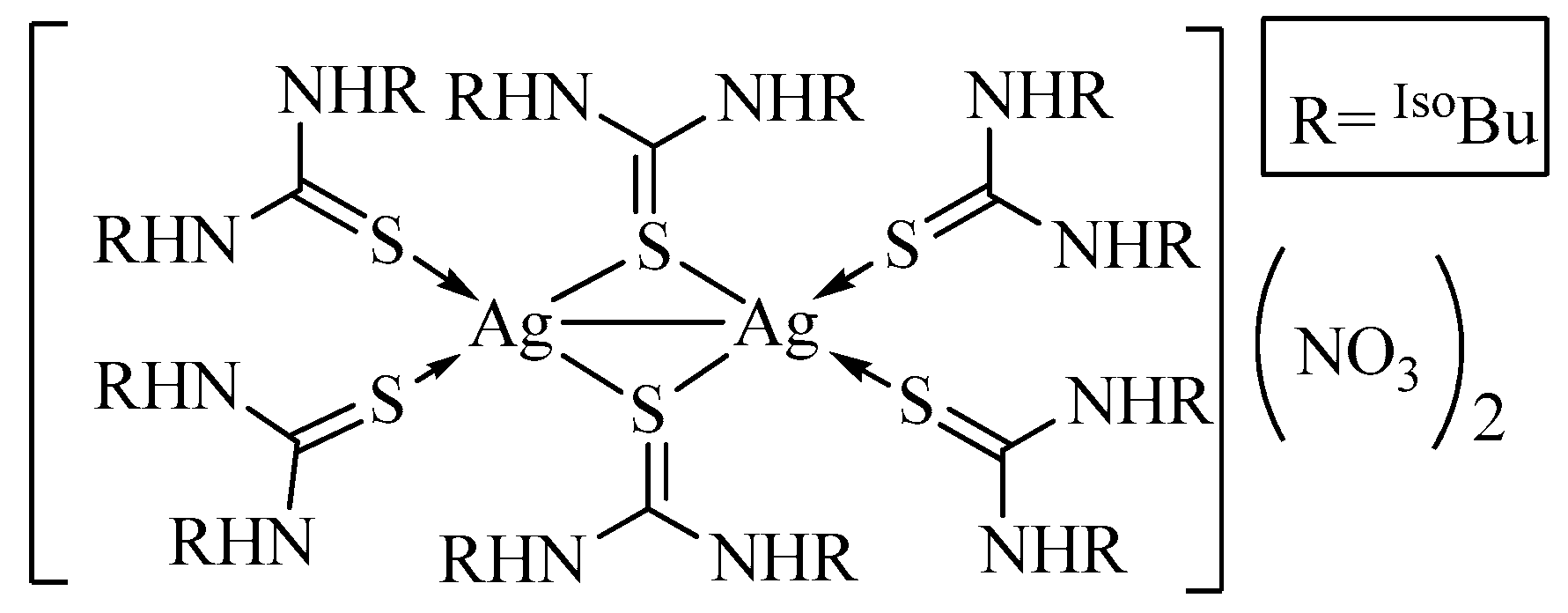
[Ag2L6](NO3)2 (2): AgNO3 (0.1 g, 0.588 mmol) in water was mixed with 1,3-diisobutyl thiourea (0.2 g, 1.06 mmol) in ethanol and the reaction mixture was stirred overnight at room temperature. Slow evaporation of the reaction mixture resulted in colorless crystals after 3 days and were characterized by X-ray structure analysis (Figure 2). Yield: 72%.
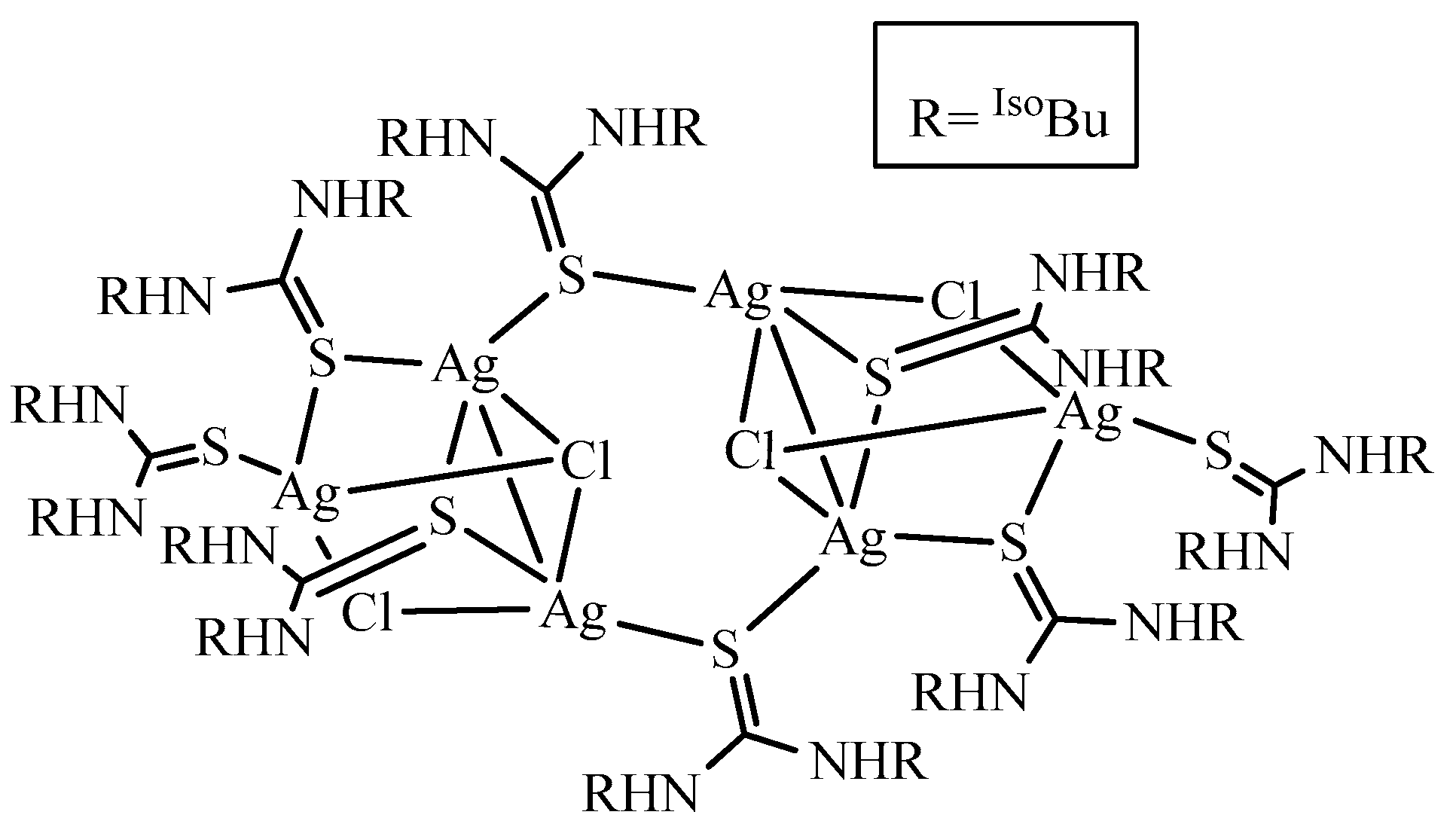
[Ag6L8Cl4] (3): A solution of AgCl (0.1 g, 0.7 mmol) was mixed with 1,3-diisobutyl thiourea (0.15 g, 0.79 mmol) in acetonitrile. After overnight stirring, the solvent was allowed to evaporate at room temperature. The colorless needle-like crystals of 3 (Figure 3) appeared in the solution after 3 days, they were separated from the mother liquor and the structure of the complex was determined by X-ray diffraction.

2.4. Hirshfeld Surface Analysis

The Hirshfeld surfaces and 2D fingerprint plots were generated using Crystal Explorer 17.50 [41]. The X-ray single-crystal crystallographic information files were used as input files. The default setting used for Hirshfeld Surface/fingerprint generation in Crystal Explorer is: property: none; resolution: High (standard). For fingerprint generation (di vs. de plot) we used; range: standard, filter: by elements and fingerprint filter options is both inside-outside elements including reciprocal contacts. The interactions with normalized contact distance in crystal structure shorter than the sum of the corresponding van der Waals radii of the atoms, are highlighted by red spots and the longer contacts with the positive dnorm value are represented in blue color.

2.5. Antioxidant Assay

Complexes 13 have been tested for their antioxidant activity. The DPPH free radical scavenging method has been employed to investigate representative compounds as antioxidant agents. When compounds donate hydrogen or electrons to the free DPPH radical, the initial absorbance of the corresponding solution decreases. These observations are taken as a key to measure radical scavenging activity. A solution of DPPH was prepared (39 mg/100 mL), absorbance was measured under normal conditions at 517 nm for blank (Ao). Specific amount of complex solution was mixed with DPPH, solutions were incubated in dark for 30 min at room temperature. The absorbance of each sample was measured at the same wavelength, 517 nm (ASample). Percent inhibition (I) % of DPPH radical was calculated for the various concentrations of compounds as given below [42].
Percent   Inhibition   I % = A 0 A A 0 × 100

3. Results and Discussion

In our previous study, we treated acetonitrile solution of the ligand with CuCl salt under normal conditions. In reaction, mononuclear complex was exclusively obtained and the reactions worked in the same way with other metal salts, namely CuI, ZnCl2 and HgCl2 [3]. When the reaction of Cu(II) salt with the identical ligand in ethanolic medium was carried out, reduction of the metal ion was observed and the trinuclear Cu(I) complex was obtained. The metal salt in repeated experiments under identical conditions with two equivalents of the ligand did not give expected mononuclear complex of general formula [CuL2Cl2]. The same ligand with AgNO3 salt in a 2:1 molar ratio, respectively, did not afford [AgL2]NO3 as an expected precursor, rather reduction of Ag(I) ion (as evident from development of black coloration in the solution) afforded binuclear complex 2, bearing general formula [Ag2L6](NO3)2. The reaction solution contained trace amounts of some unknown impurities which did not allow for collection of other data to determine exact purity of the reaction. However, crystals of suitable dimensions were selected under microscope for structure determination. The effect of Cl radical in Ag salt was totally different and following the same procedure gave a cluster containing six Ag ions, in a 6:8 molar ration, metal to ligand, respectively. The cluster [Ag6L8Cl4]Cl2 stabilization was achieved as a result of partial reduction of Ag(I) ion as was observed in complex 2. The role of both Cl and thiourea ligand as bridging ligands was found prominent in cluster formation. The work described hereunder indicates that the presence of anion and reaction medium is crucial in describing coordination chemistry around Cu and Ag metal ions. The reactions of Ag salts in the presence UV radiation can also lead to unexpected products.

3.1. Description of Crystal Structure of [Cu3L3Cl3] (1)

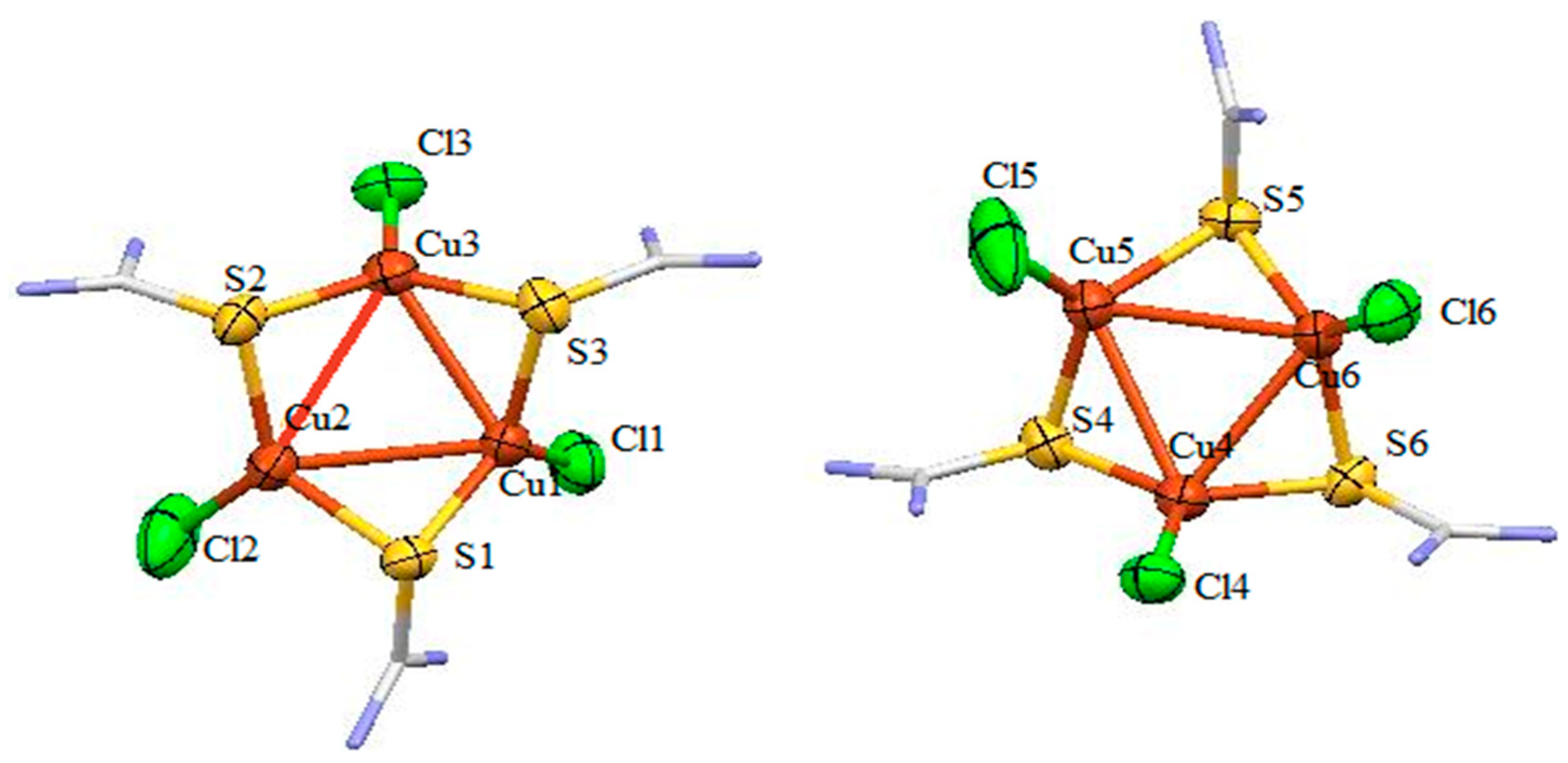
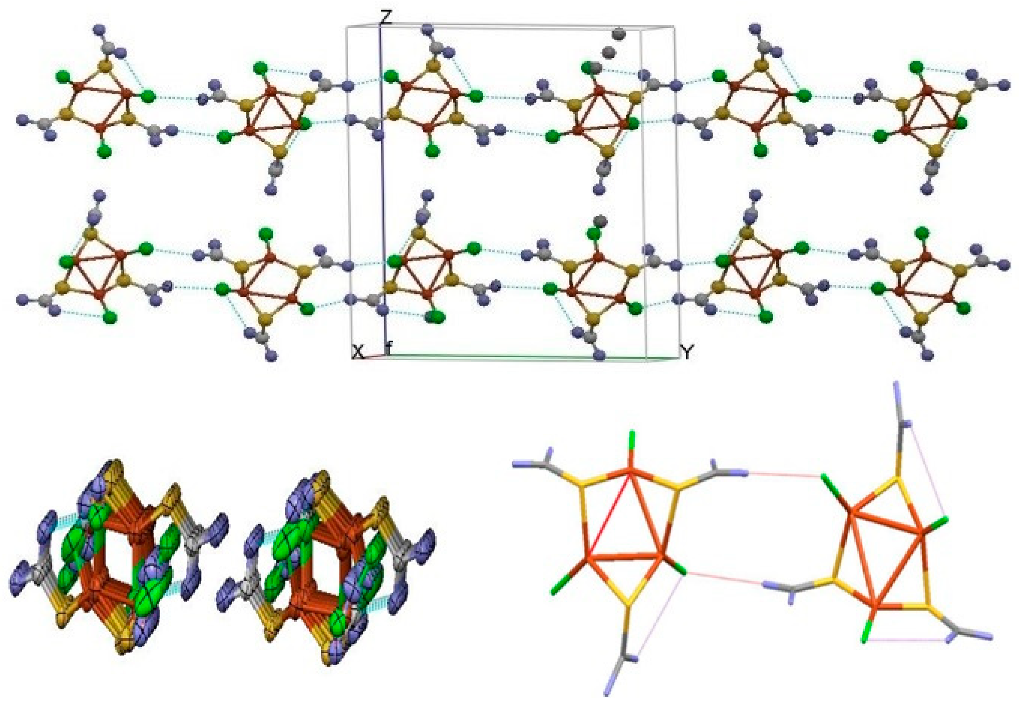
The crystal structures of complexes 1, 2 and 3 are elucidated by single crystal X-ray diffraction. The molecular crystal structure of complex 1 is given in Figure 4. Selected bond lengths and bond angles are enlisted in Table 1 and the crystallographic refinement data are given in Table 2. The tri-nuclear copper cluster crystallizes in triclinic crystal system with a space group P-1. The unit cell reveals non superimposable conformers of complex 1. According to X-ray analysis the complex 1 is trimeric having two daughter diastereomers in unit cell of the said complex, the same has also been reported in other similar complexes [17]. The monomeric portion (L-M-Cl) of the complex is connected through bridging bond via sulfur atom of C = S moiety of thiourea, providing Cu3S3 six membered metalacyclic ring adopting chair conformation. Chlorine atom is coordinated to Cu at axial position. The geometry around each Cu ion is distorted trigonal planner with varying angles around Cu center (in the range of 116.61°–124.36°). The bond distance between Cu-S is 2.258 Å and is comparable to the analogous compounds [17,43]. The Cu-Cl bond distance of 2.182 Å is shorter than that of the mononuclear metal complex [2.272(3) Å] with the identical ligand2. The trigonal arrangement of Cu-Cu in tri-nuclear unit is stabilized by Cu---Cu interaction with distance of 2.850 Å [44,45]. The supramolecular structure is stabilized via regular pattern of intra- and intermolecular hydrogen bonding N-H---Cl interactions as shown in Figure 5. The intermolecular N-H---Cl interactions [3.122 and 3.210 Å] are shorter than the intramolecular interactions [3.256 and 3.291 Å], which are in turn shorter than those for recently reported mononuclear copper complexes [3.349 Å]6. Attempts to get more insight into the strength of intermolecular interactions using Hirshfeld surface analyses were unsuccessful due to the disordered structure.

3.2. Description of Crystal Structure of [Ag2L6](NO3)2 (2)
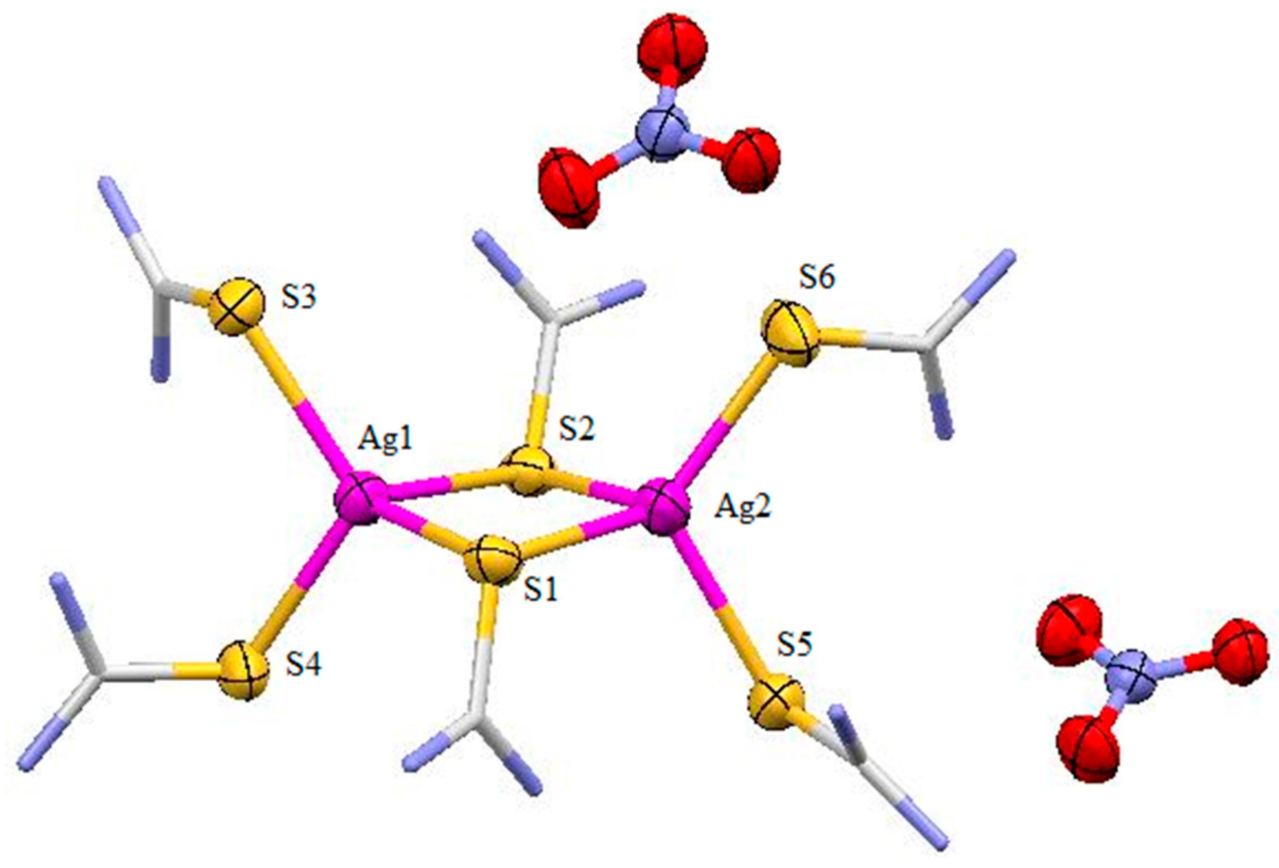
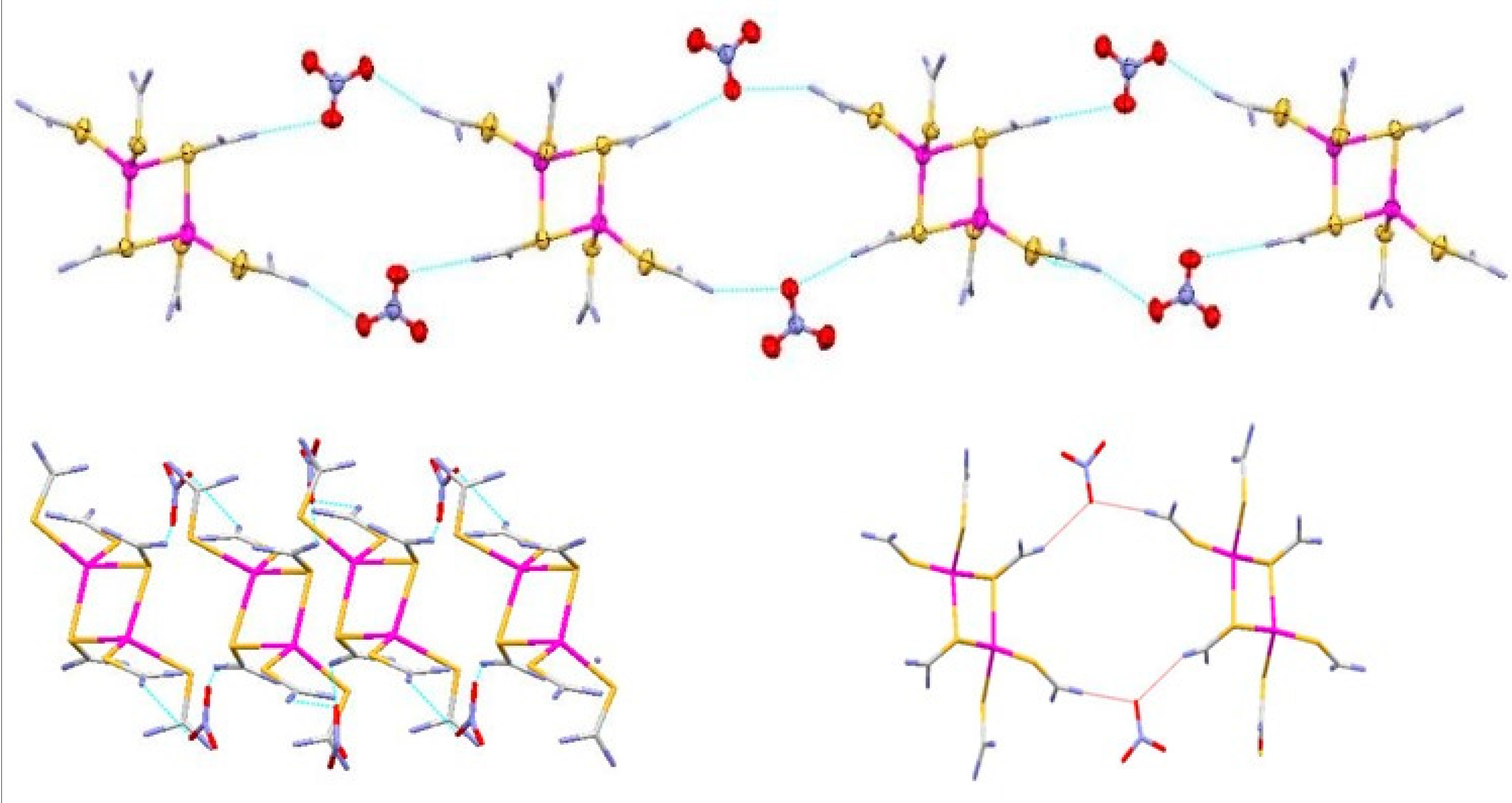
The molecular crystal structure of complex 2 is shown in Figure 6 and the selected bond lengths and bond angles are enlisted in Table 1. The bimetallic silver complex crystallizes in triclinic crystal system with space group P-1. The binuclear complex 2 consists of two Ag(I) ions and six thiourea ligands. Both silver ions are bridged by S-atom of the two 1,3-diisobutyl thiourea ligands, forming four-membered Ag-S-Ag-S ring. Thiourea derivatives have different coordination mode for silver(I) ions. Two thiourea ligands are linked in terminal fashion to each metal ions and the remaining two thiourea ligands bridged both metal ions together. In this binuclear complex each silver atom is tetrahedrally coordinated through S-atom of the two terminal thiourea and two bridging thiourea molecules. The interatomic distance between Ag and S(bridged) is 2.622 Å. As expected, this distance is longer than the respective Ag-S (terminal) bond (2.491 Å) and is because of the electron deficient bond characters between Ag and bridging thiourea ligand [46,47,48]. The plane of Ag-S (terminal) and Ag-S (bridged) are mutually twisted by 83.88°. In a crystalline state, the molecules of the complex are stabilized by regular intermolecular hydrogen bonding i.e., N-O---H-N making a supramolecular structure. Different units are interlinked through nitrate ions as shown in Figure 7.
Figure 6. The molecular structure of compound 2, only non-carbon atoms are numbered. Ellipsoids are drawn at 50% probability level, hydrogen atoms are omitted and carbons are represented by capped sticks.
Inorganics 10 00185 g006
Figure 7. 2D representation of 2, the network is stabilized by intramolecular N-H---O hydrogen bonding.
Inorganics 10 00185 g007
Figure 8. View of the three-dimensional Hirshfeld surface of 2 plotted over dnorm in the range −0.4382 to 1.6504.
Inorganics 10 00185 g008
Hirshfeld surface analyses were used to get insights into the detailed information about the strength of intermolecular interactions. The interactions with normalized contact distance in crystal structure shorter than the sum of the corresponding van der Waals radii of the atoms, are highlighted by red spots and the longer contacts with the positive dnorm value are represented in blue color (Figure 8). The significant intermolecular interactions are mapped in Figure 9. On the Hirshfeld surfaces, the H···H interactions appear as the largest region (76.1%) of the fingerprint plot. The O···H/H···O contacts with a contribution of 18.8% due to the intermolecular N–H···O [2.037, 2.307 and 2.404 Å] and C–H···O [2.554 and 2.574 Å] hydrogen bonding give rise to a pair of characteristic wings in the fingerprint plot. The pair of segregated sharp spikes represents the S···H/H···S contacts with a contribution of 4.3%. The N···H and C···H interactions are found to be negligible (0.3 and 0.1%, respectively).

3.3. Description of Crystal Structure of [Ag6L8Cl4] (3)
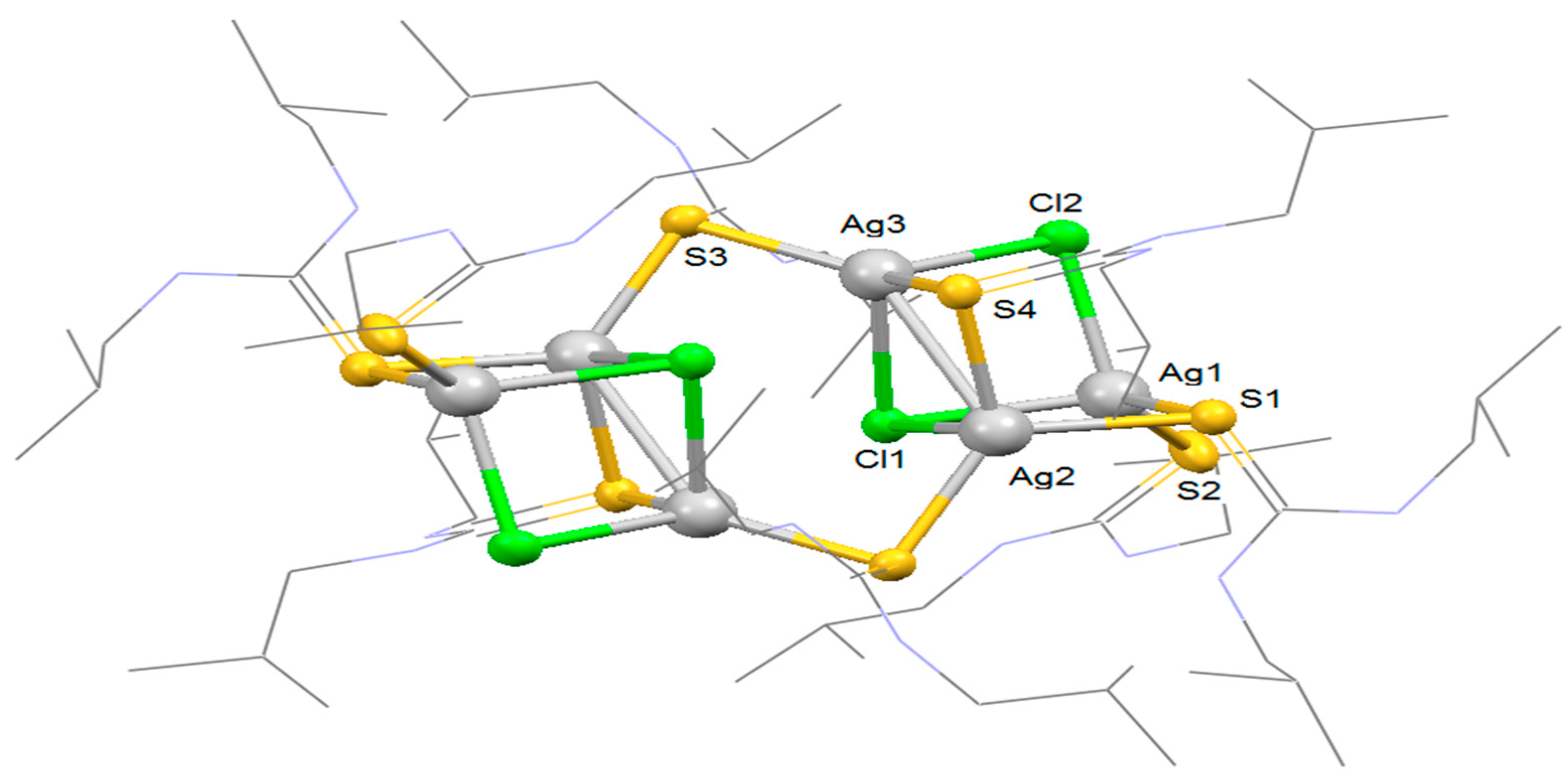
The molecular structure of polynuclear silver complex 3 was confirmed by X-ray diffraction analysis as shown in Figure 10. The complex crystallizes in monoclinic crystal system with space group P 21/c. The single crystal analysis reveals that there are two types of silver ions, one is 5-coordinated and the other is 4-coordinated. The thiourea ligands are either terminal or bridged as discussed for complex 2. The cluster structure is composed of six silver ions, four chlorine ligands and eight thiourea ligands. The chlorine ligands also show variable coordination modes, one of them bridges three silver ions and the other one is bridged to two silver ions (μ3- and μ2-Cl, respectively). The overall charge on the complex is +2 and every individual silver is present as +1 ion. Four Cl ions present in the molecule neutralize +4 charge internally leaving +2 as overall charge on the complex. The geometry around one of the silver ions is distorted tetrahedral. The complex can be regarded as dimer of the two tri-nuclear cationic clusters bridged together with the help of two thiourea ligands. The coordination environments around all silver ions (Ag1, Ag2 and Ag3) in asymmetric unit are different but geometry around silver atoms is distorted tetrahedral.
Ag1 is coordinated to one bridging thiourea (μ-S), one terminal thiourea and two bridging Cl ligands in which one chlorine is present as μ2 and the other one is μ3. Ag2 is surrounded by three bridging thiourea and a chlorine ligand (μ3-Cl) in the same manner as for Ag1. The Ag3 is coordinated to two bridging thiourea (μ-S) and two bridging Cl ligands, μ2 and μ3. The structural parameters of the complex are summarized for selected atoms in Table 1. The Ag-S bond distance around the tetrahedrally coordinated Ag1 atom is 2.446 Å for terminal thiourea and 2.602 Å for bridged thiourea ligand, respectively. The slight elongation in the later bond may be traced by the electron delocalization of a single electron pair among the three nuclei. Such bonds are also called electron deficient bonds. Ag1-Cl1 and Ag1-Cl2 bond distances are 2.644 Å and 2.642 Å, respectively. Ag2-S1, Ag2-S3 and Ag2-S4 bond distances are 2.543 Å, 2.507 Å and 2.642 Å, respectively, and Ag2-Cl1 bond distance is 2.682 Å. Similarly, the Ag3-S4 and Ag3-S3 bond distances are 2.505 and 2.529 Å. Ag3-Cl1 and Ag3-Cl2 bond distances are 2.862 and 2.711 Å, respectively. The data show that Ag3 has relatively weak interaction with Cl ligands as compared to Ag1 and Ag2 [49,50,51].
The angles around Ag1 center S2-Ag1-Cl1, S1-Ag1-Cl1, S2-Ag1-Cl2, S1-Ag1-Cl2 and S1-Ag1-S2 are 113.13°, 107.23°, 119.42°, 100.30° and 107.23°, respectively. The two asymmetric units are interconnected by bridging thiourea forming six-member ring. The crystallographic data show that the complex has Ag–Ag interactions with an average distance of 3.120 Å and the cluster is stabilized by argentophillicity. The complex formation is an unexpected result, but the data set obtained for the complex is in close agreement with the reported silver complexes bearing Ag-S and Ag-Cl ligands [29,52].
Figure 10. Molecular structure of silver cluster, complex 3. Only Ag, S and Cl atoms are labeled for an asymmetric unit, thermal ellipsoids are drawn at 40% probability level, hydrogen atoms are omitted and bonds connecting N and C are represented by wireframes for simplicity.
Inorganics 10 00185 g010
Figure 11. View of the three-dimensional Hirshfeld surface of 3 plotted over dnorm in the range −0.4772 to 2.1283.
Inorganics 10 00185 g011
Hirshfeld surface analyses (Figure 11) show that the fingerprint profiles are dominated by H···H contacts (85.1%). The contributions due to the Cl···H/H···Cl interactions are the maximum (11.1%) that have been observed as segregated sharp spikes (Figure 12). Other contributions are due to S···H/H···S and N···H/H···N interactions (1.8 and 1%, respectively).
Figure 12. Two-dimensional fingerprint plots for all intermolecular contacts in 2. The percentage of contribution is specified for each contact.
Inorganics 10 00185 g012
Table 1. List of selected bond lengths (Å) and bond angles (o) of compound 13.
CompoundAtomsBond LengthAtomsBond Angle
1Cu1-S12.221(17)S1-Cu1-Cl1123.17(7)
Cu1-Cl12.233(19)S1-Cu1-S3117.41(6)
Cu1-S32.2697(18)S3-Cu1-Cl1119.21(7)
Cu1-Cu22.889(10)Cu1-Cu2-Cu354.87(3)
2Ag1-S42.5329(15)S4-Ag1-S3113.30(5)
Ag1-S32.5568(14)S4-Ag1-S1113.51(5)
Ag1-S12.5941(14)S3-Ag1-S1115.49(5)
Ag1-S22.6451(10)S4-Ag1-S2100.16(5)
Ag1-Ag23.3539(6)S3-Ag1-S2112.00(5)
3Ag1-S12.602(15)S2-Ag1-Cl1113.13(5)
Ag1-S22.446(14)Cl1-Ag1-S1107.23(6)
Ag1-Cl12.462(13)S2-Ag1-Cl2119.42(5)
Ag1-Ag23.120(6)S1-Ag1-Cl2100.30(7)
Ag2-S12.543(11)S1-Ag1-S2107.23(5)
Ag2-S42.642(10)Ag2-S3-Ag393.88(5)
Ag2-S32.507(14)Ag3-Cl2-Ag189.43(6)
Ag3-S42.505(15)Ag1-Cl1-Ag272.01
Ag3-S32.529(12)Ag1-S1-Ag289.17
Table 2. Crystal structure determination and structure refinements of compound 13.
123
Empirical formulaC54H120N12Cu6Cl6S6C54H120N14Ag2O6S6C72H160N16Ag6Cl6S8
Formula weight1603.9714792366.57
Temperature (K)296296296
Wavelength (Å)0.710730.710730.71073
Crystal systemTriclinicTriclinicMonoclinic
Space groupP-1P-1P 21/c
Aa11.016(4)11.611(6)11.7020(11)
Bb18.227(7)15.499(8)16.5310(14)
Ac21.634(8)24.149(13)28.0730(3)
89.832(2)99.159(3)90
Ββ86.125(2)102.357(2)99.096(5)
Γγ81.288(2)108.098(3)90
Volume Å34284.0(3)3988.8(4)5362.3(9)
μ (mm−1)1.830.701.422
Z222
Density (Mg m−3)1.2431.2771.466
F (0, 0, 0)156216242432
(h, k, l) min(−13, −22, −27)−14, −18, −30(−13, −19, −33)
(h, k, l) max(13, 22, 27)14, 19, 30(13, 19, 33)
Theta (max)26.527.024.999
R (reflection)0.069 (17650)0.057 (17072)0.0853 (5485)
wR20.2360.1800.1706

3.4. Determination of Antioxidant Activities by DPPH Radical Scavenging Test

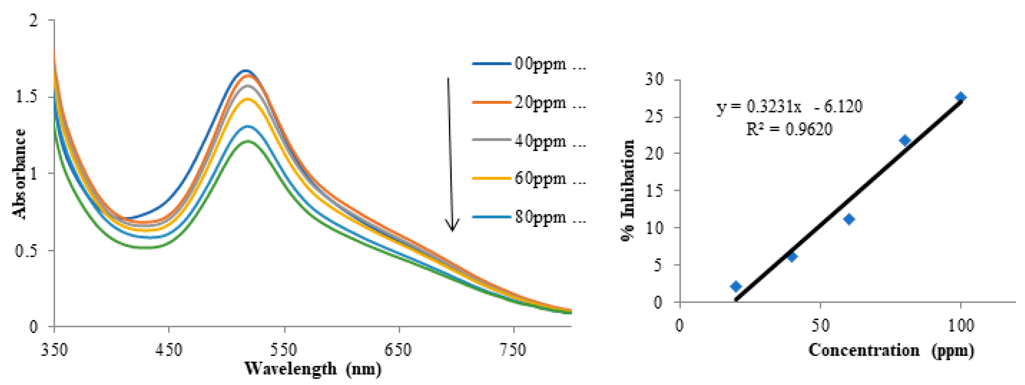
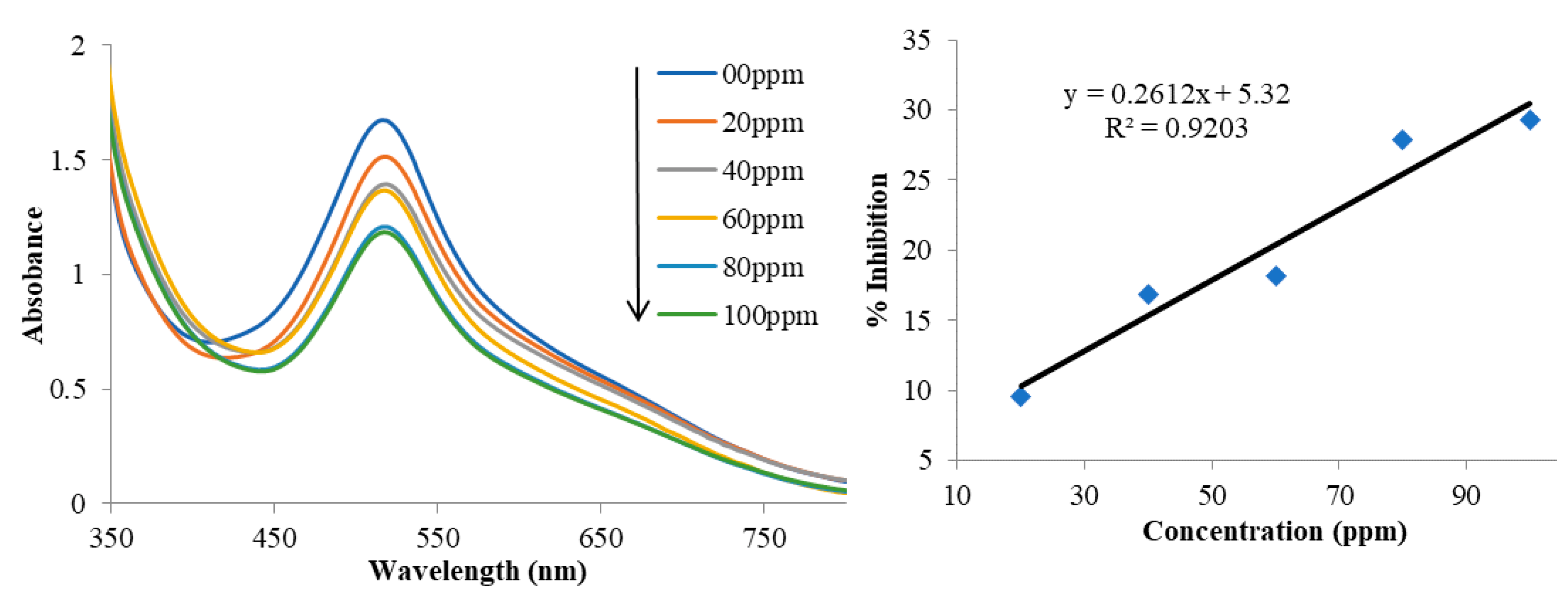
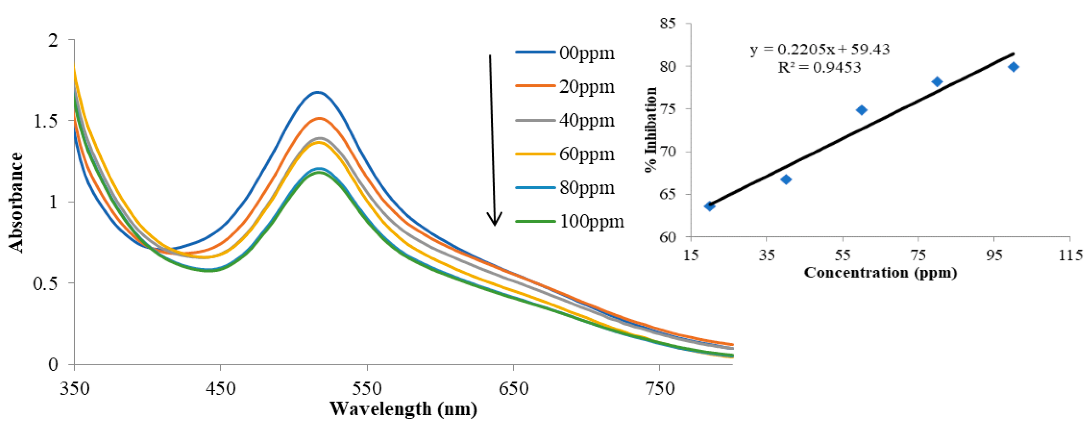
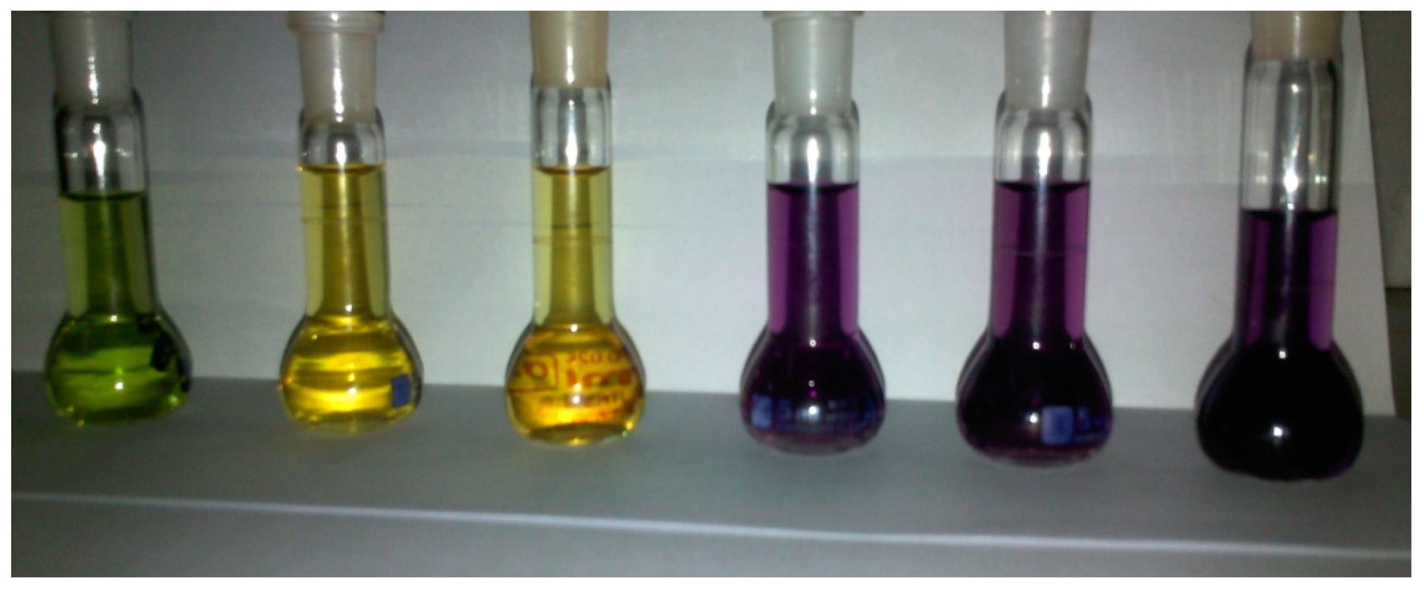
The inhibitory effect of polynuclear thiourea complexes of copper and silver (13) has been studied using DPPH as free radical. Thiourea complexes with transition metals are known for their antioxidant potentials [2]. Thus, we were interested in studying the synthesized complexes for their antioxidant activities. We observed that these complexes upon addition to the DPPH solution show a habitual variation in absorbance (concentration range 00–100 ppm) in Table 3. On addition of compounds, the absorbance of DPPH decreases with increase in concentration in a regular expected pattern. The percent radical scavenging activity (DPPH) of compounds increases from top (zero concentration of tested compound) to bottom (maximum concentration of tested compound, 100 ppm). The DPPH free radical scavenging activity of compounds (13) at 100 ppm of the compounds in reaction mixture were determined to be 19.5%, 29.3% and 19.2%, respectively. The inhibition caused by complexes (13) are diagrammatically shown in Figure 13, Figure 14 and Figure 15. In these compounds, the antioxidant ability of compound 2 is higher (29.3%) than the other compounds, which was also reflected by the color change of the solution mixture during the course of the experiment (Figure 16).
Graphical representation of the antioxidant potentials of complexes 13 reveals that the activity is dose dependent. With the increase in concentration of the complex in its solution, the scavenging activity increases. As a result of greater activity, a decrease in absorbance intensity was observed. Among the three complexes, complex 2 is comparatively more active at the concentration range of 100 ppm. The compound (2) is capable of showing naked-eye detection of the same activity, as made evident from the Figure 16. Solution without complex (blank/control) at the right is dark purple in color, there is almost no change in the color when 20 and 40 ppm complex solution is mixed. The color changes abruptly from purple to yellow with the addition of 60 and 80 ppm complex solution and to green at 100 ppm. The efficiency of the complex is predominantly because of the presence of nitrate ion out of the coordination sphere which makes the complex more soluble in the given solvent. The counter anion (chloride) in complexes 1 and 3 possesses a different coordination behavior and the activity is therefore poor.

4. Conclusion

The presence of anion in metal salts and reaction medium (solvent) plays a pivotal role in nuclearity of resultant complexes. Three new heteroleptic polynuclear complexes [Cu3L3Cl3] (1), [Ag2L6](NO3)2 (2) and [Ag6L8Cl4] (3) were successfully synthesized by treating 1,3-diisobutyl thiourea with CuCl2, AgNO3 and AgCl, respectively. The formation of 1 was independent of applied reaction conditions (temperature and concentration) and metal to ligand molar ratio. All complexes were characterized by single crystal X-ray diffraction. Crystal analyses show that 1 has two non-super imposable diastereomers in trimeric form, while 2 is dimeric with two-fold axis of rotation and 3 is consisting of two asymmetric units [Ag3L3Cl2] bridged by two thiourea ligands with two-fold axis of rotation. These complexes show moderate antioxidant potential against DPPH.

Author Contributions

Conceptualization, methodology, software, A.S., E.K. and A.N.; validation, formal analysis, E.K., A.N. and M.N.T.; investigation, A.S. and E.K.; resources, E.K. and A.N.; data curation, A.N., A.N. and E.K.; writing—original draft preparation, A.S., A.N., E.K., G.S.K., A.u.R. and M.S.; writing—review and editing, visualization, A.S., E.K. and A.N; supervision, project administration, funding acquisition, E.K. and A.N. All authors have read and agreed to the published version of the manuscript.

Funding

This work was supported by the Deanship of Scientific Research, Vice Presidency for Graduate Studies and Scientific Research, King Faisal University, Saudi Arabia [Grant No. Grant1399].

Institutional Review Board Statement

Not applicable.

Informed Consent Statement

Not applicable.

Data Availability Statement

The crystallographic data have been deposited at the Cambridge Crystallographic Data Centre as supplementary publications under CCDC Nos. 2196016 (compound 1), 2196017 (compound 2) and 2204046 (compound 3). These data can be obtained free of charge from the Cambridge Crystallographic Data Centre, www.ccdc.cam.ac.uk/datarequest/cif.

Acknowledgments

This work was supported by the Deanship of Scientific Research, Vice Presidency for Graduate Studies and Scientific Research, King Faisal University, Saudi Arabia [Grant No. Grant1399].

Conflicts of Interest

There are no conflict of interest to be declared.

References

  1. Khan, E.; Khan, S.; Gul, Z.; Muhammad, M. Medicinal Importance, Coordination Chemistry with Selected Metals (Cu, Ag, Au) and Chemosensing of Thiourea Derivatives. A Review. Crit. Rev. Anal. Chem. 2021, 51, 812–834. [Google Scholar] [CrossRef] [PubMed]
  2. Shahzad, A.; Khan, E.; Said, M.; Khan, G.S.; Syed, M.G.; Noor, A.; Zahoor, M.; Ullah, R.; Bari, A. Complexes of 1, 3-diisobutyl thiourea with copper (I), zinc (II) and mercury (II): Their antioxidant and antibacterial evaluation. Crystals 2021, 11, 989. [Google Scholar] [CrossRef]
  3. Rahman, F.U.; Bibi, M.; Altaf, A.A.; Tahir, M.N.; Ullah, F.; Khan, E. Zn, Cd and Hg complexes with unsymmetric thiourea derivatives; syntheses, free radical scavenging and enzyme inhibition essay. J. Mol. Struct. 2020, 1211, 128096. [Google Scholar] [CrossRef]
  4. Rahman, F.U.; Bibi, M.; Khan, E.; Shah, A.B.; Muhammad, M.; Tahir, M.N.; Shahzad, A.; Ullah, F.; Zahoor, M.; Alamery, S. Thiourea derivatives, simple in structure but efficient enzyme inhibitors and mercury sensors. Molecules 2021, 26, 4506. [Google Scholar] [CrossRef] [PubMed]
  5. Khan, U.A.; Badshah, A.; Tahir, M.N.; Khan, E. Gold (I), silver (I) and copper (I) complexes of 2, 4, 6-trimethylphenyl-3-benzoylthiourea; synthesis and biological applications. Polyhedron 2020, 181, 114485. [Google Scholar] [CrossRef]
  6. Noor, A.; Qayyum, S.; Jabeen, F.; Rehman, A.U. Mononuclear Tricoordinate Copper (I) and Silver (I) Halide Complexes of a Sterically Bulky Thiourea Ligand and a Computational Insight of Their Interaction with Human Insulin. Molecules 2022, 27, 4231. [Google Scholar] [CrossRef]
  7. Mykhalichko, B.M.; Temkin, O.N.; Mys’kiv, M. Polynuclear complexes of copper(I) halides: Coordination chemistry and catalytic transformations of alkynes. Russ. Chem. Rev. 2000, 69, 957–984. [Google Scholar] [CrossRef]
  8. Levitsky, M.M.; Bilyachenko, A.N.; Shul’pin, G.B. Oxidation of C-H compounds with peroxides catalyzed by polynuclear transition metal complexes in Si- or Ge-sesquioxane frameworks: A review. J. Organomet. Chem. 2017, 849–850, 201–218. [Google Scholar] [CrossRef]
  9. Roy, P.; Dhara, K.; Manassero, M.; Banerjee, P. Di-, Tetra-, and Polynuclear Copper (II) Complexes: Active Catalysts for Oxidation of Toluene and Benzene. Eur. J. Inorg. Chem. 2008, 2008, 4404–4412. [Google Scholar] [CrossRef]
  10. Musina, E.I.; Shamsieva, A.V.; Strelnik, I.D.; Gerasimova, T.P.; Krivolapov, D.B.; Kolesnikov, I.E.; Grachova, E.V.; Tunik, S.P.; Bannwarth, C.; Grimme, S.; et al. Synthesis of novel pyridyl containing phospholanes and their polynuclear luminescent copper(i) complexes. Dalton Trans. 2016, 45, 2250–2260. [Google Scholar] [CrossRef]
  11. Kobielska, P.A.; Telford, R.; Rowlandson, J.; Tian, M.; Shahin, Z.; Demessence, A.; Ting, V.P.; Nayak, S. Polynuclear Complexes as Precursor Templates for Hierarchical Microporous Graphitic Carbon: An Unusual Approach. ACS Appl. Mater. Interfaces 2018, 10, 25967–25971. [Google Scholar] [CrossRef] [PubMed] [Green Version]
  12. Puyo, M.; Lebon, E.; Vendier, L.; Kahn, M.L.; Fau, P.; Fajerwerg, K.; Lepetit, C. Topological Analysis of Ag–Ag and Ag–N Interactions in Silver Amidinate Precursor Complexes of Silver Nanoparticles. Inorg. Chem. 2020, 59, 4328–4339. [Google Scholar] [CrossRef] [PubMed]
  13. Zhang, S.; Zhao, L. Macrocycle-Encircled Polynuclear Metal Clusters: Controllable Synthesis, Reactivity Studies, and Applications. Acc. Chem. Res. 2018, 51, 2535–2545. [Google Scholar] [CrossRef] [PubMed]
  14. Bergamini, F.R.G.; Nunes, J.H.B.; de Carvalho, M.A.; Ribeiro, M.A.; de Paiva, P.P.; Banzato, T.P.; Ruiz, A.L.T.G.; de Carvalho, J.E.; Lustri, W.R.; Martins, D.O.; et al. Polynuclear copper (II) complexes with nalidixic acid hydrazones: Antiproliferative activity and selectivity assessment over a panel of tumor cells. Inorg. Chim. Acta 2019, 484, 491–502. [Google Scholar] [CrossRef]
  15. Sun, W.H.; Li, K.H.; Liu, H.; Gu, Y.T.; Zhang, Y.; You, Z.L.; Li, W. Synthesis, Characterization, Crystal Structures, and Antibacterial Activity of Polynuclear Nickel (II) and Copper (II) Complexes with Similar Tridentate Schiff Bases. Russ. J. Coord. Chem. 2017, 43, 693–699. [Google Scholar] [CrossRef]
  16. Semerci, F.; Yeşilel, O.Z.; Soylu, M.S.; Keskin, S.; Büyükgüngör, O. A two-dimensional photoluminescent cadmium (II) coordination polymer containing a new coordination mode of pyridine-2,3-dicarboxylate: Synthesis, structure and molecular simulations for gas storage and separation applications. Polyhedron 2013, 50, 314–320. [Google Scholar] [CrossRef]
  17. Mei, G.; Li, Z.; Zhang, H.; Li, G.; Meng, X. Synthesis, Structures, and Properties of Two Novel Zn (II) Polynuclear Complexes. Synth. React. Inorg. Met. Org. Nano Met. Chem. 2010, 40, 163–169. [Google Scholar] [CrossRef]
  18. Roubeau, O.; Clérac, R. Rational Assembly of High-Spin Polynuclear Magnetic Complexes into Coordination Networks: The Case of a [Mn4] Single-Molecule Magnet Building Block. Eur. J. Inorg. Chem. 2008, 2008, 4325–4342. [Google Scholar] [CrossRef]
  19. Papaefstathiou, G.S.; Perlepes, S.P. Families of Polynuclear Manganese, Cobalt, Nickel and Copper Complexes Stabilized by Various Forms of Di-2-pyridyl Ketone. Comments Inorg. Chem. 2002, 23, 249–274. [Google Scholar] [CrossRef]
  20. Yao, L.; Niu, G.; Li, J.; Gao, L.; Luo, X.; Xia, B.; Liu, Y.; Du, P.; Li, D.; Chen, C.; et al. Circularly Polarized Luminescence from Chiral Tetranuclear Copper(I) Iodide Clusters. J. Phys. Chem. Lett. 2020, 11, 1255–1260. [Google Scholar] [CrossRef]
  21. Haasnoot, J.G. Mononuclear, oligonuclear and polynuclear metal coordination compounds with 1,2,4-triazole derivatives as ligands. Coord. Chem. Rev. 2000, 200–202, 131–185. [Google Scholar] [CrossRef]
  22. Bhowmik, P.; Chattopadhyay, S.; Drew, M.G.B.; Ghosh, A. A polynuclear helical and two dinuclear copper (II) complexes of a monocondensed N, N, O donor Schiff base with pseudohalides as co-ligand. Inorg. Chim. Acta 2013, 395, 24–32. [Google Scholar] [CrossRef]
  23. Escuer, A.; Esteban, J.; Perlepes, S.P.; Stamatatos, T.C. The bridging azido ligand as a central “player” in high-nuclearity 3d-metal cluster chemistry. Coord. Chem. Rev. 2014, 275, 87–129. [Google Scholar] [CrossRef]
  24. Marvaud, V.; Decroix, C.; Scuiller, A.; Tuyèras, F.; Guyard-Duhayon, C.; Vaissermann, J.; Marrot, J.; Gonnet, F.; Verdaguer, M. Hexacyanometalate Molecular Chemistry: Di-, Tri-, Tetra-, Hexa- and Heptanuclear Heterobimetallic Complexes; Control of Nuclearity and Structural Anisotropy. Chem. A Eur. J. 2003, 9, 1692–1705. [Google Scholar] [CrossRef] [PubMed]
  25. Wang, X.-L.; Qin, C.; Wang, E.-B.; Su, Z.-M. Metal Nuclearity Modulated Four-, Six-, and Eight-Connected Entangled Frameworks Based on Mono-, Bi-, and Trimetallic Cores as Nodes. Chem. A Eur. J. 2006, 12, 2680–2691. [Google Scholar] [CrossRef]
  26. Papatriantafyllopoulou, C.; Manessi-Zoupa, E.; Escuer, A.; Perlepes, S.P. The sulfate ligand as a promising “player” in 3d-metal cluster chemistry. Inorg. Chim. Acta 2009, 362, 634–650. [Google Scholar] [CrossRef]
  27. Ahmad, S.; Isab, A.A.; Perzanowski, H.P. Silver (I) complexes of thiourea. Transit. Met. Chem. 2002, 27, 782–785. [Google Scholar] [CrossRef]
  28. Safin, D.A.; Babashkina, M.G.; Bolte, M.; Pape, T.; Hahn, F.E.; Verizhnikov, M.L.; Bashirov, A.R.; Klein, A. Polynuclear and mixed-ligand mononuclear CuI complexes with N-thiophosphorylated thioureas and 1,10-phenanthroline or PPh3. Dalton Trans. 2010, 39, 11577–11586. [Google Scholar] [CrossRef]
  29. Safin, D.A.; Babashkina, M.G.; Bolte, M.; Garcia, Y. Homoleptic polynuclear CuI and AgI complexes of N-thiophosphorylated thioureas o-RO(O)CC6H4NHC(S)NHP(S)(OiPr)2 (R = Me, Et). CrystEngComm 2012, 14, 774–778. [Google Scholar] [CrossRef]
  30. Metlushka, K.E.; Sadkova, D.N.; Nikitina, K.A.; Pashagin, A.V.; Khrizanforov, M.N.; Budnikova, Y.H.; Morozov, V.I.; Islamov, D.R.; Latypov, S.K.; Kataeva, O.N.; et al. Synthesis of the first chiral polynuclear copper(i) complex based on (R)-1-(1-phenyl) ethyl-3-(O,O-diethylthiophosphoryl)thiourea and its characterization in the solid state and solution. New J. Chem. 2020, 44, 3224–3231. [Google Scholar] [CrossRef]
  31. Li, G.; Che, D.-J.; Li, Z.-F.; Zhu, Y.; Zou, D.-P. Versatile coordination patterns in the reaction system of N-benzoyl-N′-(2-pyridyl) thiourea with CuCl2. Their reaction conditions, systematic isolation and crystal structures. New J. Chem. 2002, 26, 1629–1633. [Google Scholar] [CrossRef]
  32. Nguyen, H.H.; Thang, P.C.; Rodenstein, A.; Kirmse, R.; Abram, U. Bipodal Acylthiourea Ligands as Building Blocks for Bi-, Tetra-, and Polynuclear Oxorhenium (V) Complexes. Inorg. Chem. 2011, 50, 590–596. [Google Scholar] [CrossRef] [PubMed]
  33. Vaidya, S.; Shukla, P.; Tripathi, S.; Rivière, E.; Mallah, T.; Rajaraman, G.; Shanmugam, M. Substituted versus Naked Thiourea Ligand Containing Pseudotetrahedral Cobalt (II) Complexes: A Comparative Study on Its Magnetization Relaxation Dynamics Phenomenon. Inorg. Chem. 2018, 57, 3371–3386. [Google Scholar] [CrossRef] [PubMed]
  34. Luckay, R.C.; Sheng, X.; Strasser, C.E.; Raubenheimer, H.G.; Safin, D.A.; Babashkina, M.G.; Klein, A. Competitive bulk liquid membrane transport and solvent extraction of some metal ions using RC(S)NHP(X)(OiPr)2 (X = O, S) as ionophores. Formation of the polynuclear complex of [Ag(N≡C–NP(S)(OiPr)2)]n. Dalton Trans. 2009, 39, 8227–8236. [Google Scholar] [CrossRef] [PubMed]
  35. Altaf, A.A.; Shahzad, A.; Gul, Z.; Khan, S.A.; Badshah, A.; Tahir, M.N.; Zafar, Z.I.; Khan, E. Synthesis, crystal structure, and DFT calculations of 1, 3-diisobutyl thiourea. J. Chem. 2015, 2015, 913435. [Google Scholar] [CrossRef] [Green Version]
  36. Altomare, A.; Burla, M.C.; Camalli, M.; Cascarano, G.L.; Giacovazzo, C.; Guagliardi, A.; Moliterni, A.G.G.; Polidori, G.; Spagna, R. SIR97: A new tool for crystal structure determination and refinement. J. Appl. Crystallogr. 1999, 32, 115–119. [Google Scholar] [CrossRef]
  37. Herbst-Irmer, R.; Sheldrick, G.M. Refinement of Twinned Structures with SHELXL97. Acta Crystallogr. Sect. B 1998, 54, 443–449. [Google Scholar] [CrossRef]
  38. Farrugia, L. WinGX and ORTEP for Windows: An update. J. Appl. Crystallogr. 2012, 45, 849–854. [Google Scholar] [CrossRef]
  39. Kuhn, A.; Köhler, J.; Lotsch, B.V. Single-crystal X-ray structure analysis of the superionic conductor Li10GeP2S12. Phys. Chem. Chem. Phys. 2013, 15, 11620–11622. [Google Scholar] [CrossRef] [Green Version]
  40. Spek, A. Structure validation in chemical crystallography. Acta Crystallogr. Sect. D 2009, 65, 148–155. [Google Scholar] [CrossRef] [Green Version]
  41. Turner, M.; McKinnon, J.J.; Wolff, S.; Grimwood, D.; Spackman, P.; Jayatilaka, D.; Spackman, M. CrystalExplorer17; University of Western Australia: Perth, Australia, 2017; Volume 108, p. 76730. [Google Scholar]
  42. Yuan, C.; Du, L.; Jin, Z.; Xu, X. Storage stability and antioxidant activity of complex of astaxanthin with hydroxypropyl-β-cyclodextrin. Carbohydr. Polym. 2013, 91, 385–389. [Google Scholar] [CrossRef] [PubMed]
  43. Bombicz, P.; Mutikainen, I.; Krunks, M.; Leskelä, T.; Madarász, J.; Niinistö, L. Synthesis, vibrational spectra and X-ray structures of copper(I) thiourea complexes. Inorg. Chim. Acta 2004, 357, 513–525. [Google Scholar] [CrossRef]
  44. Battaini, G.; Granata, A.; Monzani, E.; Gullotti, M.; Casella, L. Biomimetic Oxidations by Dinuclear and Trinuclear Copper Complexes. In Advances in Inorganic Chemistry; van Eldik, R., Reedijk, J., Eds.; Academic Press: Cambridge, MA, USA, 2006; Volume 58, pp. 185–233. [Google Scholar]
  45. Ahmad, S.; Altaf, M.; Stoeckli-Evans, H.; Rüffer, T.; Lang, H.; Mufakkar, M.; Waheed, A. Crystal Structures of Trinuclear Chlorido(N,N′-diethylthiourea)copper(I) and a Second Polymorph of Iodidotris(N,N′-diethylthiourea)copper(I). J. Chem. Crystallogr. 2010, 40, 639–645. [Google Scholar] [CrossRef]
  46. Isab, A.A.; Nawaz, S.; Saleem, M.; Altaf, M.; Monim-ul-Mehboob, M.; Ahmad, S.; Evans, H.S. Synthesis, characterization and antimicrobial studies of mixed ligand silver(I) complexes of thioureas and triphenylphosphine; crystal structure of {[Ag(PPh3)(thiourea)(NO3)]2·[Ag(PPh3)(thiourea)]2(NO3)2}. Polyhedron 2010, 29, 1251–1256. [Google Scholar] [CrossRef]
  47. Mironov, I.; Tsvelodub, L. Stability of Mononuclear and Binuclear Silver (I) Complexes with Thiourea in Aqueous Solution. Russ. J. Inorg. Chem. 1996, 41, 228–232. [Google Scholar]
  48. Samadov, A.S.; Mironov, I.V.; Gorichev, I.G.; Kovalchukova, O.V.; Stepnova, A.F. Study of Silver(I) Complex Formation with Some Thiourea Derivatives in Aqueous Solution. Russ. J. Gen. Chem. 2020, 90, 2111–2114. [Google Scholar] [CrossRef]
  49. Pakawatchai, C.; Sivakumar, K.; Fun, H.-K. Bis(N,N′-dimethylthiourea-S)silver(I) Perchlorate and Tris(N,N′-dimethylthiourea-S)silver(I) Perchlorate. Acta Crystallogr. Sect. C 1996, 52, 1954–1957. [Google Scholar] [CrossRef]
  50. Choudhary, M.A.; Ahmad, S.; Aslam, M.; Saleem, M.; Tahir, M.N.; Fettouhi, M.; Isab, A.A. Synthesis, crystal structure and antimicrobial activities of a dinuclear silver (I) complex of bis (diphenylphosphano) methane and thiourea. Z. Nat. B 2019, 74, 745–750. [Google Scholar]
  51. Fernandes, J.A.; Segura, D.F.; Netto, A.V.G.; Frem, R.C.G.; Mauro, A.E.; Paz, F.A.A. A Tetranuclear Silver–Thiourea Cluster. J. Chem. Crystallogr. 2015, 45, 410–418. [Google Scholar] [CrossRef] [Green Version]
  52. Bowmaker, G.A.; Pakawatchai, C.; Saithong, S.; Skelton, B.W.; White, A.H. 1:1 complexes of silver(i) thiocyanate with (substituted) thiourea ligands. Dalton Trans. 2009, 14, 2588–2598. [Google Scholar] [CrossRef]
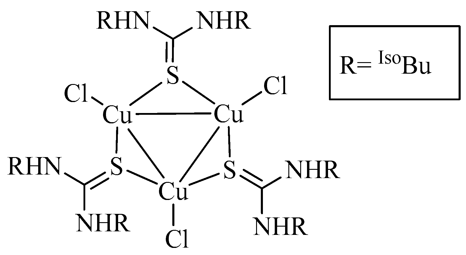
Figure 1. Trimeric copper complex, 1.
Inorganics 10 00185 g001
Figure 2. Dimeric silver complex, 2.
Inorganics 10 00185 g002
Figure 3. Silver cluster, 3.
Inorganics 10 00185 g003
Figure 4. The molecular structure of compound 1, only non-carbon atoms are numbered. Ellipsoids are drawn at 50% probability level; hydrogen atoms are omitted and carbons are represented by capped sticks.
Inorganics 10 00185 g004
Figure 5. 2D representation of 1, the network is stabilized by intermolecular and intramolecular N-H---Cl type interactions.
Inorganics 10 00185 g005
Figure 9. Two-dimensional fingerprint plots for all intermolecular contacts in 2. The percentage of contribution is specified for each contact.
Inorganics 10 00185 g009
Figure 13. Absorption spectra of compound 1. Arrow shows the decreasing change in absorption spectra of the compound/DPPH mixture on increasing concentration of the compound (left). Graphical representation of % Inhibition versus concentration of compound 1 for radical scavenging activity (right).
Inorganics 10 00185 g013
Figure 14. Absorption spectra of blank (DPPH, top) which alters by addition of compound 2 (left), graphical representation of % Inhibition versus concentration of compound 2 mixed with DPPH solution (right).
Inorganics 10 00185 g014
Figure 15. Absorption spectra of compound 3 and pure DPPH, the maximum absorbance was observed for the later one (left) and % Inhibition versus concentration of compound 3 for radical scavenging activity (right).
Inorganics 10 00185 g015
Figure 16. Naked eye detection of anti-DPPH radical scavenging activity shown by compound 2. The solution having purple color on right hand side is blank (without antioxidant agent) and that on left hand side contains 100 ppm of the antioxidant agent.
Inorganics 10 00185 g016
Table 3. DPPH radical scavenging activities (% RSA) of compound (13).
Concentration (ppm)123
205.29.46.5
4013.716.812.5
6018.218.215.6
8019.227.916.0
10019.529.319.2
Publisher’s Note: MDPI stays neutral with regard to jurisdictional claims in published maps and institutional affiliations.