ELI Gammatron Beamline: A Dawn of Ultrafast Hard X-ray Science

 , , , , , , and
Abstract
1. Introduction
2. Numerical Simulation of X/γ-ray Generation in the Gammatron Beamline
3. High-Repetition-Rate Gammatron Beamline
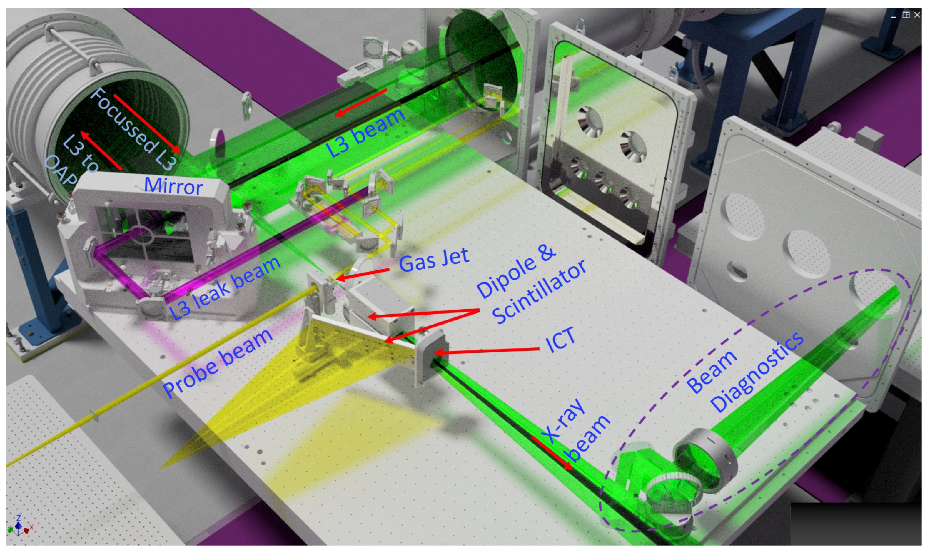
3.1. Experimental Configurations
3.2. Targetry
3.3. Laser and Plasma Diagnostics
3.3.1. Laser Diagnostics
3.3.2. Laser–Plasma Interaction Diagnostics
3.4. Particle and Radiation Diagnostics
3.4.1. Electron Diagnostics
3.4.2. X-ray Diagnostics
- Single-photon-counting X-ray CCD: In the low-photon energy range (<20 keV), a single-photon counting method using a deep depletion X-ray CCD placed a few meters from the X-ray source will be employed. Filters placed in front of the detector will attenuate the X-ray signal, allowing the detection of less than one photon per ten pixels on average. The signal generated in one detection event is therefore directly proportional to the photon energy. An array of scintillators coupled with a position-sensitive photomultiplier tube can be used for a high energy range to enable the spectral measurement in single-photon counting mode.
- Ross filter Spectrometer: In the hard X-ray range (<100 keV), spectrum measurement using the Ross filter pairs will be implemented. Each pair of Ross filters consists of two carefully selected filters of different materials and thicknesses that create closely matching transmittances differing only in their K absorption edges, which define their operational energy ranges. The difference in the signal detected by a CCD camera behind each filter of the pair is directly proportional to the number of photons in the chosen energy range. This method evaluates the spectrum up to 90 keV using 10 spectral bands, and the X-ray spectrum is reconstructed using the filter transmission values and by fitting the data with a selected function (e.g., the synchrotron radiation function). Figure 9b,c shows the ELI Beamlines high energy X-ray spectrometers based on the Ross filter technique. The high-energy spectrometer covers the X-ray spectrum from 8 to 88 keV and makes use of 18 different filters (listed in Table 1). Transmission measurements using thick filters will be implemented for X-rays > 100 keV.
- HAPG crystal spectrometer: A crystal spectrometer with high spectral resolution and high sensitivity, based on diffraction from a highly annealed pyrolytic graphite (HAPG) mosaic crystal (2d = 6.708 Å), will be implemented, in addition to the Ross-filter-based spectrometer mentioned above. The main features of the crystal spectrometer are its high spectral resolution and high sensitivity. The incoming X-ray photons are tightly focused on the CCD camera because of the cylindrical geometry of the crystal. Due to the low divergence of Betatron X-rays, a large number of emitted photons will be collected by the crystal and recorded on a narrow spectral line. The thickness and radius of curvature of the crystal were chosen to be 100 m and 103.4 mm, respectively, to obtain the appropriate spectral resolution. Both the crystal and the CCD camera will be placed in long-travel-range linear stages, allowing scanning of energy by varying the crystal’s position with respect to the source. Measuring the X-ray signal at three different positions provides six data points with first- and second-order diffraction, providing an alternative and more accurate method to measure the Betatron spectrum. In addition, this spectrometer can also be employed in static X-ray absorption spectroscopy, as reported in reference [58].
3.5. X-ray Beam Transport and Focusing Optics
3.6. Multidisciplinary User Station
3.7. X-ray Source Parameters
4. Applications of Gammatron Light Sources
4.1. Applications of Betatron X-ray Source (5–150 keV)
4.1.1. X-ray Imaging
- (a)
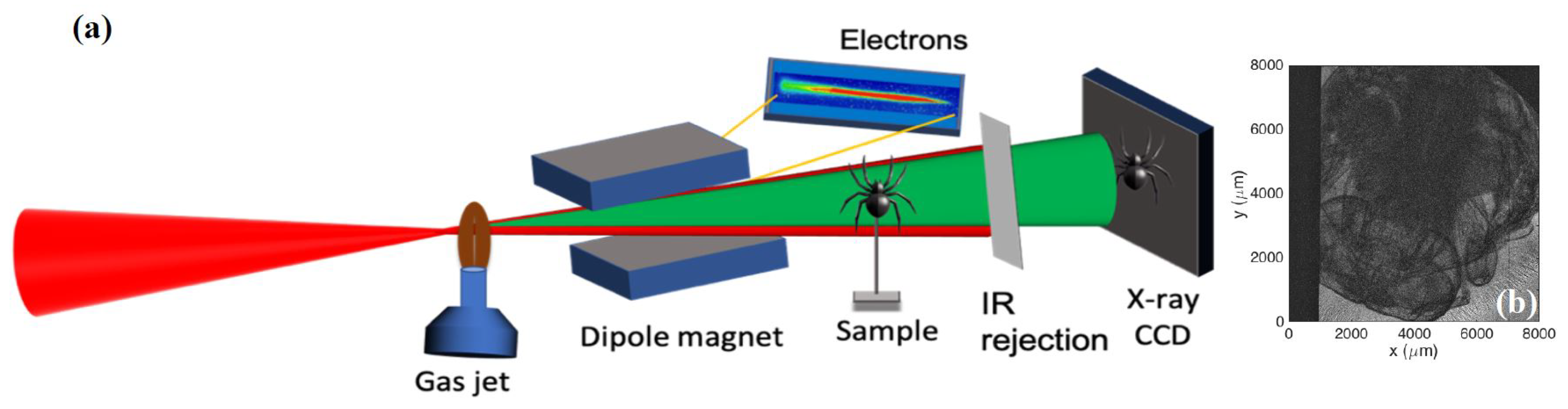
- X-ray phase-contrast imaging (XPCI): It is the most common imaging scheme performed with the partially coherent Betatron X-ray sources, as it offers superior contrast compared to the traditional absorption contrast imaging, specially for imaging specimens comprising lighter elements, such as soft biological tissues. The transverse coherence length of the X-ray source should be greater than O (1–10 m), where is the mean wavelength of the X-ray beam and D is the source-detector distance, to facilitate such imaging [61,62], which is met by Betatron sources owing to their small source size. A few instances of XPCI are already reported using Betatron sources [30,33,63,64]. The schematic scheme of XPCI and XPCI of a dry bee-fly using betatron source is shown in Figure 11. At the ELI Gammatron beamline, we will be able to generate Betatron radiation in the energy range of 10–200 keV and photon flux over 1011 photons per shot, which will be ideal for XPCI of biological samples with various thicknesses, given that 10–40 keV X-rays are used for soft-tissue absorption radiography (e.g., mammography) and 50–150 keV X-rays are used for harder-tissue (bone) radiography.Furthermore, short pulses and a high repetition rate allow motion freezing and time-lapse imaging, enabling real-time visualization of dynamics in the morphologies of biological samples (the respiration and circulatory system, the beating of a heart, etc.). Apart from the inline XPCI, it is also possible to perform speckle-based phase contrast imaging [65] and quantitative X-ray imaging techniques based on Talbot interferometry [66] using Betatron X-ray sources.
- (b)
- X-ray radiography and Microtomography: Radiography and microtomography are important methods employed in archaeology, biomedical sciences (both in vitro and in vivo), developmental biology, electronics and microdevices, food and paleontology, and various other fields [67]. A Betatron X-ray source is ideal for high-resolution radiography. The micron source size allows high-resolution measurements by exploiting high geometric magnification and relaxing the resolution requirements of the detector. Additionally, the possibility of single-shot exposure owing to the high brightness of the Betatron source helps to avoid vibration-induced artifacts in measurements, which has been recently demonstrated [35,68].
- (c)
- Subsurface probing of high-energy-density (HED) state materials: The laser-driven shock wave is an efficient way to generate dynamic high pressure for exploring the dynamical strengths of materials. Shocks are important in various fields of research, such as condensed matter physics, nuclear fusion, HED science [69,70,71,72], and laboratory astrophysics [73,74], as they provide the ability to test the extreme conditions of matter in terms of both pressure and temperature. This is extremely important in inertial confinement fusion research, where a series of shocks compress the fuel and capsules.A high-power laser pulse can generate an ultrahigh pressure (tens of megabar to one gigabar) or mechanical shock inside the irradiated material locally [69,70,71], creating an extreme state of matter with high temperatures and densities that causes structural defects, local melting and fast recrystallization, phase transitions compositional inhomogeneities, etc. The laser-driven radiative shocks can be very dense ( > 1021 cm−3), and therefore, imaging these shocks requires a radiation source with high-energy photons to penetrate high-density plasmas, and sufficient brightness to overcome the plasma self-emission. High resolution subsurface probing is a key method for a real-time diagnosis of material behavior and inhomogeneities at the microscale. The Betatron X-ray sources have tremendous potential for applications in HED science, as they have remarkable spectral, spatial, and temporal properties. Due to their micron-scale source size, they possess significant spatial coherence, allowing the phase contrast imaging of the thin shock front. The intrinsic temporal resolution of the Betatron source is in the order of 10 fs, indicating that many ultrafast processes could be probed without motion blurring and/or compromising spatial resolution. A recent report demonstrated the ability of a Betatron source to image the laser-driven shock wave propagating in matter [37]. Imaging the shock propagation in matter using ultrafast X-ray provides a unique way to understand the material’s failure mechanism and lattice defects with unprecedented resolution and provide real-time measurements of the microstructural evolution in the sample.
- (d)
- Quantum Imaging (QI): X-rays are ionizing radiation and hence can cause radiation damage to the specimen during the measurement. The damage in the specimen can be avoided by using the ultra-short pulsed X-ray sources, as proposed in the concept of “diffraction before destruction” [3]. Currently, such damage-free measurements are only possible with X-ray free-electron lasers; the other alternative to minimize the radiation-induced damage is low-dose imaging. The application of low-dose quantum imaging methods such as ghost imaging and interaction-free imaging has been well-demonstrated with visible light and recently with X-rays [75,76]. These methods are based on the intensity correlation of the reference beams and the object beam, as in the Hanbury Brown and Twiss interferometry experiments [77]. However, a major limiting factor in QI is the poor contrast of the images, which can be enhanced by employing femtosecond Betatron sources [78].
4.1.2. Time-Resolved X-ray Diffraction and Spectroscopy
- (a)
- Time-resolved X-ray Diffraction: X-ray diffraction is widely used for the structural characterization of materials and biological specimens. X-ray sources are used to provide structural details with atomic resolution. They can also reveal structural dynamics if they can deliver X-rays pulses. The picosecond time scale can be unravelled by the time-resolved experiments at synchrotrons. At the femtosecond time scale, such studies are performed with X-ray free-electron lasers, laser-driven K-alpha sources, and Betatron X-rays, and therefore, we foresee time-resolved diffraction as an important end-user experiment for the Gammatron beamline at ELI beamlines, with the possibility of the following measurements.(i) Single Crystal X-ray Diffraction: It is the method of choice in both materials science and biological science to obtain the structure of a material. Ultrafast time-resolved X-ray studies of larger single crystals, especially in materials science, have been reported previously with laser-driven K-alpha sources by following the dynamics of the angular position or the intensity of Bragg peaks [83]. In biology, single crystal structural studies with laser-driven K-alpha source were reported [84]. However, the entire structure has never been revealed. Recently, a study showed that with a direct detector operated at a kilohertz frame rate and with the flux comparable to that from the laser-driven X-ray sources, structural studies of a single crystal of the sizes of ∼50 microns can be performed [85], which emphasizes the possibility to perform single-crystal Laue diffraction using the Betatron X-ray source at the Gammatron beamline.(ii) Time-resolved powder diffraction: Crystals of both inorganic and organic molecules do not grow into sizes suitable for single crystal studies with traditional X-ray sources and hence are investigated either with X-ray free-electron lasers or with electrons [86]. Alternatively, structural studies of such small crystals can also be done with powder diffraction. Ultrafast time-resolved powder diffraction studies using various laser-driven X-ray sources have already been reported [87], proving the possibility of extending it at the Gammatron beamline too, making it a multi-functional user end station.(iii) Time-resolved small angle and wide-angle scattering (SAXS/WAXS) of solutions: For the structural studies of small molecules and macromolecules, it is preferred that they are dissolved in solution rather than in the crystalline form. Probing the structure of molecules in solution with SAXS and WAXS is a standard operation at synchrotrons [88], and a recent report demonstrated SAXS/WAXS of biological macromolecules using laboratory sources with 106 photon/s [89]. This brings a bright perspective for the Betatron source to perform time-resolved structural studies of solutions which allow investigation of bond formation and breaking dynamics, ultrafast quakes in proteins, and several other structural transitions occurring at sub-picosecond timescales.
- (b)
- Time-resolved femtosecond X-ray absorption spectroscopy: Time-resolved femtosecond X-ray spectroscopy has witnessed impressive growth and development in the past decade [90]. These developments have been specially powered by XFELs. The laser-driven X-ray sources have, however, played significant complementary roles in the development of these methods. The Betatron source has a short pulse duration and a smooth broadband spectrum, which makes it an ideal source for time-resolved X-ray absorption spectroscopy.(i) Time-resolved X-ray absorption spectroscopy (TR-XAS): Time-resolved X-ray absorption spectroscopy is a powerful tool in atomic physics, as it simultaneously reveals both electronic and atomic structures [91]. The X-ray absorption near-edge structure (XANES) and extended X-ray absorption fine structure (EXAFS) exploit a wide range of applications in chemistry, material science, studies of gas-phase systems, and/or complex biological samples. XAS has been extensively developed at synchrotrons, with X-ray energy in the range of 1–30 keV and a limited temporal resolution due to the duration of the synchrotron pulses (∼10 ps). For most processes governed by atomic motion, such as structural changes and phase transitions, the timescale of interest corresponds to one vibrational period, i.e., ≈100 fs. XFEL sources are the most commonly used sources for TR-XAS experiments at the femtosecond time scale. However, the betatron source that gathers a broadband smooth spectrum and a femtosecond duration has been demonstrated to be as a powerful complementary source [92,93,94].(ii) Time-resolved X-ray emission spectroscopy (TRXES): In addition to the information about the nature of bound ligands in a system [95], X-ray emission spectroscopy provides complementary information to X-ray absorption spectroscopy, on the electrons inside the specimen. TRXES has been demonstrated well with XFELs [96], and the broadband emission from Betatron sources can extend the capabilities of such investigations. The broadband Betatron sources can be efficiently used for simultaneous multi-excitation of the material, hence probing the charge and spin dynamics of both the light and heavy atoms inside the specimen, including both the valence and core electron dynamics.
- (c)
- Simultaneous time-resolved X-ray spectroscopy and diffraction: Micron-sized ultrafast X-ray sources increase the flexibility of designing new tools for probing various samples, as in simultaneous time-resolved X-ray spectroscopy and diffraction. While X-ray spectroscopy provides information on the electronic, spin, and magnetization states, X-ray diffraction provides structural information. For photo-excited systems with non-radiative decay pathways, the system undergoes both electronic and structural transitions in tens of femtoseconds to picoseconds. Simultaneous measurement of X-ray spectrum and diffraction enables the probing of such systems and uncouples the electronic and structural transitions [97]. Additionally, this technique would benefit the probing of systems such as hemeproteins, where the association/dissociation of the ligands induces a change in the spin state and structural quakes in the system. The Gammatron beamline has prepared a user platform to provide this experimental capacity to the users.
4.1.3. Industrial Applications
4.2. Applications of an Inverse-Compton Scattering X-ray Source (150 keV–20 MeV)
4.2.1. High-Resolution Industrial Imaging: X-ray Radiography/Tomography
4.2.2. Nuclear Resonance Fluorescence (NRF)
5. Conclusions and Perspectives
Author Contributions
Funding
Institutional Review Board Statement
Informed Consent Statement
Data Availability Statement
Acknowledgments
Conflicts of Interest
Abbreviations
| ELI | Extreme Light Infrastructure |
| XFEL | X-ray Free-Electron Laser |
| fs | Femtosecond |
| HHG | High-order Harmonic Generation |
| LWFA | Laser Wakefield Acceleration |
| LPA | Laser Plasma Accelerator |
| TS | Thomson Scattering |
| ICS | Inverse Compton Scattering |
| HAPLS | High-Repetition-Rate Advanced Petawatt Laser System |
| DUHA | Dual-beam Ultra-fast High energy OPCPA Amplifier |
| PW | Pettawatt |
| OAP | Off-Axis Parabola |
| ES | Electron Spectrometer |
| ICT | Integrated Current Transformer |
| HAPG | Highly Annealed Pyrolytic Graphite |
| KB | Kirkpatrick-Baez |
| GaUS | Gammatron User Station |
| XPCI | X-ray phase-contrast imaging |
| HED | High Energy Density |
| QI | Quantum Imaging |
| SAXS | Small-Angle X-ray Scattering |
| WAXS | Wide-Angle X-ray Scattering |
| TR-XAS | Time-Resolved X-ray Absorption Spectroscopy |
| XANES | X-ray Absorption Near-Edge Structure |
| EXAFS | Extended X-ray Absorption Fine Structure |
| LCLS | Linac Coherent Light Source |
| WDM | Warm Dense Matter |
| TRXES | Time-Resolved X-ray Emission Spectroscopy |
| NDE | Non-Destructive Evaluation |
| NRF | Nuclear Resonance Fluorescence |
References
- Sakdinawat, A.; Attwood, D. Nanoscale X-ray imaging. Nat. Photonics 2010, 4, 840–848. [Google Scholar] [CrossRef]
- Boutet, S.; Lomb, L.; Williams, G.J.; Barends, T.R.; Aquila, A.; Doak, R.B.; Weierstall, U.; DePonte, D.P.; Steinbrener, J.; Shoeman, R.L.; et al. High-resolution protein structure determination by serial femtosecond crystallography. Science 2012, 337, 362–364. [Google Scholar] [CrossRef] [PubMed]
- Neutze, R.; Wouts, R.; Van der Spoel, D.; Weckert, E.; Hajdu, J. Potential for biomolecular imaging with femtosecond X-ray pulses. Nature 2000, 406, 752–757. [Google Scholar] [CrossRef] [PubMed]
- Chapman, H.N.; Fromme, P.; Barty, A.; White, T.A.; Kirian, R.A.; Aquila, A.; Hunter, M.S.; Schulz, J.; DePonte, D.P.; Weierstall, U.; et al. Femtosecond X-ray protein nanocrystallography. Nature 2011, 470, 73–77. [Google Scholar] [CrossRef]
- Schoenlein, R.W.; Leemans, W.; Chin, A.; Volfbeyn, P.; Glover, T.; Balling, P.; Zolotorev, M.; Kim, K.J.; Chattopadhyay, S.; Shank, C. Femtosecond X-ray pulses at 0.4 Å generated by 90 Thomson scattering: A tool for probing the structural dynamics of materials. Science 1996, 274, 236–238. [Google Scholar] [CrossRef]
- Cavalleri, A.; Tóth, C.; Siders, C.W.; Squier, J.; Ráksi, F.; Forget, P.; Kieffer, J. Femtosecond structural dynamics in VO2 during an ultrafast solid–solid phase transition. Phys. Rev. Lett. 2001, 87, 237401. [Google Scholar] [CrossRef] [PubMed]
- Tajima, T.; Malka, V. Laser plasma accelerators. Plasma Phys. Control. Fusion 2020, 62, 034004. [Google Scholar] [CrossRef]
- Gaarde, M.B.; Tate, J.L.; Schafer, K.J. Macroscopic aspects of attosecond pulse generation. J. Phys. B At. Mol. Opt. Phys. 2008, 41, 132001. [Google Scholar] [CrossRef]
- Li, J.; Ren, X.; Yin, Y.; Zhao, K.; Chew, A.; Cheng, Y.; Cunningham, E.; Wang, Y.; Hu, S.; Wu, Y.; et al. 53-attosecond X-ray pulses reach the carbon K-edge. Nat. Commun. 2017, 8, 1–5. [Google Scholar] [CrossRef]
- Hort, O.; Albrecht, M.; Nefedova, V.E.; Finke, O.; Mai, D.; Reyné, S.; Giambruno, F.; Frassetto, F.; Poletto, L.; Andreasson, J.; et al. High-flux source of coherent XUV pulses for user applications. Opt. Express 2019, 27, 8871–8883. [Google Scholar] [CrossRef]
- Makos, I.; Orfanos, I.; Nayak, A.; Peschel, J.; Major, B.; Liontos, I.; Skantzakis, E.; Papadakis, N.; Kalpouzos, C.; Dumergue, M.; et al. A 10-gigawatt attosecond source for non-linear XUV optics and XUV-pump-XUV-probe studies. Sci. Rep. 2020, 10, 3759. [Google Scholar] [CrossRef] [PubMed]
- Schötz, J.; Förg, B.; Schweinberger, W.; Liontos, I.; Masood, H.; Kamal, A.; Jakubeit, C.; Kling, N.; Paasch-Colberg, T.; Biswas, S.; et al. Phase-matching for generation of isolated attosecond xuv and soft-X-ray pulses with few-cycle drivers. Phys. Rev. X 2020, 10, 041011. [Google Scholar] [CrossRef]
- Tajima, T.; Dawson, J.M. Laser electron accelerator. Phys. Rev. Lett. 1979, 43, 267. [Google Scholar] [CrossRef]
- Leemans, W.; Gonsalves, A.; Mao, H.S.; Nakamura, K.; Benedetti, C.; Schroeder, C.; Tóth, C.; Daniels, J.; Mittelberger, D.; Bulanov, S.; et al. Multi-GeV electron beams from capillary-discharge-guided subpetawatt laser pulses in the self-trapping regime. Phys. Rev. Lett. 2014, 113, 245002. [Google Scholar] [CrossRef]
- Gonsalves, A.; Nakamura, K.; Daniels, J.; Benedetti, C.; Pieronek, C.; De Raadt, T.; Steinke, S.; Bin, J.; Bulanov, S.; Van Tilborg, J.; et al. Petawatt laser guiding and electron beam acceleration to 8 GeV in a laser-heated capillary discharge waveguide. Phys. Rev. Lett. 2019, 122, 084801. [Google Scholar] [CrossRef]
- Rousse, A.; Phuoc, K.T.; Shah, R.; Pukhov, A.; Lefebvre, E.; Malka, V.; Kiselev, S.; Burgy, F.; Rousseau, J.P.; Umstadter, D.; et al. Production of a keV X-ray beam from synchrotron radiation in relativistic laser-plasma interaction. Phys. Rev. Lett. 2004, 93, 135005. [Google Scholar] [CrossRef]
- Ta Phuoc, K.; Fitour, R.; Tafzi, A.; Garl, T.; Artemiev, N.; Shah, R.; Albert, F.; Boschetto, D.; Rousse, A.; Kim, D.; et al. Demonstration of the ultrafast nature of laser produced betatron radiation. Phys. Plasmas 2007, 14, 080701. [Google Scholar] [CrossRef]
- Corde, S.; Phuoc, K.T.; Lambert, G.; Fitour, R.; Malka, V.; Rousse, A.; Beck, A.; Lefebvre, E. Femtosecond x rays from laser-plasma accelerators. Rev. Mod. Phys. 2013, 85, 1. [Google Scholar] [CrossRef]
- Ta Phuoc, K.; Corde, S.; Thaury, C.; Malka, V.; Tafzi, A.; Goddet, J.P.; Shah, R.; Sebban, S.; Rousse, A. All-optical Compton gamma-ray source. Nat. Photonics 2012, 6, 308–311. [Google Scholar] [CrossRef]
- Chen, S.; Powers, N.; Ghebregziabher, I.; Maharjan, C.; Liu, C.; Golovin, G.; Banerjee, S.; Zhang, J.; Cunningham, N.; Moorti, A.; et al. MeV-energy X rays from inverse Compton scattering with laser-wakefield accelerated electrons. Phys. Rev. Lett. 2013, 110, 155003. [Google Scholar] [CrossRef]
- Maier, A.R.; Delbos, N.M.; Eichner, T.; Hübner, L.; Jalas, S.; Jeppe, L.; Jolly, S.W.; Kirchen, M.; Leroux, V.; Messner, P.; et al. Decoding sources of energy variability in a laser-plasma accelerator. Phys. Rev. X 2020, 10, 031039. [Google Scholar] [CrossRef]
- Corde, S.; Phuoc, K.T.; Fitour, R.; Faure, J.; Tafzi, A.; Goddet, J.P.; Malka, V.; Rousse, A. Controlled betatron X-ray radiation from tunable optically injected electrons. Phys. Rev. Lett. 2011, 107, 255003. [Google Scholar] [CrossRef] [PubMed]
- Fourmaux, S.; Hallin, E.; Chaulagain, U.; Weber, S.; Kieffer, J. Laser-based synchrotron X-ray radiation experimental scaling. Opt. Express 2020, 28, 3147–3158. [Google Scholar] [CrossRef] [PubMed]
- Mourou, G.A.; Korn, G.; Sandner, W.; Collier, J.L. Eli-Extreme Light Infrastructure: Science and Technology with Ultra-Intense Lasers, Whitebook; THOSS Media GmbH: Berlin, Germany, 2011. [Google Scholar]
- ELI Beamlines—Dolní Břežany. Available online: https://www.eli-beams.eu/ (accessed on 7 November 2022).
- Nejdl, J.; Mai, D.D.; Chaulagain, U.; Hort, O.; Finke, O.; Albrecht, M.; Jurkovič, M.; Lera, R.; Karatodorov, S.; Lamač, M.; et al. Progress on laser-driven X-ray sources at ELI Beamlines. In Proceedings of the X-ray Lasers and Coherent X-ray Sources: Development and Applications XIII, San Diego, CA, USA, 12–13 August 2019; Volume 11111, pp. 44–50. [Google Scholar]
- Chaulagain, U.; Boháček, K.; Vančura, J.; Lamač, M.; Yan, W.; Gu, Y.; Kozlová, M.; Ta-Phuoc, K.; Weber, S.; Nejdl, J. LWFA-driven betatron source for Plasma Physics Platform at ELI Beamlines. In Proceedings of the International Conference on X-ray Lasers, Prague, Czech Republic, 7–12 October 2018; pp. 117–123. [Google Scholar]
- Margarone, D.; Cirrone, G.P.; Cuttone, G.; Amico, A.; Andò, L.; Borghesi, M.; Bulanov, S.S.; Bulanov, S.V.; Chatain, D.; Fajstavr, A.; et al. ELIMAIA: A laser-driven ion accelerator for multidisciplinary applications. Quantum Beam Sci. 2018, 2, 8. [Google Scholar] [CrossRef]
- Sarri, G.; Corvan, D.; Schumaker, W.; Cole, J.; Di Piazza, A.; Ahmed, H.; Harvey, C.; Keitel, C.H.; Krushelnick, K.; Mangles, S.; et al. Ultrahigh brilliance multi-MeV γ-ray beams from nonlinear relativistic Thomson scattering. Phys. Rev. Lett. 2014, 113, 224801. [Google Scholar] [CrossRef]
- Kneip, S.; McGuffey, C.; Dollar, F.; Bloom, M.; Chvykov, V.; Kalintchenko, G.; Krushelnick, K.; Maksimchuk, A.; Mangles, S.; Matsuoka, T.; et al. X-ray phase contrast imaging of biological specimens with femtosecond pulses of betatron radiation from a compact laser plasma wakefield accelerator. Appl. Phys. Lett. 2011, 99, 093701. [Google Scholar] [CrossRef]
- Ju, J.; Svensson, K.; Döpp, A.; Ferrari, H.; Cassou, K.; Neveu, O.; Genoud, G.; Wojda, F.; Burza, M.; Persson, A.; et al. Enhancement of X-rays generated by a guided laser wakefield accelerator inside capillary tubes. Appl. Phys. Lett. 2012, 100, 191106. [Google Scholar] [CrossRef]
- Wang, X.; Zgadzaj, R.; Fazel, N.; Li, Z.; Yi, S.; Zhang, X.; Henderson, W.; Chang, Y.Y.; Korzekwa, R.; Tsai, H.E.; et al. Quasi-monoenergetic laser-plasma acceleration of electrons to 2 GeV. Nat. Commun. 2013, 4, 1988. [Google Scholar] [CrossRef]
- Wenz, J.; Schleede, S.; Khrennikov, K.; Bech, M.; Thibault, P.; Heigoldt, M.; Pfeiffer, F.; Karsch, S. Quantitative X-ray phase-contrast microtomography from a compact laser-driven betatron source. Nat. Commun. 2015, 6, 7568. [Google Scholar] [CrossRef]
- Chen, L.; Yan, W.; Li, D.; Hu, Z.; Zhang, L.; Wang, W.; Hafz, N.; Mao, J.; Huang, K.; Ma, Y.; et al. Bright betatron X-ray radiation from a laser-driven-clustering gas target. Sci. Rep. 2013, 3, 1912. [Google Scholar] [CrossRef]
- Cole, J.; Wood, J.; Lopes, N.; Poder, K.; Abel, R.; Alatabi, S.; Bryant, J.; Jin, A.; Kneip, S.; Mecseki, K.; et al. Laser-wakefield accelerators as hard X-ray sources for 3D medical imaging of human bone. Sci. Rep. 2015, 5, 13244. [Google Scholar] [CrossRef] [PubMed]
- Döpp, A.; Mahieu, B.; Lifschitz, A.; Thaury, C.; Doche, A.; Guillaume, E.; Grittani, G.; Lundh, O.; Hansson, M.; Gautier, J.; et al. Stable femtosecond X-rays with tunable polarization from a laser-driven accelerator. Light. Sci. Appl. 2017, 6, e17086. [Google Scholar] [CrossRef] [PubMed]
- Wood, J.; Chapman, D.; Poder, K.; Lopes, N.; Rutherford, M.; White, T.; Albert, F.; Behm, K.; Booth, N.; Bryant, J.; et al. Ultrafast imaging of laser driven shock waves using betatron X-rays from a laser wakefield accelerator. Sci. Rep. 2018, 8, 11010. [Google Scholar] [CrossRef] [PubMed]
- Fourmaux, S.; Hallin, E.; Arnison, P.; Kieffer, J. Optimization of laser-based synchrotron X-ray for plant imaging. Appl. Phys. B 2019, 125, 1–9. [Google Scholar] [CrossRef]
- Tsung, F.S.; Narang, R.; Mori, W.B.; Joshi, C.; Fonseca, R.; Silva, L.O. Near-GeV-energy laser-wakefield acceleration of self-injected electrons in a centimeter-scale plasma channel. Phys. Rev. Lett. 2004, 93, 185002. [Google Scholar] [CrossRef]
- Bulanov, S.; Kirsanov, V.; Sakharov, A. Limiting electric field of the wakefield plasma wave. JETP Lett. 1991, 53, 565–569. [Google Scholar]
- Modena, A.; Najmudin, Z.; Dangor, A.; Clayton, C.; Marsh, K.; Joshi, C.; Malka, V.; Darrow, C.; Danson, C.; Neely, D.; et al. Electron acceleration from the breaking of relativistic plasma waves. Nature 1995, 377, 606–608. [Google Scholar] [CrossRef]
- Jackson, J.D. Classical Electrodynamics, 3rd ed.; John Wiley & Sons: New York, NY, USA, 1999. [Google Scholar]
- Thaury, C.; Guillaume, E.; Döpp, A.; Lehe, R.; Lifschitz, A.; Ta Phuoc, K.; Gautier, J.; Goddet, J.P.; Tafzi, A.; Flacco, A.; et al. Demonstration of relativistic electron beam focusing by a laser-plasma lens. Nat. Commun. 2015, 6, 6860. [Google Scholar] [CrossRef]
- Lamač, M.; Chaulagain, U.; Jurkovič, M.; Nejdl, J.; Bulanov, S. Two-color nonlinear resonances in betatron oscillations of laser accelerated relativistic electrons. Phys. Rev. Res. 2021, 3, 033088. [Google Scholar] [CrossRef]
- Kozlova, M.; Andriyash, I.; Gautier, J.; Sebban, S.; Smartsev, S.; Jourdain, N.; Chaulagain, U.; Azamoum, Y.; Tafzi, A.; Goddet, J.P.; et al. Hard X rays from laser-wakefield accelerators in density tailored plasmas. Phys. Rev. X 2020, 10, 011061. [Google Scholar] [CrossRef]
- Björklund S, J.; Guénot, D.; Ferri, J.; Ekerfelt, H.; Gallardo González, I.; Persson, A.; Svendsen, K.; Veisz, L.; Lundh, O. Low-divergence femtosecond X-ray pulses from a passive plasma lens. Nat. Phys. 2021, 17, 639–645. [Google Scholar] [CrossRef]
- Green, J.T.; Antipenkov, R.; Bakule, P. L2-DUHA 100 TW high repetition rate laser system at ELI-Beamlines. Key design considerations. Rev. Laser Eng. 2021, 49, 106–109. [Google Scholar]
- Willemsen, T.; Chaulagain, U.; Havlíčková, I.; Borneis, S.; Ebert, W.; Ehlers, H.; Gauch, M.; Groß, T.; Kramer, D.; Laštovička, T.; et al. Large area ion beam sputtered dielectric ultrafast mirrors for petawatt laser beamlines. Opt. Express 2022, 30, 6129–6141. [Google Scholar] [CrossRef] [PubMed]
- Borneis, S.; Laštovička, T.; Sokol, M.; Jeong, T.M.; Condamine, F.; Renner, O.; Tikhonchuk, V.; Bohlin, H.; Fajstavr, A.; Hernandez, J.C.; et al. Design, installation and commissioning of the ELI-Beamlines high-power, high-repetition rate HAPLS laser beam transport system to P3. High Power Laser Sci. Eng. 2021, 9, E30. [Google Scholar] [CrossRef]
- McGuffey, C.; Thomas, A.; Schumaker, W.; Matsuoka, T.; Chvykov, V.; Dollar, F.; Kalintchenko, G.; Yanovsky, V.; Maksimchuk, A.; Krushelnick, K.; et al. Ionization induced trapping in a laser wakefield accelerator. Phys. Rev. Lett. 2010, 104, 025004. [Google Scholar] [CrossRef] [PubMed]
- Huang, K.; Li, D.; Yan, W.; Li, M.; Tao, M.; Chen, Z.; Ge, X.; Liu, F.; Ma, Y.; Zhao, J.; et al. Simultaneous generation of quasi-monoenergetic electron and betatron X-rays from nitrogen gas via ionization injection. Appl. Phys. Lett. 2014, 105, 204101. [Google Scholar] [CrossRef]
- Boháček, K.; Kozlová, M.; Nejdl, J.; Chaulagain, U.; Hornỳ, V.; Krus, M.; Phuoc, K.T. Stable electron beams from laser wakefield acceleration with few-terawatt driver using a supersonic air jet. Nucl. Instrum. Methods Phys. Res. Sect. A Accel. Spectrometers Detect. Assoc. Equip. 2018, 883, 24–28. [Google Scholar] [CrossRef]
- Bulanov, S.; Naumova, N.; Pegoraro, F.; Sakai, J. Particle injection into the wave acceleration phase due to nonlinear wake wave breaking. Phys. Rev. E 1998, 58, R5257. [Google Scholar] [CrossRef]
- Nejdl, J.; Vančura, J.; Boháček, K.; Albrecht, M.; Chaulagain, U. Imaging Michelson interferometer for a low-density gas jet characterization. Rev. Sci. Instrum. 2019, 90, 065107. [Google Scholar] [CrossRef]
- Chaulagain, U.; Karatodorov, S.; Raclavskỳ, M.; Lorenz, S.; Lamač, M.; Albrecht, M.; Tomkus, V.; Dudutis, J.; Mackevičiūtė, M.; Gečys, P.; et al. Tomographic characterization of gas jets for laser-plasma acceleration with increased sensitivity. In Proceedings of the International Conference on X-ray Lasers 2020, Online, 8–10 October 2020; Volume 11886, pp. 61–67. [Google Scholar]
- Karatodorov, S.; Lera, R.; Raclavsky, M.; Lorenz, S.; Chaulagain, U.; Nejdl, J. Multi-pass probing for high-sensitivity tomographic interferometry. Sci. Rep. 2021, 11, 15072. [Google Scholar] [CrossRef]
- Glinec, Y.; Faure, J.; Guemnie-Tafo, A.; Malka, V.; Monard, H.; Larbre, J.; De Waele, V.; Marignier, J.; Mostafavi, M. Absolute calibration for a broad range single shot electron spectrometer. Rev. Sci. Instrum. 2006, 77, 103301. [Google Scholar] [CrossRef]
- Kettle, B.; Gerstmayr, E.; Streeter, M.; Albert, F.; Baggott, R.; Bourgeois, N.; Cole, J.; Dann, S.; Falk, K.; González, I.G.; et al. Single-shot multi-keV X-ray absorption spectroscopy using an ultrashort laser-wakefield accelerator source. Phys. Rev. Lett. 2019, 123, 254801. [Google Scholar] [CrossRef] [PubMed]
- Raclavský, M.; Khakurel, K.P.; Chaulagain, U.; Lamač, M.; Nejdl, J. Multi-Lane Mirror for Broadband Applications of the Betatron X-ray Source. Photonics 2021, 8, 579. [Google Scholar] [CrossRef]
- Albert, F.; Thomas, A.G. Applications of laser wakefield accelerator-based light sources. Plasma Phys. Control. Fusion 2016, 58, 103001. [Google Scholar] [CrossRef]
- Davis, T.J.; Gao, D.; Gureyev, T.; Stevenson, A.; Wilkins, S. Phase-contrast imaging of weakly absorbing materials using hard X-rays. Nature 1995, 373, 595–598. [Google Scholar] [CrossRef]
- Wilkins, S.; Gureyev, T.E.; Gao, D.; Pogany, A.; Stevenson, A. Phase-contrast imaging using polychromatic hard X-rays. Nature 1996, 384, 335–338. [Google Scholar] [CrossRef]
- Fourmaux, S.; Corde, S.; Phuoc, K.T.; Lassonde, P.; Lebrun, G.; Payeur, S.; Martin, F.; Sebban, S.; Malka, V.; Rousse, A.; et al. Single shot phase contrast imaging using laser-produced Betatron X-ray beams. Opt. Lett. 2011, 36, 2426–2428. [Google Scholar] [CrossRef]
- Chaulagain, U.; Bohacek, K.; Kozlova, M.; Nejdl, J.; Krus, M.; Horny, V.; Mahieu, B.; Ta-Phuoc, K. X-ray phase contrast imaging of biological samples using a betatron X-ray source generated in a laser wakefield accelerator. In Proceedings of the Laser Acceleration of Electrons, Protons, and Ions IV, Prague, Czech Republic, 24–26 April 2017; Volume 10240, pp. 110–114. [Google Scholar]
- Zdora, M.C. State of the art of X-ray speckle-based phase-contrast and dark-field imaging. J. Imaging 2018, 4, 60. [Google Scholar] [CrossRef]
- Momose, A.; Yashiro, W.; Harasse, S.; Kuwabara, H. Four-dimensional X-ray phase tomography with Talbot interferometry and white synchrotron radiation: Dynamic observation of a living worm. Opt. Express 2011, 19, 8423–8432. [Google Scholar] [CrossRef]
- Landis, E.N.; Keane, D.T. X-ray microtomography. Mater. Charact. 2010, 61, 1305–1316. [Google Scholar] [CrossRef]
- Döpp, A.; Hehn, L.; Götzfried, J.; Wenz, J.; Gilljohann, M.; Ding, H.; Schindler, S.; Pfeiffer, F.; Karsch, S. Quick X-ray microtomography using a laser-driven betatron source. Optica 2018, 5, 199–203. [Google Scholar] [CrossRef]
- Suzuki-Vidal, F.; Clayson, T.; Stehlé, C.; Swadling, G.; Foster, J.; Skidmore, J.; Graham, P.; Burdiak, G.; Lebedev, S.; Chaulagain, U.; et al. Counterpropagating radiative shock experiments on the orion laser. Phys. Rev. Lett. 2017, 119, 055001. [Google Scholar] [CrossRef] [PubMed]
- Ping, Y.; Coppari, F.; Hicks, D.; Yaakobi, B.; Fratanduono, D.; Hamel, S.; Eggert, J.; Rygg, J.; Smith, R.; Swift, D.; et al. Solid iron compressed up to 560 GPa. Phys. Rev. Lett. 2013, 111, 065501. [Google Scholar] [CrossRef] [PubMed]
- Tsujino, M.; Sano, T.; Sakata, O.; Ozaki, N.; Kimura, S.; Takeda, S.; Okoshi, M.; Inoue, N.; Kodama, R.; Kobayashi, K.F.; et al. Synthesis of submicron metastable phase of silicon using femtosecond laser-driven shock wave. J. Appl. Phys. 2011, 110, 126103. [Google Scholar]
- Suzuki-Vidal, F.; Clayson, T.; Stehlé, C.; Chaulagain, U.; Halliday, J.W.; Sun, M.; Ren, L.; Kang, N.; Liu, H.; Zhu, B.; et al. First radiative shock experiments on the SG-II laser. High Power Laser Sci. Eng. 2021, 9, E27. [Google Scholar] [CrossRef]
- Singh, R.; Stehlé, C.; Suzuki-Vidal, F.; Kozlova, M.; Larour, J.; Chaulagain, U.; Clayson, T.; Rodriguez, R.; Gil, J.; Nejdl, J.; et al. Experimental study of the interaction of two laser-driven radiative shocks at the PALS laser. High Energy Density Phys. 2017, 23, 20–30. [Google Scholar] [CrossRef]
- Chaulagain, U.; Stehlé, C.; Larour, J.; Kozlová, M.; Suzuki-Vidal, F.; Barroso, P.; Cotelo, M.; Velarde, P.; Rodriguez, R.; Gil, J.; et al. Structure of a laser-driven radiative shock. High Energy Density Phys. 2015, 17, 106–113. [Google Scholar] [CrossRef]
- Pelliccia, D.; Rack, A.; Scheel, M.; Cantelli, V.; Paganin, D.M. Experimental X-ray Ghost Imaging. Phys. Rev. Lett. 2016, 117, 113902. [Google Scholar] [CrossRef]
- Zhang, A.X.; He, Y.H.; Wu, L.A.; Chen, L.M.; Wang, B.B. Tabletop X-ray ghost imaging with ultra-low radiation. Optica 2018, 5, 374–377. [Google Scholar] [CrossRef]
- Brown, R.H.; Twiss, R.Q. LXXIV. A new type of interferometer for use in radio astronomy. Lond. Edinb. Dublin Philos. Mag. J. Sci. 1954, 45, 663–682. [Google Scholar] [CrossRef]
- Kim, Y.Y.; Gelisio, L.; Mercurio, G.; Dziarzhytski, S.; Beye, M.; Bocklage, L.; Classen, A.; David, C.; Gorobtsov, O.Y.; Khubbutdinov, R.; et al. Ghost imaging at an XUV free-electron laser. Phys. Rev. A 2020, 101, 013820. [Google Scholar] [CrossRef]
- Chergui, M.; Collet, E. Photoinduced structural dynamics of molecular systems mapped by time-resolved X-ray methods. Chem. Rev. 2017, 117, 11025–11065. [Google Scholar] [CrossRef] [PubMed]
- Neutze, R.; Moffat, K. Time-resolved structural studies at synchrotrons and X-ray free electron lasers: Opportunities and challenges. Curr. Opin. Struct. Biol. 2012, 22, 651–659. [Google Scholar] [CrossRef] [PubMed]
- Afshari, M.; Krumey, P.; Menn, D.; Nicoul, M.; Brinks, F.; Tarasevitch, A.; Sokolowski-Tinten, K. Time-resolved diffraction with an optimized short pulse laser plasma X-ray source. Struct. Dyn. 2020, 7, 014301. [Google Scholar] [CrossRef] [PubMed]
- Anwar, M.; Iqbal, M.; Hwang, B.; Faiyaz, M.; Mun, B.; Janulewicz, K.; Noh, D. Ultrafast X-ray absorption near edge spectroscopy of Fe3O4 using a laboratory based femtosecond X-ray source. Opt. Express 2019, 27, 6030–6036. [Google Scholar] [CrossRef] [PubMed]
- Sokolowski-Tinten, K.; Blome, C.; Blums, J.; Cavalleri, A.; Dietrich, C.; Tarasevitch, A.; Uschmann, I.; Förster, E.; Kammler, M.; Horn-von Hoegen, M.; et al. Femtosecond X-ray measurement of coherent lattice vibrations near the Lindemann stability limit. Nature 2003, 422, 287–289. [Google Scholar] [CrossRef]
- Bonvalet, A.; Darmon, A.; Lambry, J.C.; Martin, J.L.; Audebert, P. 1 kHz tabletop ultrashort hard X-ray source for time-resolved X-ray protein crystallography. Opt. Lett. 2006, 31, 2753–2755. [Google Scholar] [CrossRef]
- Khakurel, K.P.; Espinoza, S.; Savko, M.; Polovinkin, V.; Dohnalek, J.; Shepard, W.; Angelova, A.; Hajdu, J.; Andreasson, J.; Angelov, B. Kilohertz Macromolecular Crystallography Using an EIGER Detector at Low X-ray Fluxes. Crystals 2020, 10, 1146. [Google Scholar] [CrossRef]
- Khakurel, K.P.; Angelov, B.; Andreasson, J. Macromolecular nanocrystal structural analysis with electron and X-rays: A comparative review. Molecules 2019, 24, 3490. [Google Scholar] [CrossRef]
- Zamponi, F.; Ansari, Z.; Wörner, M.; Elsaesser, T. Femtosecond powder diffraction with a laser-driven hard X-ray source. Opt. Express 2010, 18, 947–961. [Google Scholar] [CrossRef]
- Graewert, M.A.; Svergun, D.I. Impact and progress in small and wide angle X-ray scattering (SAXS and WAXS). Curr. Opin. Struct. Biol. 2013, 23, 748–754. [Google Scholar] [CrossRef] [PubMed]
- Bruetzel, L.K.; Fischer, S.; Salditt, A.; Sedlak, S.M.; Nickel, B.; Lipfert, J. A Mo-anode-based in-house source for small-angle X-ray scattering measurements of biological macromolecules. Rev. Sci. Instrum. 2016, 87, 025103. [Google Scholar] [CrossRef] [PubMed]
- Chergui, M. Time-resolved X-ray spectroscopies of chemical systems: New perspectives. Struct. Dyn. 2016, 3, 031001. [Google Scholar] [CrossRef] [PubMed]
- Bressler, C.; Chergui, M. Molecular structural dynamics probed by ultrafast X-ray absorption spectroscopy. Annu. Rev. Phys. Chem. 2010, 61, 263–282. [Google Scholar] [CrossRef]
- Mo, M.; Chen, Z.; Fourmaux, S.; Saraf, A.; Otani, K.; Kieffer, J.; Tsui, Y.; Ng, A.; Fedosejevs, R. Laser wakefield generated X-ray probe for femtosecond time-resolved measurements of ionization states of warm dense aluminum. Rev. Sci. Instrum. 2013, 84, 123106. [Google Scholar] [CrossRef]
- Mahieu, B.; Jourdain, N.; Ta Phuoc, K.; Dorchies, F.; Goddet, J.P.; Lifschitz, A.; Renaudin, P.; Lecherbourg, L. Probing warm dense matter using femtosecond X-ray absorption spectroscopy with a laser-produced betatron source. Nat. Commun. 2018, 9, 3276. [Google Scholar] [CrossRef] [PubMed]
- Grolleau, A.; Dorchies, F.; Jourdain, N.; Phuoc, K.T.; Gautier, J.; Mahieu, B.; Renaudin, P.; Recoules, V.; Martinez, P.; Lecherbourg, L. Femtosecond Resolution of the Nonballistic Electron Energy Transport in Warm Dense Copper. Phys. Rev. Lett. 2021, 127, 275901. [Google Scholar] [CrossRef]
- De Groot, F. High-resolution X-ray emission and X-ray absorption spectroscopy. Chem. Rev. 2001, 101, 1779–1808. [Google Scholar] [CrossRef]
- Pushkar, Y.; Long, X.; Glatzel, P.; Brudvig, G.W.; Dismukes, G.C.; Collins, T.J.; Yachandra, V.K.; Yano, J.; Bergmann, U. Direct Detection of Oxygen Ligation to the Mn4Ca Cluster of Photosystem II by X-ray Emission Spectroscopy. Angew. Chem. Int. Ed. 2010, 49, 800–803. [Google Scholar] [CrossRef]
- Kern, J.; Alonso-Mori, R.; Tran, R.; Hattne, J.; Gildea, R.J.; Echols, N.; Glöckner, C.; Hellmich, J.; Laksmono, H.; Sierra, R.G.; et al. Simultaneous femtosecond X-ray spectroscopy and diffraction of photosystem II at room temperature. Science 2013, 340, 491–495. [Google Scholar] [CrossRef]
- Gruse, J.N.; Streeter, M.; Thornton, C.; Armstrong, C.; Baird, C.; Bourgeois, N.; Cipiccia, S.; Finlay, O.; Gregory, C.; Katzir, Y.; et al. Application of compact laser-driven accelerator X-ray sources for industrial imaging. Nucl. Instrum. Methods Phys. Res. Sect. A Accel. Spectrometers Detect. Assoc. Equip. 2020, 983, 164369. [Google Scholar] [CrossRef]
- Shearing, P.; Wu, Y.; Harris, S.J.; Brandon, N. In situ X-ray spectroscopy and imaging of battery materials. Electrochem. Soc. Interface 2011, 20, 43. [Google Scholar] [CrossRef]
- Sun, K.; Zhao, C.; Lin, C.H.; Stavitski, E.; Williams, G.J.; Bai, J.; Dooryhee, E.; Attenkofer, K.; Thieme, J.; Chen-Wiegart, Y.c.K.; et al. Operando multi-modal synchrotron investigation for structural and chemical evolution of cupric sulfide (CuS) additive in Li-S battery. Sci. Rep. 2017, 7, 12976. [Google Scholar] [CrossRef] [PubMed]
- Kulpe, S.; Dierolf, M.; Günther, B.; Busse, M.; Achterhold, K.; Gleich, B.; Herzen, J.; Rummeny, E.; Pfeiffer, F.; Pfeiffer, D. K-edge subtraction computed tomography with a compact synchrotron X-ray source. Sci. Rep. 2019, 9, 13332. [Google Scholar] [CrossRef] [PubMed]
- Döpp, A.; Guillaume, E.; Thaury, C.; Gautier, J.; Andriyash, I.; Lifschitz, A.; Malka, V.; Rousse, A.; Phuoc, K.T. An all-optical Compton source for single-exposure X-ray imaging. Plasma Phys. Control. Fusion 2016, 58, 034005. [Google Scholar] [CrossRef]
- Ma, Y.; Hua, J.; Liu, D.; He, Y.; Zhang, T.; Chen, J.; Yang, F.; Ning, X.; Yang, Z.; Zhang, J.; et al. Region-of-interest micro-focus computed tomography based on an all-optical inverse Compton scattering source. Matter Radiat. Extrem. 2020, 5, 064401. [Google Scholar] [CrossRef]
- Hajima, R.; Hayakawa, T.; Kikuzawa, N.; Minehara, E. Proposal of nondestructive radionuclide assay using a high-flux gamma-ray source and nuclear resonance fluorescence. J. Nucl. Sci. Technol. 2008, 45, 441–451. [Google Scholar] [CrossRef]
- Kikuzawa, N.; Hajima, R.; Nishimori, N.; Minehara, E.; Hayakawa, T.; Shizuma, T.; Toyokawa, H.; Ohgaki, H. Nondestructive detection of heavily shielded materials by using nuclear resonance fluorescence with a laser-compton scattering γ-ray source. Appl. Phys. Express 2009, 2, 036502. [Google Scholar] [CrossRef]
- Bertozzi, W.; Korbly, S.E.; Ledoux, R.J.; Park, W. Nuclear resonance fluorescence and effective Z determination applied to detection and imaging of special nuclear material, explosives, toxic substances and contraband. Nucl. Instrum. Methods Phys. Res. Sect. B Beam Interact. Mater. At. 2007, 261, 331–336. [Google Scholar] [CrossRef]
- Ferri, J.; Corde, S.; Döpp, A.; Lifschitz, A.; Doche, A.; Thaury, C.; Phuoc, K.T.; Mahieu, B.; Andriyash, I.; Malka, V.; et al. High-brilliance betatron γ-ray source powered by laser-accelerated electrons. Phys. Rev. Lett. 2018, 120, 254802. [Google Scholar] [CrossRef]
- Geddes, C.G.; Rykovanov, S.; Matlis, N.H.; Steinke, S.; Vay, J.L.; Esarey, E.H.; Ludewigt, B.; Nakamura, K.; Quiter, B.J.; Schroeder, C.B.; et al. Compact quasi-monoenergetic photon sources from laser-plasma accelerators for nuclear detection and characterization. Nucl. Instrum. Methods Phys. Res. Sect. B Beam Interact. Mater. At. 2015, 350, 116–121. [Google Scholar] [CrossRef]
- Banerjee, S.; Chen, S.; Powers, N.; Haden, D.; Liu, C.; Golovin, G.; Zhang, J.; Zhao, B.; Clarke, S.; Pozzi, S.; et al. Compact source of narrowband and tunable X-rays for radiography. Nucl. Instrum. Methods Phys. Res. Sect. B Beam Interact. Mater. At. 2015, 350, 106–111. [Google Scholar] [CrossRef]
- Stöhr, J.; Wu, Y.; Hermsmeier, B.; Samant, M.; Harp, G.; Koranda, S.; Dunham, D.; Tonner, B. Element-specific magnetic microscopy with circularly polarized X-rays. Science 1993, 259, 658–661. [Google Scholar] [CrossRef]
- Suzuki, M.; Yumoto, H.; Koyama, T.; Yamazaki, H.; Takeuchi, T.; Kawamura, N.; Mizumaki, M.; Osawa, H.; Kondo, Y.; Ariake, J.; et al. Magnetic microscopy using a circularly polarized hard-X-ray nanoprobe at SPring-8. Synchrotron Radiat. News 2020, 33, 4–11. [Google Scholar] [CrossRef]
- Jourdain, N.; Chaulagain, U.; Havlík, M.; Kramer, D.; Kumar, D.; Majerová, I.; Tikhonchuk, V.; Korn, G.; Weber, S. The L4n laser beamline of the P3-installation: Towards high-repetition rate high-energy density physics at ELI-Beamlines. Matter Radiat. Extrem. 2021, 6, 015401. [Google Scholar] [CrossRef]
- Edwards, J.; Rose, S. Ionization time scales in hot dense plasma. J. Phys. B At. Mol. Opt. Phys. 1993, 26, L523. [Google Scholar] [CrossRef]
- Vinko, S.; Ciricosta, O.; Cho, B.; Engelhorn, K.; Chung, H.K.; Brown, C.; Burian, T.; Chalupskỳ, J.; Falcone, R.; Graves, C.; et al. Creation and diagnosis of a solid-density plasma with an X-ray free-electron laser. Nature 2012, 482, 59–62. [Google Scholar] [CrossRef]
- Weber, S.; Bechet, S.; Borneis, S.; Brabec, L.; Bučka, M.; Chacon-Golcher, E.; Ciappina, M.; DeMarco, M.; Fajstavr, A.; Falk, K.; et al. P3: An installation for high-energy density plasma physics and ultra-high intensity laser–matter interaction at ELI-Beamlines. Matter Radiat. Extrem. 2017, 2, 149–176. [Google Scholar] [CrossRef]


| Filter Set | Foil Combination | Energy Band (keV) |
|---|---|---|
| 1 | Ge 50 m/Cu 37.5 m | 8–11 |
| 2 | Zr 45 m/Ge 100 m | 11–18 |
| 3 | Mo 50 m/Zr 90 m | 18–20 |
| 4 | Ag 72 m/Mo 100 m | 20–25.6 |
| 5 | Sn 93 m/ Ag 72 m | 25.6–29.4 |
| 6 | Nd 100 m/Sn 150 m | 29.4–43.6 |
| 7 | Dy 50 m/Nd 75 m | 43.6–53.8 |
| 8 | Ta 40 m/Dy 100 m | 53.8–67.2 |
| 9 | Au 55 m/Ta 80 m | 67.2–80.5 |
| 10 | Pb 85 m/Au 55 m | 80.5–88.1 |
| Parameters | Betatron | ICS |
|---|---|---|
| Energy range (keV) | 5–200 | 100–20,000 |
| Photon flux per shot | 1011 | 108–1010 |
| Bandwidth (%) | 100 | 10–50 |
| Peak Brightness | 1025 | 1023 |
| Photons in 0.1% BW | 109 | 106 |
| Beam divergence (mrad) | 5–20 | ∼10 |
| Source size (m) | <5 | <5 |
| Pulse duration (fs) | ∼10 | ∼10 |
| Repetition rate (Hz) | 10 | 10 |
Publisher’s Note: MDPI stays neutral with regard to jurisdictional claims in published maps and institutional affiliations. |
© 2022 by the authors. Licensee MDPI, Basel, Switzerland. This article is an open access article distributed under the terms and conditions of the Creative Commons Attribution (CC BY) license (https://creativecommons.org/licenses/by/4.0/).
Share and Cite
Chaulagain, U.; Lamač, M.; Raclavský, M.; Khakurel, K.P.; Rao, K.H.; Ta-Phuoc, K.; Bulanov, S.V.; Nejdl, J. ELI Gammatron Beamline: A Dawn of Ultrafast Hard X-ray Science. Photonics 2022, 9, 853. https://doi.org/10.3390/photonics9110853
Chaulagain U, Lamač M, Raclavský M, Khakurel KP, Rao KH, Ta-Phuoc K, Bulanov SV, Nejdl J. ELI Gammatron Beamline: A Dawn of Ultrafast Hard X-ray Science. Photonics. 2022; 9(11):853. https://doi.org/10.3390/photonics9110853
Chicago/Turabian StyleChaulagain, U., M. Lamač, M. Raclavský, K. P. Khakurel, Kavya H. Rao, K. Ta-Phuoc, S. V. Bulanov, and J. Nejdl. 2022. "ELI Gammatron Beamline: A Dawn of Ultrafast Hard X-ray Science" Photonics 9, no. 11: 853. https://doi.org/10.3390/photonics9110853
APA StyleChaulagain, U., Lamač, M., Raclavský, M., Khakurel, K. P., Rao, K. H., Ta-Phuoc, K., Bulanov, S. V., & Nejdl, J. (2022). ELI Gammatron Beamline: A Dawn of Ultrafast Hard X-ray Science. Photonics, 9(11), 853. https://doi.org/10.3390/photonics9110853