Article

Carbon Quantum Dot-Embedded SiO2: PMMA Hybrid as a Blue-Emitting Plastic Scintillator for Cosmic Ray Detection

by
Lorena Cruz León
1,
Martin Rodolfo Palomino Merino
1,*,
José Eduardo Espinosa Rosales
1,
Samuel Tehuacanero Cuapa
2,
Benito de Celis Alonso
1,
Oscar Mario Martínez Bravo
1,
Oliver Isac Ruiz-Hernandez
1,
José Gerardo Suárez García
3,
Miller Toledo-Solano
1 and
Jesús Eduardo Lugo Arce
1,3,*
1
Facultad de Ciencias Físico Matemáticas, Benemérita Universidad Autónoma de Puebla, Av. San Claudio y 18 Sur, Ciudad Universitaria, Puebla 72570, Mexico
2
Instituto de Física UNAM, Circuito de la Investigación Científica Ciudad Universitaria, Mexico City 04510, Mexico
3
Faubert Lab, School of Optometry, University of Montreal, Montreal, QC H3T1P1, Canada
*
Authors to whom correspondence should be addressed.
Photonics 2025, 12(9), 854; https://doi.org/10.3390/photonics12090854
Submission received: 24 July 2025 / Revised: 22 August 2025 / Accepted: 23 August 2025 / Published: 26 August 2025

Abstract

This work reports the synthesis and characterization of Carbon Quantum Dots (CQDs) embedded in an organic–inorganic hybrid SiO2: PMMA matrix, designed as a novel plastic scintillator material. The CQDs were synthesized through a solvo-hydrothermal method and incorporated using a sol–gel polymerization process, resulting in a mechanically durable and optically active hybrid. Structural analysis with X-ray diffraction and TEM confirmed crystalline quantum dots approximately 10 nm in size. Extensive optical characterization, including band gap measurement, photoluminescence under 325 nm UV excitation, lifetime evaluations, and quantum yield measurement, revealed a blue emission centered at 426 nm with a decay time of 3–3.6 ns. The hybrid scintillator was integrated into a compact cosmic ray detector using a photomultiplier tube optimized for 420 nm detection. The system effectively detected secondary atmospheric muons produced by low-energy cosmic rays, validated through the vertical equivalent muon (VEM) technique. These findings highlight the potential of CQD-based hybrid materials for advanced optical sensing and scintillation applications in complex environments, supporting the development of compact and sensitive detection systems.

1. Introduction

Nanometric structures with exceptional optical and semiconductor properties are at the forefront of research for technological applications. Among these, Quantum Dots (QDs) stand out for their remarkable ability to fluoresce under UV light excitation, emitting across a spectrum of wavelengths due to quantum confinement effects [1,2,3]. These nanoscale materials hold great potential, but challenges remain regarding their synthesis and toxicity. The hydrothermal method is one of the most practical and efficient techniques for synthesizing QDs [4,5,6]. This process involves high temperatures and pressures in a sealed environment, typically an autoclave, to enable chemical reactions that are difficult to achieve under normal conditions. The hydrothermal method offers several advantages, including low-cost production, scalability, and versatility in using various precursors, from organic materials to metal salts. Furthermore, it allows precise control over the size, shape, and surface functionalization of QDs, which are crucial for tuning their optical properties. The use of eco-friendly precursors, such as biomass-derived materials, in hydrothermal synthesis has increased its appeal, supporting the growing emphasis on sustainable and green chemistry practices [7].
However, the method’s dependence on high temperatures and long reaction times can be a drawback, necessitating optimization to reduce energy consumption and enhance efficiency. To address the toxicity issues related to QD, polymeric materials have proven to be effective for encapsulation, reducing harmful effects while preserving desirable optical properties. Additionally, hybrid materials that combine organic and inorganic components have gained considerable attention. These materials leverage their strengths, offering better performance than systems made of a single element. Creating such hybrids demands innovative strategies across chemistry, physics, biology, and materials science, highlighting the highly interdisciplinary nature of this field. Integrative chemistry has become a key approach, advancing the synthesis and application of hybrid materials [8,9].
Thermoplastic polymers with amorphous structures are promising materials for creating hybrid materials [10,11]. Produced through methods like free radical and emulsion polymerization, these polymers, including PMMA, offer flexible mechanical properties that can be easily adjusted. For instance, adding SiO2 to PMMA matrices enhances their hardness and strength while keeping transparency, which is essential for optical applications. This combination forms a foundation for flexible, luminescent matrices suitable for various advanced sensing and imaging uses [12].
In nanotechnology, the development of luminescent matrices has opened new possibilities, especially in biological and environmental sensing [12,13]. Researchers have created functional materials with unique luminescent properties by embedding nanostructures like QDs into amorphous or polymeric matrices. Recent trends focus on synthesizing QDs from organic matter, such as agricultural and food waste, including sugarcane bagasse, hibiscus flowers [14,15], and orange juice [16,17]. This eco-friendly approach reduces reliance on toxic precursors and promotes circular economy practices. Urea, a readily available organic compound, has also been utilized in solvothermal synthesis to produce carbon quantum dots (CQDs), yielding aqueous or ethanol-based solutions with strong luminescence and excellent encapsulation capabilities [17].
QDs and CQDs have a wide range of applications, including in biosensors, drug delivery systems, cell imaging, photovoltaic devices, and photo-stabilization agents. Their biocompatibility and low toxicity make them suitable for biomedical applications [18]. However, traditional inorganic QDs pose health and environmental risks due to toxic ions such as Cd2+, Pb2+, As3−, Se2−, and Te2− [19,20,21]. This toxicity issue has led to a shift toward greener synthesis methods, particularly for CQDs, which offer similar luminescence with significantly lower toxicity [19,20,21].
Scintillation phenomena occur in a wide range of materials, including inorganic crystals, organic polymers, and emerging metamaterials. An ideal scintillator should have a short luminescence lifetime, allowing for rapid responses when interacting with ionizing particles. The luminescence generated by such radiation can occur through dopants acting as luminescent centers, intrinsic defects, or cross-luminescence mechanisms. A suitable scintillation material should be a transparent dielectric that can emit light when traversed by a high-energy charged particle, such as a muon. This interaction produces secondary excitations, including Auger electrons, thermalized carriers, and Cherenkov radiation, all within nanosecond timeframes [22,23].
These optical phenomena are crucial for optical detection in challenging environments, such as those seen in high-energy physics, remote sensing, or biomedical imaging with ionizing radiation. In such settings, detection systems require high luminescence efficiency, rapid response times, mechanical strength, and the ability to operate in conditions with high noise, low light, or radiation [24,25,26,27,28].
In this work, we introduce a new hybrid scintillating material composed of carbon quantum dots embedded in a flexible PMMA–SiO2 matrix [29]. This material retains optical transparency and luminescence [30] while offering enhanced mechanical flexibility and easier manufacturing. The synthesis involves three steps: SiO2 sol–gel formation, PMMA polymerization, and incorporation of pre-made QDs (synthesized using ethanol, H2O, urea, and citric acid). Unlike brittle silica-based matrices, this hybrid material is durable and capable of forming large monoliths while maintaining its luminescent properties. These materials are promising for radiation detection in harsh conditions, where traditional inorganic scintillators may underperform. Recent advances in hybrid scintillators, such as Nanostructured Organosilicon Luminophores made from organosilicon and polystyrene, demonstrate the potential of this type of material for high-energy detection [31].
The following sections outline the experimental methodology, present and analyze the results, and conclude with future directions and potential applications of this work.

2. Materials and Methods

2.1. Synthesis of Carbon Quantum Dots

To develop luminescent nanomaterials suitable for optical applications under challenging environments, carbon quantum dots (CQDs) were created using hydrothermal and solvothermal methods. These methods are known for generating nanostructures with adjustable optical properties, making them excellent options for optical applications in scattering, low-light, or high-noise conditions.
All reagents used were of analytical grade and supplied by Sigma-Aldrich. Two solvent systems were employed: ethanol, which facilitates the dissolution of urea, and deionized water, which dissolves natural gelatin. These precursors were selected for their biocompatibility and ability to produce CQDs with strong photoluminescence.
The synthesis was carried out in a Teflon-lined autoclave under elevated temperature and pressure conditions. The solvothermal process, using ethanol, and the hydrothermal process, using water, were both conducted at subcritical temperatures (below 374 °C for water) to ensure controlled nucleation and growth of the quantum dots. The autoclave was sealed and heated in a KSL-1100X muffle furnace, following a programmed temperature profile: ramping from room temperature to 180 °C over 180 min, holding at 180 °C for 300 min, and then cooling over an additional 180 min.
Two formulations were prepared:
  • 20 mL ethanol + 1 g urea
  • 20 mL ethanol + 1 g urea + 0.05 g citric acid
Both mixtures were stirred at 250 rpm until fully dissolved, then sealed in the autoclave. After synthesis, samples from the first formulation showed blue luminescence under UV light. In contrast, the samples from the second formulation displayed green luminescence, indicating that variations in size and surface state were influenced by citric acid.
The solutions presented various residues, so they were subjected to a two-step purification process. First, 0.45 μm syringe filters were used to remove large particles. Then, dialysis was performed using Sigma Aldrich Pur-A-Lyzer Mega 3500 membranes. Each sample underwent four dialysis cycles in 2 L of distilled water, with the water replaced between cycles to ensure thorough removal of organic residuals. The final solutions were air-dried at room temperature for 24 h to concentrate the CQDs.
These CQDs exhibited strong UV-excited luminescence and absorption in the 230–320 nm range, with a tail extending into the visible spectrum. The size and shape of the QDs, which are influenced by synthesis parameters, directly determine their optical properties [9]. Their tunable emission and high photostability make them promising candidates for optical imaging and sensing in complex environments, such as turbid media, low-light conditions, and biological tissues.

2.2. Synthesis of Hybrid Organic–Inorganic Matrix (PMMA–SiO2)

To enhance the stability and applicability of carbon quantum dots (CQDs) in complex environments, a hybrid organic–inorganic matrix composed of polymethyl methacrylate (PMMA) and silica (SiO2) was synthesized. This matrix serves as a robust encapsulation medium, improving the photostability and environmental resilience of CQDs under challenging conditions such as scattering media, humidity, and temperature fluctuations.
The synthesis began with 4 mL of methyl methacrylate (MMA) placed in a beaker, to which 5–6 NaOH beads were added. The mixture was stirred at 70 rpm for 20 min to remove the polymerization inhibitor. Following this, 9.14 mL of tetraethyl orthosilicate (TEOS), 2.42 mL of 3-(trimethoxy-silyl)propyl methacrylate (TMSPM), and 10.42 mL of ethanol were added.
Polymerization of MMA was catalyzed using a benzoyl peroxide (BPO) solution (0.50 g BPO in 10 mL ethanol), of which 1 mL was added to the reaction mixture. The solution was stirred at 200 rpm and heated to 70 °C for 15 min. Then, 2.6 mL of deionized water was added to initiate the hydrolysis of TEOS and TMSPM, followed by an additional 5 min of stirring at 70 °C.
The initial pH of the solution was between 6 and 7, which would result in gelation over several days. To accelerate this process, the pH was adjusted to 9–10 using a NaOH solution (0.1 g NaOH in 10 mL H2O), added dropwise to prevent rapid pH shifts and opacification [28,31].
During the aging phase of the sol–gel process, CQD solutions were incorporated into the matrix to form luminescent composites. This step reduced residual alcohol and water content, yielding a solid hybrid matrix with embedded QDs. The resulting material exhibited enhanced luminescence and mechanical stability, suitable for optical applications in adverse environments.
Four hybrid matrices were synthesized:
  • A silica-based matrix via sol–gel processing.
  • A PMMA-based matrix via MMA polymerization.
  • A combined PMMA–SiO2 hybrid matrix.
  • A matrix composed solely of SiO2.
These samples were labeled systematically (see Table 1) for consistent identification in subsequent characterization and optical analyses.

2.3. Characterization of Luminescent Nanomaterials and Hybrid Matrices

To evaluate the structural and optical properties of the synthesized carbon quantum dots (CQDs) and hybrid matrices, a comprehensive set of characterization techniques was employed, targeting their suitability for optical detection in complex environments.

2.3.1. X-Ray Diffraction (XRD)

The crystalline structure of each sample was analyzed using a Panalytical Empyrean spectrometer in the Bragg-Brentano configuration (Malvern Panalytical Spectris, London, England). The measurements were conducted at 40 kV and 35 mA, with a step size of 0.0167° and a step time of 45 s, using Cu Kα radiation (λ = 1.5406 Å). For QD samples, thin films were prepared by drop-casting aqueous solutions onto sterile glass slides and then heating them to 30 °C. This process was applied to both urea-based and urea–citric acid-based CQDs. Upon solvent evaporation, a thin solid layer formed, suitable for diffraction analysis.

2.3.2. UV-VIS–NIR Spectroscopy

To investigate the interaction of energy bands and assess optical absorption characteristics, a Cary 5000 UV-VIS-NIR spectrophotometer (Agilent, Santa Clara, CA, USA) was used. This instrument offers high photometric performance across a broad spectral range (175–3300 nm) and is equipped with a PbSmart detector. Data acquisition and analysis were performed using WinUV 5.3 software.

2.3.3. Photoluminescence and Quantum Yield

The luminescent properties of the samples, including emission spectra, optical quantum yield, and fluorescence decay time, were measured using an Edinburgh FLS 1000 fluorescence spectrometer (Edinburgh Instruments, Livingston, UK). These measurements are crucial for assessing the potential of CQDs and hybrid matrices in low-light and high-noise imaging applications.

2.3.4. Transmission Electron Microscopy (TEM)

High-resolution imaging of the nanostructured CQDs was performed using a JEOL 2010F transmission electron microscope operating at 200 kV (JEOL, Peabody, MA, USA). TEM analysis provided insights into the morphology, size distribution, and crystallinity of the quantum dots, which directly influence their optical behavior [9].

2.3.5. Scintillation Testing with Cosmic Ray Detector

To assess the hybrid matrix’s performance as a scintillating material, a compact cosmic ray detector setup was used. Solid samples were placed in front of a Hamamatsu R7111 photomultiplier tube (PMT) (Hamamatsu, Shizuoka, Japan), which has a spectral response ranging from 300 to 650 nm, with a peak at 420 nm. The setup was enclosed in a light-tight black box to eliminate ambient light interference. Data acquisition was performed with a temporal resolution of 1 ns, recording 30,000 waveforms for each material (M2 and M3). This test simulates real-world detection conditions and evaluates the matrix’s responsiveness to high-energy particles.

3. Results and Discussion

3.1. Synthesis of Carbon Quantum Dots and Hybrid Organic–Inorganic Matrix (PMMA–SiO2)

Figure 1a shows the synthesized QD solutions (S1 and S2), which were subsequently incorporated into hybrid matrices (Figure 1b). Each matrix has a diameter of approximately 1 cm and a thickness of 0.3 cm, exhibiting transparency and mechanical rigidity that are suitable for handling and manipulation. Sample M3 serves as the base matrix without QDs (SiO2 + PMMA), while M1 and M2 encapsulate QD solutions S1 (urea + citric acid) and S2 (urea), respectively.

3.2. X-Ray Diffraction (XRD) Discussion
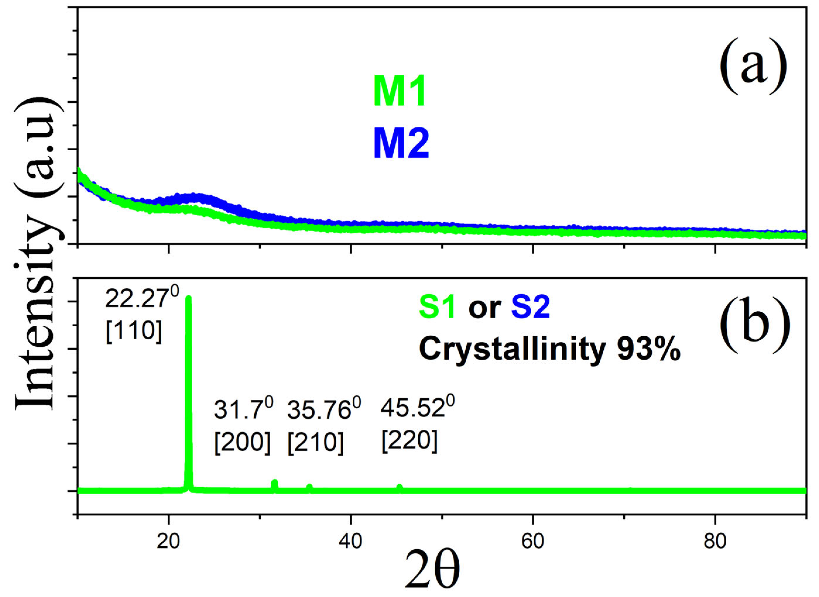
To begin characterization, liquid QD samples were deposited on glass slides for X-ray diffraction (XRD) analysis. This technique was also applied to the hybrid base matrix and QD-loaded matrices to assess their crystalline structure. Figure 2 presents the XRD spectra: samples S1 and S2 (Figure 2b) exhibit crystalline phases, while M1 and M2 (Figure 2a) show amorphous characteristics due to the hybrid matrix. Weak amorphous bands corresponding to SiO2 are visible in M1 and M2, consistent with the matrix composition [30].
The XRD spectra of S1 and S2 reveal a prominent peak at 2θ = 22.27°, 31.7°, 35.76°, and 45.52° indexed as [110], [200], [210], and [220], respectively, characteristic of crystalline urea [32,33,34]. The similarity between S1 and S2 spectra reflects their shared urea base, with S2 containing a small percentage of citric acid. Crystallinity percentages and grain sizes were calculated using DIFRAC.EVA V4.0 and VESTA 3 software [35], and Scherrer’s equation [36], yielding grain sizes of 75.58 nm and 76.08 nm, and crystallinity of 93.9% and 94.7% for S1 and S2, respectively. These grain sizes are relatively large for QDs, likely due to agglomeration that occurs during dropwise deposition and subsequent drying on the substrate.
Figure 2. (a) X-ray diffraction patterns of solid hybrid matrices M1 and M2, showing a predominantly amorphous structure with broad halos typical of silica and polymer-based hybrids. (b) XRD patterns of CQD solutions S1 and S2, revealing crystalline features with peaks near 2θ ≈ 22.27°, corresponding to the [110] plane of urea. The observed crystallinity and grain sizes (75–76 nm) reflect the aggregation of QDs during the drop-casting process.
Photonics 12 00854 g002

3.3. Transmission Electron Microscopy (TEM) Discussion

High-resolution transmission electron microscopy (HRTEM) was employed to determine the actual size of the QD particles. Figure 3a shows QDs with an approximate diameter of 10 nm. Figure 3b highlights a magnified region used for Fourier analysis via VESTA [35], with diffraction patterns shown in Figure 3c,d. Both S1 and S2 exhibit a hexagonal crystalline structure with space group P63mc and lattice parameters: a = 2.461 Å, b = 2.461 Å, c = 6.708 Å, α = 90°, β = 90°, γ = 120° [32]. These parameters were used to simulate HRTEM images using SimulaTEM 1.3.1 software [37], confirming the experimental observations. The TEM and XRD techniques complement each other; the Miller indices in Figure 2 and those calculated and shown in Figure 3c align with some of the crystal planes reported for crystals of urea [32], which is the precursor to QD synthesis. Additionally, the TEM technique allowed us to determine a size of 10 nm for the area where measurements were taken after processing the image.

3.4. UV-VIS–NIR Spectroscopy Discussion

UV-VIS spectroscopy was performed on the reference solid samples SiO2 (M5), PMMA (M4), and the hybrid matrix (M3) to determine their optical band gaps using the Tauc method [38], as shown in Figure 4a. PMMA exhibits a band gap of ~4.2 eV [39,40,41], while SiO2 shows ~3.2 eV (Insert), consistent with reported values ranging from 2.27 eV (nanoparticles) [42] to 4.3 eV (calcination-dependent) [43,44]. The hybrid matrix (M3) shows a band gap of 4.90 eV [39,40], though SiO2 band gaps can vary widely (1.7–9.6 eV) depending on synthesis and phase [45]
Figure 4b shows the band gaps of QD-doped hybrid samples: M1 (3.9 eV) and M2 (3.7 eV). These values are reduced by ~1 eV compared to M3, indicating that QD incorporation modifies the electronic structure while maintaining insulating behavior. The insert of Figure 4b shows the gaps of the QDs within the liquid samples S1 and S2, which serve as references, as explained in the following section.

3.5. Photoluminescence

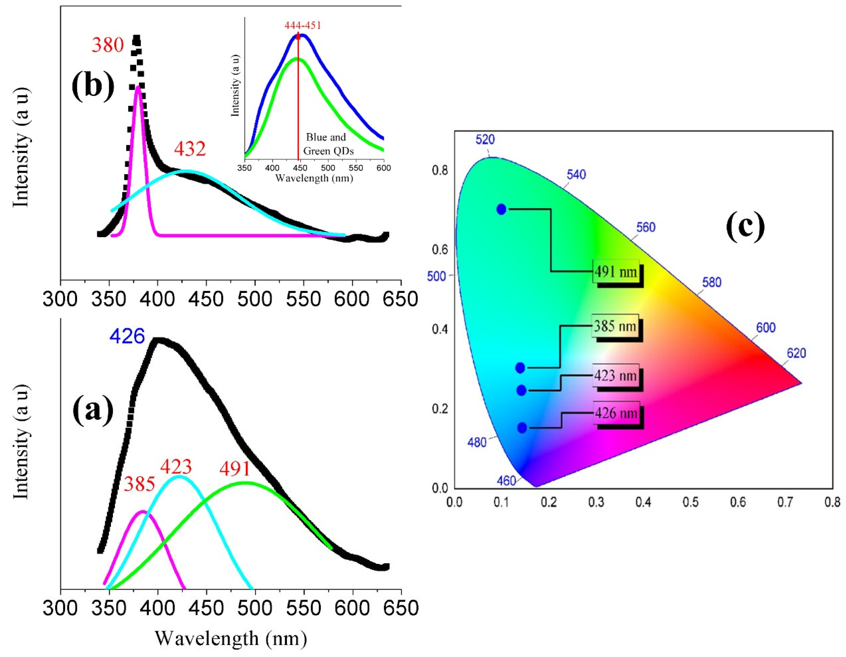
Photoluminescence measurements were conducted with excitation at λexc = 325 nm (Figure 5). The insert of Figure 5b shows emission peaks at ~444 nm for S1 and 451 nm for S2. Figure 5a presents deconvoluted emission spectra for QDs, PMMA, and SiO2 in the hybrid matrix. Three distinct bands are observed at 385 nm [46], 423 nm, and 491 nm [39,40], corresponding to the matrix components. Figure 5b shows the same sample (M1 or M2) rotated 180°, revealing two bands: one at 385 nm due to α-quartz SiO2 [44], and another at 432 nm from QDs. Figure 5c displays a chromaticity diagram, showing emission peaks at 385, 423, and 491 nm, with the overall blue emission centered at 426 nm.
Solid samples M1 and M2 display the same behavior when excited at a wavelength of 325 nm, producing emission around 426 nm that corresponds to the color shown on the chromaticity graph 5c. The process of deconvolution was performed to reveal the possible emissions of each component contributing to the total emission of the M1 or M2 samples, resulting in Figure 5a. These three components form a single solid material with structural properties characteristic of a polymer, with a band gap calculated in Figure 4. In the hybrid matrix M3, the band gap of SiO2 is 4.9 eV, and that of PMMA is 4.2 eV; additionally, the band gaps of CQDs were calculated and are shown in the insert of Figure 4, with values of 2.9 eV for S1 and 3.1 eV for S2. Since the sample was excited with 3.8 eV (325 nm), only this energy is available to stimulate the emission of the CQDs. However, the deconvolution results show emissions from the three components: SiO2 at 385 nm, CQD at 423 nm, and PMMA at 491 nm.
Within the liquid solutions, the CQDs emit light at 444 and 451 nm (insert Figure 5b). Deconvolution reveals a shift at 423 nm (Figure 5a), and the overall emission of the sample (M1 or M2) shows a final peak at 426 nm. The contribution of both SiO2 and PMMA to the total emission helps excite the CQDs, which is why it was proposed to use these samples as a plastic scintillator to operate in the visible range-our samples are suitable for blue light emission-but under much higher excitation energies with energy levels ranging from MeV to GeV, for instance. Finally, the graph in Figure 5b serves as a reference for SiO2 emission, since nanoparticles formed during the sol–gel process and the synthesis of the samples precipitated in one region, producing an emission peak corresponding to the component mentioned earlier.

3.5.1. Time Decay

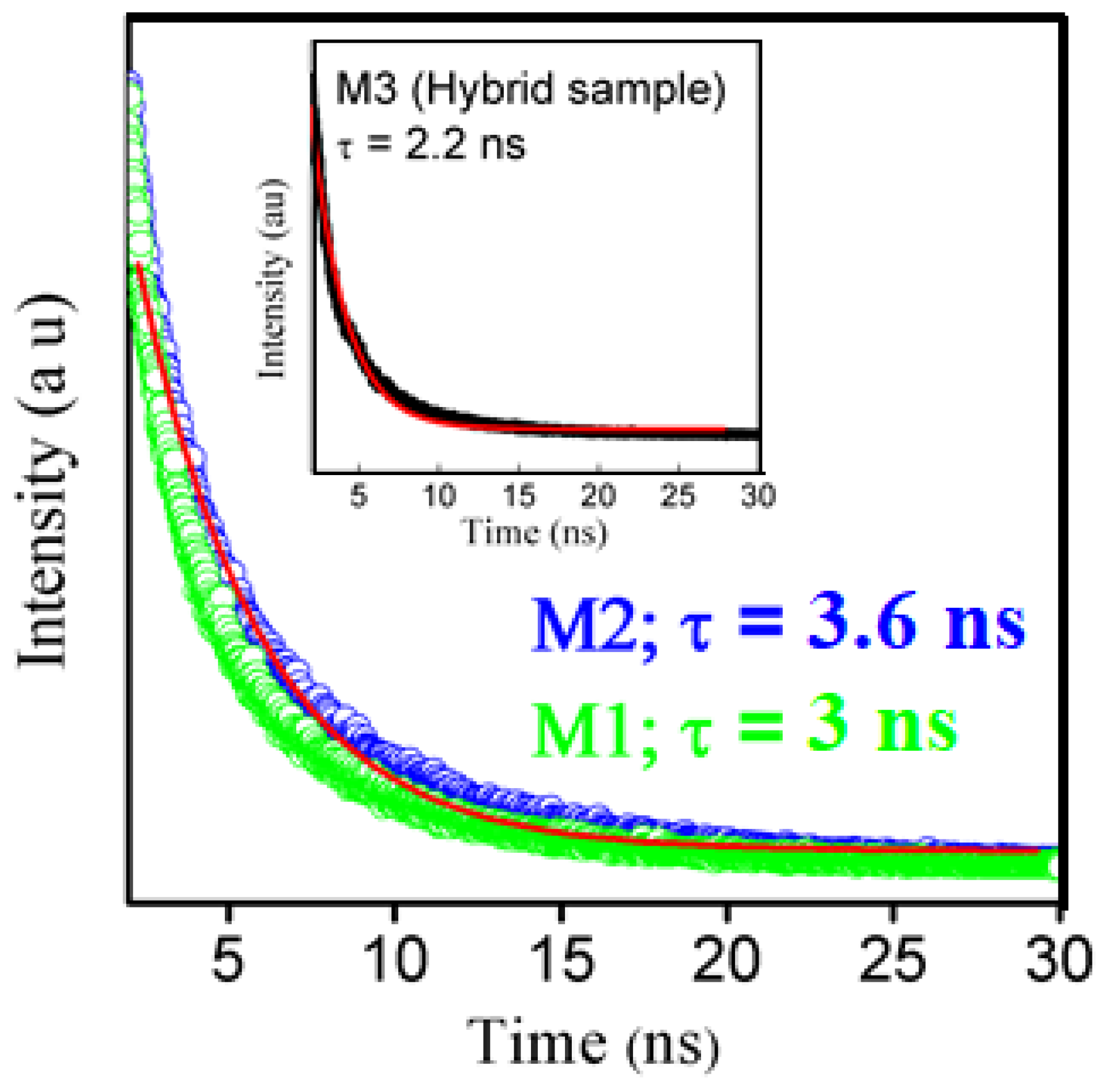
To evaluate the time-resolved luminescence behavior of the hybrid samples M1 and M2, time-decay measurements were performed. The decay curves were fitted using a single-exponential decay model, commonly applied to similar materials [47]:
y =   y 0 + A 1 e ( t t 0 ) τ ,
where:
  • (y) is the emitted light intensity,
  • (y0) is the baseline intensity,
  • (A1) is a normalization constant,
  • (t − t0) is the time elapsed since excitation,
  • (τ) is the decay time constant.
As shown in Figure 6, both M1 and M2 exhibit nearly identical decay profiles, with a single exponential fit (red line) yielding a high correlation coefficient (R2 = 0.99). The extracted decay times were 3 ns for M1 and 3.6 ns for M2. For comparison, the base matrix without QDs (insert of Figure 6) exhibited a shorter decay time of 2.2 ns.
These decay times are comparable to those of commercial plastic scintillators (PSs), such as BC408, which are widely used in radiation detection. PS materials typically consist of a polymer matrix (e.g., PMMA, polystyrene, or polyvinyl-toluene) doped with luminophore molecules, such as anthracene, naphthalene, 2,5-diphenyloxazole (PPO), or 1,4-di-(5-phenyl-2-oxazolyl)-benzene (POPOP). Notably, other studies have reported PS based on PMMA matrices doped with PPO [48,49]
The decay times of M1 and M2 fall within the range of fast-response scintillators, such as BC412, which is used for detecting alpha and beta particles [49,50] as well as X-rays, gamma rays, fast neutrons, and charged particles [51,52]. Commercial PS products from companies such as Luxium Solutions [51] and Saint-Gobain Crystals [52] (e.g., BC series) use similar matrix–luminophore combinations. Table 2 lists decay times for various PS materials, ranging from 0.7 ns (BC422Q) to 285 ns (BC444).
In terms of emission characteristics, PS materials typically emit in the UV range (~245 nm). In comparison, M1 and M2 emit in the blue region (~426 nm), offering a spectral shift that may be advantageous for specific detection systems.

3.5.2. Optical Quantum Yield (OQY)

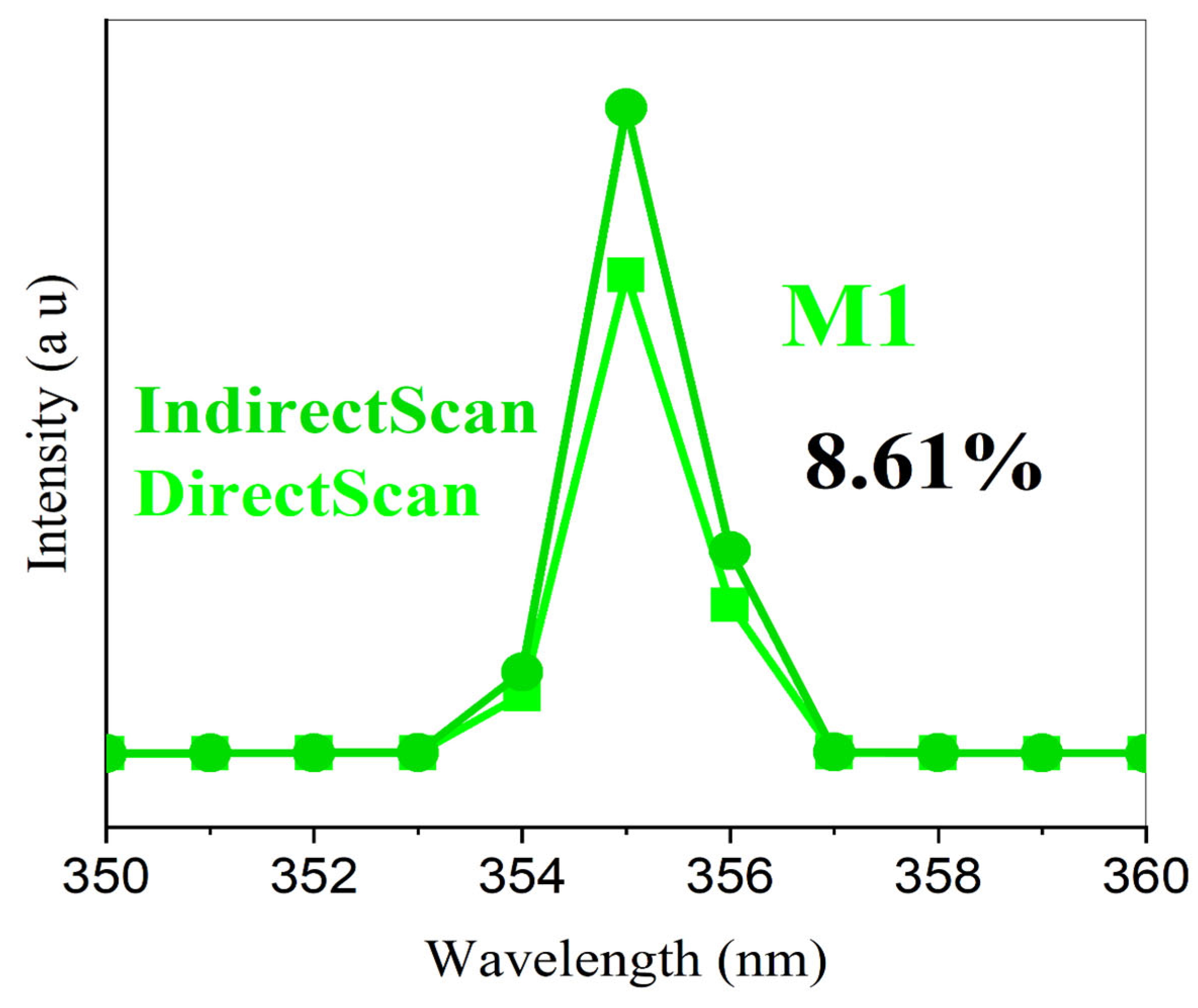
The optical quantum yield of M1 and M2 was measured using the integrating sphere module of the Edinburgh FLS 1000 spectrometer. The OQY was calculated based on both direct (EmScanDirect) and indirect (EmScanIndirect) emission measurements, considering the total number of photons emitted relative to those absorbed. The difference between the two methods is that there are only two ways to excite samples in a spectrophotometer. During direct measurement, light from the excitation monochromator reaches the sample, which is placed in a specialized container (usually quartz), and the emitted light is directed to the detector. For indirect measurement, the light from the excitation monochromator uses the integrating sphere to illuminate the sample, and the emitted light is sent to the detector. In our sample measurements, the OQYs are very similar for both methods.
As shown in Figure 7, both M1 and M2 exhibited an OQY of 8.61% under excitation at 325 nm (3.81 eV), with emission at 426 nm (M1 and M2). These values, while lower than those of commercial PS materials, are notable given the significantly lower excitation energy used in this study.
Commercial PS materials typically report light output rather than OQY, as their excitation sources are in the keV to MeV range. While M1 and M2 have lower OQY values, their blue-shifted emission, fast decay times, and compatibility with low-energy excitation make them promising candidates for optical detection in environments where traditional scintillators may be less effective.

3.6. Scintillation Testing with Cosmic Ray Detector Discussion

Cosmic rays continuously bombard the Earth’s upper atmosphere at a rate of approximately 10,000 particles per square meter per second for energies around 1 GeV, decreasing to about one particle per square meter per second at higher energies [53,54]. These primary cosmic rays interact with atmospheric nuclei, producing a cascade of secondary particles. Among these, charged pions (π+) and kaons (K+/K) are the primary sources of muons (μ+) [55]. Due to their relatively long mean lifetime (~2.2 μs) and large mass (~207 times that of the electron), muons can penetrate the atmosphere and reach the Earth’s surface, losing energy primarily through excitation and ionization processes up to energies of approximately 500 GeV [56].
Given their omnidirectional arrival, muons interact randomly with materials on the surface. When measuring the energy deposited by these background muons, the resulting histogram typically follows a Poisson distribution. However, high-energy muons that pass vertically through the center of a material—referred to as vertical-equivalent muons (VEMs)—can produce a distinct, overlapping distribution [57,58].
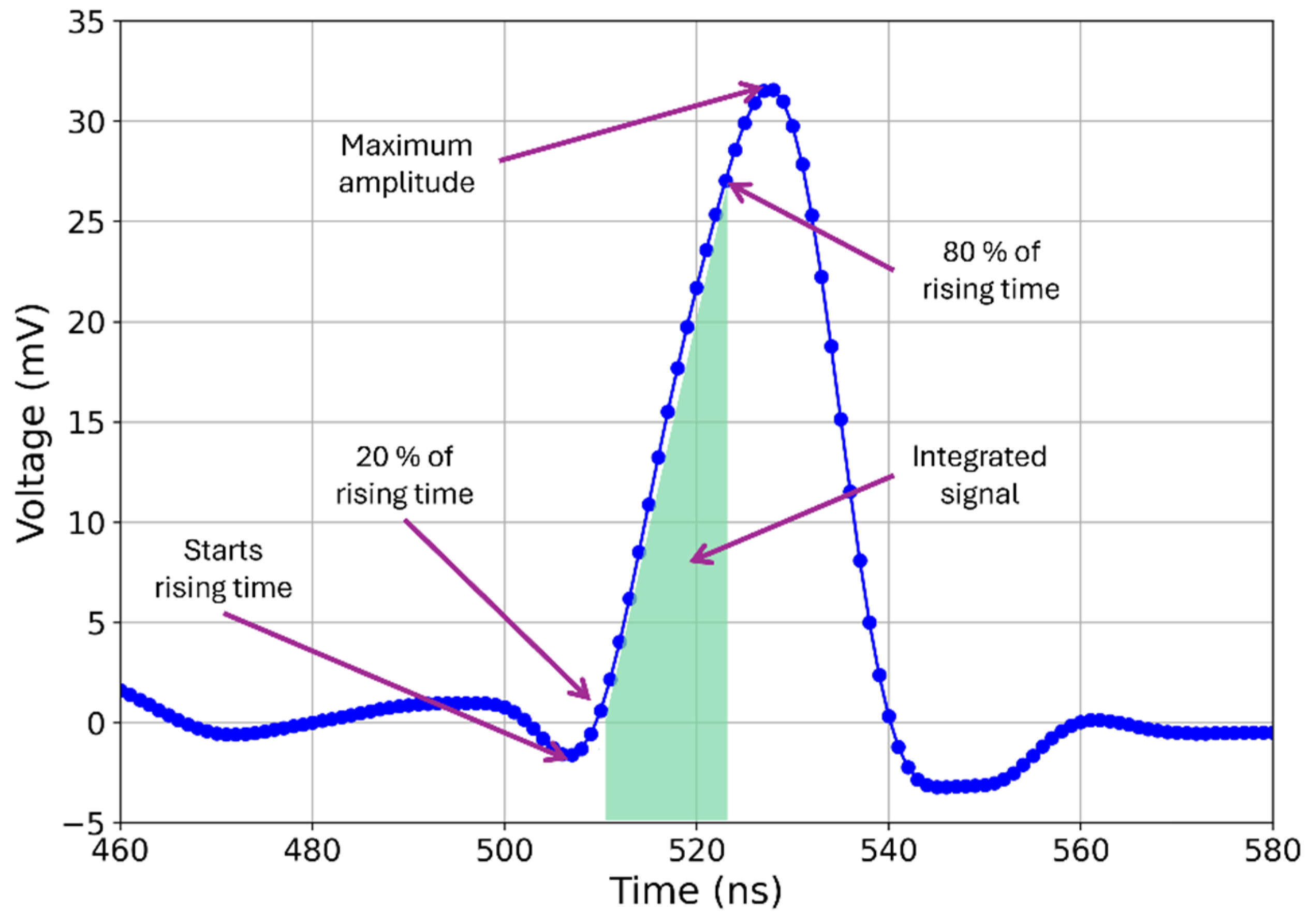
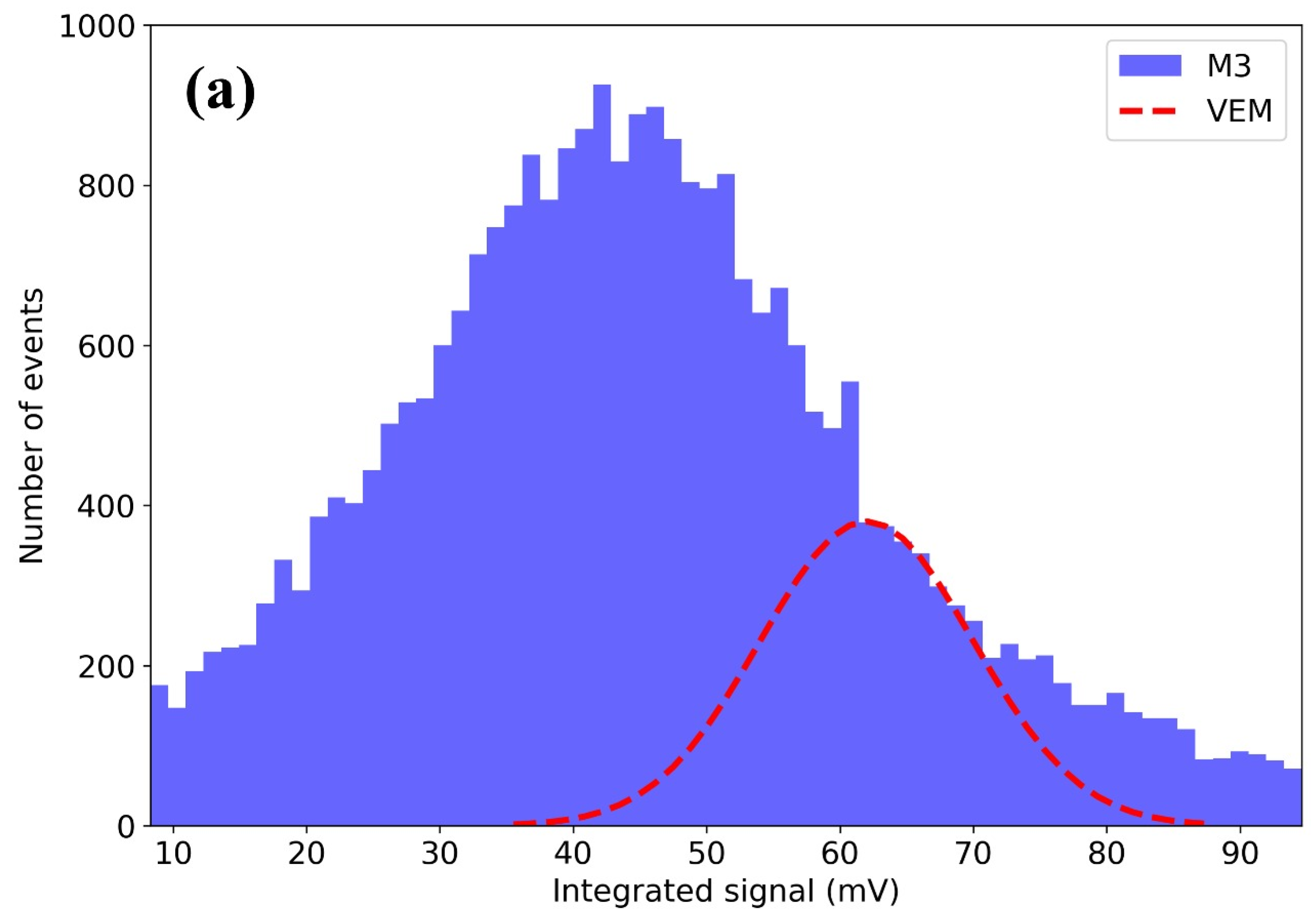
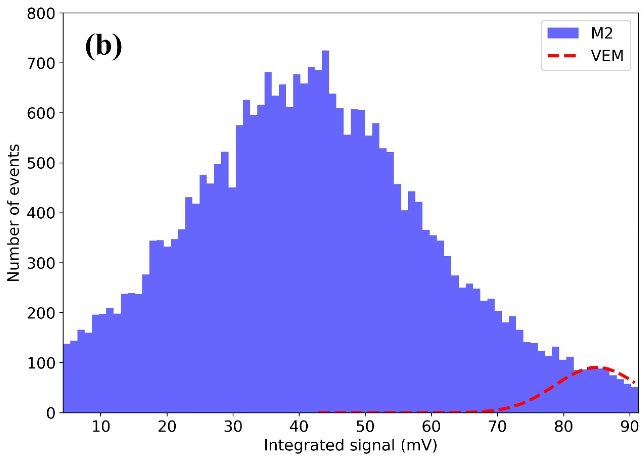
In this study, we characterized the integrated signal produced by atmospheric muons in the hybrid matrix samples. The integration was performed over the T20–T80 time window, defined as the interval during which the signal rises from 20% to 80% of its maximum amplitude. Figure 8 shows a representative signal waveform from a muon event, with the integration region marked.
Figure 9 presents histograms of the integrated signal for samples M3 (Figure 9a) and M2 (Figure 9b). The red curves represent Gaussian fits to the data. In both cases, the exponential decay of the Poisson-like background distribution is modified by the emergence of a second peak, attributed to VEMs. The Gaussian fit parameters were as follows:
  • M3: Amplitude = 380 events, Mean = 62 mV, Sigma = 8 mV
  • M2: Amplitude = 90 events, Mean = 85 mV, Sigma = 6.3 mV
The presence of a distinguishable VEM region in both samples confirms the scintillating nature of the hybrid materials. This result demonstrates the potential of these materials for passive radiation detection using naturally occurring cosmic-ray muons.
At first glance, there appears to be no significant improvement in detecting atmospheric muons using the hybrid matrix samples doped with QDs. However, for the M3 control matrix sample, the first VEM is located at about 62 mV, just under 20 mV after the peak of the Poisson-like distribution, which corresponds to background muons arriving from any direction. Around 72, 80, and 90 mV, there are additional bumps indicating changes in the slope of the distribution, likely representing second, third, and fourth VEMs. This result suggests the presence of more energetic atmospheric muons. Nonetheless, their proximity makes it challenging to perform fittings and accurately determine a maximum value for each VEM. In a typical cosmic ray experiment, it is vital to distinguish the energy of the detected muon to reconstruct the primary cosmic ray’s energy accurately. Conversely, for the M2 sample with QDs-doped matrix, the VEM occurs at 85 mV, a region separated from the distribution’s peak, making this material suitable for establishing trigger values to differentiate a muon with energy equivalent to the VEM from background muons.
Traditional inorganic scintillators may underperform in harsh conditions due to their seemingly high OQY (around 90%). Such a high figure is mainly due to testing conditions where incoming light is focused on a small surface area, which is a limitation in the present application. To address this barrier, traditional inorganic scintillators should have larger dimensions, which would also result in higher costs. For example, consider the commercial crystal scintillator LYSO: Ce (Lutetium-Yttrium Oxyortho-silicate doped with Cerium). It has small dimensions (20 × 20 × 20 mm) and costs USD 500. With that budget, we could purchase enough reagents to make about 10 blocks with a diameter of 22 cm and 4 cm thick. The OQY can be increased precisely due to the larger volume and dimensions of the scintillator. Our plastic scintillator could help improve the detection of cosmic rays because it can be shaped into different sizes and forms according to need.

4. Conclusions

In this work, we present a novel method for fabricating organic–inorganic hybrid materials incorporating Carbon Quantum Dots (CQDs) synthesized via hydrothermal and solvothermal routes. This multi-step process yields a robust, transparent, and easily shaped hybrid material, making it suitable for integration into optical and radiation detection systems. The resulting material exhibits luminescence in the blue spectral region upon UV excitation, highlighting its potential as a wavelength shifter.
The photophysical properties, including time decay (3.0–3.6 ns) and optical quantum yield (~8.6%), indicate that the material behaves similarly to plastic scintillators. These properties make it a promising candidate for fast particle detection applications, particularly in the keV to GeV energy range. The detection of atmospheric muons demonstrates an example of this type of application.
A significant advantage of the developed process is its low-cost synthesis and high adaptability. The hybrid matrix is formed from a polymerizable liquid mixture, enabling it to be cast into complex geometries such as molds, waveguides, optical circuits, or fibers. Furthermore, the method supports flexible doping, allowing the incorporation of various organic molecules, metal salts, oxides, nanoparticles, and nanofibers, provided they are compatible with the polymerization environment.
Overall, this approach offers a versatile platform for developing next-generation luminescent materials with potential applications in radiation detection, optical sensing, and photonic integration.

Author Contributions

Conceptualization, L.C.L., M.R.P.M., J.E.E.R., B.d.C.A. and J.E.L.A.; methodology, L.C.L., M.R.P.M., B.d.C.A., M.T.-S. and J.E.L.A.; software, L.C.L., S.T.C., B.d.C.A., O.M.M.B., O.I.R.-H. and J.G.S.G.; validation, L.C.L., M.R.P.M., J.E.E.R., S.T.C., J.G.S.G., M.T.-S. and J.E.L.A.; formal analysis, L.C.L., M.R.P.M., J.E.E.R., S.T.C., O.M.M.B., O.I.R.-H., M.T.-S. and J.E.L.A.; investigation, L.C.L., M.R.P.M., J.E.E.R., B.d.C.A., O.M.M.B., O.I.R.-H., J.G.S.G., M.T.-S. and J.E.L.A.; resources, L.C.L., S.T.C., B.d.C.A., O.M.M.B., O.I.R.-H., J.G.S.G. and J.E.L.A.; data curation, L.C.L., M.R.P.M., S.T.C., B.d.C.A., O.M.M.B., O.I.R.-H. and J.E.L.A.; writing—original draft preparation, L.C.L., M.R.P.M., J.E.E.R., B.d.C.A., O.M.M.B., O.I.R.-H., M.T.-S. and J.E.L.A.; writing—review and editing, L.C.L., M.R.P.M., J.E.E.R., S.T.C., B.d.C.A., O.M.M.B., O.I.R.-H., J.G.S.G. and J.E.L.A.; visualization, L.C.L., M.R.P.M., J.E.E.R., O.M.M.B., O.I.R.-H. and J.E.L.A.; supervision, L.C.L., M.R.P.M., S.T.C., O.I.R.-H. and J.E.L.A.; project administration, M.R.P.M., S.T.C. and J.E.L.A.; funding acquisition, L.C.L., M.R.P.M., B.d.C.A., O.M.M.B. and J.E.L.A. All authors have read and agreed to the published version of the manuscript.

Funding

This research was funded by Secretaría de Ciencia y Tecnología e Innovación (cátedras Project No. 3208) [Grant A1-S-38743 and 320548].

Data Availability Statement

Data are contained within the article.

Conflicts of Interest

The authors declare that they have no conflicts of interest.

References

  1. Ma, Z.; Fu, Q.; Zhang, K.; Sun, S.; Yue, M. Research on light-responsive luminescence properties of carbon dots and their applications. Mater. Horiz. 2025. Online ahead of print. [Google Scholar] [CrossRef]
  2. Liu, Y.; Huang, Z.; Wang, X.; Hao, Y.; Yang, J.; Wang, H.; Qu, S. Recent advances in highly luminescent carbon dots. Adv. Funct. Mater. 2025, 35, 2420587. [Google Scholar] [CrossRef]
  3. Wang, B.; Lu, S. The light of carbon dots: From mechanism to applications. Matter 2022, 5, 110–149. [Google Scholar] [CrossRef]
  4. Huang, Z.; Ren, L. Large scale synthesis of carbon dots and their applications: A review. Molecules 2025, 30, 774. [Google Scholar] [CrossRef]
  5. Bhatt, J.P.; Godha, N. Quantum dots: Fundamentals, synthesis and applications. In Hydrothermal Synthesis of Quantum Dots; Elsevier: Amsterdam, The Netherlands, 2023; pp. 15–34. [Google Scholar] [CrossRef]
  6. Zhu, Z.; Niu, H.; Li, R.; Yang, Z.; Wang, J.; Li, X.; Pan, P.; Liu, J.; Zhou, B. One-pot hydrothermal synthesis of fluorescent carbon quantum dots with tunable emission color for application in electroluminescence detection of dopamine. Biosens. Bioelectron. X 2022, 10, 100141. [Google Scholar] [CrossRef]
  7. Liu, M.L.; Chen, B.B.; Li, C.M.; Huang, C.Z. Carbon dots: Synthesis, formation mechanism, fluorescence origin and sensing applications. Green Chem. 2019, 21, 449–471. [Google Scholar] [CrossRef]
  8. Kickelbick, G. (Ed.) Hybrid Materials: Synthesis, Characterization and Applications; Wiley-VCH: Weinheim, Germany, 2007; ISBN 978-3-527-31299-3. [Google Scholar]
  9. Gomez-Romero, P.; Pokhriyal, A.; Rueda-García, D.; Bengoa, L.N.; González-Gil, R.M. Hybrid materials: A metareview. Chem. Mater. 2024, 36, 8–27. [Google Scholar] [CrossRef]
  10. Olabisi, O.; Adewale, K. (Eds.) Handbook of Thermoplastics, 2nd ed.; CRC Press: Boca Raton, FL, USA, 2016; ISBN 3: 978-1-4665-7723-7. [Google Scholar]
  11. Yuan, X.W.; Jung, D.D.; Bhattacharyya, D.; Easteal, A.J. Hybrid thermoplastic composites using ceramic reinforcements. Plast. Rubber Compos. 2005, 34, 85–92. [Google Scholar] [CrossRef]
  12. Park, W.; Shin, H.; Choi, B.; Rhim, W.K.; Na, K.; Han, D.K. Advanced hybrid nanomaterials for biomedical applications. Prog. Mater. Sci. 2020, 114, 100686. [Google Scholar] [CrossRef]
  13. Song, J.; Vikulina, A.S.; Parakhonskiy, B.V.; Skirtach, A.G. Hierarchy of hybrid materials. Part-II: The place of organics-on-inorganics in it, their composition and applications. Front. Chem. 2023, 11, 1078840. [Google Scholar] [CrossRef]
  14. Rojas-Valencia, O.G.; Regules-Carrasco, M.; Hernández-Fuentes, J.; Reza-San Germán, C.M.; Estrada-Flores, M.; Villagarcía-Chávez, E. Synthesis of blue emissive carbon quantum dots from Hibiscus Sabdariffa flower: Surface functionalization analysis by FT-IR spectroscopy. Materialia 2021, 19, 101182. [Google Scholar] [CrossRef]
  15. Yalshetti, S.; Thokchom, B.; Bhavi, S.M.; Singh, S.R.; Patil, S.R.; Harini, B.P.; Sillanpää, M.; Manjunatha, J.G.; Srinath, B.S.; Yarajarla, R.B. Microwave-assisted synthesis, characterization and in vitro biomedical applications of Hibiscus rosa-sinensis Linn.-mediated carbon quantum dots. Sci. Rep. 2024, 14, 9915. [Google Scholar] [CrossRef]
  16. Kundu, A.; Maity, B.; Basu, S. Orange pomace-derived fluorescent carbon quantum dots: Detection of dual analytes in the nanomolar range. ACS Omega 2023, 8, 22178–22189. [Google Scholar] [CrossRef]
  17. Gholipour, A.; Rahmani, S. The green synthesis of carbon quantum dots through one-step hydrothermal approach by orange juice for rapid, and accurate detection of dopamine. J. Fluoresc. 2024, 34, 2665–2677. [Google Scholar] [CrossRef]
  18. Abdellatif, A.A.H.; Younis, M.A.; Alsharidah, M.; Al Rugaie, O.; Tawfeek, H.M. Biomedical applications of quantum dots: Overview, challenges, and clinical potential. Int. J. Nanomed. 2022, 17, 1951–1970. [Google Scholar] [CrossRef] [PubMed]
  19. Derfus, A.M.; Chan, W.C.W.; Bhatia, S.N. Probing the cytotoxicity of semiconductor quantum dots. Nano Lett. 2004, 4, 11–18. [Google Scholar] [CrossRef]
  20. Lisi, F.; Sawayama, J.; Gautam, S.; Rubanov, S.; Duan, X.; Kirkwood, N. Re-Examination of the Polymer Encapsulation of Quantum Dots for Biological Applications. ACS Appl. Nano Mater. 2023, 6, 4046–4055. [Google Scholar] [CrossRef]
  21. Wang, Y.; Hu, R.; Lin, G.; Roy, I.; Yong, K.-T. Functionalized Quantum Dots for Biosensing and Bioimaging and Concerns on Toxicity; ACS Appl. Mater. Interfaces 2013, 5, 2786–2799. [Google Scholar] [CrossRef]
  22. Korzhik, M.; Tamulaitis, G.; Vasil’ev, A.N. Physics of Fast Processes in Scintillators; Springer: Berlin/Heidelberg, Germany, 2020; Volume 49. [Google Scholar] [CrossRef]
  23. Lecoq, P. Development of new scintillators for medical applications. Nucl. Instrum. Methods Phys. Res. A 2016, 809, 130–139. [Google Scholar] [CrossRef]
  24. Weninger, L.; Morana, A.; Ouerdane, Y.; Marin, E.; Boukenter, A.; Girard, S. Distributed optical fiber-based radiation detection using an ultra-low-loss optical fiber. Radiation 2024, 4, 167–182. [Google Scholar] [CrossRef]
  25. Ntoupis, V.; Michail, C.; Kalyvas, N.; Bakas, A.; Kandarakis, I.; Fountos, G.; Valais, I. Luminescence efficiency and spectral compatibility of cerium fluoride (CeF3) inorganic scintillator with various optical sensors in the diagnostic radiology X-ray energy range. Inorganics 2024, 12, 230. [Google Scholar] [CrossRef]
  26. Zhao, J.; Wang, K.; Chen, W.; Li, D.; Lei, L. Controlled synthesis of Cs2NaYF6: Tb nanoparticles for high-resolution X-ray imaging and molecular detection. Nanomaterials 2025, 15, 728. [Google Scholar] [CrossRef]
  27. Gong, S.; Ma, Z.; Li, X. UMFNet: Frequency-guided multi-scale fusion with dynamic noise suppression for robust low-light object detection. Appl. Sci. 2025, 15, 5362. [Google Scholar] [CrossRef]
  28. Buchner, A.; Hadrath, S.; Burkard, R.; Kolb, F.M.; Ruskowski, J.; Ligges, M.; Grabmaier, A. Analytical evaluation of signal-to-noise ratios for avalanche- and single-photon avalanche diodes. Sensors 2021, 21, 2887. [Google Scholar] [CrossRef]
  29. Sun, J.; Akdogan, E.K.; Klein, L.C.; Safari, A. Characterization and optical properties of sol–gel processed PMMA/SiO2 hybrid monoliths. J. Non-Cryst. Solids 2007, 353, 2807–2812. [Google Scholar] [CrossRef]
  30. Gil-Kowalczyk, M.; Łyszczek, R.; Jusza, A.; Piramidowicz, R. Thermal, Spectroscopy and Luminescent Characterization of Hybrid PMMA/Lanthanide Complex Materials. Materials 2021, 14, 3156. [Google Scholar] [CrossRef] [PubMed]
  31. Ponomarenko, S.A.; Surin, N.M.; Borshchev, O.V.; Luponosov, Y.N.; Akimov, D.Y.; Alexandrov, I.S.; Burenkov, A.A.; Kovalenko, A.G.; Stekhanov, V.N.; Kleymyuk, E.A.; et al. Nanostructured organosilicon luminophores and their application in highly efficient plastic scintillators. Sci. Rep. 2014, 4, 6549. [Google Scholar] [CrossRef]
  32. Hanawalt, J.D.; Rinn, H.W.; Frevel, L.K. Chemical analysis by X-ray diffraction. Ind. Eng. Chem. Anal. Ed. 1938, 10, 457–512. [Google Scholar] [CrossRef]
  33. Madhurambal, G.; Mariappan, M. Growth and characterization of urea-thiourea non-linear optical organic mixed crystal. Indian J. Pure Appl. Phys. 2010, 48, 264–270. [Google Scholar]
  34. Adams, R.; Balyuzi, H.H.M.; Burge, R.E. X-ray diffraction studies of aqueous solutions of urea. J. Appl. Cryst. 1977, 10, 256–261. [Google Scholar] [CrossRef]
  35. Momma, K.; Izumi, F. VESTA 3 for three-dimensional visualization of crystal, volumetric and morphology data. J. Appl. Cryst. 2011, 44, 1272–1276. [Google Scholar] [CrossRef]
  36. Cullity, B.D.; Stock, S.R. Elements of X-Ray Diffraction, 3rd ed.; Prentice-Hall Inc.: Wilmington, DE, USA, 2001; pp. 96–102. ISBN 0-201-61091-4. [Google Scholar]
  37. Gómez-Rodríguez, A.; Beltrán-Del-Río, L.M.; Herrera-Becerra, R. SimulaTEM: Multislice simulations for general objects. Ultramicroscopy 2010, 110, 95–104. [Google Scholar] [CrossRef] [PubMed]
  38. Makuła, P.; Pacia, M.; Macyk, W. How to correctly determine the band gap energy of modified semiconductor photocatalysts based on UV–Vis spectra. J. Phys. Chem. Lett. 2018, 9, 6814–6817. [Google Scholar] [CrossRef]
  39. Trejo-García, P.M.; Palomino-Merino, R.; De la Cruz, J.; Espinosa, J.E.; Aceves, R.; Moreno-Barbosa, E.; Portillo Moreno, O. Luminescent properties of Eu3+-doped hybrid SiO2–PMMA material for photonic applications. Micromachines 2018, 9, 441. [Google Scholar] [CrossRef] [PubMed]
  40. De la Cruz, J.; Palomino Merino, R.; Trejo-García, P.; Espinosa, J.E.; Aceves Torres, R.; Moreno-Barbosa, E.; Gervacio-Arciniega, J.J.; Soto, E. Luminescent properties of a hybrid SiO2–PMMA matrix doped with terbium. Opt. Mater. 2019, 87, 42–47. [Google Scholar] [CrossRef]
  41. Mergen, Ö.B.; Arda, E.; Kara, S.; Pekcan, Ö. Effects of GNP addition on optical properties and band gap energies of PMMA films. Polym. Compos. 2019, 40, 1862–1869. [Google Scholar] [CrossRef]
  42. Wardani, D.A.P.; Hariyanto, B.; Kurniawati, N.; Har, N.P.; Darmawan, N. Irzaman, Functional groups, band gap energy, and morphology properties of annealed silicon dioxide (SiO2). Egypt. J. Chem. 2023, 66, 529–535. [Google Scholar] [CrossRef]
  43. Alibe, I.M.; Matori, K.A.; Saion, E.; Alibe, A.M.; Zaid, M.H.M.; Engku, E.A.A.G. A facile synthesis of amorphous silica nanoparticles by simple thermal treatment route. Dig. J. Nanomater. Biostruct. 2016, 11, 1155–1164. [Google Scholar]
  44. Rai, C.; Pandey, P.; Haque, F.Z. Optical, dielectric and impedance studies of SiO2/MWCNT nanocomposite synthesized through in-situ ultrasonication-assisted sol–gel method. J. Adv. Phys. 2014, 3, 179–193. [Google Scholar] [CrossRef]
  45. Seguini, G.; Schamm-Chardon, S.; Pellegrino, P.; Perego, M. The energy band alignment of Si in SiO2. Appl. Phys. Lett. 2011, 99, 082107. [Google Scholar] [CrossRef]
  46. Trukhin, A.; Truhins, K. Luminescence of α Quartz. arXiv 2012, arXiv:1209.4200. [Google Scholar] [CrossRef]
  47. Yan, Y.-L.; Liu, Y.; Fang, C.-H.; Zhu, J.-J.; Lin, S.-T.; Liu, S.-K.; Tang, C.-J.; Wang, C.-L.; Xing, H.-Y. Preparation and characterization of polystyrene-based plastic scintillators as a self-vetoing structural material. J. Polym. Res. 2025, 32, 19. [Google Scholar] [CrossRef]
  48. Salimgareeva, V.N.; Kolesov, S.V. Plastic scintillators based on polymethyl methacrylate: A review. Instrum. Exp. Tech. 2005, 48, 273–282. [Google Scholar] [CrossRef]
  49. Bhakta, S.; Raman, A.; Ajish, J.K. Development of plastic scintillator with better performance than the commercial counterpart (UPS-923A) and effect of fluorophore composition on light output. Appl. Radiat. Isot. 2024, 212, 111453. [Google Scholar] [CrossRef]
  50. Efremova, O.A.; Brylev, K.A.; Vorotnikov, Y.A.; Vejsadova, L.; Shestopalov, M.A.; Chimonides, G.F.; Mikes, P.; Topham, P.D.; Kim, S.-J.; Kitamura, N.; et al. Photoluminescent materials based on PMMA and a highly-emissive octahedral molybdenum metal cluster complex. J. Mater. Chem. C 2016, 4, 497–503. [Google Scholar] [CrossRef]
  51. Luxium Solutions, Solid Plastic Scintillators, Data Sheets. Available online: https://luxiumsolutions.com/radiation-detection-scintillators/plastic-scintillators/bc400-bc404-bc408-bc412-bc416 (accessed on 5 May 2025).
  52. Saint Gobain Crystals, BC400, BC404, BC408, BC412, BC416 Premium Plastic Scintillators, Data Sheet. Available online: https://www.hep.phy.cam.ac.uk/~lester/teaching/SparkChamber/SGC_BC400_404_408_412_416_Data_Sheet.pdf (accessed on 5 May 2025).
  53. Grieder, P.K.F. Extensive Air Showers—High Energy Phenomena and Astrophysical Aspects, in Cosmic Rays at Earth. Volume 1. 2010. Available online: http://www.sciencedirect.com/science/article/pii/B9780444507105500038 (accessed on 10 May 2025).
  54. Tomassetti, N. Direct measurements of galactic cosmic rays. In Proceedings of the 27th European Cosmic Ray Symposium (ECRS), Nijmegen, The Netherlands, 25–29 July 2022; Volume 423, p. 007. [Google Scholar] [CrossRef]
  55. Gaisser, T.K.; Engel, R.; Resconi, E. (Eds.) Atmospheric Muons and Neutrinos, in Cosmic Rays and Particle Physics, 2nd ed.; Cambridge University Press: Cambridge, UK, 2016; pp. 126–148. [Google Scholar] [CrossRef]
  56. Procureur, S. Muon imaging: Principles, technologies and applications. Nucl. Instrum. Methods Phys. Res. A 2017, 878, 169–179. [Google Scholar] [CrossRef]
  57. Etchegoyen, A.; Bauleo, P.; Bertou, X.; Bonifazi, C.B.; Filevich, A.; Medina, M.C.; Melo, D.G.; Rovero, A.C.; Supanitsky, A.D.; Tamashiro, A. Muon-track studies in a water Cherenkov detector. Nucl. Instrum. Methods Phys. Res. A 2005, 545, 602–612. [Google Scholar] [CrossRef]
  58. Bertou, X.; Allison, P.S.; Bonifazi, C.; Bauleo, P.; Grunfeld, C.M.; Aglietta, M.; Arneodo, F.; Barnhill, D.; Beatty, J.J.; Busca, N.G.; et al. Calibration of the surface array of the Pierre Auger Observatory. Nucl. Instrum. Methods Phys. Res. A 2006, 568, 839–846. [Google Scholar] [CrossRef]
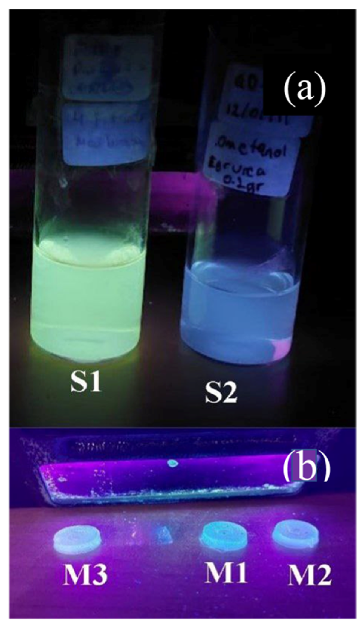
Figure 1. (a) Carbon Quantum Dot (CQD) solutions synthesized via hydrothermal/solvothermal methods: S1 (urea + citric acid) and S2 (urea), showing luminescence under 365 nm UV light. (b) Solid hybrid matrices M1 and M2 containing encapsulated QDs, also under UV illumination. The matrices are optically transparent and mechanically stable. M3 (base matrix of SiO2 + PMMA), M4 (PMMA), and M5 (SiO2) are excluded for clarity.
Photonics 12 00854 g001
Figure 3. (a) HRTEM image of CQDs in sample S1 showing nanoparticles with average sizes around 10 nm. (b) Zoomed-in image highlighting a single QD; the red box denotes the region analyzed. (c) FFT-derived diffraction pattern identifying lattice spacing and zone axes, confirming a hexagonal crystal lattice. (d) Simulated HRTEM image and diffraction pattern of urea crystal using VESTA and SimulaTEM, validating experimental results.
Photonics 12 00854 g003
Figure 4. (a) Tauc plots for the base hybrid matrix (M3), along with an insert for PMMA (M4, 4.2 eV) and SiO2 (M5, 3.2 eV), confirming typical optical band gaps of the constituent materials. (b) Band gap estimation for QD-doped hybrid matrices: M1 (3.7 eV) and M2 (3.9 eV), indicating QD-induced narrowing of the gap while retaining insulating behavior. Insert (b) shows the gaps of the QDs within the liquid samples S1 (2.9 eV) and S2 (3.1 eV).
Photonics 12 00854 g004
Figure 5. (a) Deconvoluted photoluminescence spectra of QD-doped hybrid matrix (M1 or M2), with distinct contributions from PMMA (423 nm), SiO2 (385 nm), and QDs (491 nm). (b) Rotated sample emission spectrum emphasizing SiO2 (α-quartz) and QD contributions. (Insert) PL spectra of CQD solutions S1 and S2 with peaks at 444 nm and 451 nm, respectively. (c) Chromaticity diagram indicating resultant blue emission centered at 426 nm.
Photonics 12 00854 g005
Figure 6. Time-resolved photoluminescence decay curves for QD-doped hybrid samples M1 and M2, showing fitted exponential decay with τ values of 3.0 ns and 3.6 ns, respectively. Insert: Control sample M3 exhibits a shorter decay time (2.2 ns), confirming the role of QDs in modifying luminescence lifetime.
Photonics 12 00854 g006
Figure 7. Optical Quantum Yield (OQY) measurement of sample M1 using an integrating sphere in the Edinburgh FLS1000 system. The OQY was calculated at 8.61% under 325 nm excitation (3.81 eV). M2 showed a similar yield, confirming consistent performance between the QD-doped samples.
Photonics 12 00854 g007
Figure 8. Typical waveform of a scintillation signal induced by an atmospheric muon interaction with the hybrid matrix. The shaded region indicates the T20–T80 rising time window used to calculate the integrated pulse area, which is indicative of the deposited energy.
Photonics 12 00854 g008
Figure 9. Histograms of integrated signals from cosmic-ray-induced muon interactions: (a) Sample M3 (hybrid matrix), (b) Sample M2 (QD-doped matrix). The red curve represents a Gaussian fit; the presence of a distinct secondary distribution suggests the existence of vertical-equivalent muons (VEM), confirming scintillation behavior in both samples.
Photonics 12 00854 g009aPhotonics 12 00854 g009b
Table 1. Nomenclature used for labeling all liquid and solid samples.
Liquid SamplesLabel
QDs Urea GreenS1
QDs Urea BlueS2
Solid Samples
Hybrid + QDs Urea GreenM1
Hybrid + QDs Urea BlueM2
Hybrid SampleM3
PMMAM4
SiO2M5
Table 2. Optical Quantum Yield and decay time comparison between samples M1 and M2, and some commercial plastic scintillators.
Plastic
Scintillator
Time
Decay (ns)
Quantum
Yield (%)
Light
Output (%)
Maximum Emission
Wavelength (nm)
Reference
BC422Q0.7-11370[42,43]
Polystyrene+ POPOP1.593--[41]
BC4082.122.964425[42,43]
BC4002.4-65423[42,43]
M13.08.61-426This work
BC4123.3-60434[42,43]
M23.68.61-426This work
PMMA + 4’vinyl-2,5-diphenyloxadiazole-1,3,44.268--[41]
PMMA+PPO12.442--[41]
BC444285-41428[42,43]
Disclaimer/Publisher’s Note: The statements, opinions and data contained in all publications are solely those of the individual author(s) and contributor(s) and not of MDPI and/or the editor(s). MDPI and/or the editor(s) disclaim responsibility for any injury to people or property resulting from any ideas, methods, instructions or products referred to in the content.