Article

Hunting for Monolayer Black Phosphorus with Photoluminescence Microscopy

by
Chenghao Pan
1,
Yixuan Ma
1,
Quan Wan
1,
Boyang Yu
1,
Shenyang Huang
1,2,* and
Hugen Yan
1,*
1
State Key Laboratory of Surface Physics, Key Laboratory of Micro and Nano-Photonic Structures (Ministry of Education), Shanghai Key Laboratory of Metasurfaces for Light Manipulation, Department of Physics, Fudan University, Shanghai 200433, China
2
Shanghai Frontiers Science Research Base of Intelligent Optoelectronics and Perception, Institute of Optoelectronics, Fudan University, Shanghai 200433, China
*
Authors to whom correspondence should be addressed.
Photonics 2024, 11(9), 866; https://doi.org/10.3390/photonics11090866
Submission received: 9 August 2024 / Revised: 9 September 2024 / Accepted: 13 September 2024 / Published: 14 September 2024
(This article belongs to the Special Issue Recent Advances in Infrared Photodetection and Imaging)

Abstract

:
Monolayer black phosphorus (BP) holds great promise for naturally hyperbolic polaritons and correlated states in rectangular moiré superlattices. However, preparing and identifying high-quality monolayer BP are challenging due to its instability and high transparency, which limits extensive studies. In this study, we developed a method for rapidly and nondestructively identifying monolayer BP and its crystal orientation simultaneously using modified photoluminescence (PL) microscopy. The optical contrast of monolayer BP has been significantly increased by at least twenty times compared to previous reports, making it visible even on a transparent substrate. The polarization dependence of optical contrast also allows for the in situ determination of crystal orientation. Our study facilitates the identification of monolayer BP, expediting more extensive research on and potential industrial applications of this material.

1. Introduction

Recently, there has been growing interest in black phosphorus (BP), a new infrared two-dimensional (2D) material exhibiting many exotic properties such as relatively high carrier mobility [1,2], natural in-plane anisotropy [3,4,5], and a highly tunable direct bandgap [6,7,8,9,10,11,12]. The bandgap of BP is always direct in the range of 0.3–1.7 eV from bulk to monolayer, which covers the mid-infrared to visible light spectrum and fills the gap between zero-gap graphene and transition metal dichalcogenides (TMDCs) in the visible region [7]. Additionally, BP has a puckered hexagonal lattice structure (Figure 1b) that forms an anisotropic rectangular unit cell with two non-equivalent directions in the layer plane, namely armchair (AC, parallel to the pucker) and zigzag (ZZ, perpendicular to the pucker), leading to a lot of intrinsic anisotropic properties and significantly distinguishing BP from isotropic 2D materials. Consequently, BP is a promising candidate for infrared electronic and optoelectronic applications including transistors [13], photodetectors [14], ultrafast lasers [15], solar cells [16], light-emitting diodes (LEDs) [17,18], and gas sensors [19]. Significant progress has been made in both fundamental research and novel device design [5].
However, most of these studies focus on few-layer BP or its bulk counterpart, with very little research on monolayer BP. In the monolayer limit, the Coulomb interactions are prominent due to strong quantum confinement and reduced screening, making monolayer BP an ideal platform to investigate anisotropic excitonic effect [3,20,21,22] and many-body interaction [20,23]. Moreover, the exotic properties of monolayer BP, such as hyperbolic plasmons [24], hyperbolic exciton polaritons [22], and exciton states [25], remain to be experimentally investigated. Furthermore, due to its unique anisotropic structure and Γ-point electronic band extrema, monolayer BP holds great promise for novel correlated quantum phenomena in rectangular BP moiré superlattices [26,27,28,29,30] or in other BP heterostructures [31].
To advance research in this area, high-quality monolayer BP is highly desired, making its fabrication and identification essential. In terms of fabrication, several methods have been developed, including mechanical exfoliation [2], liquid-phase exfoliation [32], electrochemical exfoliation [33], chemical vapor deposition (CVD) [34], plasma thinning [35], and so on. Among them, mechanical exfoliation [13,22,36] is still the most common way. However, when it comes to identification, rapidly and nondestructively identifying monolayer BP is extremely challenging due to its instability and high transparency [23,37]. Since mono- and few-layer BP degrades easily under ambient conditions [38,39], common methods used to determine the layer number of 2D materials, such as atomic force microscopy (AFM), Raman spectroscopy, photoluminescence (PL), or absorption spectroscopy, are not convenient anymore for determining the thickness of BP. To avoid BP degradation during thickness measurements, protective procedures are necessary, such as encapsulating BP with boron nitride (BN), placing BP in a box purged with inert gas, or surface passivation [18].
Based on optical contrast, optical microscopy (which can be placed in a glove box) provides a simpler, more efficient, and noninvasive way to rapidly estimate the layer number of BP flakes. This method has been extensively applied to other 2D materials [40,41]. Several optical microscopy-based approaches are currently used to identify high-quality few-layer and monolayer BP, including optical reflection [40], optical interferometry [23], and substrate modification [42,43]. Among these, the optical reflection method is the most commonly used. This method highly depends on the properties of the substrate [41,43]. Generally, a Si substrate with dozens to hundreds of nanometers of SiO2 on top is preferred, as it can increase the optical contrast. While for monolayer or few-layer BP exfoliation, a polydimethylsiloxane (PDMS) substrate is mostly chosen due to its higher yield of thin BP flakes. However, on this substrate, the white-light optical contrast of monolayer BP is less than 3%, much less than the 10% contrast in monolayer TMDCs under the same conditions [22,44], making the identification of monolayer BP extremely difficult. A study by Jiong Yang et al. [23] shows that optical interferometry can make monolayer BP easily identifiable because of multiple interfacial light reflections, but the measurement of optical interferometry is time-consuming and does not happen in real time, requiring sample scanning and subsequent data analysis. Moreover, the measurement area is also limited by the instrument. Thus, an economical method for rapidly identifying monolayer BP is highly desirable.
In this study, we propose a real-time, noninvasive, and low-cost approach to identify monolayer BP by converting an optical microscope into a PL microscope. The optical contrast of monolayer BP on a transparent substrate under the PL microscope significantly increases to ~80%, which is more than twenty times higher compared to that under an optical microscope (less than 3%) [22,44]. Such high optical contrast makes transparent monolayer BP well marked even on a transparent substrate (e.g., PDMS, quartz…), greatly facilitating its identification. Moreover, the optical contrast is polarization-dependent due to the anisotropy of monolayer BP. Thus, the crystal orientation of monolayer BP can be determined simultaneously by plugging in a linear polarizer, as confirmed by the polarization-dependent PL spectra. Our study paves the way for the extensive exploration of new fundamental properties of monolayer BP.

2. Materials and Methods

Sample fabrication: High-quality monolayer BP samples were obtained by mechanically exfoliating bulk BP crystals (HQ Graphene Inc., Groningen, The Netherlands) onto a PDMS substrate (Gelpak, Hayward, CA, USA) using Scotch tape. A piece of bulk BP on the tape was lightly pressed against a PDMS substrate and peeled off rapidly. The BP flakes were then manually spotted under a PL optical microscope under 10× objectives with 300 ms exposure time and 10× analog gain. Sample fabrication was performed inside a glove box (Lab2000, Etelux, Beijing, China) filled with N2 (O2 and H2O < 1 ppm) to prevent degradation.
Polarized PL optical microscopy: The PL imaging of BP was carried out using a commercial bright-field microscope (CX40M, Sunny Optical Technology Group Co., Ltd., Ningbo, China). The original white-light source of the microscope was replaced by a blue LED (XML2-U3, CREE, Durham, NC, USA). A 470 nm narrow bandpass filter was used to block the low-energy emission from the blue LED. A polarizer was used to convert the light emitted by LED into linearly polarized light, which can be rotated to change the polarization direction of the incident light. The PL signal was filtered by 600 nm and 650 nm long-pass filters (FELH0650, Thorlabs, Newton, NJ, USA) and collected by a complementary metal-oxide semiconductor (CMOS, E3ISPM06300KPB, Shenzhen Oyfly Electronic Technology Co., Ltd., Shenzhen, China). The hot mirror in front of the detection chip in the CMOS was removed to allow near-infrared PL imaging. The spectral response range of the CMOS is 400–1000 nm.
AFM characterization: An AFM (NX10, Park Systems, Suwon, Republic of Korea) system in a glove box was used to confirm the layer number of BP flakes by measuring the thickness in tapping mode. The data were analyzed using free software Gwyddion (version 2.66).
Crystal orientation measurement: The PL images of monolayer BP were recorded using a 50× objective with a 1500 ms acquisition and 50× analog gain on the camera in a dark room, with only the angle of the polarizer changed for each shot. All data were recorded within 10 min to avoid the severe degradation of the sample. The optical images are composed of three separate color channels (RGB: red, green, and blue). The images were processed using the open-source software ImageJ (version 1.54 g, National Institutes of Health, Bethesda, MD, USA). The grayscale images of R, G, and B channels were extracted by using the “Split Channels” command from “Image > Color > Split channel” in the menu bar. Then, we used “Threshold” from “Image > Adjust > Threshold” to determine the measurement area. Finally, we used “measure” from “Analyze > measure” to obtain the mean gray value within the area, which was proportional to the PL intensity of the sample.
PL spectral measurement: The spectral PL measurements of monolayer BP were conducted using a commercial confocal Raman spectrometer (HR EVOLUTION, Horiba, Kyoto, Japan) with an excitation energy of 2.33 eV (532 nm) at room temperature. A microscope with a 50× objective was used to focus the laser spot to ~2 μm. The excitation laser was linearly polarized and the laser power was kept below 10 μW to avoid sample damage. The BP sample was placed into a chamber (INSTEC, Inc., Boulder, CO, USA) filled with nitrogen gas to prevent degradation.

3. Results

Recently, it has been demonstrated that a standard optical microscope equipped with optical filters can be utilized for the rapid identification of TMDC monolayers by PL imaging [45]. Here, we optimized this PL imaging method to serve as an effective approach not only to identify monolayer BP but also to determine its in-plane crystal orientation. Figure 1a illustrates the schematic diagram of our PL imaging setup (see technical details in Section 2). The system is based on a standard optical microscope, equipped with a digital camera (CMOS) and several plug-in optical elements (filters and polarizer). Because monolayer BP exhibits a strong PL signal at approximately 1.7 eV (730 nm) [7], attributed to the high radiative excitonic recombination rate [46] resulting from direct interband transitions at the Γ point (Figure 1c), the CMOS can effectively capture the PL signal of monolayer BP at room temperature. Two long-pass filters (LP) with working wavelengths of 600 nm and 650 nm, respectively, are inserted before the CMOS to filter the light from the illumination source or ambient light. To achieve better PL imaging of monolayer BP, an original white-LED source in the reflective optical path of the microscope is replaced by a commercial blue-LED, with a 470 nm bandpass filter (BF) in front. Additionally, a linear polarizer can be inserted in the incident light path to produce polarized light. Arising from the structural anisotropy (Figure 1b), the absorption of monolayer BP depends on the excitation light polarization, which means the PL intensity varies as the excitation polarization changes [3], enabling the determination of its crystal orientation.
Figure 2a,b show the typical images of the monolayer BP sample on the PDMS substrate under different microscopy modes with a 20× objective (a photo with a 10× objective is shown in Figure S1). The layer number of BP flakes was confirmed by AFM imaging (Figure 2c), revealing a height of approximately 0.9 nm (Figure 2f). This value is slightly larger than the theoretical thickness of 0.53 nm [44] but less than the bilayer thickness of 1.06 nm, which could be attributed to an interfacial layer of adsorbates that increase the measured height. As shown in Figure 2a, under the PL imaging mode, even a linear-shaped monolayer BP flake with a width of only 1   μ m can be clearly observed. However, these monolayer BP flakes are almost invisible under a conventional optical reflection mode due to small optical contrast (see Figure 2b). The improvement in finding monolayer BP by PL imaging is significant. To have a quantitative comparison, Figure 2d,e show the gray value differences between the monolayer BP region and the PDMS substrate obtained from their color images. The optical contrast C can be defined as follows [47]:
C = I f l a k e I s u b s t r a t e I f l a k e + I s u b s t r a t e
where I f l a k e and I s u b s t r a t e are average gray values of the corresponding area, which can be directly measured from the color image by using ImageJ software. According to Equation (1), the optical contrast of monolayer BP in Figure 2a is calculated to be 79.9%, which is more than 20 times the reported optical contrast [44] of 3% under an optical transmission mode. On the contrary, the gray value curve of the monolayer BP in Figure 2e (imaged under an optical reflection mode) is almost impossible to identify. Although a higher optical contrast can be obtained by choosing appropriate RGB (red, green, blue) channels [48,49,50,51], it is still difficult to identify monolayer BP in such a reflection mode (see Figure S2).
Due to the detection range limitation of the CMOS (400–1000 nm), only the PL signal from monolayer BP (~730 nm) can be detected. Bilayer BP and BP with larger layer sizes with corresponding ~1130 nm and longer wavelength emissions are dark under our PL microscope. This indicates that monolayer BP can be rapidly and exclusively identified. Furthermore, only monolayer BP with high crystal quality can be detected under our PL microscope, as degraded monolayers are filtered out due to their lower brightness (see Figure S3). It should be noted that luminous spots can occasionally appear due to scattering effects under PL microscopy. This interference factor can be excluded by simply switching the imaging mode from reflection to transmission (see Figure S4). Thus, our optimized PL microscopy provides a powerful tool for rapidly identifying high-quality monolayer BP.
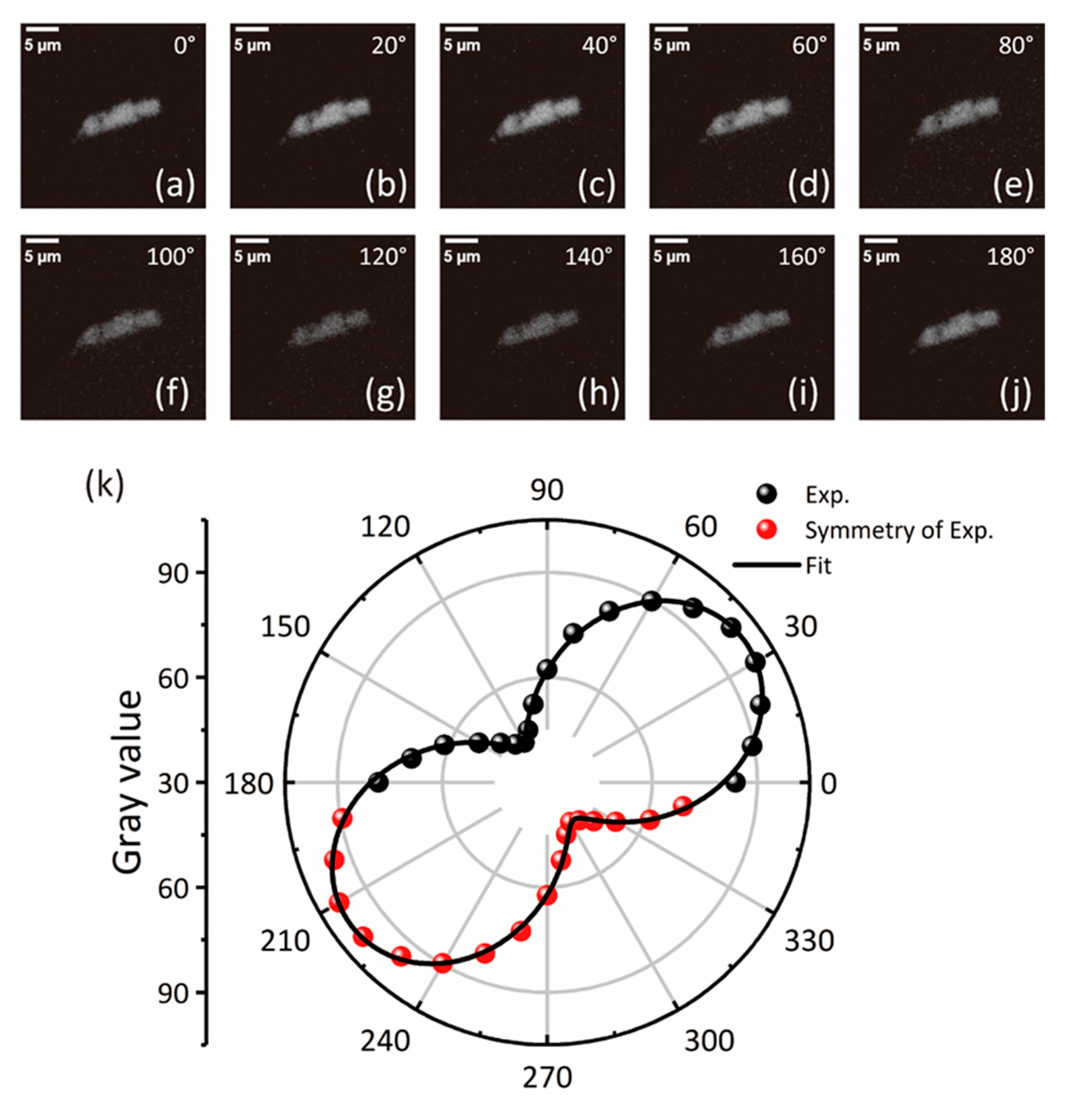
In addition to identifying monolayer BP, our PL microscopy can also simultaneously determine its crystal orientation. Previously, the crystal orientation of BP was commonly determined by PL, absorption, or Raman spectroscopy, which required expensive spectrometers and protective procedures for the sample during measurements. In contrast, with our modified PL microscope, the crystal orientation of monolayer BP can be determined in situ by simply inserting a linear polarizer in the incident light path. The polarization-dependent brightness of the sample, due to its anisotropic absorption [3], determines the crystal orientation, making this method much more convenient than the previous ones. Figure 3a–j show the R channel images of monolayer BP under different polarization angles ranging from 0° to 180°. Since the PL energy of monolayer BP (~1.7 eV) is close to the near-infrared region, we only focus on the gray value of the R channel image to avoid the higher energy noise from the G and B channels. The relationship between the brightness of monolayer BP (extracted as the average gray value of the sample area) and the polarization angle is plotted in Figure 3k. The maximum (minimum) brightness corresponds to polarization along the AC (ZZ) directions of monolayer BP. The data in Figure 3k can be well fitted by A c o s 2 θ φ + B , where θ denotes the polarizer rotation angle and A ,     φ ,     B are all fitting parameters, with values of 56.3 ± 0.8 ,   35.5 ± 0.4 ,   43.1 ± 0.5 , respectively. The minimum gray value (B) along the ZZ direction of monolayer BP is not zero due to incompletely linear polarized incident light or background noise from ambient conditions. Despite this, the fitting allows us to unambiguously determine the crystal orientation of monolayer BP. The AC and ZZ directions of this sample are shown in Figure 4a.
To further confirm the crystal orientation of monolayer BP determined by PL microscopy, we measured the polarization dependent PL spectra by a spectrometer. As shown in Figure 4b, a PL peak at ~722 nm (1.72 eV) exhibits the maximum (minimum) intensity when the excited light is polarized along the AC (ZZ) direction labeled in Figure 4a, verifying the accuracy of crystal orientation determination by our PL microscopy.

4. Discussion

Since mechanical exfoliation under microscope is mainly performed manually, the speed of identifying monolayer BP is limited by the time needed for visual judgment and the scanning frame rate. In the former case, the limit factor is the optical contrast of the sample. Our PL imaging significantly enhances the optical contrast of monolayer BP, greatly reducing the time required for visual identification. In the latter case, the limit factor is the exposure time. Longer exposure times increase the image capture time, lowering the frame rate and slowing stage movement, which hinders the rapid search for samples as well. In our study, the exposure time was set between 300 and 500 ms, which may not be fast enough for researchers experienced in exfoliation techniques. However, with the enhanced optical contrast, low magnification objectives (such as the 10× objective used in our study) can be used to find monolayer BP, offering a larger field of view that allows for the identification of more samples within a single image compared to higher magnification objectives (a 50× objective is commonly needed to spot monolayer BP [22]). This compensates the slower stage movement. Additionally, exposure time can be further reduced by increasing the analog gain, using a higher-power LED, or selecting a bandpass with higher transmittance, all of which could further speed up sample identification.
This system is also versatile. It has been reported that this method can be used for the identification of monolayer and few-layer TMDCs [45] by changing the optical filters or light source. The effectiveness of this method in other 2D semiconductors with strong PL emission such as 2D perovskite [52], InSe [53,54], etc., requires further experiments to verify. Furthermore, the interlayer coupling in van der Waals heterostructures can also be imaged by this technique [45]. Furthermore, under the reflection PL mode, various substrates used in mechanical exfoliation (e.g., PDMS, Si/SiO2, polycarbonate) are acceptable, since the reflected light from the substrate is not collected by the CMOS. In addition, this technology can be further optimized by introducing automated systems and image recognition algorithms, enabling the real-time monitoring of the large-area synthesis of high-quality monolayer BP for commercial applications [55].
In conclusion, we developed a rapid, nondestructive, and low-cost approach for identifying monolayer BP as well as its crystal orientation using polarized PL optical microscopy. Due to the low cost and high feasibility of this method, it will be accessible to most laboratories. Our study paves the way for more extensive research on and potential industrial applications of monolayer BP.

Supplementary Materials

The following supporting information can be downloaded at https://www.mdpi.com/article/10.3390/photonics11090866/s1, Figure S1: full image of monolayer BP under PL microscope. Figure S2: RGB images of monolayer BP and corresponding gray value profiles. Figure S3: PL intensity of monolayer BP as a function of time. Figure S4: illustration of scattering contaminants.

Author Contributions

Conceptualization, H.Y., S.H. and C.P.; methodology, C.P. and Y.M.; software, C.P.; validation, C.P.; formal analysis, C.P.; investigation, C.P.; resources, C.P.; data curation, C.P., Y.M., Q.W. and B.Y.; writing—original draft preparation, C.P.; writing—review and editing, H.Y., S.H. and C.P.; visualization, C.P.; supervision, H.Y. and S.H.; project administration, H.Y. and S.H.; funding acquisition, H.Y. All authors have read and agreed to the published version of the manuscript.

Funding

This research was funded by the National Key Research and Development Program of China (Grant Nos.2022YFA1404700, 2021YFA1400100), the National Natural Science Foundation of China (Grant No. 12074085), and the Natural Science Foundation of Shanghai (Grant Nos.23XD1400200, 23JC1401100).

Institutional Review Board Statement

Not applicable.

Informed Consent Statement

Not applicable.

Data Availability Statement

The data that support the findings of this study are available from the corresponding author upon request.

Acknowledgments

We would like to thank Yuanbo Zhang and his student Mingyan Luo for assisting us with AFM measurement of our samples. Part of the experimental work was carried out in the Fudan Nanofabrication Lab.

Conflicts of Interest

The authors declare no conflicts of interest.

References

  1. Li, L.; Yu, Y.; Ye, G.J.; Ge, Q.; Ou, X.; Wu, H.; Feng, D.; Chen, X.H.; Zhang, Y. Black Phosphorus Field-Effect Transistors. Nat. Nanotechnol. 2014, 9, 372–377. [Google Scholar] [CrossRef] [PubMed]
  2. Liu, H.; Neal, A.T.; Zhu, Z.; Luo, Z.; Xu, X. Phosphorene: An Unexplored 2D Semiconductor with a High Hole Mobility. ACS Nano 2014, 8, 4033–4041. [Google Scholar] [CrossRef] [PubMed]
  3. Wang, X.; Jones, A.M.; Seyler, K.L.; Tran, V.; Jia, Y.; Zhao, H.; Wang, H.; Yang, L.; Xu, X.; Xia, F. Highly Anisotropic and Robust Excitons in Monolayer Black Phosphorus. Nat. Nanotechnol. 2015, 10, 517–521. [Google Scholar] [CrossRef]
  4. Lee, S.; Yang, F.; Suh, J.; Yang, S.; Lee, Y.; Li, G.; Sung Choe, H.; Suslu, A.; Chen, Y.; Ko, C.; et al. Anisotropic In-Plane Thermal Conductivity of Black Phosphorus Nanoribbons at Temperatures Higher than 100 K. Nat. Commun. 2015, 6, 8573. [Google Scholar] [CrossRef]
  5. Huang, S.; Wang, C.; Xie, Y.; Yu, B.; Yan, H. Optical Properties and Polaritons of Low Symmetry 2D Materials. Photonics Insights 2023, 2, R03. [Google Scholar] [CrossRef]
  6. Kim, J.; Baik, S.S.; Ryu, S.H.; Sohn, Y.; Park, S.; Park, B.-G.; Denlinger, J.; Yi, Y.; Choi, H.J.; Kim, K.S. Observation of Tunable Band Gap and Anisotropic Dirac Semimetal State in Black Phosphorus. Science 2015, 349, 723–726. [Google Scholar] [CrossRef]
  7. Li, L. Direct Observation of the Layer-Dependent Electronic Structure in Phosphorene. Nat. Nanotechnol. 2017, 12, 21–25. [Google Scholar] [CrossRef] [PubMed]
  8. Huang, S.; Zhang, G.; Fan, F.; Song, C.; Wang, F.; Xing, Q.; Wang, C.; Wu, H.; Yan, H. Strain-Tunable van Der Waals Interactions in Few-Layer Black Phosphorus. Nat. Commun. 2019, 10, 2447. [Google Scholar] [CrossRef]
  9. Huang, S.; Lu, Y.; Wang, F.; Lei, Y.; Song, C.; Zhang, J.; Xing, Q.; Wang, C.; Xie, Y.; Mu, L.; et al. Layer-Dependent Pressure Effect on the Electronic Structure of 2D Black Phosphorus. Phys. Rev. Lett. 2021, 127, 186401. [Google Scholar] [CrossRef]
  10. Huang, S.; Wang, F.; Zhang, G.; Song, C.; Lei, Y.; Xing, Q.; Wang, C.; Zhang, Y.; Zhang, J.; Xie, Y.; et al. From Anomalous to Normal: Temperature Dependence of the Band Gap in Two-Dimensional Black Phosphorus. Phys. Rev. Lett. 2020, 125, 156802. [Google Scholar] [CrossRef]
  11. Lei, Y.; Ma, J.; Luo, J.; Huang, S.; Yu, B.; Song, C.; Xing, Q.; Wang, F.; Xie, Y.; Zhang, J.; et al. Layer-Dependent Exciton Polarizability and the Brightening of Dark Excitons in Few-Layer Black Phosphorus. Nat. Commun. 2023, 14, 5314. [Google Scholar] [CrossRef] [PubMed]
  12. Zhang, G.; Huang, S.; Wang, F.; Yan, H. Layer-Dependent Electronic and Optical Properties of 2D Black Phosphorus: Fundamentals and Engineering. Laser Photonics Rev. 2021, 15, 2000399. [Google Scholar] [CrossRef]
  13. Li, W.; Tao, Q.; Li, Z.; Yang, G.; Lu, Z.; Chen, Y.; Wen, Y.; Wang, Y.; Liao, L.; Liu, Y.; et al. Monolayer Black Phosphorus and Germanium Arsenide Transistors via van Der Waals Channel Thinning. Nat. Electron. 2023, 7, 131–137. [Google Scholar] [CrossRef]
  14. Huang, M.; Wang, M.; Chen, C.; Ma, Z.; Li, X.; Han, J.; Wu, Y. Broadband Black-Phosphorus Photodetectors with High Responsivity. Adv. Mater. 2016, 28, 3481–3485. [Google Scholar] [CrossRef] [PubMed]
  15. Wu, W.; Zhou, Y.; Wang, J.; Shao, Y.; Kong, D.; Gao, Y.; Wang, Y. The Pump Fluence and Wavelength-Dependent Ultrafast Carrier Dynamics and Optical Nonlinear Absorption in Black Phosphorus Nanosheets. Nanophotonics 2020, 9, 2033–2043. [Google Scholar] [CrossRef]
  16. Yang, Y.; Gao, J.; Zhang, Z.; Xiao, S.; Xie, H.; Sun, Z.; Wang, J.; Zhou, C.; Wang, Y.; Guo, X.; et al. Black Phosphorus Based Photocathodes in Wideband Bifacial Dye-Sensitized Solar Cells. Adv. Mater. 2016, 28, 8937–8944. [Google Scholar] [CrossRef] [PubMed]
  17. Zong, X.; Hu, H.; Ouyang, G.; Wang, J.; Shi, R.; Zhang, L.; Zeng, Q.; Zhu, C.; Chen, S.; Cheng, C.; et al. Black Phosphorus-Based van Der Waals Heterostructures for Mid-Infrared Light-Emission Applications. Light Sci. Appl. 2020, 9, 114. [Google Scholar] [CrossRef]
  18. Higashitarumizu, N.; Tajima, S.; Kim, J.; Cai, M.; Javey, A. Long Operating Lifetime Mid-Infrared LEDs Based on Black Phosphorus. Nat. Commun. 2023, 14, 4845. [Google Scholar] [CrossRef]
  19. Kou, L.; Frauenheim, T.; Chen, C. Phosphorene as a Superior Gas Sensor: Selective Adsorption and Distinct I−V Response. J. Phys. Chem. Lett. 2014, 5, 2675–2681. [Google Scholar] [CrossRef]
  20. Rodin, A.S.; Carvalho, A.; Neto, A.H.C. Excitons in Anisotropic Two-Dimensional Semiconducting Crystals. Phys. Rev. B 2014, 90, 075429. [Google Scholar] [CrossRef]
  21. Xu, R.; Zhang, S.; Wang, F.; Yang, J.; Wang, Z.; Pei, J.; Myint, Y.W.; Xing, B.; Yu, Z.; Fu, L.; et al. Extraordinarily Bound Quasi-One-Dimensional Trions in Two-Dimensional Phosphorene Atomic Semiconductors. ACS Nano 2016, 10, 2046–2053. [Google Scholar] [CrossRef] [PubMed]
  22. Wang, F.; Wang, C.; Chaves, A.; Song, C.; Zhang, G.; Huang, S.; Lei, Y.; Xing, Q.; Mu, L.; Xie, Y.; et al. Prediction of Hyperbolic Exciton-Polaritons in Monolayer Black Phosphorus. Nat. Commun. 2021, 12, 5628. [Google Scholar] [CrossRef] [PubMed]
  23. Yang, J.; Xu, R.; Pei, J.; Myint, Y.W.; Wang, F.; Wang, Z.; Zhang, S.; Yu, Z.; Lu, Y. Optical Tuning of Exciton and Trion Emissions in Monolayer Phosphorene. Light Sci. Appl. 2015, 4, e312. [Google Scholar] [CrossRef]
  24. Nemilentsau, A.; Low, T.; Hanson, G. Anisotropic 2D Materials for Tunable Hyperbolic Plasmonics. Phys. Rev. Lett. 2016, 116, 066804. [Google Scholar] [CrossRef]
  25. Biswas, S.; Wong, J.; Pokawanvit, S.; Yang, W.-C.D.; Zhang, H.; Akbari, H.; Watanabe, K.; Taniguchi, T.; Davydov, A.V. Edge-Confined Excitons in Monolayer Black Phosphorus. ACS Nano 2023, 17, 23692–23701. [Google Scholar] [CrossRef]
  26. Guo, H.; Zhang, X.; Lu, G. Pseudo-Heterostructure and Condensation of 1D Moiré Excitons in Twisted Phosphorene Bilayers. Sci. Adv. 2023, 9, eadi5404. [Google Scholar] [CrossRef]
  27. Soltero, I.; Guerrero-Sánchez, J.; Mireles, F.; Ruiz-Tijerina, D.A. Moiré Band Structures of Twisted Phosphorene Bilayers. Phys. Rev. B 2022, 105, 235421. [Google Scholar] [CrossRef]
  28. Pan, D.; Wang, T.-C.; Xiao, W.; Hu, D.; Yao, Y. Simulations of Twisted Bilayer Orthorhombic Black Phosphorus. Phys. Rev. B 2017, 96, 041411. [Google Scholar] [CrossRef]
  29. Zhao, S.; Wang, E.; Üzer, E.A.; Guo, S.; Qi, R.; Tan, J.; Watanabe, K.; Taniguchi, T.; Nilges, T.; Gao, P.; et al. Anisotropic Moiré Optical Transitions in Twisted Monolayer/Bilayer Phosphorene Heterostructures. Nat. Commun. 2021, 12, 3947. [Google Scholar] [CrossRef]
  30. Kang, P.; Zhang, W.-T.; Michaud-Rioux, V.; Kong, X.-H.; Hu, C.; Yu, G.-H.; Guo, H. Moiré Impurities in Twisted Bilayer Black Phosphorus: Effects on the Carrier Mobility. Phys. Rev. B 2017, 96, 195406. [Google Scholar] [CrossRef]
  31. Cao, T.; Li, Z.; Qiu, D.Y.; Louie, S.G. Gate Switchable Transport and Optical Anisotropy in 90° Twisted Bilayer Black Phosphorus. Nano Lett 2016, 16, 5542–5546. [Google Scholar] [CrossRef] [PubMed]
  32. Guo, Z.; Zhang, H.; Lu, S.; Wang, Z.; Tang, S.; Shao, J.; Sun, Z.; Xie, H.; Wang, H.; Yu, X.-F.; et al. From Black Phosphorus to Phosphorene: Basic Solvent Exfoliation, Evolution of Raman Scattering, and Applications to Ultrafast Photonics. Adv. Funct. Mater. 2015, 25, 6996–7002. [Google Scholar] [CrossRef]
  33. Erande, M.B.; Pawar, M.S.; Late, D.J. Humidity Sensing and Photodetection Behavior of Electrochemically Exfoliated Atomically Thin-Layered Black Phosphorus Nanosheets. ACS Appl. Mater. Interfaces 2016, 8, 11548–11556. [Google Scholar] [CrossRef] [PubMed]
  34. Smith, J.B. Growth of 2D Black Phosphorus Film from Chemical Vapor Deposition. Nanotechnology 2016, 27, 215602. [Google Scholar] [CrossRef]
  35. Jia, J.; Jang, S.K.; Lai, S.; Xu, J.; Choi, Y.J.; Park, J.-H.; Lee, S. Plasma-Treated Thickness-Controlled Two-Dimensional Black Phosphorus and Its Electronic Transport Properties. ACS Nano 2015, 9, 8729–8736. [Google Scholar] [CrossRef]
  36. Zhang, W.; Zhang, X.; Ono, L.K.; Qi, Y.; Oughaddou, H. Recent Advances in Phosphorene: Structure, Synthesis, and Properties. Small 2024, 20, 202303115. [Google Scholar] [CrossRef]
  37. Zhao, T.; Zhang, S.; Guo, Y.; Wang, Q. Photocarrier Dynamics in Monolayer Phosphoreneand Bulk Black Phosphorus. Nanoscale 2016, 8, 233–242. [Google Scholar] [CrossRef]
  38. Favron, A.; Gaufrès, E.; Fossard, F.; Phaneuf-L’Heureux, A.-L.; Tang, N.Y.-W.; Lévesque, P.L.; Loiseau, A.; Leonelli, R.; Francoeur, S.; Martel, R. Photooxidation and Quantum Confinement Effects in Exfoliated Black Phosphorus. Nat. Mater. 2015, 14, 826–832. [Google Scholar] [CrossRef] [PubMed]
  39. Wang, F. Electronic Structures of Air-Exposed Few-Layer Black Phosphorus by Optical Spectroscopy. Phys. Rev. B 2019, 99, 075427. [Google Scholar] [CrossRef]
  40. Li, H.; Wu, J.; Huang, X.; Lu, G.; Yang, J.; Lu, X.; Xiong, Q.; Zhang, H. Rapid and Reliable Thickness Identification of Two-Dimensional Nanosheets Using Optical Microscopy. ACS Nano 2013, 7, 10344–10353. [Google Scholar] [CrossRef]
  41. Zhang, B.; Zhang, Z.; Han, H.; Ling, H.; Zhang, X.; Wang, Y.; Wang, Q.; Li, H.; Zhang, Y.; Zhang, J.; et al. A Universal Approach to Determine the Atomic Layer Numbers in Two-Dimensional Materials Using Dark-Field Optical Contrast. Nano Lett. 2023, 23, 9170–9177. [Google Scholar] [CrossRef] [PubMed]
  42. Tian, R.; Fei, R.; Hu, S.; Li, T.; Zheng, B.; Shi, Y.; Zhao, J.; Zhang, L.; Gan, X.; Wang, X. Observation of Excitonic Series in Monolayer and Few-Layer Black Phosphorus. Phys. Rev. B 2020, 101, 235407. [Google Scholar] [CrossRef]
  43. Rubio-Bollinger, G.; Guerrero, R.; De Lara, D.; Quereda, J.; Vaquero-Garzon, L.; Agraït, N.; Bratschitsch, R.; Castellanos-Gomez, A. Enhanced Visibility of MoS2, MoSe2, WSe2 and Black-Phosphorus: Making Optical Identification of 2D Semiconductors Easier. Electronics 2015, 4, 847–856. [Google Scholar] [CrossRef]
  44. Castellanos-Gomez, A.; Vicarelli, L.; Prada, E.; Island, J.O.; Narasimha-Acharya, K.L.; Blanter, S.I.; Groenendijk, D.J.; Buscema, M.; Steele, G.A.; Alvarez, J.V.; et al. Isolation and Characterization of Few-Layer Black Phosphorus. 2D Mater. 2014, 1, 025001. [Google Scholar] [CrossRef]
  45. Alexeev, E.M.; Catanzaro, A.; Skrypka, O.V.; Nayak, P.K.; Ahn, S.; Pak, S.; Lee, J.; Sohn, J.I.; Novoselov, K.S.; Shin, H.S.; et al. Imaging of Interlayer Coupling in van Der Waals Heterostructures Using a Bright-Field Optical Microscope. Nano Lett 2017, 17, 5342–5349. [Google Scholar] [CrossRef]
  46. Higashitarumizu, N.; Uddin, S.Z.; Weinberg, D.; Azar, N.S.; Reaz Rahman, I.K.M.; Wang, V.; Crozier, K.B.; Rabani, E.; Javey, A. Anomalous Thickness Dependence of Photoluminescence Quantum Yield in Black Phosphorous. Nat. Nanotechnol. 2023, 18, 507–513. [Google Scholar] [CrossRef]
  47. Castellanos-Gomez, A.; Agraït, N.; Rubio-Bollinger, G. Optical Identification of Atomically Thin Dichalcogenide Crystals. Appl. Phys. Lett. 2010, 96, 213116. [Google Scholar] [CrossRef]
  48. Bing, D.; Wang, Y.; Bai, J.; Du, R.; Wu, G.; Liu, L. Optical Contrast for Identifying the Thickness of Two-Dimensional Materials. Opt. Commun. 2018, 406, 128–138. [Google Scholar] [CrossRef]
  49. Brotons-Gisbert, M.; Sánchez-Royo, J.F.; Martínez-Pastor, J.P. Thickness Identification of Atomically Thin InSe Nanoflakes on SiO2/Si Substrates by Optical Contrast Analysis. Appl. Surf. Sci. 2015, 354, 453–458. [Google Scholar] [CrossRef]
  50. Zhao, Q.; Puebla, S.; Zhang, W.; Wang, T.; Frisenda, R.; Castellanos-Gomez, A. Thickness Identification of Thin InSe by Optical Microscopy Methods. Adv. Photonics Res. 2020, 1, 200025. [Google Scholar] [CrossRef]
  51. Wang, Y.Y.; Gao, R.X.; Ni, Z.H.; He, H.; Guo, S.P.; Yang, H.P.; Cong, C.X.; Yu, T. Thickness Identification of Two-Dimensional Materials by Optical Imaging. Nanotechnology 2012, 23, 495713. [Google Scholar] [CrossRef] [PubMed]
  52. Li, J.; Wang, J.; Zhang, Y.; Wang, H.; Lin, G.; Xiong, X.; Zhou, W.; Luo, H.; Li, D. Fabrication of Single Phase 2D Homologous Perovskite Microplates by Mechanical Exfoliation. 2D Mater. 2018, 5, 021001. [Google Scholar] [CrossRef]
  53. Brotons-Gisbert, M.; Andres-Penares, D.; Suh, J.; Hidalgo, F.; Abargues, R.; Rodr, P.J.; Ordejon, P.; Wu, J.; Mart, J.P. Nanotexturing To Enhance Photoluminescent Response of Atomically Thin Indium Selenide with Highly Tunable Band Gap. Nano Lett. 2016, 16, 3221–3229. [Google Scholar] [CrossRef] [PubMed]
  54. Song, C.; Huang, S.; Wang, C.; Luo, J.; Yan, H. The Optical Properties of Few-Layer InSe. J. Appl. Phys. 2020, 128, 060901. [Google Scholar] [CrossRef]
  55. Severs Millard, T.; Genco, A.; Alexeev, E.M.; Randerson, S.; Ahn, S.; Jang, A.-R.; Suk Shin, H.; Tartakovskii, A.I. Large Area Chemical Vapour Deposition Grown Transition Metal Dichalcogenide Monolayers Automatically Characterized through Photoluminescence Imaging. Npj 2D Mater. Appl. 2020, 4, 12. [Google Scholar] [CrossRef]
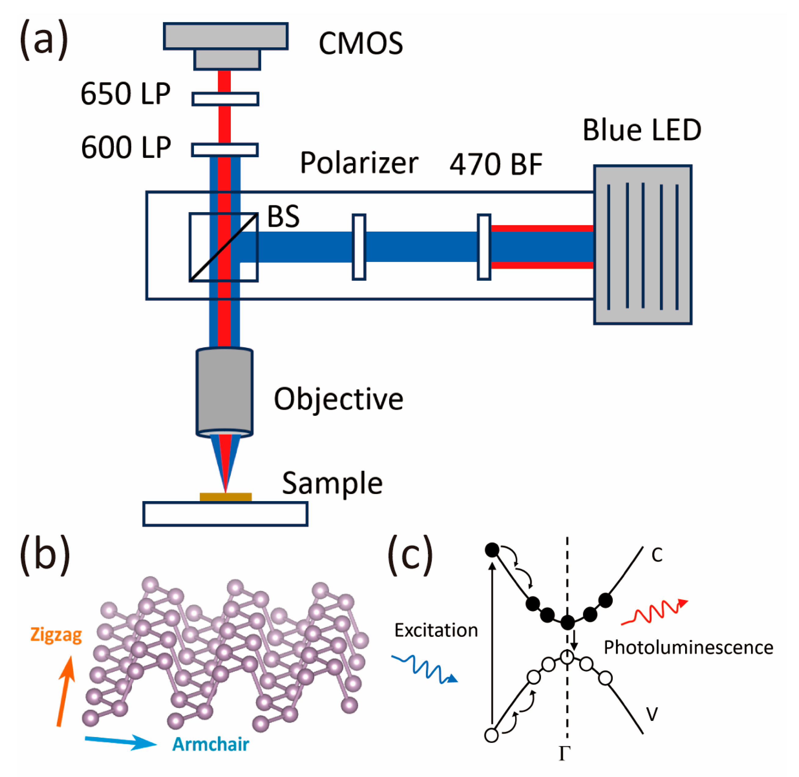
Figure 1. (a) Schematic illustration of the PL microscope optimized on a standard optical microscope. The PL microscope is composed of an objective, beam splitter (BS), light source (blue LED), digital camera (CMOS), linear polarizer, and filters, including a 470 nm narrow bandpass filter (BF) and 600 nm and 650 nm long-pass filters (LPs). (b) Schematic illustration of the crystal structure of monolayer BP. (c) Schematic illustration of the photoluminescence process in monolayer BP.
Photonics 11 00866 g001
Figure 2. (a) PL images of the monolayer BP sample (selected in the blue dotted area) under a 20× objective with a 300 ms acquisition time and 10× analog gain on the camera. (b) Corresponding bright-field image of the monolayer BP under conventional reflection mode with 50 ms acquisition time. (c) Corresponding AFM image of the monolayer BP sample. The inset shows the zoomed image in the yellow dotted box. (d,e) Gray value profile obtained along the green dashed lines in (a,b). (f) Height profile along the white line in the inset in (c).
Photonics 11 00866 g002
Figure 3. Crystal orientation identification of monolayer BP. (aj) R channel images of monolayer BP at different polarizer rotation angles from 0° to 180°. (k) Polar plots of polarized gray values extracted from (aj) as a function of the polarizer rotation angle. The solid line is fitted using A c o s 2 θ φ + B ( θ denotes the polarizer rotation angle and A ,     φ ,     B are all fitting parameters).
Photonics 11 00866 g003
Figure 4. (a) PL image of the monolayer BP sample in Figure 3. Light blue and yellow arrows indicate the AC and ZZ crystal orientations obtained from the fitting respectively. (b) PL spectrum of monolayer BP sample in (a) excited in the AC (black line) and ZZ (red line) directions.
Photonics 11 00866 g004
Disclaimer/Publisher’s Note: The statements, opinions and data contained in all publications are solely those of the individual author(s) and contributor(s) and not of MDPI and/or the editor(s). MDPI and/or the editor(s) disclaim responsibility for any injury to people or property resulting from any ideas, methods, instructions or products referred to in the content.