Review

Probing G-Quadruplexes Conformational Dynamics and Nano-Mechanical Interactions at the Single Molecule Level: Techniques and Perspectives

1
Department of Science and High Technology, University of Insubria, Via Valleggio 11, 22100 Como, Italy
2
CLIP-Como Lake Institute of Photonics, Via Valleggio 11, 22100 Como, Italy
3
Department of Pharmaceutical and Pharmacological Science, University of Padua, Via Marzolo 5, 35131 Padua, Italy
*
Author to whom correspondence should be addressed.
Photonics 2024, 11(11), 1061; https://doi.org/10.3390/photonics11111061
Submission received: 16 September 2024 / Revised: 31 October 2024 / Accepted: 6 November 2024 / Published: 13 November 2024
(This article belongs to the Special Issue Photonics in Single Molecule Detection and Analysis Techniques)

Abstract

:
The analysis of nucleic acid structures, topologies, nano-mechanics and interactions with ligands and other biomacromolecules (most notably proteins) at the single molecule level has become a fundamental topic in molecular biophysics over the last two decades. Techniques such as molecular tweezers, single-molecule fluorescence resonance energy transfer, and atomic force microscopy have enabled us to disclose an unprecedented insight into the mechanisms governing gene replication, transcription and regulation. In this minireview, we survey the main working principles and discuss technical caveats of the above techniques, using as a fil-rouge the history of their achievements in dissecting G-quadruplexes. The revised literature offers a clear example of the superior ability of single-molecule techniques with respect to ensemble techniques to unveil the structural and functional diversity of the several polymorphs corresponding to a single G-quadruplex folding sequence, thus shedding new light on the extreme complexity of these fascinating non-Watson–Crick structures.

1. Introduction

G-quadruplexes (G4s) are non-canonical secondary structures of nucleic acids that can occur at sequences containing at least four runs of consecutive guanines. At the genomic level, these motifs are significantly enriched at some functional sites, such as telomeres, promoters, and 5’-UTR domains. In addition, they appear to be significantly conserved throughout evolution. At first, G4 functional roles were addressed at telomeres where long single-stranded G-rich domains are present. Here, their G4 folding was found to impair the telomerase elongation activity. Later on, bioinformatic, in vitro and in-cell studies highlighted their occurrence at multiple genomic sites with an evolutionarily conserved enrichment at the promoter level, thus pointing them as switches for transcription. In particular, at these positions, the G4 formation has been proven to be able to alter the transcription factors recruitment [1]. Interestingly, for several oncogenes (MYC, KIT, BLC2, etc.), such a mechanism was found to cause a reduction in the protein expression level, although alternative patterns are not excluded [2]. These data, along with several in-cell and in vivo studies, extensively confirm their fundamental roles in many cellular regulatory processes [3]. This scenario is fully consistent with their exploitation as targets in oncology, neurological disorders and, more recently, antiviral treatments. The extreme versatility of G4s in acting as signaling elements in metabolic pathways [4,5,6,7] largely depends on their conformational polymorphism. First, it is worth noting that, from a structural point of view, the key architectural element of G4s is the pairing of four guanines through Hoogsteen hydrogen bonds, which drives the formation of a tetra-helical arrangement. Consequently, their features are distinct from the more common double helix. Moreover, in G4s, the four strands can be arranged with different mutual orientations and, in the monomeric structures, they are interconnected by three loops that can vary in terms of length and composition. Finally, different G4s can assume multiple topological arrangements [8,9,10], and even the same sequence can fold into different structures [11,12].
The full landscape of these complex structural equilibria depends on environmental conditions [13,14], exertion of forces and/or torques on DNA by proteins [15,16,17] and interaction with ligands [18,19,20,21,22]. This latter point is of particular interest due to the fact that such ligands may tune the biological functions of the targeted G4, and thus are promising to act as novel therapeutics for several diseases [1,23,24,25,26,27,28].
From this picture, it is easy to derive potential intriguing correlations between the molecular features of G4s and a wealth of fundamental aspects of cell life, such as metabolism, self-regulation, senescence, survival, and pathological pathways. For these reasons, it is of pivotal importance to set up biophysical tools capable of yielding detailed information about the conformational dynamics of G4-folding in DNA and RNA sequences, as well as probing the effects of nanomechanical forces and torques that mimic those solicitations which are physiologically exerted, e.g., by proteins. Due to the extreme conformational diversity of G4s, samples are expected to present dramatic heterogeneity. As a result, probing the above-mentioned features at the single-molecule level is mandatory to faithfully reconstruct the statistical distributions of either the structures assumed or the nanomechanical interactions acting on a G4-folding sequence. Here, we report on advanced techniques developed for this purpose, review the fundamental literature on the topic, and critically assess their strengths and weaknesses.

2. Conformational Studies — Single-Molecule Fluorescence Resonance Energy Transfer

Historically, biomolecular conformations have been extensively and successfully investigated by exploiting the fluorescence resonance energy transfer (FRET) phenomenon. Theorized in 1949 by T. Förster [29], FRET is a quantum dipole–dipole resonance interaction occurring between two chromophores, in this context called donor (D) and acceptor (A). The fluorescence spectrum of D is partially overlapped with the absorption spectrum of A. Upon illumination in the absorption band of D, the excitation energy can be exchanged between the two dyes, resulting in a reduced fluorescence emission of D and a concomitant promotion of A to an electronically excited state. If A is also a fluorophore, its emission can be observed (see Figure 1b). As the efficiency of the energy transfer drops with the sixth power of the D-to-A distance, FRET can be exploited as a precise “spectroscopic ruler” to measure intramolecular distances in suitably labelled biomolecules [30].
The amount of energy transferred from the D to the A is usually evaluated in terms of the FRET efficiency, which is the ratio between the intensity of the fluorescence emitted by the A to the total fluorescence emitted by A and D (measured in number of photons per unit time):
E = I A I A + γ I D
The correction factor γ takes into account the different fluorescence quantum yields of the D and the A, as well as different detection efficiencies in the two channels, and depends on the setup and fluorophores used.
At the ensemble level, the FRET technique has been operatively implemented on systems of biological relevance since the early 1970s (for a review of early experiments, see [32,33]). However, the ensemble approach can be tricky in heterogeneous samples [34], where, as a result of conformational polymorphism and dynamics of the labelled biomolecule, the distance between the sites to which the D and the A are attached varies in time as well as from a molecule to another. It is worth noting that in this instance, the limitation of the technique reseeds in the unfeasibility of extrapolating FRET efficiency distributions from the data, which inherently provide only a mean FRET efficiency value consisting in the average over long times (compared with those proper of molecular dynamics) and high numbers of molecules of the actual ones. Implementation of FRET at the single-molecule level dates back to the 1990s [35,36,37,38]. It allows for overcoming the above limitation and provides an invaluable tool to probe the structural heterogeneity of a biomolecular specimen. Indeed, single-molecule FRET (SM-FRET) is nowadays a preferred technique to reveal the number of different structures assumed by a peptide [39,40,41,42,43] or nucleic acid sequence [44], as well as to unravel complex formation with defined partners. Accordingly, it can be optimally exploited to investigate the conformational diversity, dynamics, and transitions of G4s under strictly controlled experimental conditions. It should be noted that, by themselves, SM-FRET data can only yield indirect information on the conformational dynamics of a biomolecule. Indeed, the observable which is directly measured in an SM-FRET experiment is the D-to-A distance, which cannot always be straightforwardly connected with conformational features of the spacer (i.e., the labelled biomolecule of interest). However, with the advent of computer sciences, SM-FRET data can be interpreted in the light of dedicated molecular dynamics simulations, which offer a boosted comprehension by providing the needed insight to bridge the gap between the D-to-A distance datum and the corresponding structural conformation. Exemplary studies have been performed on the topic in the last few years [45,46,47,48]. Pioneering attempts to address the G4 topologies of the folded human telomeric sequence (TTAGGG)4 were made as early as 2003 [49]. Since then, the SM-FRET technique has been systematically applied to probe telomeric G4s under various experimental conditions [14,50,51,52,53,54,55,56,57,58], and it has been extended to additional G4-forming sequences, such as those occurring at gene promoters [59,60,61,62,63,64], in both their wild-type and mutated forms, and in the presence or absence of partner proteins [65].
Currently, two different implementations of SM-FRET exist: total internal reflection microscopy-based (TIRF-FRET) and confocal microscopy-based (CONF-FRET). These methods have been extensively described in excellent papers by Ha [36,66,67,68,69] and applied to characterize G4s. Each method has its advantages and drawbacks. TIRF-FRET allows for real-time monitoring of the conformational dynamics of single DNA molecules tethered to a surface for several seconds, which is relatively long compared to the timescales of conformational transitions [61,62]. In contrast, CONF-FRET, especially in its standard implementation, captures snapshots of molecules freely diffusing in and out of the confocal observation volume [70], thus producing fluorescence bursts (see Figure 1). The conformational distribution of species in the specimen is determined by sampling several of such independent fluorescence burst events. Notably, the time for an oligonucleotide [71,72], oligopeptide [43,73,74], or even a high molecular weight protein [63,75] to cross the observation volume is typically on the order of hundreds of microseconds. Given that the fastest folding–unfolding dynamics and interconversion kinetics between differently folded conformations occur on timescales of hundreds of milliseconds [41,61,62,76], negligible conformational changes are typically assumed during diffusion time.
Furthermore, CONF-FRET is minimally invasive and can monitor system evolution over hours [77], whereas the observation time in TIRF-FRET experiments is inherently limited by dye photobleaching, which typically occurs within tens of seconds [66,78,79,80]. Moreover, the environment experienced by freely diffusing biomolecules in CONF-FRET closely resembles physiological conditions compared to TIRF-FRET experiments, where biomolecules can interact with the surface to which they are tethered. This is particularly critical for oligonucleotide probes, which behave as polyelectrolytes and require surface passivation [66,81]. Additionally, the linking procedure requires careful design of the oligonucleotide construct [67], and the surface must undergo thorough cleaning procedures to minimize background interference [81,82,83]. The photophysics of donor and acceptor chromophores can also be perturbed unpredictably by proximity to a solid surface [84,85].
As to a direct comparison between results yielded by the two SM-FRET variants, to the best of our knowledge, there exists only one instance in which the same G4-forming sequence has been probed by means of both methods. Namely, in 2007, Balasubramanian and collaborators [59] performed a characterization of oligonucleotides mimicking the sequences of the G4s c-kit1 and c-kit2 embedded within the c-KIT oncogene promoter, exploiting both TIRF- and CONF-FRET. Although their findings appear self-consistent from a qualitative standpoint, the FRET efficiency distributions obtained on homologous samples with the two setups are remarkably different. However, it must be noted that the same authors published a similar study on the same G4-folding sequences in 2010 [60], using the CONF-FRET setup only, and they obtained different results with respect to the homologous measurements they had previously published. This fact indicates that many environmental aspects concur in determining the actual folding of a G4. Thus, the difference recorded in the 2007 experiments on the two setups might be reconducted for reasons other than the SM-FRET option chosen (e.g., slight variations in the buffer or temperature).
TIRF-FRET has been more extensively applied than CONF-FRET to probe G4s so far. Besides investigating the conformations and folding/unfolding kinetics of telomeric G4s under various environmental conditions, specifically different concentrations of cations [14,49,50,52,55,57,58], and the role of small ligands and proteins in tuning such equilibria [54,56,62,86,87,88,89,90,91], analyses have been extended to G4s formed within the promoters of several genes [91], including the oncogenes c-KIT [88,90], c-MYC [61,62,82,87,88,92], and PAX9 [88].
However, in recent years, significant attention has been devoted to designing experimental models that more closely resemble the physiological genomic context, where the G4-forming domain is embedded within a more complex elongated framework. It has been found that the presence of either single- or double-stranded DNA tracts located at the ends of the G4-folding sequence significantly affects the conformational equilibrium of the tetrahelical domain [77,93]. This paves the way for SM-FRET studies aimed at dissecting the structural properties of G4s encompassed within oligonucleotide constructs that better mimic the features of the native DNA environment. For this purpose, CONF-FRET setups offer superior flexibility, thus this approach is expected to obtain more attention in the near future.

3. Probing the G4 Mechanical Stability and Mimicking Traction and Torsion Stresses Exerted on G4 by Proteins in Physiological Environment

As discussed above, in addition to the genomic context, the forces exerted by proteins play pivotal roles in inducing the conversion of double-stranded DNA into a G4. Therefore, valuable information about G4 folding can be extracted from their nanomechanical manipulation using single-molecule force spectroscopy techniques [94,95].

3.1. Optical Tweezers

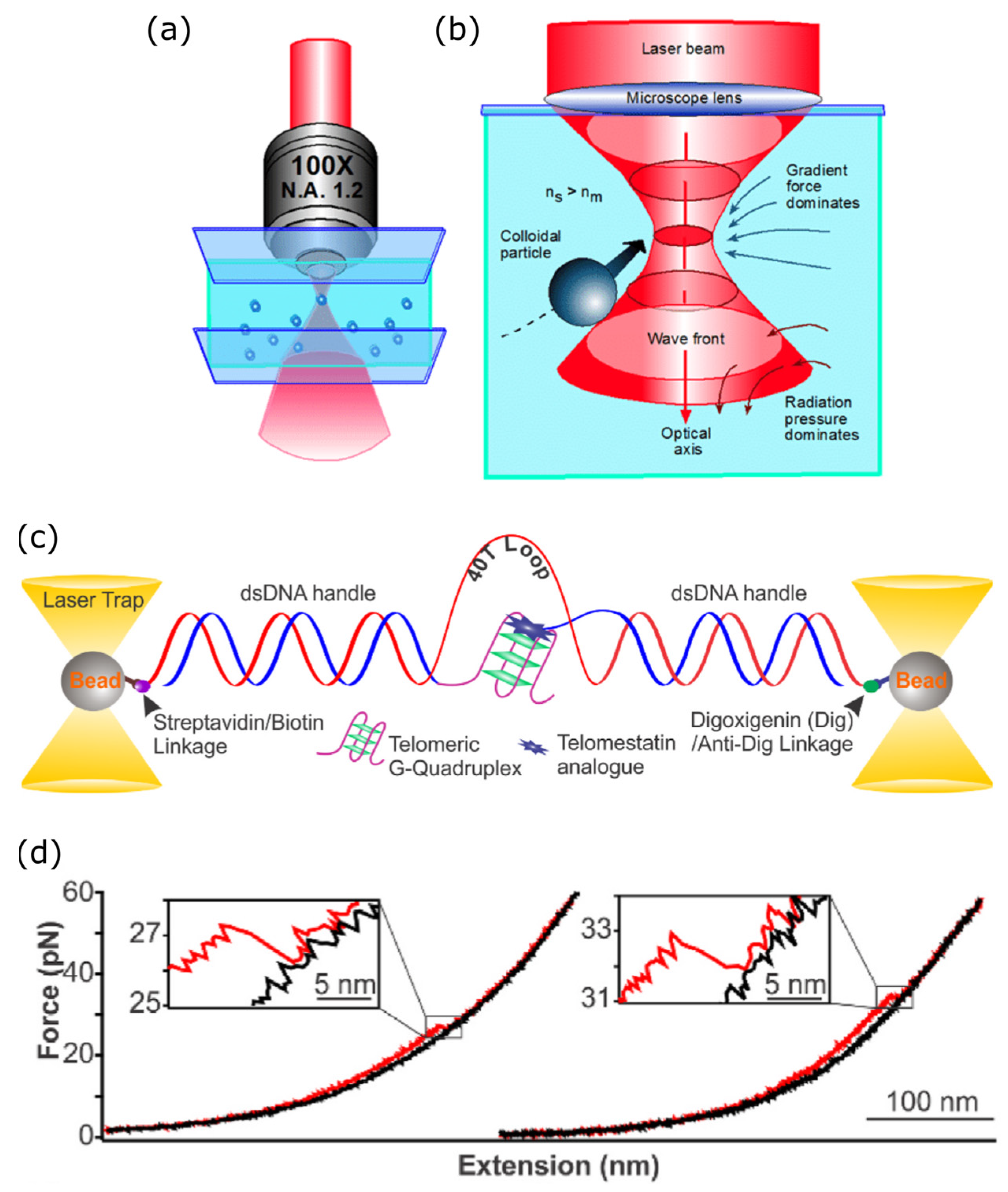
The unfolding free energy landscapes of biomolecular structures can be determined by inducing controlled traction forces using optical tweezers (OT). In an OT setup, radiation pressure allows a microscopic bead to be held in a focused laser beam (Figure 2). In this trapping mode, it is possible to generate spring-like forces ranging from a few piconewtons to nanonewtons on biomolecules properly functionalized with the bead(s) [96,97]. Since the pioneering works of Ashkin [98,99] in the late 1980s, this technique has found wide applications in biophysics [100,101]. In its simpler implementation for DNA experiments, OT involves tethering the free end of the DNA molecule under investigation to a surface and attaching a dielectric bead [102,103,104]. Alternatively, more than one bead can be attached to the opposite ends of a freely diffusing DNA molecule, creating multiple traps and offering more experimental flexibility [105,106,107,108] (see Figure 2). Overall, these beads serve as probes to measure the forces acting on the DNA molecule (e.g., due to interactions with proteins, environmental or structural changes) or to study its mechanical response using the laser beam as a force actuator. In the field of G4 research, optical tweezers (OT) were originally applied to probe the nanomechanical stability of single G4s by determining their folding free energy. Specifically, Mao’s group studied a G-rich sequence present in the insulin-linked polymorphic region (ILPR), a DNA domain of the insulin gene promoter. The G4-folding sequence was inserted between two double-stranded DNA tracts, either as a single-stranded linker [109] or counterfaced by its complementary cytosine-rich sequence [110]. Under acidic conditions, the complementary strand of a G4-forming site can fold into a different non-canonical secondary structure called an I-motif (iM), a tetrahelix supported by intercalated C − C + base pairs. The authors demonstrated that the G4 was more resistant to mechanical denaturation than the iM. However, they also showed that under conditions compatible with the folding of both structures, only one could be formed at a time due to steric hindrance. These results were confirmed by independent investigations by de Messieres [111].
Expanding on the OT technique, Mao’s group applied it to probe the interaction between neighboring G4s within the ILPR [112], pairs of telomeric G4s [113], and later several repeats [114]. Additionally, they elucidated the stabilizing forces of telomeric G4s in both DNA [115,116,117] and RNA [118], interactions with small ligands [113,114,119,120,121,122,123], folding–unfolding equilibria of G4s in the hTERT promoter [124,125], and the effects of crowding agents on G4 stability [126].
Other groups have focused their efforts on analyzing the effects of condensation on the stability of telomeric RNA G4s [127] and assessing G4-protein interactions [128].
Figure 2. Scheme and principle of operation of an optical tweezer. (a) A trapping laser beam is expanded to fill the back aperture of a high-NA microscope objective and focused into the sample, creating a trapping optical field. White light is transmitted through the sample, collected from the objective and sent to a CCD or CMOS camera through a lens to capture images of the focal plane of the objective in the sample. (b) Detail of the geometry of the trapping beam: inside the sample, the focused laser beam produces a force gradient attracting particles with a refractive index larger than that of the medium in the position of the focus. (c) Example of a force spectroscopy measurement employing two optical tweezers to trap and pull two beads connected to a G4 through a pair of dsDNA handles. The handles are connected to the beads through biotin/streptavidin and digoxigenin/anti-digoxigenin links. Note that this kind of experiment can be performed with a single optical tweezer, by fixing one end of the construct under study to the coverslip or to a micropipette. (d) Example of a force-extension measurement performed with the setup depicted in (c): the distance between the ends of the construct under study is changed over time, and the force (a,b) from [129], used under license; (c,d) from [114], © 2024 Elsevier B.V., its licensors, and contributors, used with permission.
Photonics 11 01061 g002

3.2. Magnetic Tweezers

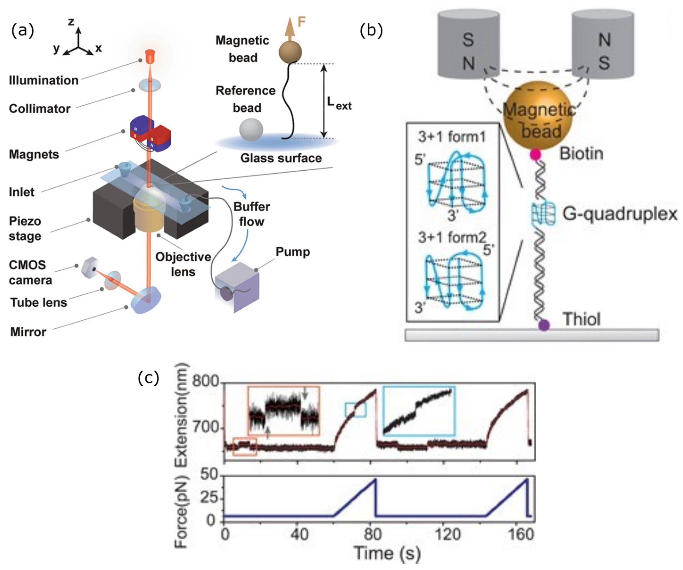
A valuable alternative to OT for probing DNA nanomechanics is represented by magnetic tweezers (MT), which are particularly suitable for assessing the role of mechanical stress in the folding/unfolding of G4s. In the case of MT, magnetic fields generated by either permanent magnets or electromagnets are used to induce forces on biomolecules tethered to magnetic particles [130]. Specifically, an MT apparatus appears as an evolution of a traditional microscope (Figure 3). It is basically composed of an imaging system, an image recording system (usually a CCD camera) that tracks the confined Brownian motion of the magnetic beads, and a magnet mounted on a piezoelectric actuator providing translational and rotational degrees of freedom. The movement of the magnet alters the magnetic field in which the beads are immersed, thereby setting them into motion. These forces are transmitted to the biomolecules to which they are tethered. MT lacks the extreme spatial resolution and flexibility of OT, and applying forces exceeding tens of piconewtons is challenging. However, the ability to easily rotate the magnets constitutes a notable advantage of MT over OT, particularly useful in G4 studies. Decoupling the rotation and translation of the magnets allows for simultaneous, independent, and strictly controllable torsional and tensile stress on DNA single molecules [131]. This capability is particularly relevant because negative supercoiling has been widely reported to enhance the transition of double-stranded DNA to G4 structures [132,133,134].
In order to enable the inspection of G4s through MT experiments, a panel of non-trivial biotechnological solutions must be devised. In a typical MT experiment on DNA, the sample is contained in a flow chamber allowing to vary the DNA substrate features in a controlled way. For instance, the pH and ionic strength of the buffer solution can be tuned while measuring the force-extension curve. Additionally, specific ions (most importantly K+ in the case of G4s analyses), ligands or proteins can be injected. The flow chamber may consist either of a capillary tube or in the interstitial space between two coverslips sealed with, e.g., parafilm. The sample must be tethered to the chamber’s internal surface. To this end, both chemical (e.g., thiol/silane) and biochemical (mainly digoxygenin/antidigoxygenin) molecular recognition systems have been exploited. The functionalization of the opposite end of the DNA molecules with micrometric-size superparamagnetic beads typically relies on the binding of biotinylated bases with streptavidin, coating the bead. In order to avoid streptavidin adherence to the glass surface of the chamber, after the adhesion of the DNA sample, but prior to their labeling with the beads, the chamber walls must be passivated. One standard solution is incubation with bovine serum albumin. In experiments in which the G4s are submitted to traction forces, the G4-folding tract is encompassed within two several kbase long double-stranded flanking ends, connecting it to the chamber surface and to the magnetic bead, respectively. Since such specimen is not torsionally constrained, to inspect the double-strand to G4 transition and equilibrium under exertion of external torques double-stranded samples encoding a central tract replicating one or more adjacent sequences capable of folding into G4s have been devised.
MT has been extensively exploited in the last decade to probe the conformational stability of telomeric [136,137] and promoter [137,138,139,140] G4s. Interestingly, MT has proven particularly effective in studies focused on G4s embedded in gene promoters. In vivo, these G4s are counteracted by a complementary strand and exist in equilibrium with double-stranded structures. Besides specific ion abundance, the G4/double-strand equilibrium is primarily dictated by superhelicity, which is locally regulated in vivo by proteins such as helicases or gyrases. MT allow mimicking the effects of such proteins and studying the double-helix to G4 transition under different superhelicity conditions induced by controlled torques applied through magnets onto magnetic beads [140].
Several studies aimed to rationalize or establish benchmarks in the folding pathways of G4s. Specifically, the precise measurement of the work exerted by the magnetic field on the bead to unfold a G4 enabled the reconstruction of unfolding energy landscapes and folding pathways of telomeric G4s [141], as well as the measurement of unfolding energies of telomeric G4s [142] and G4s embedded in the KIT oncogene promoter [140]. The folding kinetics and dynamics of various G4 structures [136,137,138,139] were monitored. Moreover, Cheng and colleagues systematically studied superior mechanical stability and slower unfolding rates of parallel versus antiparallel and hybrid G4s [137,143]. The same group also investigated the role of bulges in tuning folding pathways of typical parallel G4s through the formation of folding intermediates upon the gradual release of tension forces [143]. MT has shown particular versatility also in probing the effects of helicases on the stability of G4s [144], as well as the torques exerted by these proteins on DNA upon binding to G4 structures [145]. The effects of the binding of cisplatin on the folding kinetics of telomeric G4s have also been evaluated with MT [146].
Finally, the role of G4–G4 interactions in tuning the folding equilibrium of both telomeric [147] and promoter [140] G4s has also been recently addressed.
In several instances, MT and OT have been implemented together to combine spatial resolution with the ability to exert torques [133,134,148]. Moreover, to enhance the sensitivity of force-spectroscopy assays regarding different conformational arrangements of the investigated G4s, both MT [149] and OT [150,151] have been performed concurrently with SM-FRET.
Additionally, the MT technique has been used in combination with electron microscopy to demonstrate the compaction of single-stranded telomeric repeats into a bead-like structure upon G4 formation [152].

3.3. Atomic Force Microscopy

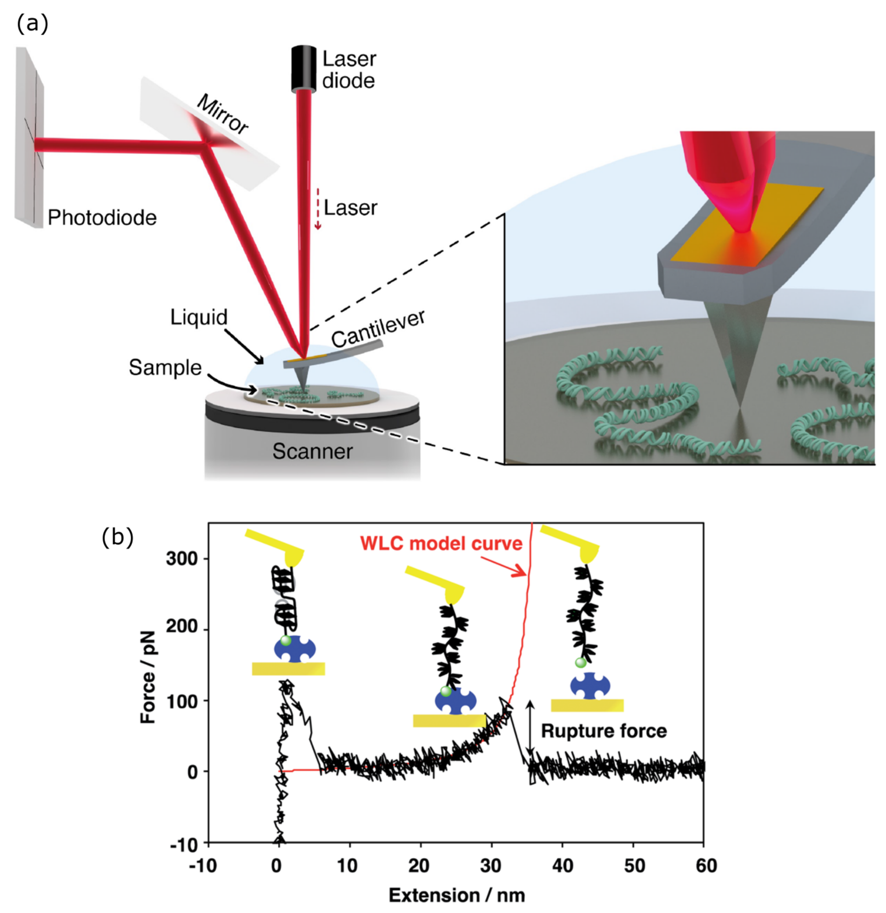
Atomic force microscopy (AFM) is a third technique that allows the exertion of pico- to nanonewton forces on single biomolecules, including DNA. The working principle of the atomic force microscope involves monitoring the interaction of a cantilever with the sample surface. As illustrated in Figure 4, this interaction causes tiny flexions of the cantilever tip, which can be detected by measuring the deflections of a laser beam focused on the cantilever. The cantilever can be moved across a sample deposited on a smooth surface (usually a mica layer) in a horizontal plane, using the raster-scanning mode. This mode provides a sub-nanometer resolution map of the surface roughness, giving topological information about the sample [153].
Alternatively, AFM allows the exertion of traction forces up to nanonewtons on objects deposited onto a smooth surface when the cantilever is operated in tapping mode. In tapping mode, scanning a sample surface entails lowering the tip onto the surface sequentially to apply controlled pressure, followed by lifting the cantilever either at a constant velocity or by applying a constant pulling force. The surface is scanned (fishing mode), searching for deposited objects. When the cantilever tip encounters an object, the contact is detected as a change in pressure. Moreover, the stiffness of the obstacle can be measured based on the tip bending at different applied forces.
For the purpose of this review, it is worth noting that even single nucleic acid or peptide molecules fixed onto a mica surface can be “fished” by the cantilever. In this case, adhesion forces cause one end of the biopolymer to bind to the cantilever tip. Consequently, controlled work can be exerted on single molecules by lifting the cantilever at a constant force. Such work can disrupt intramolecular interactions responsible for folding, thereby precisely determining the unfolding energetics [153,157].
In such force-spectroscopy modality, AFM has been applied to probe the stability of either telomeric [158,159] or aptameric [160] G4s, and their binding force to proteins [160]. The technique also allowed the authors to quantify the G4-stabilizing effect of a drug, namely a telomerase inhibitor [156].
On the other hand, AFM has been used also in the imaging mode to certify the formation of G4s in different DNA constructs at varying environmental properties. To the best of our knowledge, the first attempt in this sense was made by Thalhammer and coworkers as early as 2004 [161]. The authors unraveled the interaction of model palindromic G-rich oligonucleotides leading to disruption of hairpins and the formation of a tetrameric G4. Later, similar experiments were conducted by Oliveira-Brett et al. on different sequences at varying cation concentrations in the reaction buffer [162]. In 2009, Edwardson et al. managed to visualize by AFM imaging the formation of a promotorial G4 within a fragment of murine Sg3 switch region amplified from a plasmid [163]. Moreover, the authors thoroughly characterized the morphology of the counterfacing DNA trait, unraveling four different topological arrangements. In a subsequent study [164], some of them also probed the interactions of the same DNA specimen with the nuclear protein PARP-1 and the antibody HF1. Furthermore, they have shown that small ligands can tune in either sense of these interactions. Another study by Wang et al. [165] probed the distribution of folded G4s in a realistic mimic of the telomeric single-strand tail, namely an oligonucleotide sequence consisting of sixteen repeats of the GGGTTA motif. The authors discovered that, on average, only two out of the four possible G4 structures that could theoretically form coexist. Additionally, POT1 was shown to disrupt pre-folded G4s more efficiently than antisense oligonucleotides.
Finally, in a series of studies, Endo and collaborators exploited the DNA-origami technique combined with fast-scanning AFM imaging to monitor in real time the formation of an interstrand G4 with the telomeric repeat [166,167] and the interconversion kinetics between the double-stranded structure and a bubble stabilized by concomitant formation of a G4 counterfaced by an I-motif [168].

4. Conclusions

In this mini-review, we described the main single-molecule biophysical techniques that were exploited to probe the conformational dynamics, folding energetics, binding interactions, and reactivity to nanomechanical stresses of G4-forming sequences.
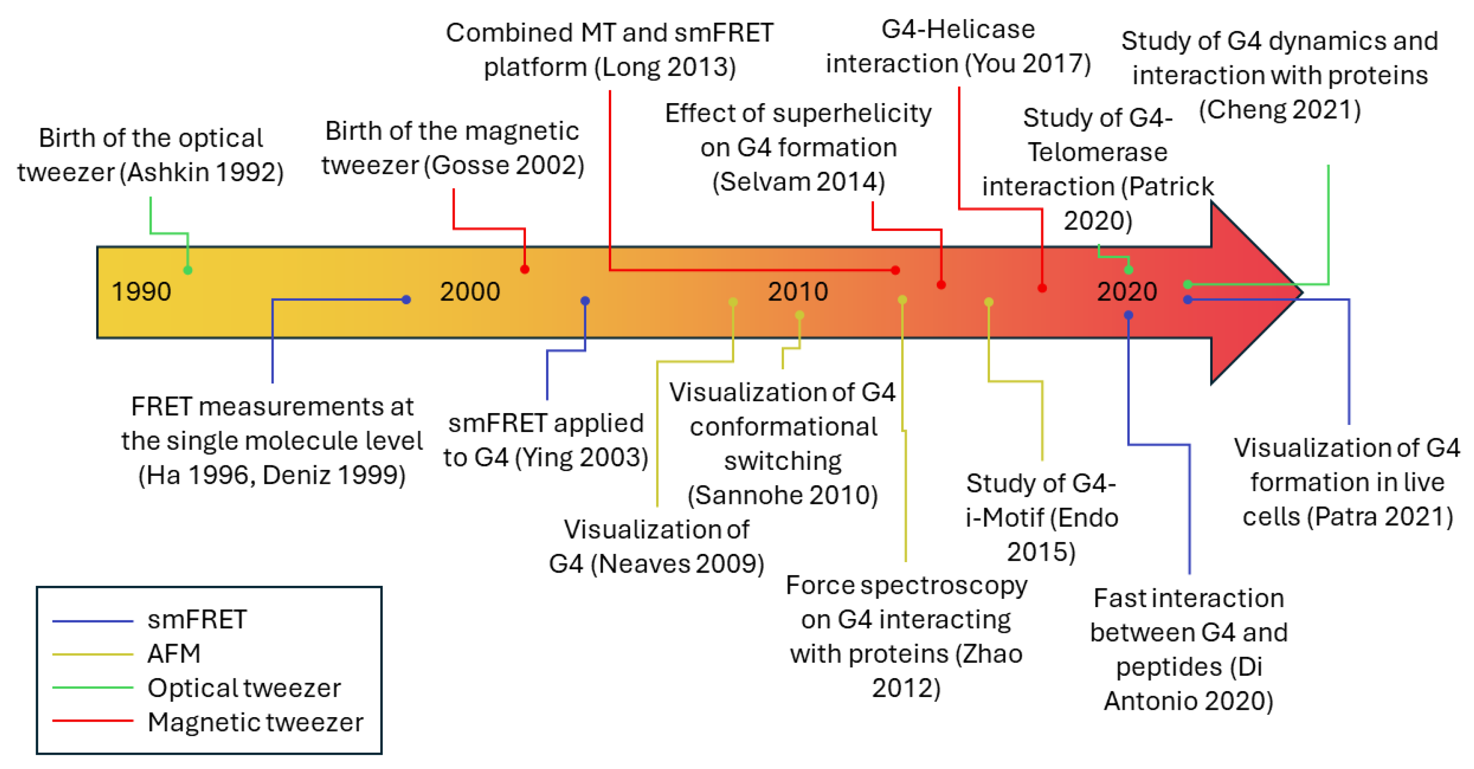
We revised the main literature on the topic. The milestones of this literature are summarized in Figure 5. Based on this excursus, we critically examined the strengths and pitfalls of each technique. The resulting pattern suggests that a single-molecule approach for the study of G4 folding equilibria, dynamics, and energetics is indispensable to grasp the essence of related biology and offers valuable information for new-conception pharmacological strategies based on the selective targeting of these fascinating secondary structures. At the same time, the combination of several single-molecule methods with more consolidated and standardized biochemical assays as well as state-of-art whole genome and whole-organism investigation protocols is mandatory to achieve a full comprehension of physiological and pathological structure–function relationships and in-cell interactions of G4.
In conclusion, although the single-molecule approach has been extensively and successfully applied to the elucidation of the folding features of selected guanine-rich sequences, mainly the telomeric one and those corresponding to a panel of G4s forming within the promotors of a few oncogenes, wide avenues remain to be explored in this field of investigation, including extending the analysis to a multitude of other sequences, each of those behaving uniquely and being a fundamental piece of the metabolic puzzle. The main frontier remaining, however, is the design of specimens which, whilst lending themselves to single-molecule analysis, mimic the physiological milieu of the G4-forming sequences under quest with increasing reliability and detail.

Author Contributions

Conceptualization, L.N. and C.S.; writing—original draft preparation, L.N., M.L. and R.R.; writing—review and editing, C.S.; supervision, L.N. and C.S. All authors have read and agreed to the published version of the manuscript.

Funding

This research received no external funding.

Institutional Review Board Statement

Not applicable.

Informed Consent Statement

Not applicable.

Data Availability Statement

No new data were created.

Conflicts of Interest

The authors declare no conflicts of interest.

References

  1. Figueiredo, J.; Mergny, J.-L.; Cruz, C. G-Quadruplex Ligands in Cancer Therapy: Progress, Challenges, and Clinical Perspectives. Life Sci. 2024, 340, 122481. [Google Scholar] [CrossRef]
  2. Liano, D.; Monti, L.; Chowdhury, S.; Raguseo, F.; Antonio, M.D. Long-Range DNA Interactions: Inter-Molecular G-Quadruplexes and Their Potential Biological Relevance. Chem. Commun. 2022, 58, 12753–12762. [Google Scholar] [CrossRef] [PubMed]
  3. Varshney, D.; Spiegel, J.; Zyner, K.; Tannahill, D.; Balasubramanian, S. The Regulation and Functions of DNA and RNA G-Quadruplexes. Nat. Rev. Mol. Cell Biol. 2020, 21, 459–474. [Google Scholar] [CrossRef]
  4. Wu, S.; Jiang, L.; Lei, L.; Fu, C.; Huang, J.; Hu, Y.; Dong, Y.; Chen, J.; Zeng, Q. Crosstalk between G-Quadruplex and ROS. Cell Death Dis. 2023, 14, 37. [Google Scholar] [CrossRef] [PubMed]
  5. Sato, K.; Knipscheer, P. G-Quadruplex Resolution: From Molecular Mechanisms to Physiological Relevance. DNA Repair 2023, 130, 103552. [Google Scholar] [CrossRef]
  6. Linke, R.; Limmer, M.; Juranek, S.A.; Heine, A.; Paeschke, K. The Relevance of G-Quadruplexes for DNA Repair. Int. J. Mol. Sci. 2021, 22, 12599. [Google Scholar] [CrossRef] [PubMed]
  7. Gorini, F.; Ambrosio, S.; Lania, L.; Majello, B.; Amente, S. The Intertwined Role of 8-oxodG and G4 in Transcription Regulation. Int. J. Mol. Sci. 2023, 24, 2031. [Google Scholar] [CrossRef]
  8. Huppert, J.L. Four-Stranded Nucleic Acids: Structure, Function and Targeting of G-Quadruplexes. Chem. Soc. Rev. 2008, 37, 1375–1384. [Google Scholar] [CrossRef]
  9. Lightfoot, H.L.; Hagen, T.; Tatum, N.J.; Hall, J. The Diverse Structural Landscape of Quadruplexes. FEBS Lett. 2019, 593, 2083–2102. [Google Scholar] [CrossRef]
  10. Burge, S.; Parkinson, G.N.; Hazel, P.; Todd, A.K.; Neidle, S. Quadruplex DNA: Sequence, Topology and Structure. Nucleic Acids Res. 2006, 34, 5402–5415. [Google Scholar] [CrossRef]
  11. Dettler, J.M.; Buscaglia, R.; Le, V.H.; Lewis, E.A. DSC Deconvolution of the Structural Complexity of C-MYC P1 Promoter G-Quadruplexes. Biophys. J. 2011, 100, 1517–1525. [Google Scholar] [CrossRef] [PubMed]
  12. Harkness, R.W.; Mittermaier, A.K. G-Quadruplex Dynamics. Biochim. Biophys. Acta (BBA)—Proteins Proteom. 2017, 1865, 1544–1554. [Google Scholar] [CrossRef] [PubMed]
  13. Dai, J.; Carver, M.; Punchihewa, C.; Jones, R.A.; Yang, D. Structure of the Hybrid-2 Type Intramolecular Human Telomeric G-Quadruplex in K+ Solution: Insights into Structure Polymorphism of the Human Telomeric Sequence. Nucleic Acids Res. 2007, 35, 4927–4940. [Google Scholar] [CrossRef] [PubMed]
  14. Noer, S.L.; Preus, S.; Gudnason, D.; Aznauryan, M.; Mergny, J.-L.; Birkedal, V. Folding Dynamics and Conformational Heterogeneity of Human Telomeric G-Quadruplex Structures in Na+ Solutions by Single Molecule FRET Microscopy. Nucleic Acids Res. 2016, 44, 464–471. [Google Scholar] [CrossRef]
  15. Troisi, R.; Sica, F. Structural Overview of DNA and RNA G-Quadruplexes in Their Interaction with Proteins. Curr. Opin. Struct. Biol. 2024, 87, 102846. [Google Scholar] [CrossRef]
  16. Sahoo, B.R.; Kocman, V.; Clark, N.; Myers, N.; Deng, X.; Wong, E.L.; Yang, H.J.; Kotar, A.; Guzman, B.B.; Dominguez, D.; et al. Protein G-Quadruplex Interactions and Their Effects on Phase Transitions and Protein Aggregation. Nucleic Acids Res. 2024, 52, 4702–4722. [Google Scholar] [CrossRef]
  17. Lu, Z.; Xie, S.; Su, H.; Han, S.; Huang, H.; Zhou, X. Identification of G-Quadruplex-Interacting Proteins in Living Cells Using an Artificial G4-Targeting Biotin Ligase. Nucleic Acids Res. 2024, 52, e37. [Google Scholar] [CrossRef]
  18. Sengupta, A.; Ganguly, A.; Chowdhury, S. Promise of G-Quadruplex Structure Binding Ligands as Epigenetic Modifiers with Anti-Cancer Effects. Molecules 2019, 24, 582. [Google Scholar] [CrossRef]
  19. Che, T.; Wang, Y.-Q.; Huang, Z.-L.; Tan, J.-H.; Huang, Z.-S.; Chen, S.-B. Natural Alkaloids and Heterocycles as G-Quadruplex Ligands and Potential Anticancer Agents. Molecules 2018, 23, 493. [Google Scholar] [CrossRef]
  20. Ruggiero, E.; Richter, S.N. G-Quadruplexes and G-Quadruplex Ligands: Targets and Tools in Antiviral Therapy. Nucleic Acids Res. 2018, 46, 3270–3283. [Google Scholar] [CrossRef]
  21. Asamitsu, S.; Obata, S.; Yu, Z.; Bando, T.; Sugiyama, H. Recent Progress of Targeted G-Quadruplex-Preferred Ligands Toward Cancer Therapy. Molecules 2019, 24, 429. [Google Scholar] [CrossRef] [PubMed]
  22. Balasubramanian, S.; Hurley, L.H.; Neidle, S. Targeting G-quadruplexes in gene promoters: A novel anticancer strategy? Nat. Rev. Drug Discov. 2011, 10, 261–275. [Google Scholar] [CrossRef] [PubMed]
  23. Debbarma, S.; Acharya, P.C. Targeting G-Quadruplex DNA for Cancer Chemotherapy. Curr. Drug Discov. Technol. 2022, 19, 13–25. [Google Scholar] [CrossRef]
  24. Iachettini, S.; Biroccio, A.; Zizza, P. Therapeutic Use of G4-Ligands in Cancer: State-of-the-Art and Future Perspectives. Pharmaceuticals 2024, 17, 771. [Google Scholar] [CrossRef] [PubMed]
  25. Neidle, S. A Phenotypic Approach to the Discovery of Potent G-Quadruplex Targeted Drugs. Molecules 2024, 29, 3653. [Google Scholar] [CrossRef]
  26. Shu, H.; Zhang, R.; Xiao, K.; Yang, J.; Sun, X. G-Quadruplex-Binding Proteins: Promising Targets for Drug Design. Biomolecules 2022, 12, 648. [Google Scholar] [CrossRef]
  27. Frasson, I.; Pirota, V.; Richter, S.N.; Doria, F. Multimeric G-Quadruplexes: A Review on Their Biological Roles and Targeting. Int. J. Biol. Macromol. 2022, 204, 89–102. [Google Scholar] [CrossRef]
  28. Choudhury, S.D.; Kumar, P.; Choudhury, D. Bioactive Nutraceuticals as G4 Stabilizers: Potential Cancer Prevention and Therapy—A Critical Review. Naunyn Schmiedebergs Arch. Pharmacol. 2024, 397, 3585–3616. [Google Scholar] [CrossRef]
  29. Förster, T. Experimentelle Und Theoretische Untersuchung Des Zwischenmolekularen Übergangs von Elektronenanregungsenergie. Z. Naturforschung A 1949, 4, 321–327. [Google Scholar] [CrossRef]
  30. Stryer, L. Fluorescence Energy Transfer as a Spectroscopic Ruler. Annu. Rev. Biochem. 1978, 47, 819–846. [Google Scholar] [CrossRef]
  31. Bandyopadhyay, D.; Mishra, P.P. Decoding the Structural Dynamics and Conformational Alternations of DNA Secondary Structures by Single-Molecule FRET Microspectroscopy. Front. Mol. Biosci. 2021, 8, 725541. [Google Scholar] [CrossRef] [PubMed]
  32. Clegg, R.M. Fluorescence Resonance Energy Transfer. Curr. Opin. Biotechnol. 1995, 6, 103–110. [Google Scholar] [CrossRef] [PubMed]
  33. Selvin, P.R. The Renaissance of Fluorescence Resonance Energy Transfer. Nat. Struct. Mol. Biol. 2000, 7, 730–734. [Google Scholar] [CrossRef] [PubMed]
  34. Weiss, S. Fluorescence Spectroscopy of Single Biomolecules. Science 1999, 283, 1676–1683. [Google Scholar] [CrossRef]
  35. Ha, T.; Enderle, T.; Ogletree, D.F.; Chemla, D.S.; Selvin, P.R.; Weiss, S. Probing the Interaction between Two Single Molecules: Fluorescence Resonance Energy Transfer between a Single Donor and a Single Acceptor. Proc. Natl. Acad. Sci. USA 1996, 93, 6264–6268. [Google Scholar] [CrossRef]
  36. Deniz, A.A.; Dahan, M.; Grunwell, J.R.; Ha, T.; Faulhaber, A.E.; Chemla, D.S.; Weiss, S.; Schultz, P.G. Single-Pair Fluorescence Resonance Energy Transfer on Freely Diffusing Molecules: Observation of Förster Distance Dependence and Subpopulations. Proc. Natl. Acad. Sci. USA 1999, 96, 3670–3675. [Google Scholar] [CrossRef]
  37. Deniz, A.A.; Laurence, T.A.; Dahan, M.; Chemla, D.S.; Schultz, P.G.; Weiss, S. Ratiometric Single-Molecule Studies of Freely Diffusing Biomolecules. Annu. Rev. Phys. Chem. 2001, 52, 233–253. [Google Scholar] [CrossRef]
  38. Murphy, M.C.; Rasnik, I.; Cheng, W.; Lohman, T.M.; Ha, T. Probing Single-Stranded DNA Conformational Flexibility Using Fluorescence Spectroscopy. Biophys. J. 2004, 86, 2530–2537. [Google Scholar] [CrossRef]
  39. Schuler, B.; Lipman, E.A.; Eaton, W.A. Probing the Free-Energy Surface for Protein Folding with Single-Molecule Fluorescence Spectroscopy. Nature 2002, 419, 743–747. [Google Scholar] [CrossRef]
  40. Nettels, D.; Gopich, I.V.; Hoffmann, A.; Schuler, B. Ultrafast Dynamics of Protein Collapse from Single-Molecule Photon Statistics. Proc. Natl. Acad. Sci. USA 2007, 104, 2655–2660. [Google Scholar] [CrossRef]
  41. Schuler, B.; Eaton, W.A. Protein Folding Studied by Single-Molecule FRET. Curr. Opin. Struct. Biol. 2008, 18, 16–26. [Google Scholar] [CrossRef] [PubMed]
  42. Borgia, A.; Borgia, M.B.; Bugge, K.; Kissling, V.M.; Heidarsson, P.O.; Fernandes, C.B.; Sottini, A.; Soranno, A.; Buholzer, K.J.; Nettels, D.; et al. Extreme Disorder in an Ultrahigh-Affinity Protein Complex. Nature 2018, 555, 61–66. [Google Scholar] [CrossRef] [PubMed]
  43. Nüesch, M.F.; Ivanović, M.T.; Claude, J.-B.; Nettels, D.; Best, R.B.; Wenger, J.; Schuler, B. Single-Molecule Detection of Ultrafast Biomolecular Dynamics with Nanophotonics. J. Am. Chem. Soc. 2022, 144, 52–56. [Google Scholar] [CrossRef] [PubMed]
  44. Phelps, C.; Lee, W.; Jose, D.; Von Hippel, P.H.; Marcus, A.H. Single-Molecule FRET and Linear Dichroism Studies of DNA Breathing and Helicase Binding at Replication Fork Junctions. Proc. Natl. Acad. Sci. USA 2013, 110, 17320–17325. [Google Scholar] [CrossRef]
  45. Šponer, J.; Islam, B.; Stadlbauer, P.; Haider, S. Chapter Seven—Molecular Dynamics Simulations of G-Quadruplexes: The Basic Principles and Their Application to Folding and Ligand Binding. In Annual Reports in Medicinal Chemistry; Neidle, S., Ed.; Quadruplex Nucleic Acids as Targets For Medicinal Chemistry; Academic Press: Cambridge, MA, USA, 2020; Volume 54, pp. 197–241. [Google Scholar]
  46. Roy, S.; Ali, A.; Bhattacharya, S. Theoretical Insight into the Library Screening Approach for Binding of Intermolecular G-Quadruplex RNA and Small Molecules through Docking and Molecular Dynamics Simulation Studies. J. Phys. Chem. B 2021, 125, 5489–5501. [Google Scholar] [CrossRef]
  47. Lemkul, J.A. Same Fold, Different Properties: Polarizable Molecular Dynamics Simulations of Telomeric and TERRA G-Quadruplexes. Nucleic Acids Res. 2020, 48, 561–575. [Google Scholar] [CrossRef]
  48. Jurkowski, M.; Kogut, M.; Czub, J. Stability Differences between Right- and Left-Handed G-Quadruplexes—Explained by Molecular Dynamics Simulations. Biophys. J. 2024, 123, 80a. [Google Scholar] [CrossRef]
  49. Ying, L.; Green, J.J.; Li, H.; Klenerman, D.; Balasubramanian, S. Studies on the Structure and Dynamics of the Human Telomeric G Quadruplex by Single-Molecule Fluorescence Resonance Energy Transfer. Proc. Natl. Acad. Sci. USA 2003, 100, 14629–14634. [Google Scholar] [CrossRef]
  50. Lee, J.Y.; Okumus, B.; Kim, D.S.; Ha, T. Extreme Conformational Diversity in Human Telomeric DNA. Proc. Natl. Acad. Sci. USA 2005, 102, 18938–18943. [Google Scholar] [CrossRef]
  51. Patra, S.; Claude, J.-B.; Naubron, J.-V.; Wenger, J. Fast Interaction Dynamics of G-Quadruplex and RGG-Rich Peptides Unveiled in Zero-Mode Waveguides. Nucleic Acids Res. 2021, 49, 12348–12357. [Google Scholar] [CrossRef]
  52. Paudel, B.P.; Moye, A.L.; Abou Assi, H.; El-Khoury, R.; Cohen, S.B.; Holien, J.K.; Birrento, M.L.; Samosorn, S.; Intharapichai, K.; Tomlinson, C.G.; et al. A Mechanism for the Extension and Unfolding of Parallel Telomeric G-Quadruplexes by Human Telomerase at Single-Molecule Resolution. eLife 2020, 9, e56428. [Google Scholar] [CrossRef] [PubMed]
  53. Lee, H.-T.; Sanford, S.; Paul, T.; Choe, J.; Bose, A.; Opresko, P.L.; Myong, S. Position-Dependent Effect of Guanine Base Damage and Mutations on Telomeric G-Quadruplex and Telomerase Extension. Biochemistry 2020, 59, 2627–2639. [Google Scholar] [CrossRef] [PubMed]
  54. Ray, S.; Bandaria, J.N.; Qureshi, M.H.; Yildiz, A.; Balci, H. G-Quadruplex Formation in Telomeres Enhances POT1/TPP1 Protection against RPA Binding. Proc. Natl. Acad. Sci. USA 2014, 111, 2990–2995. [Google Scholar] [CrossRef]
  55. Lee, J.Y.; Kim, D.S. Dramatic Effect of Single-Base Mutation on the Conformational Dynamics of Human Telomeric G-Quadruplex. Nucleic Acids Res. 2009, 37, 3625–3634. [Google Scholar] [CrossRef]
  56. Jena, P.V.; Shirude, P.S.; Okumus, B.; Laxmi-Reddy, K.; Godde, F.; Huc, I.; Balasubramanian, S.; Ha, T. G-Quadruplex DNA Bound by a Synthetic Ligand Is Highly Dynamic. J. Am. Chem. Soc. 2009, 131, 12522–12523. [Google Scholar] [CrossRef]
  57. Okamoto, K.; Sannohe, Y.; Mashimo, T.; Sugiyama, H.; Terazima, M. G-Quadruplex Structures of Human Telomere DNA Examined by Single Molecule FRET and BrG-Substitution. Bioorganic Med. Chem. 2008, 16, 6873–6879. [Google Scholar] [CrossRef] [PubMed]
  58. Long, X.; Stone, M.D. Kinetic Partitioning Modulates Human Telomere DNA G-Quadruplex Structural Polymorphism. PLoS ONE 2013, 8, e83420. [Google Scholar] [CrossRef]
  59. Shirude, P.S.; Okumus, B.; Ying, L.; Ha, T.; Balasubramanian, S. Single-Molecule Conformational Analysis of G-Quadruplex Formation in the Promoter DNA Duplex of the Proto-Oncogene C-Kit. J. Am. Chem. Soc. 2007, 129, 7484–7485. [Google Scholar] [CrossRef]
  60. Fegan, A.; Shirude, P.S.; Ying, L.; Balasubramanian, S. Ensemble and Single Molecule FRET Analysis of the Structure and Unfolding Kinetics of the C-Kit Promoter Quadruplexes. Chem. Commun. 2010, 46, 946–948. [Google Scholar] [CrossRef]
  61. Tippana, R.; Xiao, W.; Myong, S. G-Quadruplex Conformation and Dynamics Are Determined by Loop Length and Sequence. Nucleic Acids Res. 2014, 42, 8106–8114. [Google Scholar] [CrossRef]
  62. Tippana, R.; Hwang, H.; Opresko, P.L.; Bohr, V.A.; Myong, S. Single-Molecule Imaging Reveals a Common Mechanism Shared by G-Quadruplex–Resolving Helicases. Proc. Natl. Acad. Sci. USA 2016, 113, 8448–8453. [Google Scholar] [CrossRef] [PubMed]
  63. Vesco, G.; Lualdi, M.; Fasano, M.; Nardo, L.; Alberio, T. Demonstration of Fibrinogen-FcRn Binding at Acidic pH by Means of Fluorescence Correlation Spectroscopy. Biochem. Biophys. Res. Commun. 2021, 536, 32–37. [Google Scholar] [CrossRef] [PubMed]
  64. Shirude, P.S.; Balasubramanian, S. Single Molecule Conformational Analysis of DNA G-Quadruplexes. Biochimie 2008, 90, 1197–1206. [Google Scholar] [CrossRef] [PubMed]
  65. Ray, S.; Qureshi, M.H.; Malcolm, D.W.; Budhathoki, J.B.; Çelik, U.; Balci, H. RPA-Mediated Unfolding of Systematically Varying G-Quadruplex Structures. Biophys. J. 2013, 104, 2235–2245. [Google Scholar] [CrossRef]
  66. Roy, R.; Hohng, S.; Ha, T. A Practical Guide to Single-Molecule FRET. Nat. Methods 2008, 5, 507–516. [Google Scholar] [CrossRef]
  67. Joo, C.; Ha, T. Single-Molecule FRET with Total Internal Reflection Microscopy. Cold Spring Harb. Protoc. 2012, 2012, pdb.top072058. [Google Scholar] [CrossRef]
  68. Ha, T. Single-Molecule Fluorescence Resonance Energy Transfer. Methods 2001, 25, 78–86. [Google Scholar] [CrossRef]
  69. Ha, T. Single-Molecule Fluorescence Methods for the Study of Nucleic Acids. Curr. Opin. Struct. Biol. 2001, 11, 287–292. [Google Scholar] [CrossRef]
  70. Wang, Q.; Goldsmith, R.H.; Jiang, Y.; Bockenhauer, S.D.; Moerner, W.E. Probing Single Biomolecules in Solution Using the Anti-Brownian Electrokinetic (ABEL) Trap. Acc. Chem. Res. 2012, 45, 1955–1964. [Google Scholar] [CrossRef]
  71. Kim, J.; Doose, S.; Neuweiler, H.; Sauer, M. The Initial Step of DNA Hairpin Folding: A Kinetic Analysis Using Fluorescence Correlation Spectroscopy. Nucleic Acids Res. 2006, 34, 2516–2527. [Google Scholar] [CrossRef]
  72. Nardo, L.; Lamperti, M.; Salerno, D.; Cassina, V.; Missana, N.; Bondani, M.; Tempestini, A.; Mantegazza, F. Effects of Non-CpG Site Methylation on DNA Thermal Stability: A Fluorescence Study. Nucleic Acids Res. 2015, 43, 10722–10733. [Google Scholar] [CrossRef] [PubMed]
  73. Nag, S.; Chen, J.; Irudayaraj, J.; Maiti, S. Measurement of the Attachment and Assembly of Small Amyloid-β Oligomers on Live Cell Membranes at Physiological Concentrations Using Single-Molecule Tools. Biophys. J. 2010, 99, 1969–1975. [Google Scholar] [CrossRef]
  74. Nardo, L.; Re, F.; Brioschi, S.; Cazzaniga, E.; Orlando, A.; Minniti, S.; Lamperti, M.; Gregori, M.; Cassina, V.; Brogioli, D.; et al. Fluorimetric Detection of the Earliest Events in Amyloid β Oligomerization and Its Inhibition by Pharmacologically Active Liposomes. Biochim. Biophys. Acta (BBA)—Gen. Subj. 2016, 1860, 746–756. [Google Scholar] [CrossRef] [PubMed]
  75. Strömqvist, J.; Nardo, L.; Broekmans, O.; Kohn, J.; Lamperti, M.; Santamato, A.; Shalaby, M.; Sharma, G.; Di Trapani, P.; Bondani, M.; et al. Binding of Biotin to Streptavidin: A Combined Fluorescence Correlation Spectroscopy and Time-Resolved Fluorescence Study. Eur. Phys. J. Spec. Top. 2011, 199, 181–194. [Google Scholar] [CrossRef]
  76. Rigo, R.; Dean, W.L.; Gray, R.D.; Chaires, J.B.; Sissi, C. Conformational Profiling of a G-Rich Sequence within the c-KIT Promoter. Nucleic Acids Res. 2017, 45, 13056–13067. [Google Scholar] [CrossRef]
  77. Vesco, G.; Lamperti, M.; Salerno, D.; Marrano, C.A.; Cassina, V.; Rigo, R.; Buglione, E.; Bondani, M.; Nicoletto, G.; Mantegazza, F.; et al. Double-Stranded Flanking Ends Affect the Folding Kinetics and Conformational Equilibrium of G-Quadruplexes Forming Sequences within the Promoter of KIT Oncogene. Nucleic Acids Res. 2021, 49, 9724–9737. [Google Scholar] [CrossRef]
  78. Sabanayagam, C.R.; Eid, J.S.; Meller, A. High-Throughput Scanning Confocal Microscope for Single Molecule Analysis. Appl. Phys. Lett. 2004, 84, 1216–1218. [Google Scholar] [CrossRef]
  79. Kapanidis, A.N.; Weiss, S. Fluorescent Probes and Bioconjugation Chemistries for Single-Molecule Fluorescence Analysis of Biomolecules. J. Chem. Phys. 2002, 117, 10953–10964. [Google Scholar] [CrossRef]
  80. Eggeling, C.; Widengren, J.; Rigler, R.; Seidel, C.A.M. Photobleaching of Fluorescent Dyes under Conditions Used for Single-Molecule Detection:  Evidence of Two-Step Photolysis. Anal. Chem. 1998, 70, 2651–2659. [Google Scholar] [CrossRef]
  81. Kudalkar, E.M.; Deng, Y.; Davis, T.N.; Asbury, C.L. Coverslip Cleaning and Functionalization for Total Internal Reflection Fluorescence Microscopy. Cold Spring Harb. Protoc. 2016, 2016, pdb.prot085548. [Google Scholar] [CrossRef]
  82. Di Antonio, M.; Ponjavic, A.; Radzevičius, A.; Ranasinghe, R.T.; Catalano, M.; Zhang, X.; Shen, J.; Needham, L.-M.; Lee, S.F.; Klenerman, D.; et al. Single-Molecule Visualization of DNA G-Quadruplex Formation in Live Cells. Nat. Chem. 2020, 12, 832–837. [Google Scholar] [CrossRef] [PubMed]
  83. Cras, J.J.; Rowe-Taitt, C.A.; Nivens, D.A.; Ligler, F.S. Comparison of Chemical Cleaning Methods of Glass in Preparation for Silanization. Biosens. Bioelectron. 1999, 14, 683–688. [Google Scholar] [CrossRef]
  84. Wang, L.; Gaigalas, A.K.; Reipa, V. Optical Properties of AlexaTM 488 and CyTM5 Immobilized on a Glass Surface. BioTechniques 2005, 38, 127–132. [Google Scholar] [CrossRef]
  85. Axelrod, D.; Hellen, E.H. Chapter 15 Emission of Fluorescence at an Interface. In Methods in Cell Biology; Taylor, D.L., Wang, Y.-L., Eds.; Fluorescence Microscopy of Living Cells in Culture Part B. Quantitative Fluorescence Microscopy—Imaging and Spectroscopy; Academic Press: Cambridge, MA, USA, 1989; Volume 30, pp. 399–416. [Google Scholar]
  86. Aznauryan, M.; Noer, S.L.; Pedersen, C.W.; Mergny, J.-L.; Teulade-Fichou, M.-P.; Birkedal, V. Ligand Binding to Dynamically Populated G-Quadruplex DNA. ChemBioChem 2021, 22, 1811–1817. [Google Scholar] [CrossRef]
  87. Xue, Z.-Y.; Wu, W.-Q.; Zhao, X.-C.; Kumar, A.; Ran, X.; Zhang, X.-H.; Zhang, Y.; Guo, L.-J. Single-Molecule Probing the Duplex and G4 Unwinding Patterns of a RecD Family Helicase. Int. J. Biol. Macromol. 2020, 164, 902–910. [Google Scholar] [CrossRef]
  88. Lim, G.; Hohng, S. Single-Molecule Fluorescence Studies on Cotranscriptional G-Quadruplex Formation Coupled with R-Loop Formation. Nucleic Acids Res. 2020, 48, 9195–9203. [Google Scholar] [CrossRef]
  89. Wu, C.G.; Spies, M. G-Quadruplex Recognition and Remodeling by the FANCJ Helicase. Nucleic Acids Res. 2016, 44, 8742–8753. [Google Scholar] [CrossRef] [PubMed]
  90. Chatterjee, S.; Zagelbaum, J.; Savitsky, P.; Sturzenegger, A.; Huttner, D.; Janscak, P.; Hickson, I.D.; Gileadi, O.; Rothenberg, E. Mechanistic Insight into the Interaction of BLM Helicase with Intra-Strand G-Quadruplex Structures. Nat. Commun. 2014, 5, 5556. [Google Scholar] [CrossRef]
  91. Qureshi, M.H.; Ray, S.; Sewell, A.L.; Basu, S.; Balci, H. Replication Protein A Unfolds G-Quadruplex Structures with Varying Degrees of Efficiency. J. Phys. Chem. B 2012, 116, 5588–5594. [Google Scholar] [CrossRef]
  92. Kreig, A.; Calvert, J.; Sanoica, J.; Cullum, E.; Tipanna, R.; Myong, S. G-Quadruplex Formation in Double Strand DNA Probed by NMM and CV Fluorescence. Nucleic Acids Res. 2015, 43, 7961–7970. [Google Scholar] [CrossRef]
  93. Chen, J.; Cheng, M.; Salgado, G.F.; Stadlbauer, P.; Zhang, X.; Amrane, S.; Guédin, A.; He, F.; Šponer, J.; Ju, H.; et al. The Beginning and the End: Flanking Nucleotides Induce a Parallel G-Quadruplex Topology. Nucleic Acids Res. 2021, 49, 9548–9559. [Google Scholar] [CrossRef] [PubMed]
  94. Neuman, K.C.; Nagy, A. Single-Molecule Force Spectroscopy: Optical Tweezers, Magnetic Tweezers and Atomic Force Microscopy. Nat. Methods 2008, 5, 491–505. [Google Scholar] [CrossRef] [PubMed]
  95. Woodside, M.T.; Block, S.M. Reconstructing Folding Energy Landscapes by Single-Molecule Force Spectroscopy. Annu. Rev. Biophys. 2014, 43, 19–39. [Google Scholar] [CrossRef]
  96. Pesce, G.; Jones, P.H.; Maragò, O.M.; Volpe, G. Optical Tweezers: Theory and Practice. Eur. Phys. J. Plus 2020, 135, 949. [Google Scholar] [CrossRef]
  97. Molloy, J.E.; Padgett, M.J. Lights, Action: Optical Tweezers. Contemp. Phys. 2002, 43, 241–258. [Google Scholar] [CrossRef]
  98. Ashkin, A.; Dziedzic, J.M.; Bjorkholm, J.E.; Chu, S. Observation of a Single-Beam Gradient Force Optical Trap for Dielectric Particles. Opt. Lett. OL 1986, 11, 288–290. [Google Scholar] [CrossRef]
  99. Ashkin, A. Forces of a Single-Beam Gradient Laser Trap on a Dielectric Sphere in the Ray Optics Regime. Biophys. J. 1992, 61, 569–582. [Google Scholar] [CrossRef] [PubMed]
  100. Bustamante, C.; Alexander, L.; Maciuba, K.; Kaiser, C.M. Single-Molecule Studies of Protein Folding with Optical Tweezers. Annu. Rev. Biochem. 2020, 89, 443–470. [Google Scholar] [CrossRef]
  101. Bustamante, C.J.; Chemla, Y.R.; Liu, S.; Wang, M.D. Optical Tweezers in Single-Molecule Biophysics. Nat. Rev. Methods Primers 2021, 1, 25. [Google Scholar] [CrossRef]
  102. Wang, M.D.; Yin, H.; Landick, R.; Gelles, J.; Block, S.M. Stretching DNA with Optical Tweezers. Biophys. J. 1997, 72, 1335–1346. [Google Scholar] [CrossRef]
  103. McCauley, M.J.; Williams, M.C. Mechanisms of DNA Binding Determined in Optical Tweezers Experiments. Biopolymers 2007, 85, 154–168. [Google Scholar] [CrossRef] [PubMed]
  104. Bockelmann, U.; Thomen, P.; Essevaz-Roulet, B.; Viasnoff, V.; Heslot, F. Unzipping DNA with Optical Tweezers: High Sequence Sensitivity and Force Flips. Biophys. J. 2002, 82, 1537–1553. [Google Scholar] [CrossRef] [PubMed]
  105. Heller, I.; Hoekstra, T.P.; King, G.A.; Peterman, E.J.G.; Wuite, G.J.L. Optical Tweezers Analysis of DNA–Protein Complexes. Chem. Rev. 2014, 114, 3087–3119. [Google Scholar] [CrossRef] [PubMed]
  106. King, G.A.; Spakman, D.; Peterman, E.J.G.; Wuite, G.J.L. Generating Negatively Supercoiled DNA Using Dual-Trap Optical Tweezers. In Optical Tweezers: Methods and Protocols; Gennerich, A., Ed.; Methods in Molecular Biology; Springer: New York, NY, USA, 2022; pp. 243–272. ISBN 978-1-07-162229-2. [Google Scholar]
  107. Choudhary, D.; Mossa, A.; Jadhav, M.; Cecconi, C. Bio-Molecular Applications of Recent Developments in Optical Tweezers. Biomolecules 2019, 9, 23. [Google Scholar] [CrossRef]
  108. Cheng, Y.; Zhang, Y.; You, H. Characterization of G-Quadruplexes Folding/Unfolding Dynamics and Interactions with Proteins from Single-Molecule Force Spectroscopy. Biomolecules 2021, 11, 1579. [Google Scholar] [CrossRef]
  109. Yu, Z.; Schonhoft, J.D.; Dhakal, S.; Bajracharya, R.; Hegde, R.; Basu, S.; Mao, H. ILPR G-Quadruplexes Formed in Seconds Demonstrate High Mechanical Stabilities. J. Am. Chem. Soc. 2009, 131, 1876–1882. [Google Scholar] [CrossRef]
  110. Dhakal, S.; Yu, Z.; Konik, R.; Cui, Y.; Koirala, D.; Mao, H. G-Quadruplex and i-Motif Are Mutually Exclusive in ILPR Double-Stranded DNA. Biophys. J. 2012, 102, 2575–2584. [Google Scholar] [CrossRef]
  111. de Messieres, M.; Chang, J.-C.; Brawn-Cinani, B.; La Porta, A. Single-Molecule Study of $G$-Quadruplex Disruption Using Dynamic Force Spectroscopy. Phys. Rev. Lett. 2012, 109, 058101. [Google Scholar] [CrossRef]
  112. Schonhoft, J.D.; Bajracharya, R.; Dhakal, S.; Yu, Z.; Mao, H.; Basu, S. Direct Experimental Evidence for Quadruplex–Quadruplex Interaction within the Human ILPR. Nucleic Acids Res. 2009, 37, 3310–3320. [Google Scholar] [CrossRef]
  113. Abraham Punnoose, J.; Ma, Y.; Li, Y.; Sakuma, M.; Mandal, S.; Nagasawa, K.; Mao, H. Adaptive and Specific Recognition of Telomeric G-Quadruplexes via Polyvalency Induced Unstacking of Binding Units. J. Am. Chem. Soc. 2017, 139, 7476–7484. [Google Scholar] [CrossRef]
  114. Pokhrel, P.; Sasaki, S.; Hu, C.; Karna, D.; Pandey, S.; Ma, Y.; Nagasawa, K.; Mao, H. Single-Molecule Displacement Assay Reveals Strong Binding of Polyvalent Dendrimer Ligands to Telomeric G-Quadruplex. Anal. Biochem. 2022, 649, 114693. [Google Scholar] [CrossRef] [PubMed]
  115. Koirala, D.; Mashimo, T.; Sannohe, Y.; Yu, Z.; Mao, H.; Sugiyama, H. Intramolecular Folding in Three Tandem Guanine Repeats of Human Telomeric DNA. Chem. Commun. 2012, 48, 2006–2008. [Google Scholar] [CrossRef] [PubMed]
  116. Koirala, D.; Ghimire, C.; Bohrer, C.; Sannohe, Y.; Sugiyama, H.; Mao, H. Long-Loop G-Quadruplexes Are Misfolded Population Minorities with Fast Transition Kinetics in Human Telomeric Sequences. J. Am. Chem. Soc. 2013, 135, 2235–2241. [Google Scholar] [CrossRef] [PubMed]
  117. Ghimire, C.; Park, S.; Iida, K.; Yangyuoru, P.; Otomo, H.; Yu, Z.; Nagasawa, K.; Sugiyama, H.; Mao, H. Direct Quantification of Loop Interaction and π–π Stacking for G-Quadruplex Stability at the Submolecular Level. J. Am. Chem. Soc. 2014, 136, 15537–15544. [Google Scholar] [CrossRef]
  118. Yangyuoru, P.M.; Zhang, A.Y.Q.; Shi, Z.; Koirala, D.; Balasubramanian, S.; Mao, H. Mechanochemical Properties of Individual Human Telomeric RNA (TERRA) G-Quadruplexes. ChemBioChem 2013, 14, 1931–1935. [Google Scholar] [CrossRef]
  119. Koirala, D.; Dhakal, S.; Ashbridge, B.; Sannohe, Y.; Rodriguez, R.; Sugiyama, H.; Balasubramanian, S.; Mao, H. A Single-Molecule Platform for Investigation of Interactions between G-Quadruplexes and Small-Molecule Ligands. Nat. Chem. 2011, 3, 782–787. [Google Scholar] [CrossRef]
  120. Jonchhe, S.; Ghimire, C.; Cui, Y.; Sasaki, S.; McCool, M.; Park, S.; Iida, K.; Nagasawa, K.; Sugiyama, H.; Mao, H. Binding of a Telomestatin Derivative Changes the Mechanical Anisotropy of a Human Telomeric G-Quadruplex. Angew. Chem. Int. Ed. 2019, 58, 877–881. [Google Scholar] [CrossRef]
  121. Mandal, S.; Hoque, M.E.; Mao, H. Single-Molecule Investigations of G-Quadruplex. In G-Quadruplex Nucleic Acids: Methods and Protocols; Yang, D., Lin, C., Eds.; Methods in Molecular Biology; Springer: New York, NY, USA, 2019; pp. 275–298. ISBN 978-1-4939-9666-7. [Google Scholar]
  122. Pandey, S.; Li, Y.; Young, M.D.; Mandal, S.; Lu, L.; Shelley, J.T.; Mao, H. Cooperative Heteroligand Interaction with G-Quadruplexes Shows Evidence of Allosteric Binding. Biochemistry 2020, 59, 3438–3446. [Google Scholar] [CrossRef]
  123. Yangyuoru, P.M.; Di Antonio, M.; Ghimire, C.; Biffi, G.; Balasubramanian, S.; Mao, H. Dual Binding of an Antibody and a Small Molecule Increases the Stability of TERRA G-Quadruplex. Angew. Chem. Int. Ed. 2015, 54, 910–913. [Google Scholar] [CrossRef]
  124. Yu, Z.; Gaerig, V.; Cui, Y.; Kang, H.; Gokhale, V.; Zhao, Y.; Hurley, L.H.; Mao, H. Tertiary DNA Structure in the Single-Stranded hTERT Promoter Fragment Unfolds and Refolds by Parallel Pathways via Cooperative or Sequential Events. J. Am. Chem. Soc. 2012, 134, 5157–5164. [Google Scholar] [CrossRef]
  125. Selvam, S.; Yu, Z.; Mao, H. Exploded View of Higher Order G-Quadruplex Structures through Click-Chemistry Assisted Single-Molecule Mechanical Unfolding. Nucleic Acids Res. 2016, 44, 45–55. [Google Scholar] [CrossRef] [PubMed]
  126. Shrestha, P.; Jonchhe, S.; Emura, T.; Hidaka, K.; Endo, M.; Sugiyama, H.; Mao, H. Confined Space Facilitates G-Quadruplex Formation. Nat. Nanotech. 2017, 12, 582–588. [Google Scholar] [CrossRef] [PubMed]
  127. Gutiérrez, I.; Garavís, M.; de Lorenzo, S.; Villasante, A.; González, C.; Arias-Gonzalez, J.R. Single-Stranded Condensation Stochastically Blocks G-Quadruplex Assembly in Human Telomeric RNA. J. Phys. Chem. Lett. 2018, 9, 2498–2503. [Google Scholar] [CrossRef]
  128. Patrick, E.M.; Slivka, J.D.; Payne, B.; Comstock, M.J.; Schmidt, J.C. Observation of Processive Telomerase Catalysis Using High-Resolution Optical Tweezers. Nat. Chem. Biol. 2020, 16, 801–809. [Google Scholar] [CrossRef]
  129. Hernandez, R.J.H. Optical Trapping and Manipulation Exploiting Liquid Crystalline Systems. Ph. D. Thesis, Università della Calabria, Arcavacata, Italy, 2012. [Google Scholar]
  130. Gosse, C.; Croquette, V. Magnetic Tweezers: Micromanipulation and Force Measurement at the Molecular Level. Biophys. J. 2002, 82, 3314–3329. [Google Scholar] [CrossRef]
  131. Kilinc, D.; Lee, G.U. Advances in Magnetic Tweezers for Single Molecule and Cell Biophysics. Integr. Biol. 2014, 6, 27–34. [Google Scholar] [CrossRef]
  132. Sun, D.; Hurley, L.H. The Importance of Negative Superhelicity in Inducing the Formation of G-Quadruplex and i-Motif Structures in the c-Myc Promoter: Implications for Drug Targeting and Control of Gene Expression. J. Med. Chem. 2009, 52, 2863–2874. [Google Scholar] [CrossRef] [PubMed]
  133. Selvam, S.; Koirala, D.; Yu, Z.; Mao, H. Quantification of Topological Coupling between DNA Superhelicity and G-Quadruplex Formation. J. Am. Chem. Soc. 2014, 136, 13967–13970. [Google Scholar] [CrossRef] [PubMed]
  134. Selvam, S.; Mandal, S.; Mao, H. Quantification of Chemical and Mechanical Effects on the Formation of the G-Quadruplex and i-Motif in Duplex DNA. Biochemistry 2017, 56, 4616–4625. [Google Scholar] [CrossRef]
  135. Dulin, D. An Introduction to Magnetic Tweezers. In Single Molecule Analysis: Methods and Protocols; Heller, I., Dulin, D., Peterman, E.J.G., Eds.; Springer: New York, NY, USA, 2024; pp. 375–401. ISBN 978-1-07-163377-9. [Google Scholar]
  136. You, H.; Zeng, X.; Xu, Y.; Lim, C.J.; Efremov, A.K.; Phan, A.T.; Yan, J. Dynamics and Stability of Polymorphic Human Telomeric G-Quadruplex under Tension. Nucleic Acids Res. 2014, 42, 8789–8795. [Google Scholar] [CrossRef]
  137. Cheng, Y.; Zhang, Y.; Gong, Z.; Zhang, X.; Li, Y.; Shi, X.; Pei, Y.; You, H. High Mechanical Stability and Slow Unfolding Rates Are Prevalent in Parallel-Stranded DNA G-Quadruplexes. J. Phys. Chem. Lett. 2020, 11, 7966–7971. [Google Scholar] [CrossRef] [PubMed]
  138. You, H.; Wu, J.; Shao, F.; Yan, J. Stability and Kinetics of C-MYC Promoter G-Quadruplexes Studied by Single-Molecule Manipulation. J. Am. Chem. Soc. 2015, 137, 2424–2427. [Google Scholar] [CrossRef] [PubMed]
  139. Cheng, Y.; Tang, Q.; Li, Y.; Zhang, Y.; Zhao, C.; Yan, J.; You, H. Folding/Unfolding Kinetics of G-Quadruplexes Upstream of the P1 Promoter of the Human BCL-2 Oncogene. J. Biol. Chem. 2019, 294, 5890–5895. [Google Scholar] [CrossRef] [PubMed]
  140. Buglione, E.; Salerno, D.; Marrano, C.A.; Cassina, V.; Vesco, G.; Nardo, L.; Dacasto, M.; Rigo, R.; Sissi, C.; Mantegazza, F. Nanomechanics of G-Quadruplexes within the Promoter of the KIT Oncogene. Nucleic Acids Res. 2021, 49, 4564–4573. [Google Scholar] [CrossRef] [PubMed]
  141. Li, W.; Hou, X.-M.; Wang, P.-Y.; Xi, X.-G.; Li, M. Direct Measurement of Sequential Folding Pathway and Energy Landscape of Human Telomeric G-Quadruplex Structures. J. Am. Chem. Soc. 2013, 135, 6423–6426. [Google Scholar] [CrossRef]
  142. You, H.; Guo, S.; Le, S.; Tang, Q.; Yao, M.; Zhao, X.; Yan, J. Two-State Folding Energy Determination Based on Transition Points in Nonequilibrium Single-Molecule Experiments. J. Phys. Chem. Lett. 2018, 9, 811–816. [Google Scholar] [CrossRef]
  143. Zhang, Y.; Cheng, Y.; Chen, J.; Zheng, K.; You, H. Mechanical Diversity and Folding Intermediates of Parallel-Stranded G-Quadruplexes with a Bulge. Nucleic Acids Res. 2021, 49, 7179–7188. [Google Scholar] [CrossRef]
  144. You, H.; Lattmann, S.; Rhodes, D.; Yan, J. RHAU Helicase Stabilizes G4 in Its Nucleotide-Free State and Destabilizes G4 upon ATP Hydrolysis. Nucleic Acids Res. 2017, 45, 206–214. [Google Scholar] [CrossRef]
  145. You, H.; Zhou, Y.; Yan, J. Using Magnetic Tweezers to Unravel the Mechanism of the G-Quadruplex Binding and Unwinding Activities of DHX36 Helicase. In RNA Remodeling Proteins: Methods and Protocols; Boudvillain, M., Ed.; Methods in Molecular Biology; Springer: New York, NY, USA, 2021; pp. 175–191. ISBN 978-1-07-160935-4. [Google Scholar]
  146. Ju, H.-P.; Wang, Y.-Z.; You, J.; Hou, X.-M.; Xi, X.-G.; Dou, S.-X.; Li, W.; Wang, P.-Y. Folding Kinetics of Single Human Telomeric G-Quadruplex Affected by Cisplatin. ACS Omega 2016, 1, 244–250. [Google Scholar] [CrossRef]
  147. Wang, Y.-Z.; Hou, X.-M.; Ju, H.-P.; Xiao, X.; Xi, X.-G.; Dou, S.-X.; Wang, P.-Y.; Li, W. Interaction between Human Telomeric G-Quadruplexes Characterized by Single Molecule Magnetic Tweezers. Chin. Phys. B 2018, 27, 068701. [Google Scholar] [CrossRef]
  148. Tran, P.L.T.; Rieu, M.; Hodeib, S.; Joubert, A.; Ouellet, J.; Alberti, P.; Bugaut, A.; Allemand, J.-F.; Boulé, J.-B.; Croquette, V. Folding and Persistence Times of Intramolecular G-Quadruplexes Transiently Embedded in a DNA Duplex. Nucleic Acids Res. 2021, 49, 5189–5201. [Google Scholar] [CrossRef] [PubMed]
  149. Long, X.; Parks, J.W.; Bagshaw, C.R.; Stone, M.D. Mechanical Unfolding of Human Telomere G-Quadruplex DNA Probed by Integrated Fluorescence and Magnetic Tweezers Spectroscopy. Nucleic Acids Res. 2013, 41, 2746–2755. [Google Scholar] [CrossRef] [PubMed]
  150. Mitra, J.; Ha, T. Streamlining Effects of Extra Telomeric Repeat on Telomeric DNA Folding Revealed by Fluorescence-Force Spectroscopy. Nucleic Acids Res. 2019, 47, 11044–11056. [Google Scholar] [CrossRef] [PubMed]
  151. Mitra, J.; Makurath, M.A.; Ngo, T.T.M.; Troitskaia, A.; Chemla, Y.R.; Ha, T. Extreme Mechanical Diversity of Human Telomeric DNA Revealed by Fluorescence-Force Spectroscopy. Proc. Natl. Acad. Sci. USA 2019, 116, 8350–8359. [Google Scholar] [CrossRef]
  152. Kar, A.; Jones, N.; Arat, N.Ö.; Fishel, R.; Griffith, J.D. Long Repeating (TTAGGG)n Single-Stranded DNA Self-Condenses into Compact Beaded Filaments Stabilized by G-Quadruplex Formation. J. Biol. Chem. 2018, 293, 9473–9485. [Google Scholar] [CrossRef]
  153. Li, S.; Wang, X.; Li, Z.; Huang, Z.; Lin, S.; Hu, J.; Tu, Y. Research Progress of Single Molecule Force Spectroscopy Technology Based on Atomic Force Microscopy in Polymer Materials: Structure, Design Strategy and Probe Modification. Nano Sel. 2021, 2, 909–931. [Google Scholar] [CrossRef]
  154. Haynes, P.J.; Main, K.H.S.; Pyne, A. Atomic Force Microscopy of DNA and DNA-Protein Interactions. In Chromosome Architecture: Methods and Protocols; Springer: New York, NY, USA, 2020. [Google Scholar]
  155. Pyne, A.L.B.; Noy, A.; Main, K.H.S.; Velasco-Berrelleza, V.; Piperakis, M.M.; Mitchenall, L.A.; Cugliandolo, F.M.; Beton, J.G.; Stevenson, C.E.M.; Hoogenboom, B.W.; et al. Base-Pair Resolution Analysis of the Effect of Supercoiling on DNA Flexibility and Major Groove Recognition by Triplex-Forming Oligonucleotides. Nat. Commun. 2021, 12, 1053. [Google Scholar] [CrossRef]
  156. Funayama, R.; Nakahara, Y.; Kado, S.; Tanaka, M.; Kimura, K. A Single-Molecule Force-Spectroscopic Study on Stabilization of G-Quadruplex DNA by a Telomerase Inhibitor. Analyst 2014, 139, 4037–4043. [Google Scholar] [CrossRef]
  157. Müller, D.J.; Dumitru, A.C.; Lo Giudice, C.; Gaub, H.E.; Hinterdorfer, P.; Hummer, G.; De Yoreo, J.J.; Dufrêne, Y.F.; Alsteens, D. Atomic Force Microscopy-Based Force Spectroscopy and Multiparametric Imaging of Biomolecular and Cellular Systems. Chem. Rev. 2021, 121, 11701–11725. [Google Scholar] [CrossRef]
  158. Lynch, S.; Baker, H.; Byker, S.G.; Zhou, D.; Sinniah, K. Single molecule force spectroscopy on G-quadruplex DNA. Chemistry 2009, 15, 8113. Available online: https://chemistry-europe.onlinelibrary.wiley.com/doi/full/10.1002/chem.200901390 (accessed on 25 October 2023). [CrossRef]
  159. Zhang, X.; Zhang, Y.; Zhang, W. Dynamic Topology of Double-Stranded Telomeric DNA Studied by Single-Molecule Manipulation in Vitro. Nucleic Acids Res. 2020, 48, 6458–6470. [Google Scholar] [CrossRef] [PubMed]
  160. Zhao, X.-Q.; Wu, J.; Liang, J.-H.; Yan, J.-W.; Zhu, Z.; Yang, C.J.; Mao, B.-W. Single-Molecule Force Spectroscopic Studies on Intra- and Intermolecular Interactions of G-Quadruplex Aptamer with Target Shp2 Protein. J. Phys. Chem. B 2012, 116, 11397–11404. [Google Scholar] [CrossRef] [PubMed]
  161. Costa, L.T.; Kerkmann, M.; Hartmann, G.; Endres, S.; Bisch, P.M.; Heckl, W.M.; Thalhammer, S. Structural Studies of Oligonucleotides Containing G-Quadruplex Motifs Using AFM. Biochem. Biophys. Res. Commun. 2004, 313, 1065–1072. [Google Scholar] [CrossRef]
  162. Chiorcea-Paquim, A.-M.; Viegas Santos, P.; Eritja, R.; Maria Oliveira-Brett, A. Self-Assembled G-Quadruplex Nanostructures: AFM and Voltammetric Characterization. Phys. Chem. Chem. Phys. 2013, 15, 9117–9124. [Google Scholar] [CrossRef]
  163. Neaves, K.J.; Huppert, J.L.; Henderson, R.M.; Edwardson, J.M. Direct Visualization of G-Quadruplexes in DNA Using Atomic Force Microscopy. Nucleic Acids Res. 2009, 37, 6269–6275. [Google Scholar] [CrossRef]
  164. Mela, I.; Kranaster, R.; Henderson, R.M.; Balasubramanian, S.; Edwardson, J.M. Demonstration of Ligand Decoration, and Ligand-Induced Perturbation, of G-Quadruplexes in a Plasmid Using Atomic Force Microscopy. Biochemistry 2012, 51, 578–585. [Google Scholar] [CrossRef] [PubMed]
  165. Wang, H.; Nora, G.J.; Ghodke, H.; Opresko, P.L. Single Molecule Studies of Physiologically Relevant Telomeric Tails Reveal POT1 Mechanism for Promoting G-Quadruplex Unfolding. J. Biol. Chem. 2011, 286, 7479–7489. [Google Scholar] [CrossRef]
  166. Sannohe, Y.; Endo, M.; Katsuda, Y.; Hidaka, K.; Sugiyama, H. Visualization of Dynamic Conformational Switching of the G-Quadruplex in a DNA Nanostructure. J. Am. Chem. Soc. 2010, 132, 16311–16313. [Google Scholar] [CrossRef]
  167. Endo, M.; Sugiyama, H. Single-Molecule Imaging of Dynamic Motions of Biomolecules in DNA Origami Nanostructures Using High-Speed Atomic Force Microscopy. Acc. Chem. Res. 2014, 47, 1645–1653. [Google Scholar] [CrossRef]
  168. Endo, M.; Xing, X.; Zhou, X.; Emura, T.; Hidaka, K.; Tuesuwan, B.; Sugiyama, H. Single-Molecule Manipulation of the Duplex Formation and Dissociation at the G-Quadruplex/i-Motif Site in the DNA Nanostructure. ACS Nano 2015, 9, 9922–9929. [Google Scholar] [CrossRef]
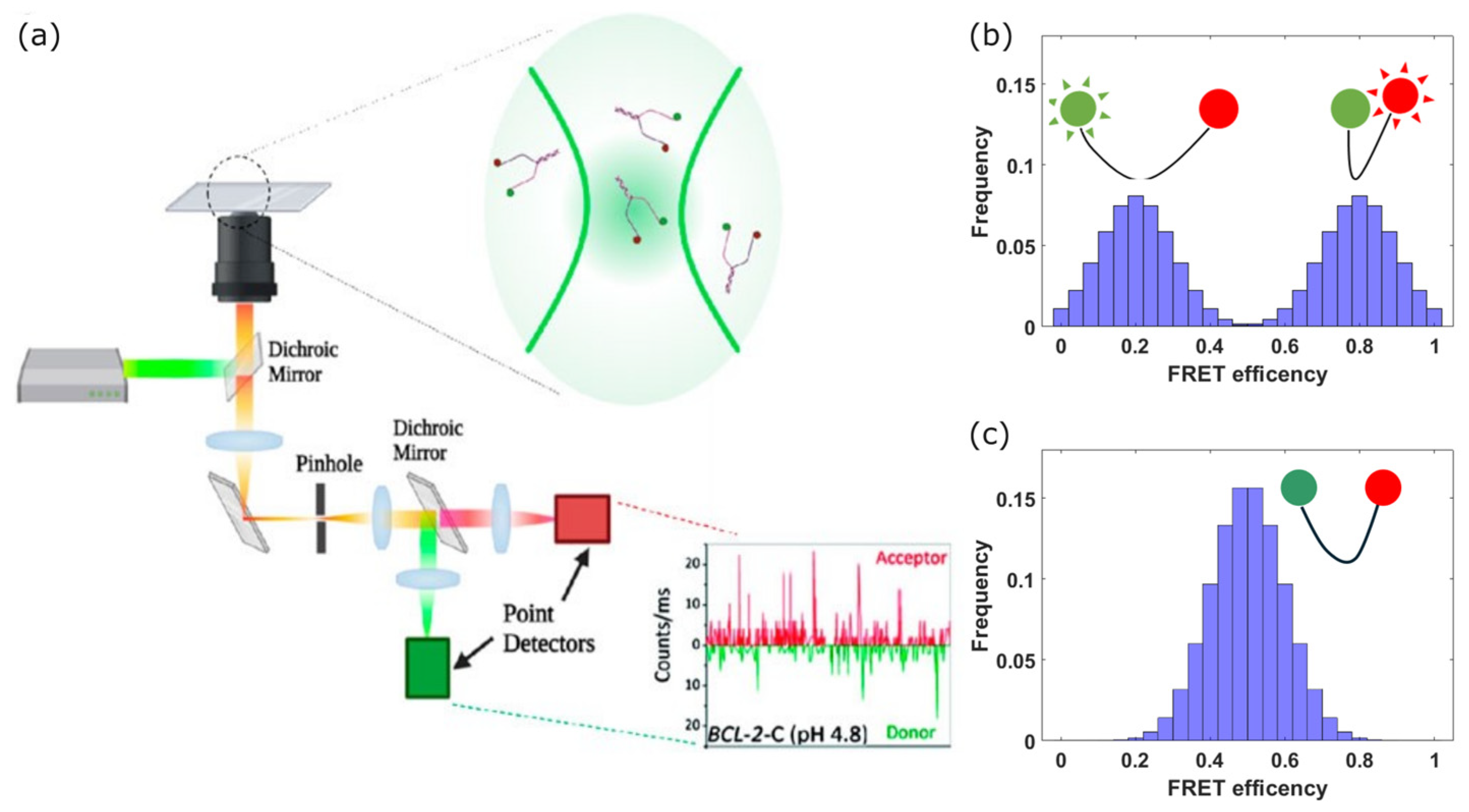
Figure 1. Scheme and principle of a CONF-FRET setup. (a) Scheme of a confocal microscope. A laser beam (green) is expanded to fill the back aperture of the microscope objective and focused into the sample. The fluorescence collected through the same objective is transmitted by the dichroic mirror and focused onto a pinhole. Only the fluorescence light emitted from the confocal volume passes through the pinhole, while the emission from the rest of the sample is filtered. The fluorescence transmitted by the pin hole is further split to separate the emission spectra of the FRET pair fluorophores, and detected by single-photon detectors. The numerical aperture of the objective and the size of the pinhole determine the transverse and longitudinal size of the confocal volume. Single-photon detection events are integrated with a fixed time gate to obtain donor and acceptor emission traces. (b) FRET efficiencies obtained from the emission traces are usually visualized through histograms. Here, we plot a simulated single-molecule FRET histogram with an average FRET efficiency of 0.5, resulting from two populations with FRET efficiencies of 0.2 and 0.8. (c) Simulated single-molecule FRET histogram form a single population with FRET efficiency of 0.5, which would be indistinguishable from the distribution in (b) if a bulk measurement is performed. (a) adapted from [31], used under Creative Commons CC-BY license.
Photonics 11 01061 g001
Figure 3. Scheme of a typical magnetic tweezer setup. (a) An optical microscope is coupled to a pair of magnets positioned onto a piezoelectric stage enabling their vertical translation and rotation with respect to the sample stage. The light emitted by an LED collimated and sent through the sample is diffracted by the magnetic bead. The diffraction pattern is imaged through the microscope onto a camera. Upon calibration of the imaging system from the recorded diffraction pattern it is possible to retrieve the bead height. (b) Magnetic tweezers permit the application of pulling forces and torques to the construct under study, a combination which is difficult or impossible to obtain with AFM and optical tweezers. The construct (here a G4 with dsDNA handles) is attached to the coverslip on one end, and to the magnetic bead on the other end. Pulling forces can be modified by changing the distance between the magnets and the sample, and monitored by measuring the Brownian motion of the bead. (c) The folding/unfolding kinetics of the G4 can be investigated either at constant force (red rectangle and inset) or during a force ramp (blue rectangle and inset) where an abrupt change in the extension of the construct corresponds to folding or unfolding of the structure under study. This investigation can be repeated as a function of the twisting angle imposed on the construct by rotating the magnets before the pulling force is applied. (a) Adapted from [135], used under Creative Commons 4.0 CC-BY license; (b,c) from [136], used under Creative Commons 3.0 CC-BY license.
Photonics 11 01061 g003
Figure 4. Scheme of an atomic force microscope. (a) A cantilever with a sharp tip is placed close to the surface of the sample to be investigated, and a laser beam is reflected off the back of the tip. Any tilt of the cantilever due to forces between the tip and the sample surface induces a deflection of the reflected laser beam, which is measured by a quadrant photodiode. A scanner moves the sample in three dimensions, allowing it to acquire an image of the sample surface. (b) To perform force spectroscopy, the cantilever is modified by attaching the construct to be studied at its tip. After bringing the tip close to the functionalized substrate so that the construct links to the substrate (here through a biotin-streptavidin bound), the tip is pulled at constant velocity while measuring the pulling force. A force–extension curve is thus obtained, where unfolding events or rupture of the link to the substrate can be identified. (a) Figure from [154,155], used under Creative Commons 4.0 CC-BY license; (b) figure from [156], © The Royal Society of Chemistry 2024, reused with permission.
Photonics 11 01061 g004
Figure 5. Timeline of milestones in the use of single-molecule techniques to study G4s. Text is connected to the timeline through colored lines which represent the corresponding technique: blue for smFRET, gold for AFM, green for optical tweezers, and red for magnetic tweezers (see also legend). References in chronologic order: (Ashkin 1992): [99], (Ha 1996): [35], (Deniz 1999): [36], (Gosse 2002): [130], (Ying 2003): [49], (Neaves 2009): [163], (Sannohe 2010): [166], (Zhao 2012): [160], (Long 2013): [149], (Selvam 2014): [133], (Endo 2015): [168], (You 2017): [144], (Di Antonio 2020): [82], (Patrick 2020): [128], (Cheng 2021): [108], (Patra 2021): [51].
Photonics 11 01061 g005
Disclaimer/Publisher’s Note: The statements, opinions and data contained in all publications are solely those of the individual author(s) and contributor(s) and not of MDPI and/or the editor(s). MDPI and/or the editor(s) disclaim responsibility for any injury to people or property resulting from any ideas, methods, instructions or products referred to in the content.