Article

Passively Mode-Locked Erbium-Doped Fiber Laser and Application in Laser Thrombolysis

1
Key Laboratory of Precision Opto-Mechatronics Technology of Education Ministry, School of Instrumentation and Optoelectronic Engineering, Beihang University, Beijing 100191, China
2
Key Laboratory of Big Data-Based Precision Medicine, Ministry of Industry and Information Technology, School of Engineering Medicine, Beihang University, Beijing 100191, China
*
Author to whom correspondence should be addressed.
Photonics 2024, 11(11), 1006; https://doi.org/10.3390/photonics11111006
Submission received: 2 September 2024 / Revised: 10 October 2024 / Accepted: 23 October 2024 / Published: 25 October 2024
(This article belongs to the Special Issue Photonics: 10th Anniversary)

Abstract

:
Fiber lasers have been widely used in surgery with the development of fiber photonics. Since the human body is prone to myocardial infarction caused by blood clots, laser thrombolysis was proposed as a safe and efficient treatment. Mode-locked fiber lasers have high peak power and narrow pulse width. In order to observe the effect of laser thrombolysis with mode-locked fiber lasers, a 1.5 µm mode-locked fiber laser based on carbon nanotubes was built, showing a pulse width of 1.46 ps, a 3 dB bandwidth of 1.65 nm, and a repetition rate of 29.5 MHz. The output pulses were amplified by an erbium-doped fiber amplifier to the hundred-milliwatt level and were applied to the surface of a self-made thrombus. The influences of lasing power and time on the damage diameter of the thrombus surface were evaluated. A low threshold damage power of 45 mW was observed, which resulted from the high peak power of the mode-locked pulses. These results demonstrate that high ablation efficiency can be achieved by using mode-locked pulses with a narrow pulse width and high peak power.

1. Introduction

Fiber lasers are widely used in biomedical and surgical techniques due to the advantages of high beam quality, extreme power efficiency, and low cost [1,2,3]. Ultrafast fiber lasers possess specific advantages compared to continuous wave lasers, which can be used in medical surgeries [4] and photoacoustic imaging application [5]. At present, the technology of passive mode-locking is recognized as a key method for the generation of ultrashort pulses [6]. A saturable absorber (SA) is the key component in passively mode-locked fiber lasers, including the aspects of semiconductor saturable absorber mirrors [7], graphene [8,9], carbon nanotubes (CNTs) [10], MXene [11,12], black phosphorus [13], heterostructure [14], and topological insulator [15,16]. CNTs are one-dimensional nanomaterials with excellent properties, including electrical and thermal conductivity. At the same time, they have significant nonlinear fiber properties and are often used as mode-locked devices in ultrafast fiber lasers [17].
The application of pulsed fiber lasers in laser medicine is a significant research topic. Diseases such as cerebral infarction and stroke are normally caused by blood clots, for which laser thrombolysis is an advanced treatment method with the advantages of less bleeding and fewer complications. The ablation mechanism interaction between the laser and tissue is a photothermolysis effect [18]. This mechanism suggests that ablation occurs as a consequence of vaporization, which is triggered by the laser energy absorbed in the tissue [19].
The advancement of pulsed laser technology has promoted the evolution of laser-based medical treatments. Over the past few years, researchers have investigated the impact of pulsed lasers across various laser parameters on the process of laser-induced thrombolysis. In 2014, Bidinger et al. investigated the feasibility of femtosecond laser thrombolysis with wavelengths of 800 and 1030 nm, and the results indicated that the thresholds for laser ablation were different with 800 and 1030 nm [20], which demonstrated the influence of laser wavelength. In 2017, Herzog et al. introduced a 355 nm solid-state laser for arterial disease treatment. They utilized a hybrid catheter to deliver nanosecond pulses for clinical case analysis. The study demonstrated the efficacy of 355 nm laser technology in atherectomy procedures [21]. In 2018, Li et al. evaluated the effect of laser spot size on the thermal denaturation zone of skin, which demonstrated that, the smaller the diameter, the higher the power density and the more concentrated the energy. As the spot size increases, the heated area of the tissue increases, but the peak temperature decreases significantly [22]. This provides a reference for the influence of spot size on thermal effects. In 2022, Zhang et al. studied the mechanism of thrombus ablation with a femtosecond laser. A simulation model of the interaction between the femtosecond laser and the thrombus was established. The burst-mode femtosecond laser with a wavelength of 1035 nm and a repetition frequency of 1 MHz was used for ablation, and the high efficiency of the burst mode for thrombus ablation was verified [23]. The effects of wavelength, spot size, and burst mode have been studied. However, there are few studies on laser thrombolysis using 1.5 µm mode-locked fiber lasers, and the influences of power and time have not been explored. Water absorption is strong in the 1.5 µm band, and there will be a significant thermal effect after the thrombus absorbs the laser energy under the action of the laser, which has important applications for the thrombolysis mechanism using thermal decomposition. The 1550 nm used in this experiment has the advantages of a simple structure, low cost, and easy maintenance compared to the previous 355 nm [24]. In general, 355 nm represents Q-switched pulses, which have wider pulse widths and higher ablation thresholds compared to mode-locked pulses [25]. Therefore, the investigation of the ablation effects induced by 1.5 µm mode-locking pulses on the thrombus is significant. Accordingly, the assessment of the impact of power and time on laser thrombolysis efficacy is necessary.
Here, we used CNTs as the SA to generate mode-locked pulses in a 1.5 µm fiber laser. A low pump power threshold for the mode-locked pulses was achieved. The mode-locked pulses were obtained when the pump power was 45 mW. The 1.5 µm mode-locked pulses were applied to the surface of a self-made thrombus for laser thrombolysis, and the application of a near-infrared pulsed laser in laser thrombolysis was studied. The effects of laser power and time on the damage range of the thrombus surface were observed, and the variations in damage size with power and time were determined. This provides guidance for the selection of light sources for subsequent clinical laser thrombolysis.

2. Results and Discussion

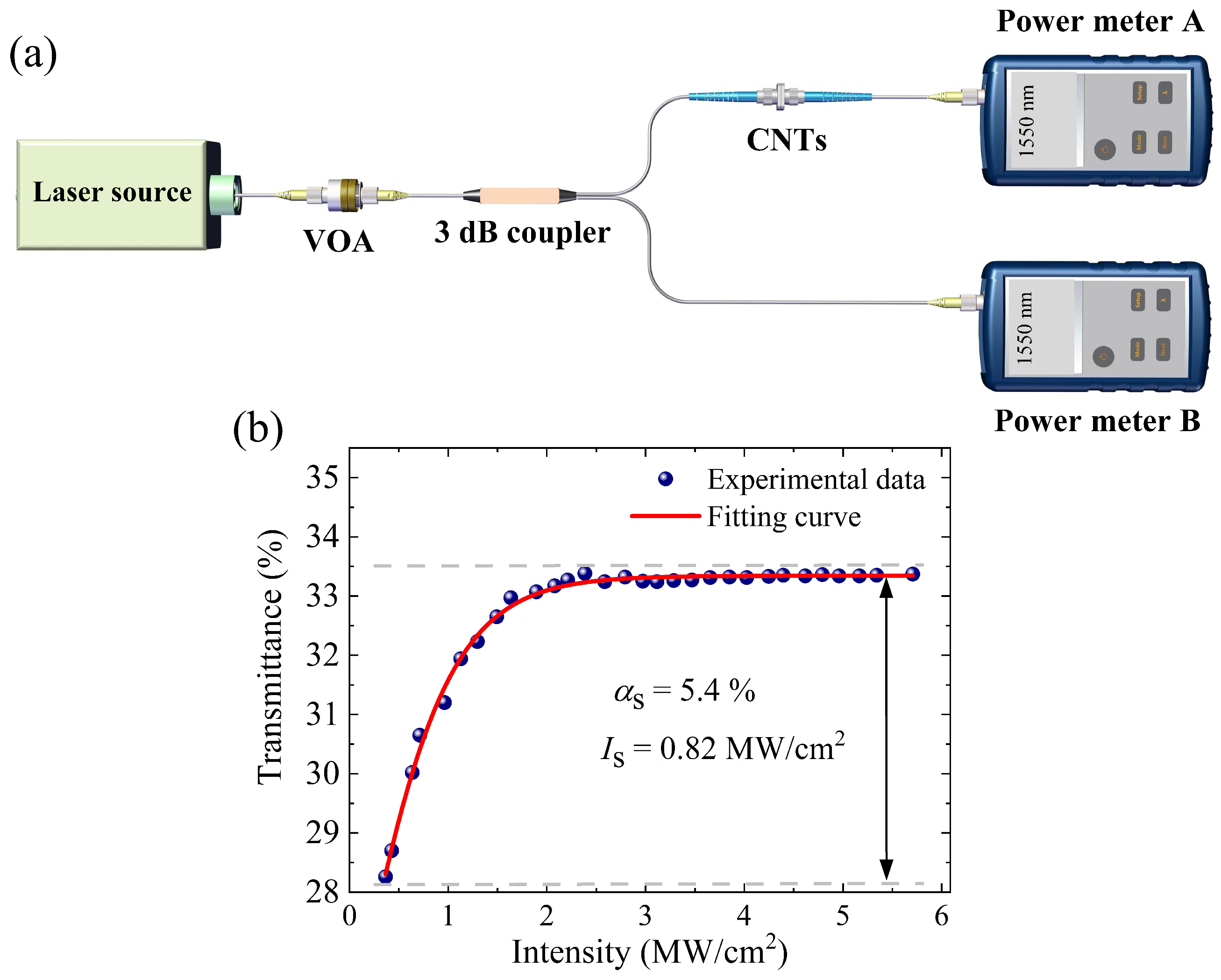
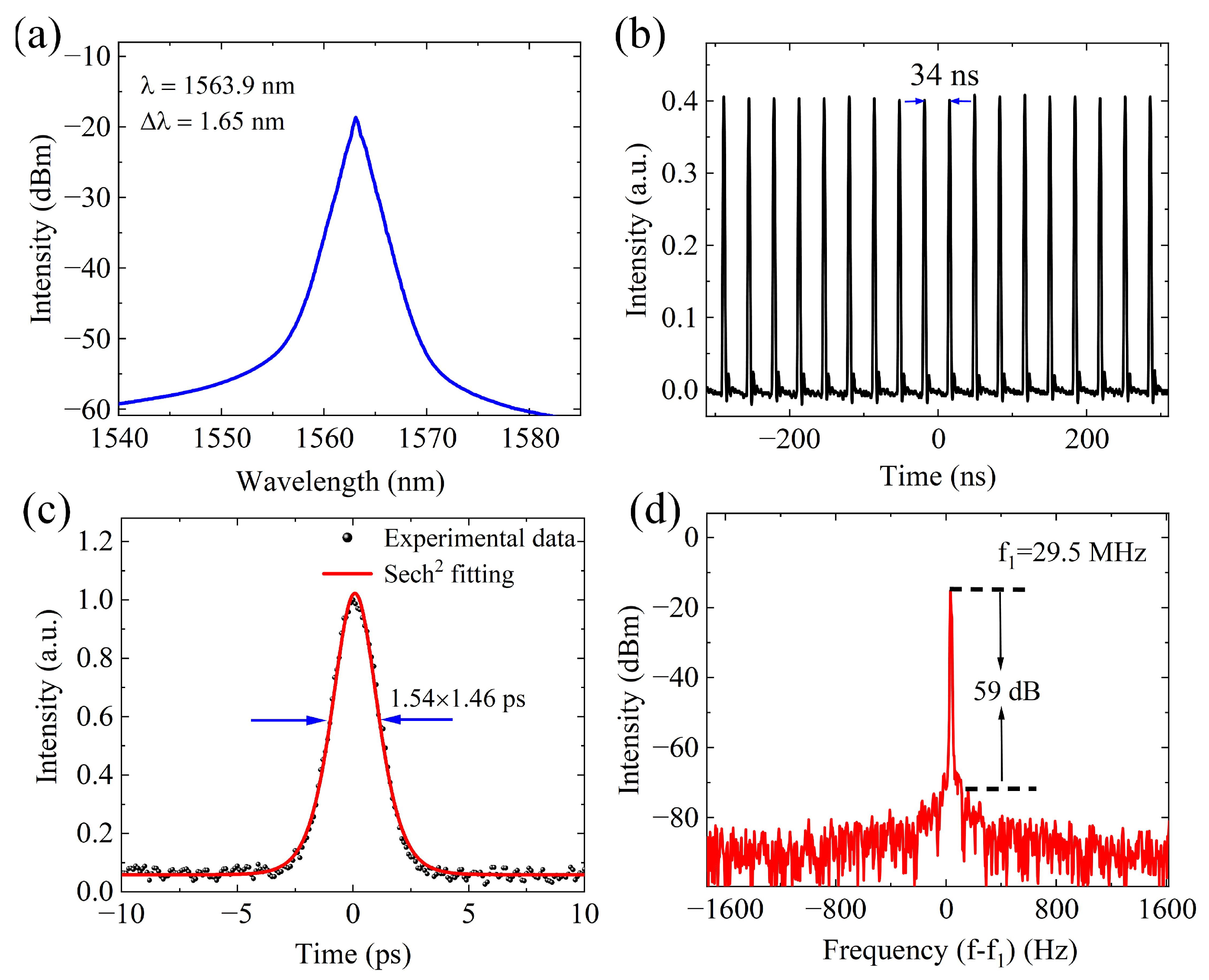
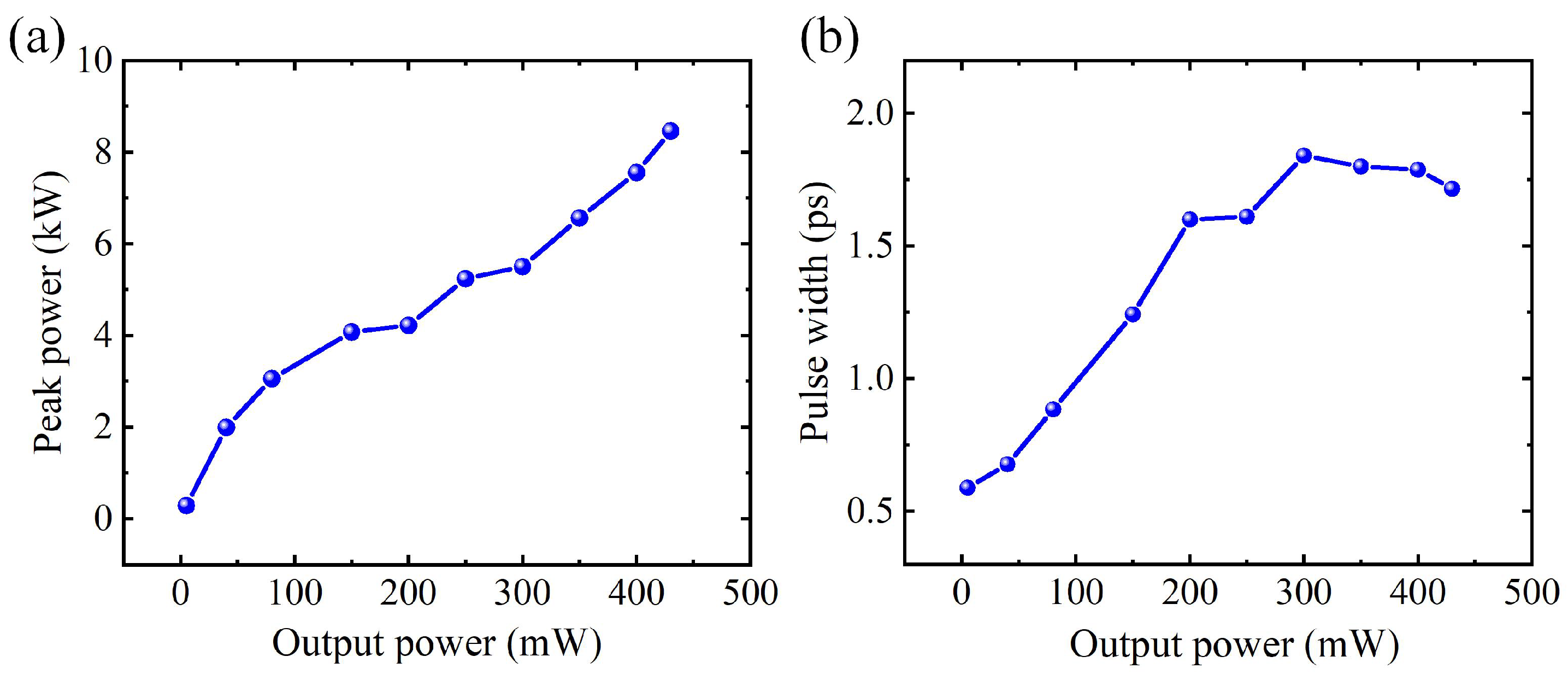
2.1. Mode-Locked Fiber Laser Based on CNTs

The experimental setup of the laser is shown in Figure 1. The ∼0.5 m erbium-doped fiber was pumped by a 980 nm laser diode (LD) through a 980/1550 nm wavelength-division multiplexer (WDM). The operating direction was ensured by a polarization-insensitive isolator (PI-ISO). A polarization controller (PC) was used to control the polarization state to optimize the output pulses. The pulses were generated by using CNTs as the SA. The laser passes through a 20:80 output coupler (OC), where 20 % of the output laser pulses is used for measurement. The cavity length was ∼7 m, and the fiber used in the laser is single-mode fiber (SMF-28). The output pulses were measured by an optical spectrum analyzer (Agilent 86142B), an autocorrelator (APE pulseCheck NX50), an oscilloscope (Siglent SDS6104 H10 Pro), and a radio-frequency (RF) signal analyzer (Keysight N9020A).
The CNT SA was constructed by the sandwich method due to the advantages of feasibility and effectiveness. The nonlinear saturable absorption properties of the CNTs were measured by a balanced twin detector measurement system, as shown in Figure 2a. A mode-locked fiber laser served as the laser source, which was connected with a variable optical attenuator (VOA). The laser output, after being attenuated, was split into two distinct paths by a 3 dB coupler. One path routed through a CNT SA before reaching power meter A for measurement, while the other was directly assessed with power meter B. Power meter A was the test port, and power meter B was the reference port. The transmittance of the CNT SA was the power ratio of the test port to the reference port at different output powers by tuning the VOA, as shown in Figure 2b.
The nonlinear curve was fit by the conventional saturable absorption model T = 1 [ α s / ( 1 + I / I sat ) ] α ns [26], where T, α s , and α ns represent transmittance, modulation depth, and non-saturable loss, respectively. I and I sat are incident light intensity and saturation intensity, respectively. Initially, the transmittance increased with the incident light intensity. Subsequently, when the transmittance reached a certain value and remained consistent, the CNTs were saturated. The modulation depth α s is 5.4% and the saturation intensity is 0.82 MW/cm2 according to the fitting curve, as shown in Figure 2b. A low saturation intensity is useful for the generation of mode-locked operation in low power, and further for a narrower pulse width [27].
Mode-locked pulses were generated at the pump power of 45 mW when the CNT SA was connected to the cavity. The group velocity dispersion of the whole cavity structure is about −0.12 ps2. The output power was 0.5 mW when the pump power was 60 mW. We measured the characteristics of mode-locked pulses, as shown in Figure 3. The optical spectrum is shown in Figure 3a, with a central wavelength of 1563.9 nm, and the 3 dB bandwidth was 1.65 nm. The relevant pulse train is depicted in Figure 3b, with a pulse separation of 34 ns. The pulse width is shown in Figure 3c, which was measured by an autocorrelator. The experimental data were fitted by the sech2 function, and the pulse width was 1.46 ps. Figure 3d displays the characteristics of the radio frequency (RF) spectrum, which was measured with a resolution bandwidth of 100 Hz. Pulses with a repetition rate of 29.5 MHz and signal-to-noise ratio of 59 dB were achieved, showing the stable operation of mode-locking. The pulse peak power was calculated to be 11.6 W at this output. In order to further increase the peak power, the fourth-order soliton method can be used in subsequent experiments [28,29]. The pulse interval and basic repetition rate both correspond to the cavity length, indicating that the laser was operating in a stable mode-locked state.

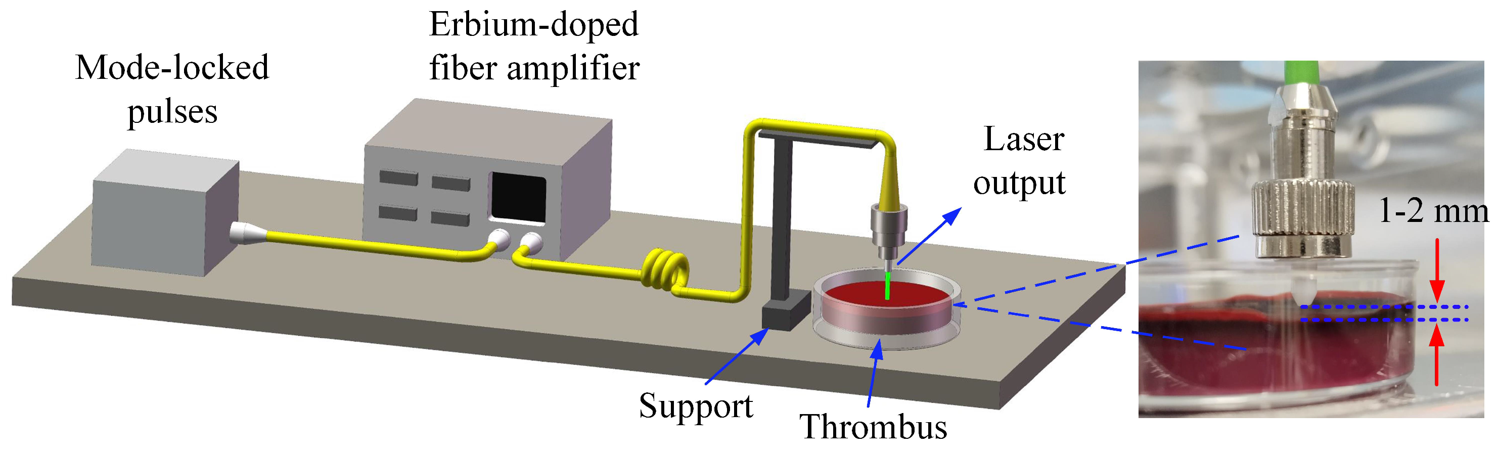
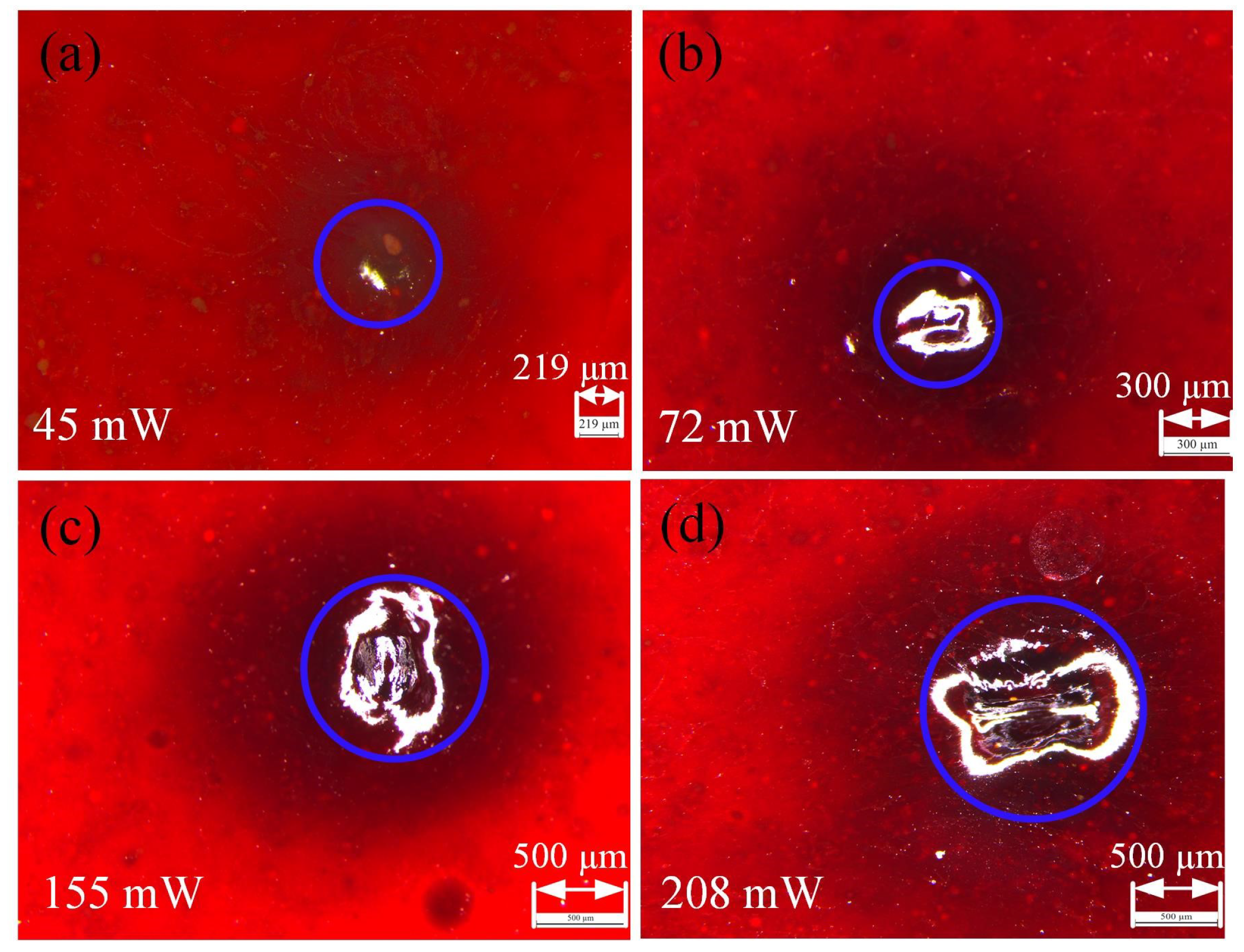
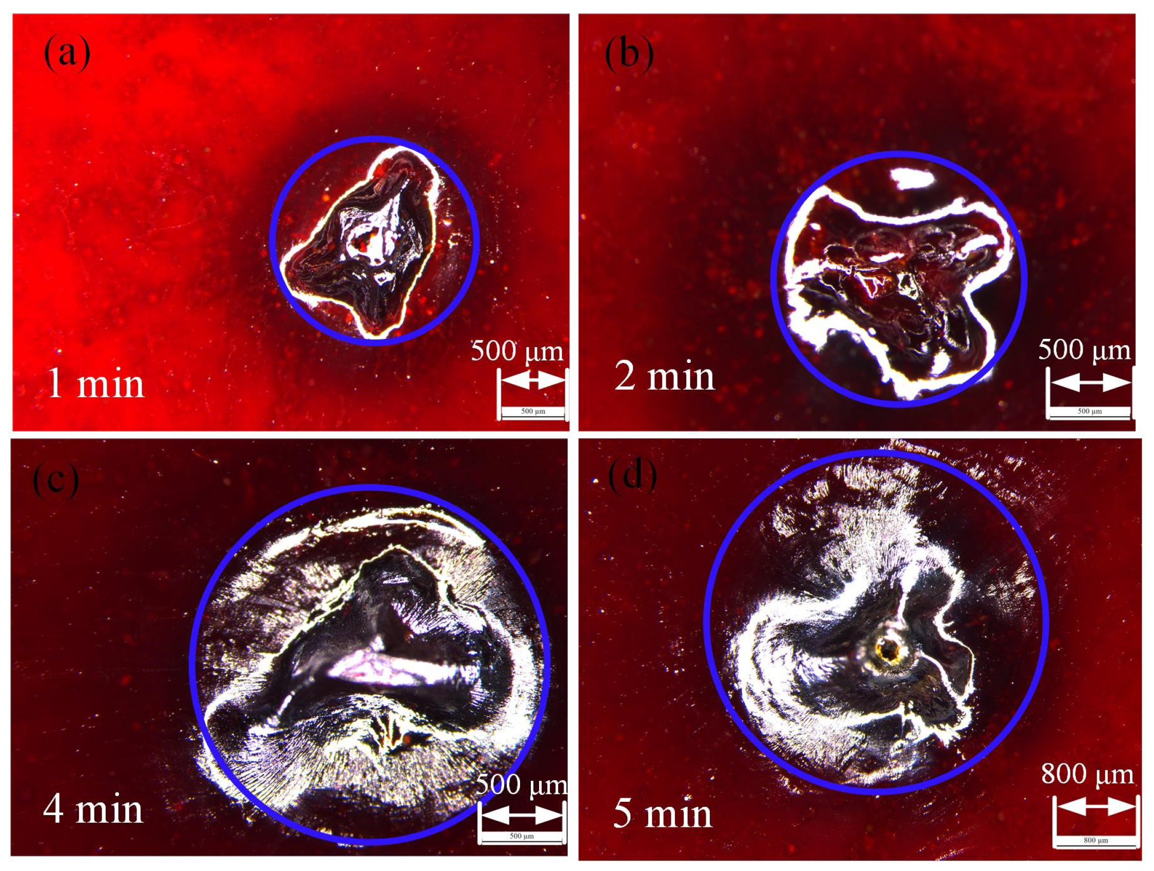
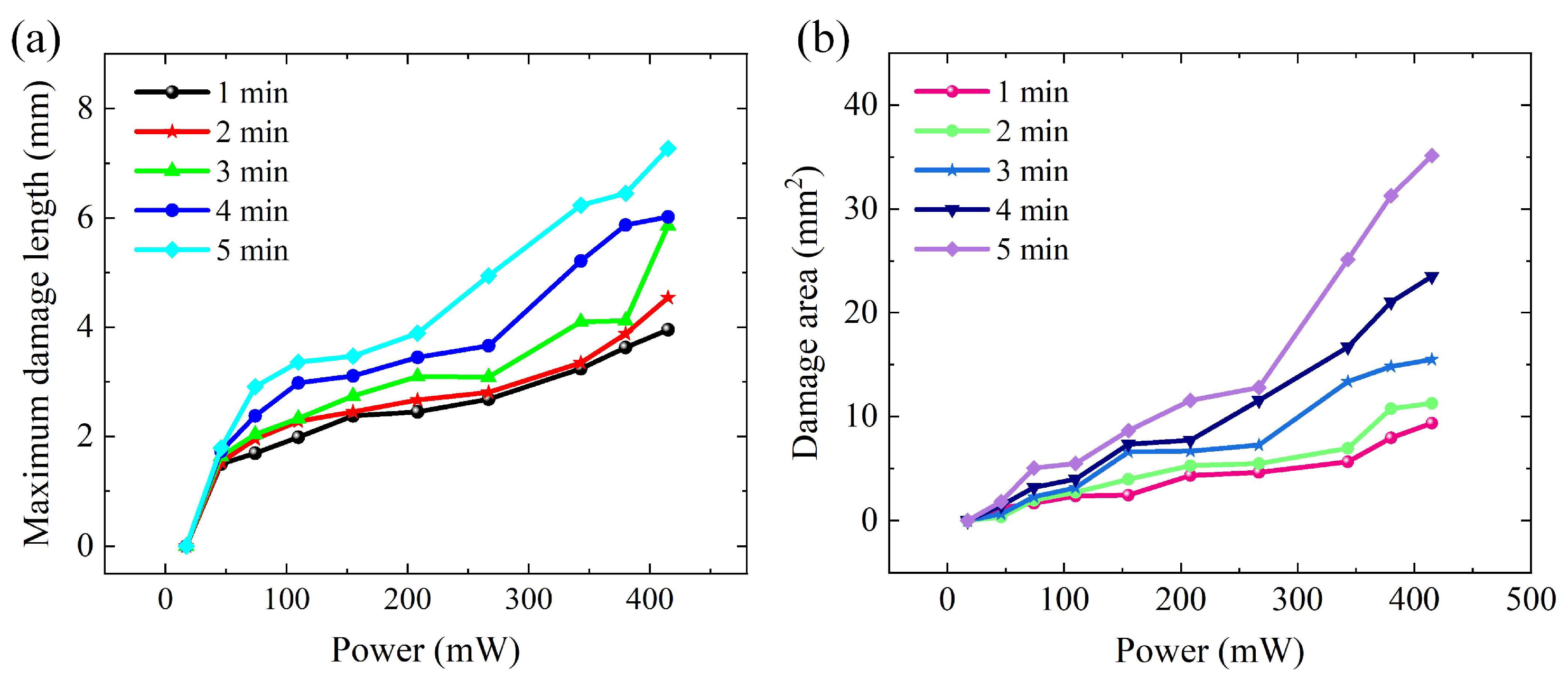
2.2. Laser Thrombolysis Application

In this experiment, the thrombus was prepared by using fresh pig blood and left to set for 12 h [30]. The self-made thrombus was mainly composed of erythrocyte, and the other ingredients include sodium chloride, calcium chloride, and normal saline. A thrombus fabricated with this method can simulate the human body in terms of composition. Before laser thrombolysis, the smooth surfaces of the thrombus in the culture dish were selected for the experiment.
The output mode-locked pulses were amplified by an erbium-doped fiber amplifier, and variable power was achieved. The parameters of the laser pulses after amplification are depicted in Figure 4. The peak power values of the amplified pulses are shown in Figure 4a, and the pulse widths of the amplified pulses are shown in Figure 4b.
The amplified mode-locking pulses were transmitted to the thrombus through the support. The surface thrombus was irradiated vertically with different powers, and the characteristics of the thrombus were observed with a microscope. The schematic diagram of the laser thrombolysis is shown in Figure 5. The laser was placed 1–2 mm away from the thrombus surface, as shown in the inset of Figure 5.
The laser was directed onto the thrombus surface, where the water contained within the thrombus absorbed the laser energy, transforming it into heat. According to the Pennes heat transfer equation, power and time are the main factors affecting heat generation [31]. The power was increased from 17 to 400 mW, and the time was kept constant at 1 min. The surface began to denature and coagulate (the surface turns dark) at 35 mW, and ablation appeared at 45 mW, as shown in Figure 6a. Figure 6b–d show that the ablation gradually worsened when the power increased, and, finally, carbonization occurred. The black part around the central point of action was protein denaturation, which was the surrounding damage caused by heat diffusion. The occurrence of this process mainly depends on thermal effects because its power density is much smaller than the conditions for plasma formation [32].
The characteristics of the surface thrombus were also observed by a microscope. The power was maintained at 307 mW under different times, as shown in Figure 7. At this power, the phenomenon is obvious. A burnt smell appeared as soon as irradiation started, and soon the surface of the thrombus was ablated, as shown in Figure 7a. The time was increased to 2 min, the ablation range was significantly expanded, and a burn hole even appeared in the irradiation center, as shown in Figure 7b. As the irradiation time continued to increase, the ablation range expanded further and the carbonization phenomenon became more serious, as shown in Figure 7c. The irradiation time was 5 min, and the ablation hole became obviously larger, as shown in Figure 7d. In the actual laser thrombolysis process, the ablation time cannot be continuously increased to improve the ablation efficiency; otherwise, it is easy to cause additional thermal damage. Instead, it is necessary to comprehensively consider the combined effects of multiple parameters to obtain the best ablation effect.
The heat generation can be transferred to the adjacent tissues instead of gathering at one point, which will cause thermal damage to the surrounding tissues. Therefore, after the laser irradiation to the thrombus, observing and measuring the damage range is an important step to understand the extent of the damage. It can be seen from Figure 6 and Figure 7 that the damage shape is irregular, which may be caused by the influence of the surrounding environment and the insufficient stability of the output pulses. In order to improve the stability further, the laser structure can be improved in subsequent experiments, such as cascaded Mamyshev regeneration [33], a figure-eight fiber laser [34], and self-mode locking technology [35].
The maximum damage length and damage area were measured to describe the damage accurately, as shown in Figure 8. It can be seen that the maximum damage length increases gradually with power, as shown in Figure 8a. Figure 8b shows the damage area, including the denatured part of the protein. It can be concluded that the damage area increases with the increase in time. At high power, the damage area increases sharply. This can be attributed to the diffusion of energy accumulated inside the thrombus to the surrounding area, which expands the damage area at high power. The efficiency of laser thrombolysis is based on the ablation area divided by the total thermal change area [36]. According to this formula, we can approximate the efficiency of thrombolysis by experimental phenomena and results. The results show that a greater ablation effect is obtained at a lower pulse energy, which indicates that the ablation efficiency is high. In addition to the above factors, the pulse width also has a great influence on the thrombolytic effect. In the experiment, the pulse width was at the ps level, and the pulse width affected the ablation threshold and penetration depth. A larger pulse width has a higher ablation threshold and larger penetration depth [25].
In the present study, we investigated the application of ultrafast pulses with near-infrared wavelengths in laser thrombolysis and compared the effects of different powers and times on the thrombolytic effect. Laser thrombolysis using the thermal effect of the strong absorption of water in the 1.5 µm band is an important mechanism of medical surgery. The application of near-infrared ultrafast lasers in the laser ablation of thrombi has been studied in depth [36,37]. The study of an ultrafast pulse as a light source can further improve accuracy and reduce additional damage. The wavelength primarily influences the degree of thermal effect, whereas the pulse width impacts the ablation threshold and penetration depth. In this study, the influences of two important parameters, power and time, on the surface ablation of a thrombus were compared. The thrombus model was created by using fresh pig blood, and the model was more similar to the human thrombus sample by mixing the solution. At pulse widths of the order of ps, the effect of a narrow pulse width on the other biological tissues during laser ablation has been expanded [25].

3. Conclusions

In this work, a mode-locked fiber laser was established by using a CNT SA. The nonlinear optical properties of the CNT SA were measured by a two-detector measurement system, showing a low saturable intensity of 0.82 MW/cm2, which is useful for generating mode-locked pulses. A low pump threshold power of 45 mW was achieved for the mode-locked pulses. The output properties of wavelength, pulse train, pulse width, and repetition rate were measured in 60 mW, proving the stability of the laser. Further, the application of ultrafast pulses at the near-infrared 1.5 µm band in laser thrombolysis was studied. A laser was irradiated onto the surface of a self-made thrombus, and the damage was observed with a microscope. The two main factors that affect surface damage are power and time. The variations in maximum damage length and damage area with power were ascertained. This experiment can provide guidance for subsequent clinical laser thrombolysis.

Author Contributions

Conceptualization, X.Z.; methodology, X.Z.; software, L.C.; formal analysis, X.Z.; investigation, X.Z.; writing—original draft preparation, X.Z.; writing—review and editing, X.Z., C.Z. (Congyu Zhang), C.Z. (Chenxi Zhang) and I.A.; supervision, B.F.; project administration, B.F.; funding acquisition, B.F. All authors have read and agreed to the published version of the manuscript.

Funding

This work is supported by the State Key Laboratory of Advanced Optical Communication Systems and Networks, China, College Students’ Innovative Entrepreneurial Training Plan Program, the National Natural Science Foundation of China (grant 62071016).

Institutional Review Board Statement

Not applicable.

Informed Consent Statement

Not applicable.

Data Availability Statement

Data is contained within the article.

Conflicts of Interest

The authors declare no conflicts of interest.

References

  1. Sroka, R.; Stepp, H.; Hennig, G.; Brittenham, G.M.; Rühm, A.; Lilge, L. Medical laser application: Translation into the clinics. J. Biomed. Opt. 2015, 20, 061110. [Google Scholar] [CrossRef] [PubMed]
  2. Chamkouri, H.; Si, J.; Chen, P.; Niu, C.; Chen, L. Biomedical optics and photonics for advanced clinical technologies. Opt. Laser Technol. 2024, 179, 111265. [Google Scholar] [CrossRef]
  3. Pierce, M.C.; Jackson, S.D.; Golding, P.S.; Dickinson, B.; Dickinson, M.R.; King, T.A.; Sloan, P. Development and application of fiber lasers for medical applications. In Proceedings of the Optical Fibers and Sensors for Medical Applications, San Jose, CA, USA, 20–21 January 2001; Volume 4253, pp. 144–154. [Google Scholar]
  4. Gursel, A.T. Fiber lasers and their medical applications. Opt.-Amplifiers-Few Differ. Dimens. 2018, 10, 17–23. [Google Scholar]
  5. Piao, Z.; Zeng, L.; Chen, Z.; Kim, C.S. Q-switched erbium-doped fiber laser at 1600 nm for photoacoustic imaging application. Appl. Phys. Lett. 2016, 108, 14. [Google Scholar] [CrossRef]
  6. Zhao, X.; Jin, H.; Liu, J.; Chao, J.; Liu, T.; Zhang, H.; Wang, G.; Lyu, W.; Wageh, S.; Al-Hartomy, O.A.; et al. Integration and applications of nanomaterials for ultrafast photonics. Laser Photonics Rev. 2022, 16, 2200386. [Google Scholar] [CrossRef]
  7. Bogusławski, J.; Sterczewski, Ł.; Stachowiak, D.; Soboń, G. Intracavity filtering in SESAM mode-locked fiber lasers: Soliton effects and noise performance. Opt. Express 2023, 31, 27667–27676. [Google Scholar] [CrossRef]
  8. Liu, Z.B.; He, X.; Wang, D. Passively mode-locked fiber laser based on a hollow-core photonic crystal fiber filled with few-layered graphene oxide solution. Opt. Lett. 2011, 36, 3024–3026. [Google Scholar] [CrossRef]
  9. Fu, B.; Hua, Y.; Xiao, X.; Zhu, H.; Sun, Z.; Yang, C. Broadband graphene saturable absorber for pulsed fiber lasers at 1, 1.5, and 2 µm. IEEE J. Sel. Top. Quantum Electron. 2014, 20, 411–415. [Google Scholar]
  10. Zhou, Y.; Lin, J.; Zhang, X.; Xu, L.; Gu, C.; Sun, B.; Wang, A.; Zhan, Q. Self-starting passively mode-locked all fiber laser based on carbon nanotubes with radially polarized emission. Photonics Res. 2016, 4, 327–330. [Google Scholar] [CrossRef]
  11. Sun, J.; Cheng, H.; Xu, L.; Fu, B.; Liu, X.; Zhang, H. Ag/MXene composite as a broadband nonlinear modulator for ultrafast photonics. ACS Photonics 2023, 10, 3133–3142. [Google Scholar] [CrossRef]
  12. Fu, B.; Sun, J.; Wang, C.; Shang, C.; Xu, L.; Li, J.; Zhang, H. MXenes: Synthesis, optical properties, and applications in ultrafast photonics. Small 2021, 17, 2006054. [Google Scholar] [CrossRef] [PubMed]
  13. Ma, C.; Tian, X.; Gao, B.; Wu, G. Dynamic evolution of the dissipative soliton in passively mode-locked fiber laser based on black phosphorus as a new saturable absorber. Opt. Commun. 2018, 406, 177–182. [Google Scholar] [CrossRef]
  14. Lyu, W.; An, J.; Lin, Y.; Qiu, P.; Wang, G.; Chao, J.; Fu, B. Fabrication and applications of heterostructure materials for broadband ultrafast photonics. Adv. Opt. Mater. 2023, 11, 2300124. [Google Scholar] [CrossRef]
  15. Liu, H.; Zheng, X.W.; Liu, M.; Zhao, N.; Luo, A.P.; Luo, Z.C.; Xu, W.C.; Zhang, H.; Zhao, C.J.; Wen, S.C. Femtosecond pulse generation from a topological insulator mode-locked fiber laser. Opt. Express 2014, 22, 6868–6873. [Google Scholar] [CrossRef]
  16. Yan, P.; Lin, R.; Ruan, S.; Liu, A.; Chen, H.; Zheng, Y.; Chen, S.; Guo, C.; Hu, J. A practical topological insulator saturable absorber for mode-locked fiber laser. Sci. Rep. 2015, 5, 8690. [Google Scholar] [CrossRef]
  17. Dai, L.; Huang, Z.; Huang, Q.; Zhao, C.; Rozhin, A.; Sergeyev, S.; Al Araimi, M.; Mou, C. Carbon nanotube mode-locked fiber lasers: Recent progress and perspectives. Nanophotonics 2020, 10, 749–775. [Google Scholar] [CrossRef]
  18. Anderson, R.R.; Parrish, J.A. Selective photothermolysis: Precise microsurgery by selective absorption of pulsed radiation. Science 1983, 220, 524–527. [Google Scholar] [CrossRef]
  19. Jiao, J.; Guo, Z. Modeling of ultrashort pulsed laser ablation in water and biological tissues in cylindrical coordinates. Appl. Phys. B 2011, 103, 195–205. [Google Scholar] [CrossRef]
  20. Bidinger, J.; Ackermann, R.; Cattaneo, G.; Kammel, R.; Nolte, S. A feasibility study on femtosecond laser thrombolysis. Photomed. Laser Surg. 2014, 32, 17–22. [Google Scholar] [CrossRef]
  21. Herzog, A.; Oszkinis, G.; Planer, D.; Ziaja, K.; Kruszyna, Ł.; Stanisić, M.G.; Ziaja, D.; Ishaaya, A.A.; Kuczmik, W. Atherectomy using a solid-state laser at 355 nm wavelength. J. Biophotonics 2017, 10, 1271–1278. [Google Scholar] [CrossRef]
  22. Li, C.; Wang, K.; Huang, J. Simulation of the effect of spot size on temperature field and weld forming in laser tissue welding. Optik 2018, 155, 315–323. [Google Scholar] [CrossRef]
  23. Zhang, H.; Liu, X.; Li, Y.; Wu, W.; Gu, Y.; Zhang, T. Study on the mechanism of thrombus ablation in vitro by burst-mode femtosecond laser. J. Biophotonics 2022, 15, e202200197. [Google Scholar] [CrossRef] [PubMed]
  24. Xu, S.; Zhai, S.; Li, B.; Gao, S. High power and conversion efficiency intracavity ultraviolet laser at 355 nm. Infrared Phys. Technol. 2023, 128, 104485. [Google Scholar] [CrossRef]
  25. Kim, B.M.; Feit, M.D.; Rubenchik, A.M.; Joslin, E.J.; Celliers, P.M.; Eichler, J.r.; Da Silva, L.B. Influence of pulse duration on ultrashort laser pulse ablation of biological tissues. J. Biomed. Opt. 2001, 6, 332–338. [Google Scholar] [CrossRef] [PubMed]
  26. Garmire, E. Resonant optical nonlinearities in semiconductors. IEEE J. Sel. Top. Quantum Electron. 2000, 6, 1094–1110. [Google Scholar] [CrossRef]
  27. Lau, K.; Hou, D. Recent research and advances of material-based saturable absorber in mode-locked fiber laser. Opt. Laser Technol. 2021, 137, 106826. [Google Scholar] [CrossRef]
  28. Yang, S.; Zhu, Z.; Qi, Y.; Jin, L.; Li, L.; Lin, X. Internal motion within pulsating pure-quartic soliton molecules in a fiber laser. Chaos Solitons Fractals 2023, 172, 113544. [Google Scholar] [CrossRef]
  29. Runge, A.F.; Hudson, D.D.; Tam, K.K.; de Sterke, C.M.; Blanco-Redondo, A. The pure-quartic soliton laser. Nat. Photonics 2020, 14, 492–497. [Google Scholar] [CrossRef]
  30. Janis, A.D.; Buckley, L.A.; Nyara, A.N.; Prahl, S.A.; Gregory, K. A reconstituted in vitro clot model for evaluating laser thrombolysis. J. Thromb. Thrombolysis 2002, 13, 167–175. [Google Scholar] [CrossRef]
  31. Pennes, H.H. Analysis of tissue and arterial blood temperatures in the resting human forearm. J. Appl. Physiol. 1948, 1, 93–122. [Google Scholar] [CrossRef]
  32. Niemz, M. Laser-Tissue Interactions; Elsevier: Amsterdam, The Netherlands, 2007. [Google Scholar]
  33. Liu, Z.; Ziegler, Z.M.; Wright, L.G.; Wise, F.W. Megawatt peak power from a Mamyshev oscillator. Optica 2017, 4, 649–654. [Google Scholar] [CrossRef] [PubMed]
  34. Tsun, T.; Islam, M.; Chu, P. High-energy femtosecond figure-eight fiber laser. Opt. Commun. 1997, 141, 65–68. [Google Scholar] [CrossRef]
  35. Zheng, J.; Xie, J.; Liu, Q.; He, C.; Qi, Y.; Li, L.; He, L.; Liu, C.; Lin, X.; Yang, S. Generation of high-energy self-mode-locked pulses in a Tm-doped fiber laser. Appl. Phys. Lett. 2024, 125, 5. [Google Scholar] [CrossRef]
  36. Alagha, H.Z.; Gülsoy, M. Photothermal ablation of liver tissue with 1940-nm thulium fiber laser: An ex vivo study on lamb liver. J. Biomed. Opt. 2016, 21, 015007. [Google Scholar] [CrossRef] [PubMed]
  37. Huang, Y.; Jivraj, J.; Zhou, J.; Ramjist, J.; Wong, R.; Gu, X.; Yang, V.X. Pulsed and CW adjustable 1942 nm single-mode all-fiber Tm-doped fiber laser system for surgical laser soft tissue ablation applications. Opt. Express 2016, 24, 16674–16686. [Google Scholar] [CrossRef]
Figure 1. Schematic diagram of mode-locked EDF based on CNTs as SA. LD: laser diode; WDM: wavelength division multiplexer; EDF: erbium-doped fiber; PI-ISO: polarization-insensitive isolator; PC: polarization controller; CNTs: carbon nanotubes; SA: saturable absorber; OC: output coupler.
Photonics 11 01006 g001
Figure 2. Experimental setup and nonlinear optical properties of CNT SA. (a) Setup for measurement of the nonlinear transmittance of CNT SA. (b) Nonlinear transmittance of CNT SA in 1.5 µm. VOA: variable optical attenuator.
Photonics 11 01006 g002
Figure 3. Mode-locked results of the erbium-doped fiber laser. (a) Spectrum. (b) Pulse train. (c) Pulse profile. (d) RF spectrum.
Photonics 11 01006 g003
Figure 4. Parameters of laser pulses after amplification. (a) Peak power. (b) Pulse width.
Photonics 11 01006 g004
Figure 5. Schematic diagram of the laser thrombolysis process. The physical picture is shown in the inset.
Photonics 11 01006 g005
Figure 6. Mode-locked laser thrombolysis under the same time of 1 min and different powers of (a) 45 mW, (b) 72 mW, (c) 155 mW, and (d) 208 mW.
Photonics 11 01006 g006
Figure 7. Mode-locked laser thrombolysis under the same output power of 307 mW and different times of (a) 1 min, (b) 2 min, (c) 4 min, and (d) 5 min.
Photonics 11 01006 g007
Figure 8. Mode-locked laser thrombolysis under different power and time. (a) Maximum damage length. (b) Damage area.
Photonics 11 01006 g008
Disclaimer/Publisher’s Note: The statements, opinions and data contained in all publications are solely those of the individual author(s) and contributor(s) and not of MDPI and/or the editor(s). MDPI and/or the editor(s) disclaim responsibility for any injury to people or property resulting from any ideas, methods, instructions or products referred to in the content.

