Article

Caries Preventive Action of Nd:YAG and Fluoride in Three Different pH Conditions: FTIR Spectroscopy and SEM Evaluation

by
Amanda Caramel-Juvino
1,
Nathalia A. Zanini
1,
Sabrina Gardiano Avelino
1,
Yasmin Reis Fontes-Oliveira
1,
Gleice Conceição Mendonça Germano
1,
Carlos de Paula Eduardo
2 and
Denise Maria Zezell
1,*
1
Center for Lasers and Applications, Nuclear and Energy Research Institute, 2242 Prof. Lineu Prestes Avenue, Sao Paulo 05508-000, SP, Brazil
2
Special Laboratory for Laser Dentistry-LELO, School of Dentistry, 2227 Prof. Lineu Prestes Avenue, Sao Paulo 05508-000, SP, Brazil
*
Author to whom correspondence should be addressed.
Photonics 2023, 10(9), 985; https://doi.org/10.3390/photonics10090985
Submission received: 28 June 2023 / Revised: 21 July 2023 / Accepted: 31 July 2023 / Published: 29 August 2023

Abstract

:
This in vitro study aimed to evaluate the preventive action of topical fluoride application combined with laser irradiation under different pH conditions using Fourier Transform Infrared Spectroscopy (FTIR) and Scanning Electron Microscopy (SEM). A total of 180 samples of human dental enamel were prepared and divided into groups: Negative Control, Fluoride (FFA 12.300 µF/g), Laser (Nd:YAG 84.9 J/cm2), and Laser + Fluoride (Nd:YAG 84.9 J/cm2 + FFA 12.300 µF/g). The pH cycling was performed at three different pH conditions: pH 5 (below the critical pH for hydroxyapatite), pH 4.5 (below the critical pH in the presence of fluorapatite), and pH 4 (investigating acid resistance of hydroxyapatite and fluorapatite forms with laser irradiation). In the FTIR analysis, the Laser + Fluoride group demonstrated statistically significant differences compared to the Negative Control group and Fluoride group at pH 4.5 and pH 4 when evaluating the phosphate bands. Similar results were observed in the SEM analysis, where the Laser + Fluoride group exhibited lower demineralization compared to the other treatments at pH 4.5 and pH 4. In conclusion, the Laser + Fluoride group demonstrated a significant reduction in demineralization even at pH levels below the critical threshold for fluorapatite, highlighting its superior acid resistance compared to fluoride alone.

1. Introduction

Despite significant advancements in the study and prevention of dental caries, this disease remains highly prevalent [1]. According to the WHO Global Oral Health Status Report (2022), an estimated 3.5 billion people worldwide are affected by dental caries, making it the most common disease globally, as reported by the Global Burden of Disease 2019 [2]. Considering this situation, continuous progress in caries prevention and control is strongly encouraged.
Dental caries is a multifactorial chronic disease that occurs due to an imbalance in the demineralization and remineralization processes that take place on the tooth surface. The process of demineralization and remineralization is influenced by the composition of dental tissues, which primarily consist of inorganic components, enabling ionic exchanges between the oral structure and oral fluids. As a result, the dental structure can experience the loss, gain, or maintenance of ions, depending on variations in pH levels and their impact on the structure’s solubility [3].
Demineralization occurs when dental tissues lose minerals such as calcium and phosphate due to the medium being undersaturated with respect to these minerals, leading to their dissolution. On the other hand, remineralization is a natural protective process through which existing minerals in saliva become incorporated into the teeth, especially when the medium is supersaturated with the minerals, facilitating their precipitation. The development of caries lesions takes place when the demineralization process surpasses the capacity for remineralization [3,4].
Fluoride plays a crucial role in caries prevention by interfering with the demineralization and remineralization processes [5,6]. By utilizing high-concentration fluoride products such as gels and varnishes, fluorine ions are incorporated into the existing hydroxyapatite crystal, resulting in the formation of fluorapatite. Fluoride ions being smaller in size than hydroxyl ions (which are a component of hydroxyapatite), allow for improved ion organization; as a result, fluorapatite has lower solubility then hydroxyapatite. Another beneficial aspect is that it can lead to the creation of calcium fluoride, which remains on the surface and serves as a fluoride reservoir during both the demineralization and remineralization processes [3,5].
Lasers have been present in dental research evaluating changes in hard tissues since 1964 [7]. High-powered lasers such as neodymium:yttrium–aluminum–garnet (Nd:YAG) have been extensively studied as an alternative for caries prevention since the same time. They enhance the acid resistance of tooth structure and exhibit a synergistic effect with fluoride, leading to reduced enamel solubility [8]. The solubility change occurs due to a reduction in water and carbonate content, an increase in the proportion of hydroxyl component, alterations in the shape and size of hydroxyapatite crystals, formation of pyrophosphates, protein decomposition, and the development of new crystalline phases [8,9,10].
Studies involving the use of Nd:YAG lasers and fluoride products such as fluoride varnishes and gels have demonstrated positive results [9,10,11,12]. Both fluoride varnishes and fluoride gels have been applied on an outpatient basis and have shown the ability to create calcium fluoride reservoirs within the tooth structure, and they exhibit similar tolerances for prolonged periods [13]. The use of acidulated fluoridated gel is particularly beneficial, as its acidic pH promotes better availability of calcium ions, leading to positive effects on the tooth surface [3].
Whereas fluoride treatment makes the surface more resistant to demineralization, the critical pH of the surface is altered. The critical pH for enamel is the pH value that is below the limit in which tooth demineralization occurs. The generally accepted value for dental enamel is pH = 5.5; however, in the presence of fluoride this value changes to 4.5 [3]. Therefore, analyzing different preventive treatments for dental caries under varying pH conditions appears to be an effective approach for comparing their preventive potential. In addition, it is useful for evaluating whether the critical pH of the surface treated with Nd:YAG laser and fluoride can be altered.
Considering the structural characteristics of dental elements and the demineralization process present in dental caries, chemical analyses are valuable for evaluating this process. Fourier Transform Infrared Spectroscopy (FTIR) has been widely recognized for its utility in chemically characterization of biological tissues [8,12,14,15,16]. By evaluating the interaction between electromagnetic radiation and tissues, it allows for quantitative assessment of tissue composition in a quick, non-destructive, and non-invasive way [12,16].
The objective of this study was to evaluate the preventive action of topical application of fluoride (FFA 12.300 µF/g) combined with laser irradiation (Nd:YAG, 84.9 J/cm2) under different pH conditions using FTIR and Scanning Electron Microscopy (SEM).

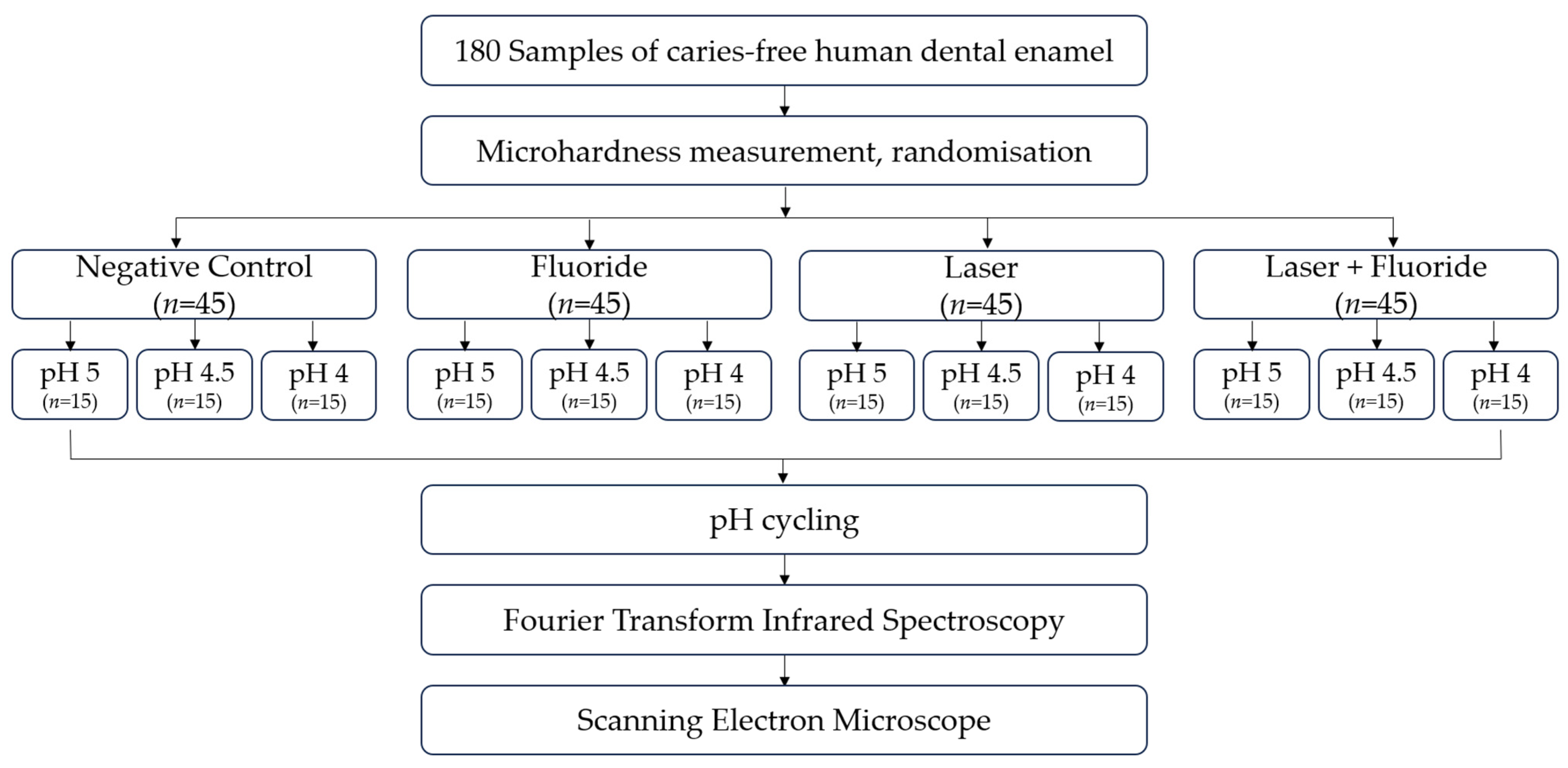
2. Materials and Methods

2.1. Experimental Design

Samples of caries-free human dental enamel (n = 180) homogenized by surface microhardness were divided into four treatment groups (n = 45): Negative Control, Fluoride, Laser, and Laser + Fluoride. The objective was to evaluate the preventive action of these treatments under different pH conditions; thus, the groups were subdivided to simulate in vitro caries at varied pH levels. The groups that underwent fluoride or laser treatment followed established parameters from the existing literature. After preparation and treatment, the groups were further subdivided into three groups for pH cycling: pH 5, pH 4.5, and pH 4 (Figure 1). Subsequently, samples were evaluated using Fourier Transform Infrared Spectroscopy and Scanning Electron Microscopy.

2.2. Sample Preparation

Forty-five permanent human molars extracted by orthodontic indication were selected for the study after approval by the Research Ethics Committee (CAAE: 02854118.3.0000.0075). All samples were examined by visual inspection, and teeth with visual defects on the enamel surface such as decay and enamel fractures were eliminated. The teeth were disinfected according to the White Protocol [17] and transformed into 180 enamel blocks (4 × 4 × 2 mm) using a cutting machine (Accutom-5, Struers, Ballerup, Denmark). The samples were polished with an abrasive disc under decreasing granulation: 400-grit, 600-grit, and 1200-grit (Carbimed Paper Discs, Buehler, Lake Bluff, IL, USA) under pressure 1 N and cooling with water in a polisher (EcoMet® 250/300 Grinder Polisher, Buehler, Lake Bluff, IL, USA), followed by a 1 μm. diamond suspension-embedded polishing cloth (METADI Diamond suspension, Color Polishing Water-spray, Buehler, USA). The samples were sonicated in distilled and deionized water for 3 min (Cuba Ultrassom Cristofoli, Sao Paulo, Brazil) and stored in relative humidity at 4 °C throughout the experiment.
The surface microhardness (SMH) is one of the standard measurements used for baseline analysis [18]. The baseline surface hardness was assessed using a microhardness tester (HMZ-2000, Shimadzu, Kyoto, Japan) with a Knoop diamond under 25 g loading for 5 s, making ten indentations in the center of the sample, from which the mean SMH was calculated. Samples with SMH between 360 and 420 Knoop hardness were selected for study.

2.3. Experimental Groups

The samples were randomly assigned to four treatment groups (n = 45/group):
  • Negative Control: Non-treated
  • Fluoride: Topical application of fluoride (FFA 12.300 µF/g)
  • Laser: Irradiation with Nd:YAG (84.9 J/cm2)
  • Laser + Flouride: Irradiation with Nd:YAG (84.9 J/cm2) and topical application of fluoride (FFA 12.300 µF/g)
Each group was subdivided into three for pH cycling: pH 5, pH 4.5, and pH 4 (n = 5/subgroup), for three similar conditions:
  • pH 5: The enamel surface tends to demineralize in the form of hydroxyapatite below a critical pH, but not in the form of fluorapatite, which is more acid resistant.
  • pH 4.5: The enamel surface tends to demineralize in the form of hydroxyapatite and in the form of fluorapatite below a critical pH.
  • pH 4: This pH was used to investigate the acid resistance of hydroxyapatite and fluorapatite forms in the presence of laser irradiation.

2.4. Treatment of Samples

Fluoride was applied at a concentration of 12.300 µF/g (Biodinamica, Ibipora, Brazil), in an amount of 1 mL per sample and was maintained for 4 min. After 30 min, the surface was washed with deionized water.
Irradiations was performed with an Nd:YAG laser (PulseMaster 1000 ADT, Corpus Christi, TX, USA), presenting a wavelength of 1064 nm and temporal width of 100 μs. The energy was delivered through a fiber-optic system with a spot size of 300 μm. Previously, the samples received a layer of photoabsorbent in the form of a pure coal solution diluted in equal parts deionized water and 99% ethanol, then applied with a brush.
The parameters used for irradiation were selected based on positive results from previous studies and their demonstrated safety for pulp tissue [10,12,19,20]. The Nd:YAG laser was used at 60 mJ energy per pulse (84.9 J/cm2), with a repetition rate of 10 Hz and a power of 0.6 W for a duration of 30 s for each sample. Considering that irradiation occurred in contact mode, no collimator was used during the process. For increased precision during irradiation, a high-precision motorized translator (ESP300, Newport Corporation, Irvine, CA, USA) was used and the energy per pulse was calibrated with an energy/power meter (FieldMaster, Coherent, Santa Clara, CA, USA). The group samples were irradiated with intercalation for distribution of the experimental error and temperature variations of the equipment.

2.5. pH Cycling

The pH cycling method was originally proposed by Featherstone et al. (1986) [21] and later modified by Argenta et al. (2003) [22]. The samples were exposed to the demineralizing solution for 3 h and to the remineralizing solution for 21 h over a period of 10 days.
The demineralizing solution consisted of 2.0 mM Ca, 2.0 mM P, and 0.030 ppm F in 0.075 M acetate buffer solution. The remineralizing solution (pH 7.4) contained 1.5 mM Ca, 0.9 mM P, 150 mmol KCl, and 0.050 ppm F in 20 mM cacodylate buffer.
For the purposes of this research, the pH cycling regime was modified. The demineralizing solution was adjusted to three different pH conditions (pH 5, pH 4.5, and pH 4) by using HCl to increase the acidity of the solution. The pH measurements were conducted using a pH/ISE Meter (Dual Star, Thermo Scientific Orion, Waltham, MA, USA).

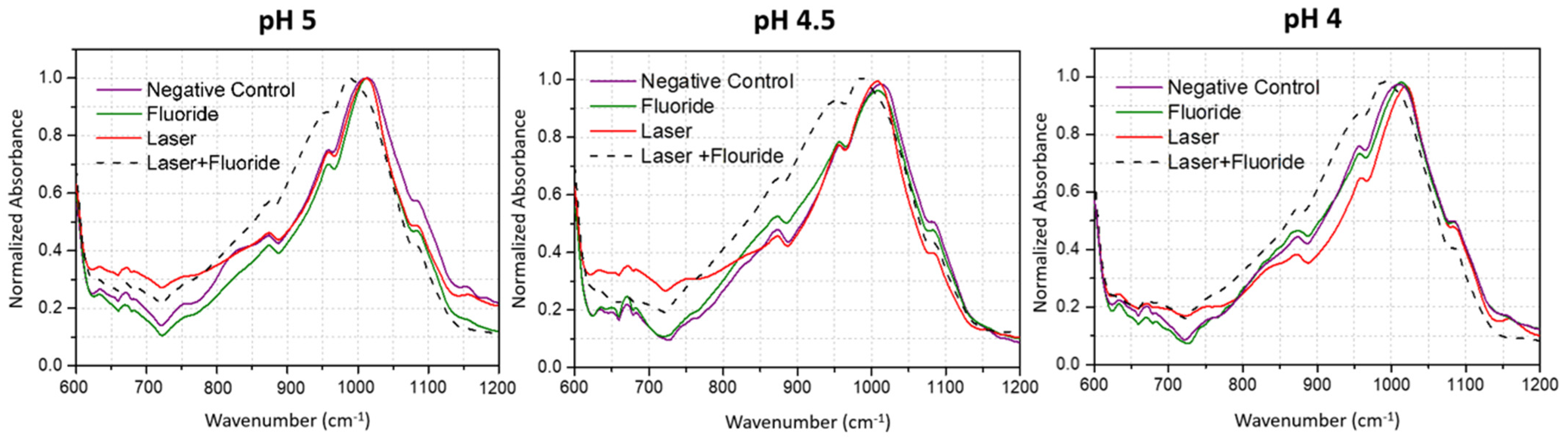
2.6. Fourier Transform Infrared Spectroscopy

A Fourier Transform Infrared spectrophotometer (Nicolet 6700, Thermo) equipped with an attenuated total reflection accessory (ATR; Smart Orbit, Thermo Scientific) with a diamond crystal of 2.25 mm2 was utilized. The spectra were obtained with a resolution of 4 cm−1. Each sample underwent 100 scans within the interval of 4000–400 cm−1, and the averaged spectrum of each sample was subsequently derived from the scans.
To minimize uncontrolled errors, samples from each group were randomly interspersed during the analysis. The measurement environment was maintained at a constant temperature of 20 °C and air humidity control was in place. The analysis table was cleaned using a tissue soaked in 70% alcohol.
OMINIC software was employed for data processing. The background spectrum was initially collected in order to subtract any environmental contributions, enhancing the reliability of subsequent sample analysis. The background measurement process lasted approximately 10 min, after which a new background spectrum was collected with the environmental conditions. Subsequently, the data were processed using software developed by the IR Hard Tissue group (BR512019001094-9, IPEN, Sao Paulo, Brazil).

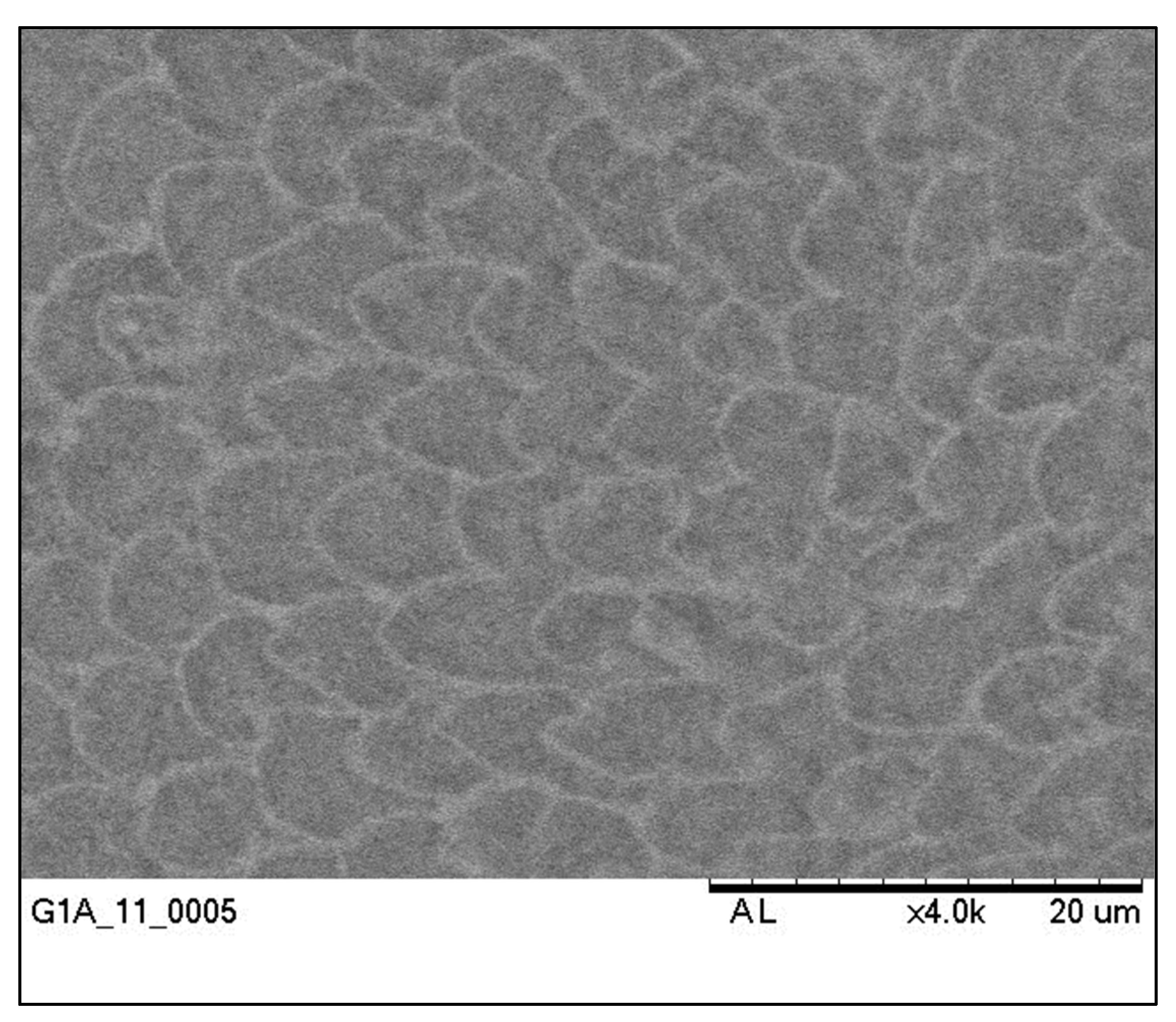
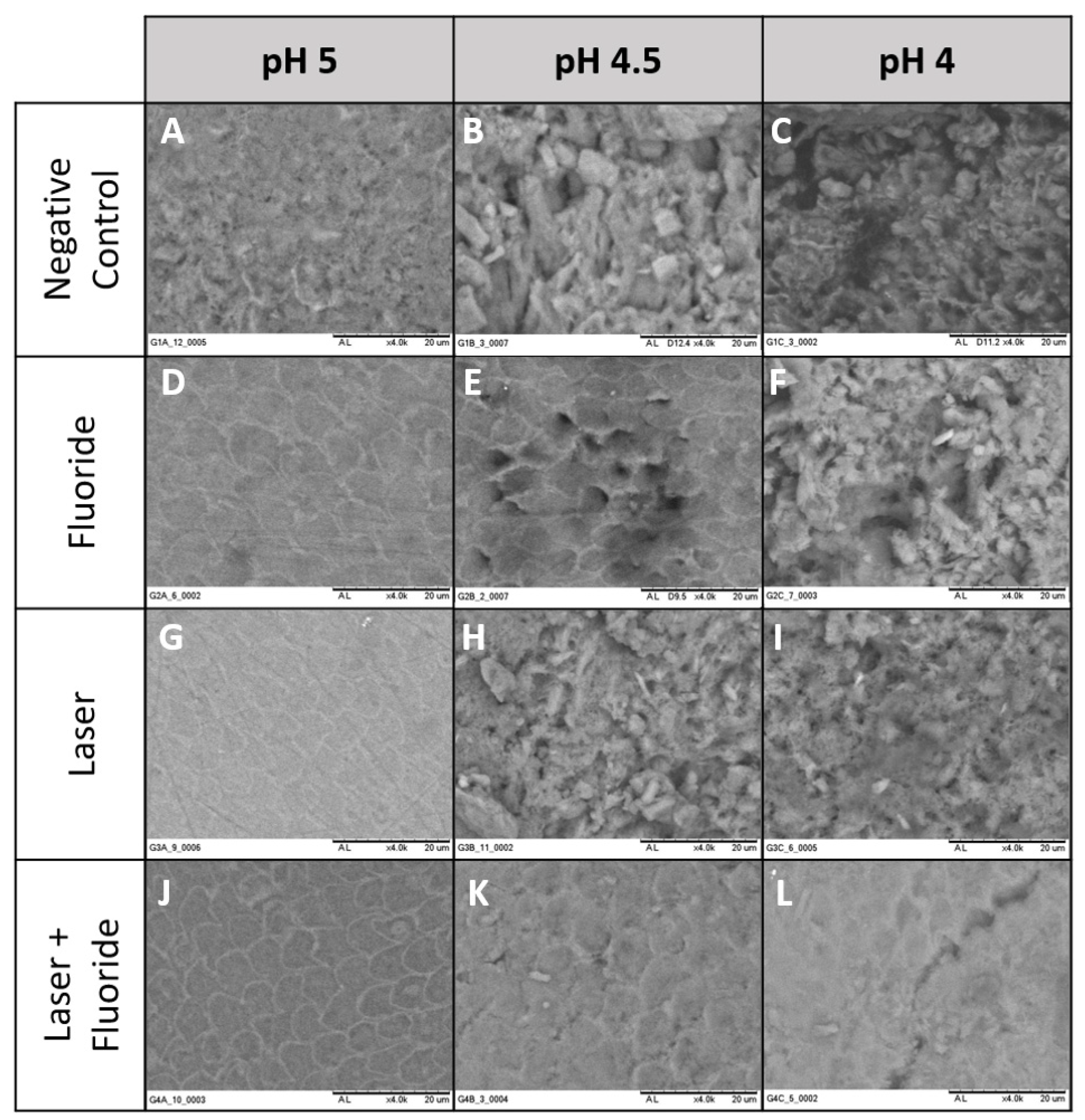
2.7. Scanning Electron Microscope

All samples were analyzed using a Scanning Electron Microscope (SEM; TM 3000 Tabletop Microscope, Hitachi, Chiyoda, Japan) at 15 kV. The analysis was conducted at two time points: before the treatments and cycling, and after the cycling. Both images were captured at a magnification of 4000×. All laboratory experiments were carried out by the same operator.

2.8. Statistical Tests

The outlier values were removed using ROUT outlier analysis (Q = 1%). The normality of the FTIR data was assessed using the Shapiro–Wilk test. The data demonstrated a parametric distribution, and groups were compared using ANOVA with Dunnett’s multiple comparisons test. The software utilized was GraphPad Prism 5®, and a significance level of 5% was adopted.

3. Results

3.1. Fourier Transform Infrared Spectroscopy

The bands analyzed in this study were Phosphate (900–1200 cm−1) and Carbonate (1350–1450 cm−1). After obtaining the spectrum of each sample, an average spectrum was obtained (Figure 2). Subsequently, the area over each band was calculated for statistical analysis (Table 1).

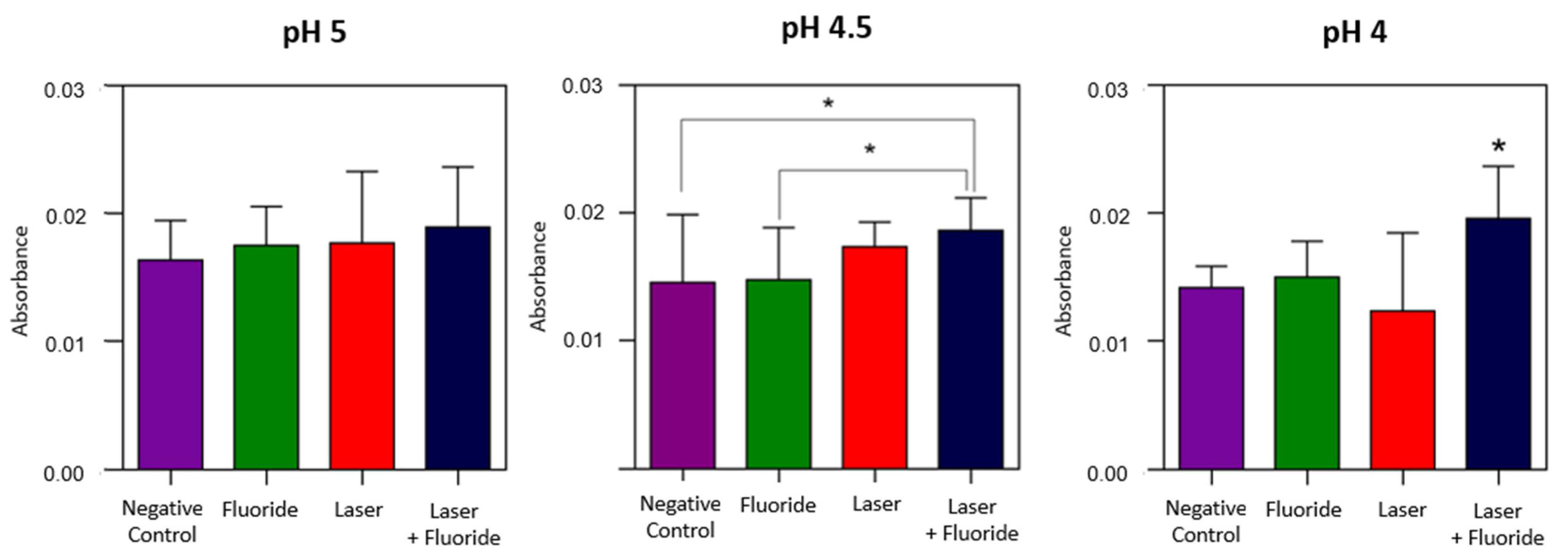
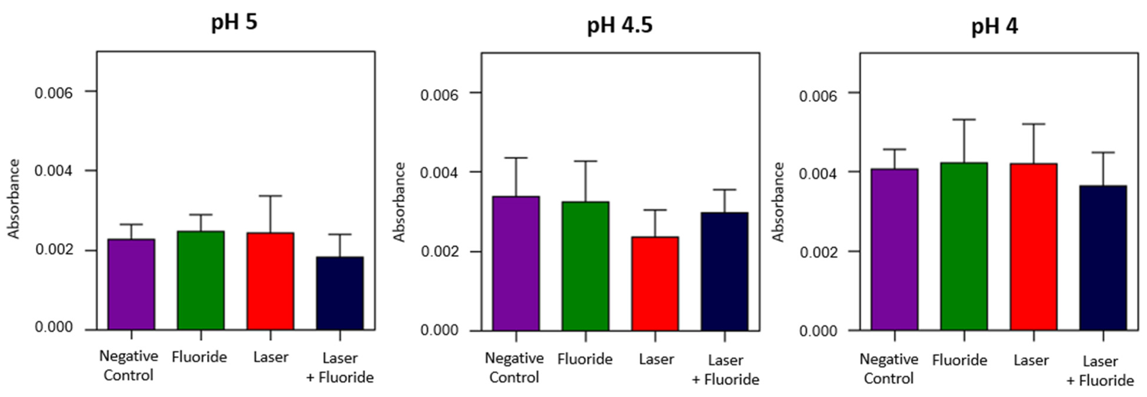
Phosphate and Carbonate Analysis

When comparing the phosphate content at different pH levels, it was observed that the values tended to decrease in more acidic conditions irrespective of the treatment. At pH 5, which is considered critical for enamel but below the critical value when fluoride is present, no significant differences were found between the groups. However, at pH 4.5 the Laser + Fluoride treatment showed a statistically significant difference compared to the Negative Control and Fluoride groups. At pH 4, the proposed Laser + Fluoride treatment exhibited a significant difference when compared to all other groups. At pH 4, neither the Fluoride treatment nor the Laser treatment alone were able to prevent phosphate loss. There was no significant difference observed compared to the Negative Control group (Figure 3).
The evaluated carbonate exhibited higher values in more acidic pH conditions. Specifically, the values ranged from 0.0018 to 0.0022 at pH 5, 0.0023 to 0.0033 at pH 4.5, and 0.0036 to 0.0042 at pH 4 (Table 1). However, there were no significant differences observed among these values (Figure 4).

3.2. Scanning Electron Microscope

In the image below, taken at an original magnification of 4000×, the enamel appears to be in a healthy state before the treatment. No cracks or defects are observed, and the enamel surface exhibits appropriate polishing. Additionally, enamel prisms are clearly visible in the image (Figure 5).
After the treatment and pH cycling, all samples were subjected to SEM analysis. A representative image from each group was carefully selected and included in Figure 4 (Figure 6).

4. Discussion

The prevention of dental caries is an ongoing area of research and there is a constant need to enhance preventive measures given that it remains a significant global public health issue. The literature supports the preventive effect of Nd:YAG laser application, which is attributed to its ability to increase acid resistance [9,11,12,23,24,25,26,27]. This demonstrates that the combination of Nd:YAG laser treatment and other preventive strategies is already an established reality based on the existing scientific literature.
The increase in acid resistance after irradiation with the Nd:YAG laser is due to chemical and microstructural modification of the enamel resulting from the thermal effect, causing fusion and resolidification of the enamel crystals [8,25,28]. When considering the protection of the pulp tissue against this heating effect, it is crucial to understand that the Nd:YAG laser emits a wavelength of 1064 nm, which is poorly absorbed by water and hydroxyapatite in the enamel, leading to its transmission through the enamel. To address this issue, conventional practice involves using photosensitizers, such as pure charcoal, to enhance surface absorption and control pulpal damage, thereby enabling safe irradiation within the prescribed parameters (84 J/cm2) [9,10,26].
The irradiated enamel undergoes an alteration in the organic matrix characterized by the evaporation of water and carbonate [26,29]. Another alteration is the formation of small amounts of new crystallographic phases such as tetracalcium phosphate in the α- and β-phases. In addition to chemical modification, it is possible to identify an increase in the average size of hydroxyapatite crystals [8,12]. The new crystalline phases are less resistant to demineralization; therefore, it is possible that the main factor associated with a higher acid resistance is the alteration of the carbonate. The evaporation of water and carbonate decreases the proportion of both in the mineral structure, increasing the proportion of other minerals in the enamel. The reduction of carbonate is beneficial because it is a more soluble mineral and makes the surface more prone to demineralization [8,26].
The use of fluoride in caries prevention aims to enhance resistance against acid challenges and facilitate remineralization. When topically applied in high concentrations, it enables the formation of fluoride reservoirs on the dental surface. Moreover, when combined with high-power laser irradiation the effect on acid resistance is significantly amplified. The interaction between fluoride and laser contributes to reducing the solubility of the enamel [10]. The irradiation process enhances fluoride absorption by increasing the contact surface resulting from melting [30]. Another potential mechanism involves the prolonged retention of fluorine on the irradiated surface as compared to using fluorine alone [8]. Additionally, it has been suggested that surface heating may induce the formation of fluorapatite through the incorporation of fluorine into molten enamel layers [9].
The increase in phosphate content after Nd:YAG laser irradiation is already evident in the literature [10,11,12,16,24]. The analysis of phosphate absorbance under different pH conditions in this study complements the existing literature. At pH 5, a noncritical condition for enamel treated with fluoride, the performed treatments showed no statistical differences compared to the control group. At pH 4.5, a critical condition for fluoride-treated enamel, it was possible to differentiate treatment with fluoride alone, where there was no significant difference with the negative control, from the Laser + Fluoride group, which did show significant differences. This condition suggests that the increased acid resistance conferred by the Laser + Fluoride treatment alters the critical pH of the tooth structure. At pH 4, even below the critical pH, the combined treatment group showed a significant difference in mineral content. Considering that this measurement was made after pH cycling, meaning that there was a higher phosphate content, these results verify the lower demineralization present in the Laser + Fluoride group.
The change in carbonate absorbance has been evidenced in other studies using the same laser with the same parameters; however, in these studies measurements were made after treatment without pH cycling [12,26]. In one study that evaluated the absorbance after pH cycling, the data corroborate those found in our study, showing no statistical difference after cycling [12]. It is possible that the effect on carbonate reduction is altered by the induction of demineralization.
Less demineralization can be evidenced in the morphological analysis performed by SEM. Our data corroborate those of Harazaki et al., who found less demineralization in SEM analysis of the enamel surface after treatment with Nd:YAG [31]. Another study demonstrated that high energy levels can alter the surface in an undesirable way, causing cracks and glazed surfaces [9]. These alterations were not observed in the microscopic images in this work. Yamada et al. found these alterations at 120 mJ energy [32].
It was not possible to observe any melting on the irradiated surface in the images obtained after pH cycling. Hossain et al. found melting on the surface of irradiated enamel immediately after Nd:YAG treatment; this surface modification was not seen after pH cycling [28]. Andrade et al. found a decrease in the elevation of the margins and the fused appearance of the enamel after dissolution of the superficial layer of enamel after simulating caries [33].
Morphological analysis under different pH conditions allowed us to evaluate the structural differences in demineralization. The Negative Control group showed the beginnings of demineralization even at pH 5, with a smoky pattern that was different from the other groups. At pH 4.5, the Negative Control and Laser group showed demineralization, the Fluoride group lost some central portions of the prisms, and the Laser + Fluoride group remained healthy. The difference between treatments was even more evident at pH 4, where the Laser + Fluoride group was the only group with a mostly healthy structure.
In vivo studies have demonstrated that the combination of Nd:YAG laser (84.9 J/cm2) and topical application of fluoride (FFA) is a clinically viable practice with positive results. In a study involving 33 volunteers, this combined approach led to a remarkable 39.2% reduction in caries incidence compared to the control group. Moreover, the group receiving Nd:YAG laser and fluoride application exhibited significantly smaller numbers of white spots compared to the fluoride-only group [10]. Another in vivo study with 52 children at high risk for caries evaluated the effects of Nd:YAG laser (73.9 J/cm2). The results showed that this treatment effectively prevented lesions in the occlusion of teeth for one year [9].
This treatment appears promising for individuals with a high risk of caries, including patients undergoing radiotherapy and orthodontic treatment. A clinical study involving patients undergoing orthodontic treatment revealed that isolated Nd:YAG laser (40 J/cm2) treatment led to a mean increase of 141% in white spots in the irradiated group after one year, while the control group had a twofold increase (287%) [31].
In addition to its preventive applications, laser irradiation has proven beneficial in treating tooth hypersensitivity, preventing tooth erosion, and addressing various other oral health conditions. However, to further advance preventive research and optimize the utilization of laser irradiation it is crucial to conduct additional studies focusing on long-term effects and comprehensive clinical evaluations. These research efforts will contribute to a deeper understanding of the potential benefits and safety aspects of laser treatments for oral health, paving the way for broader and more effective applications in dental care.

5. Conclusions

Combined Nd:YAG laser (84.9 J/cm2) treatment and topical application of fluoride demonstrated promising results in terms of dental mineral content and demineralization prevention. The study showed higher phosphate absorbance in FTIR, indicating increased surface mineral content and reduced demineralization at both pH 4.5 and pH 4.
Morphological analysis in SEM revealed that less demineralization occurred in the Fluoride group and the Laser + Fluoride group at pH 4.5. Remarkably, at pH 4 the Laser + Fluoride group displayed the most significant reduction in demineralization compared to other groups.
The data suggest that the proposed combined treatment effectively reduces demineralization even under conditions that are typically considered to be below the critical threshold for fluoride-treated enamel. Moreover, the acid resistance provided by this treatment approach may potentially alter the critical pH of the enamel with regard to demineralization, offering a promising avenue for caries prevention.

Author Contributions

Conceptualization, A.C.-J. and D.M.Z.; methodology, A.C.-J., N.A.Z., Y.R.F.-O., S.G.A. and C.d.P.E.; formal analysis, A.C.-J. and D.M.Z.; writing—original draft preparation, G.C.M.G. and A.C.-J.; writing—review and editing, C.d.P.E., G.C.M.G. and D.M.Z.; supervision, D.M.Z.; project administration, D.M.Z.; funding acquisition, D.M.Z. All authors have read and agreed to the published version of the manuscript.

Funding

This research was funded by Fundação de Amparo à Pesquisa do Estado de São Paulo (FAPESP) (21/00633-0 and 17/50332-0), Coordenação de Aperfeiçoamento de Pessoal de Nível Superior (CAPES) (Finance Code 001), and Conselho Nacional de Desenvolvimento Científico e Tecnológico (CNPq) (INCT-465763/2014-6, INCT 406761/2022-1, Sisfoton 440228/2021-2, and PQ 314517/2021-9).

Institutional Review Board Statement

The study was conducted in accordance with the Declaration of Helsinki and approved by Ethics Committee of School of Dentistry/Sao Paulo (protocol code 02854118.3.0000.0075 of 4 February 2019).

Informed Consent Statement

Informed consent was obtained from all subjects involved in the study.

Data Availability Statement

The data presented in this study are available on request from the corresponding author.

Acknowledgments

We express our gratitude to the Special Laboratory for Laser Dentistry-LELO for their invaluable support during the research.

Conflicts of Interest

The authors declare no conflict of interest.

References

  1. Gomes, A.P.M.; Da Silva, E.G.; Gonçalves, S.H.F.; Huhtala, M.F.R.L.; Martinho, F.C.; Gonçalves, S.E.D.P.; Torres, C.R.G. Relationship between patient’s education level and knowledge on oral health preventive measures. Int. Dent. Med. J. Adv. Res. 2015, 1, 1–7. [Google Scholar] [CrossRef]
  2. World Health Organization. Oral Health. 2023. Available online: https://www.who.int/news-room/fact-sheets/detail/oral-health (accessed on 23 May 2023).
  3. Fejerskov, O.; Nyvad, B.; Kidd, E. Dental Caries: The Disease and Its Clinical Management, 3rd ed.; Wiley Blackwell: Oxford, UK, 2015. [Google Scholar]
  4. Buzalaf, M.A.R.; Pessan, J.P.; Honorio, H.M.; Ten Cate, J.M. Mechanisms of action of fluoride for caries control. Monogr. Oral. Sci. 2011, 22, 97–114. Available online: https://www.karger.com/Article/Abstract/325151 (accessed on 1 June 2023).
  5. Tenuta, L.M.A.; Cury, J.A. Fluoride: Its role in dentistry. Braz. Oral Res. 2010, 24, 9–17. [Google Scholar] [CrossRef]
  6. Robinson, C. Fluoride and the caries lesion: Interactions and mechanism of action. Eur. Arch. Paediatr. Dent. 2009, 10, 136–140. [Google Scholar] [CrossRef]
  7. Neev, J.; White, J.M.; Goodis, H.E. Thermal characteristics during Nd:YAG and CO2 laser application to enamel and dentin. Laser Surg. Adv. Charact. Ther. Syst. IV 1994, 2128, 424–430. [Google Scholar] [CrossRef]
  8. Ana, P.A.; Bachmann, L.; Zezell, D.M. Lasers effects on enamel for caries prevention. Laser Phys. 2006, 16, 865–875. [Google Scholar] [CrossRef]
  9. Raucci-Neto, W.; de Castro-Raucci, L.M.S.; Lepri, C.P.; Faraoni-Romano, J.J.; da Silva, J.M.G.; Palma-Dibb, R.G. Nd:YAG laser in occlusal caries prevention of primary teeth: A randomized clinical trial. Lasers Med. Sci. 2013, 30, 761–768. [Google Scholar] [CrossRef]
  10. Zezell, D.M.; Boari, H.G.D.; Ana, P.A.; Eduardo, C.d.P.; Powell, G.L. Nd:YAG laser in caries prevention: A clinical trial. Lasers Surg. Med. 2009, 41, 31–35. [Google Scholar] [CrossRef]
  11. Zezell, D.M.; Silva, M.R.; Castro, P.A.A.; Silva, T.M.; Goncalves, S.E.P. Nd:YAG laser on dental enamel in the reduction of artificial caries demineralization. In Optics InfoBase Conference Papers; Optica Publishing Group: Washington, DC, USA, 2019. [Google Scholar] [CrossRef]
  12. Dias-Moraes, M.C.; Castro, P.A.A.; Pereira, D.L.; Ana, P.A.; Freitas, A.Z.; Zezell, D.M. Assessment of the preventive effects of Nd:YAG laser associated with fluoride on enamel caries using optical coherence tomography and FTIR spectroscopy. PLoS ONE 2021, 16, e0254217. [Google Scholar] [CrossRef]
  13. Agnol, M.A.D.; Battiston, C.; Tenuta, L.M.A.; Cury, J.A. Fluoride Formed on Enamel by Fluoride Varnish or Gel Application: A Randomized Controlled Clinical Trial. Caries Res. 2021, 56, 73–80. [Google Scholar] [CrossRef]
  14. Rabelo, J.S.; Ana, P.A.; Benetti, C.; Valério, M.E.G.; Zezell, D.M. Changes in dental enamel oven heated or irradiated with Er,Cr:YSGG laser. Analysis by FTIR. Laser Phys. 2010, 20, 871–875. [Google Scholar] [CrossRef]
  15. Corrêa-Afonso, A.M.; Bachmann, L.; de Almeida, C.G.; Corona, S.A.M.; Borsatto, M.C. FTIR and SEM analysis of CO2 laser irradiated human enamel. Arch. Oral Biol. 2012, 57, 1153–1158. [Google Scholar] [CrossRef]
  16. Lopes, C.D.C.A.; Limirio, P.H.J.O.; Novais, V.R.; Dechichi, P. Fourier transform infrared spectroscopy (FTIR) application chemical characterization of enamel, dentin and bone. Appl. Spectrosc. Rev. 2018, 53, 747–769. [Google Scholar] [CrossRef]
  17. White, D.; Featherstone, J. A Longitudinal Microhardness Analysis of Fluoride Dentifrice Effects on Lesion Progression in vitro. Caries Res. 1987, 21, 502–512. [Google Scholar] [CrossRef]
  18. Amaechi, B.T.; Tenuta, L.M.A.; Filho, A.P.R.; Cury, J.A. Protocols to Study Dental Caries In Vitro: Microbial Caries Models. Methods Mol. Biol. 2019, 1922, 357–368. [Google Scholar] [CrossRef]
  19. Boari, H.G.D.; Ana, P.A.; Eduardo, C.P.; Powell, G.L.; Zezell, D.M. Absorption and thermal study of dental enamel when irradiated with Nd:YAG laser with the aim of caries prevention. Laser Phys. 2009, 19, 1463–1469. [Google Scholar] [CrossRef]
  20. Seino, P.Y.; Freitas, P.M.; Marques, M.M.; de Souza Almeida, F.C.; Botta, S.B.; Moreira, M.S.N.A. Influence of CO2 (10.6 μm) and Nd:YAG laser irradiation on the prevention of enamel caries around orthodontic brackets. Lasers Med. Sci. 2015, 30, 611–616. [Google Scholar] [CrossRef]
  21. Featherstone, J.; O’Reilly, M.; Shariati, M.; Brugler, S. Enhancement of remineralization in vitro and in vivo. In Leach SA. Factors Affecting de- and Remineralization of the Teeth; IRL Press: Oxford, UK, 1986; pp. 23–24. [Google Scholar]
  22. Argenta, R.M.O.; Tabchoury, C.P.M.; Cury, J.A. A modified pH-cycling model to evaluate fluoride effect on enamel demineralization. Pesqui. Odontol. Bras. 2003, 17, 241–246. [Google Scholar] [CrossRef]
  23. Raghis, T.R.; Mahmoud, G.; Hamadah, O. Effectiveness of laser irradiation in preventing enamel demineralization during orthodontic treatment: A systematic review. Dent. Med. Probl. 2018, 55, 321–332. [Google Scholar] [CrossRef]
  24. Pereira, D.; Freitas, A.; Bachmann, L.; Benetti, C.; Zezell, D.; Ana, P. Variation on Molecular Structure, Crystallinity, and Optical Properties of Dentin Due to Nd:YAG Laser and Fluoride Aimed at Tooth Erosion Prevention. Int. J. Mol. Sci. 2018, 19, 433. [Google Scholar] [CrossRef]
  25. Chen, C.-C.; Huang, S.-T. The Effects of Lasers and Fluoride on the Acid Resistance of Decalcified Human Enamel. Photomed. Laser Surg. 2009, 27, 447–452. [Google Scholar] [CrossRef]
  26. Corrêa-Afonso, A.M.; Bachmann, L.; de Almeida, C.G.; Dibb, R.G.P.; Borsatto, M.C. Loss of structural water and carbonate of Nd:YAG laser-irradiated human enamel. Lasers Med. Sci. 2014, 30, 1183–1187. [Google Scholar] [CrossRef]
  27. Castellan, C.S.; Luiz, A.C.; Bezinelli, L.M.; Lopes, R.M.; Mendes, F.M.; Eduardo, C.D.P.; Freitas, P.M.D. In Vitro Evaluation of Enamel Demineralization after Er:YAG and Nd:YAG Laser Irradiation on Primary Teeth. Photomed. Laser Surg. 2007, 25, 85–90. [Google Scholar] [CrossRef]
  28. Nakamura, Y.; Kimura, Y.; Yamada, Y.; Kawanaka, T.; Matsumoto, K.; de Oliveira, R.M.; de Souza, V.M.; Esteves, C.M.; Lima-Arsati, Y.B.d.O.; Cassoni, A.; et al. Effect of Pulsed Nd:YAG Laser Irradiation on Acid Demineralization of Enamel and Dentin. J. Clin. Laser Med. Surg. 2001, 19, 105–108. [Google Scholar] [CrossRef]
  29. Harris, D.M.; Fried, D. Pulsed Nd:YAG laser selective ablation of surface enamel caries: I. Photoacoustic response and FTIR spectroscopy. In Lasers in Dentistry VI; SPIE: San Jose, CA, USA, 2000; Volume 3910, pp. 164–171. [Google Scholar] [CrossRef]
  30. Huang, G.F.; Lan, W.H.; Guo, M.K.; Chiang, C.-P. Synergistic effect of Nd:YAG laser combined with fluoride varnish on inhibition of caries formation in dental pits and fissures in vitro. J. Formos. Med. Assoc. 2001, 100, 181–186. Available online: http://www.fma.org.tw/jfma/PDF/2001-100/issue_3/Article_6.pdf (accessed on 1 June 2023).
  31. Harazaki, M.; Hayakawa, K.; Fukui, T.; Isshiki, Y.; Powell, L.G. The Nd-YAG Laser is Useful in Prevention of Dental Caries During Orthodontic Treatment. Bull. Tokyo Dent. Coll. 2001, 42, 79–86. [Google Scholar] [CrossRef]
  32. Yamada, M.K.; Uo, M.; Ohkawa, S.; Akasaka, T.; Watari, F. Three-dimensional topographic scanning electron microscope and Raman spectroscopic analyses of the irradiation effect on teeth by Nd:YAG, Er: YAG, and CO2 lasers. J. Biomed. Mater. Res. 2004, 71B, 7–15. [Google Scholar] [CrossRef]
  33. Deandrade, L.E.H.; Lizarelli, R.F.Z.; Pelino, J.E.P.; Bagnato, V.S.; Oliveira, O.B., Jr. Enamel caries resistance accidentally irradiated by the Nd:YAG laser. Laser Phys. Lett. 2007, 4, 457–463. [Google Scholar] [CrossRef]
Figure 1. Schematic representation of the experimental design.
Photonics 10 00985 g001
Figure 2. The average spectra obtained for each treatment at pH 5, 4.5, and 4 were analyzed in the Phosphate band region (900–1200 cm−1). In the three analyzed pHs, all groups (Negative Control, Fluorine, and Laser) presented similar regions and amplitudes. However, the Laser + Fluorine group presented an average spectrum with a visibly larger area within the analyzed band.
Photonics 10 00985 g002
Figure 3. Absorbance of Phosphate at pH 4, 4.5, and 4. * Indicates statistically significant differences (ANOVA test + Dunnett’ multiple comparisons test, p < 0.05).
Photonics 10 00985 g003
Figure 4. Absorbance of Carbonate at pH 4, 4.5 and 4. No statistically significant differences were found (ANOVA test + Dunnett’ multiple comparisons test, p < 0.05).
Photonics 10 00985 g004
Figure 5. SEM image, at 4.000× original magnification.
Photonics 10 00985 g005
Figure 6. Representative images of each group. (A) Smoky appearance, suggestive of the initial stages of demineralization. (B,C) Advanced demineralization, with a significant loss of structural continuity. (D) Healthy, without signs of demineralization or structural damage. (E) Loss of the central portion of the enamel prisms. (F) Advanced demineralization. (G) Healthy. (H) Demineralization without loss of enamel continuity. (I) advanced demineralization. (J) Healthy. (K) Small points of demineralization, without loss of the central portion of the prism. (L) Small points of demineralization, without loss of the central portion of the prism.
Photonics 10 00985 g006
Table 1. The mean percentage ± standard deviation of the area over the phosphate and carbonate bands analyzed at different cycling pHs.
Phosphate
GrouppH 5pH 4.5pH 4
Negative Control0.0136 ± 0.00310.0146 ± 0.0052 a0.0142 ± 0.0017 a
Fluoride0.0175 ± 0.00300.0148 ± 0.0041 a 0.0150 ± 0.0028 a
Laser0.0177 ± 0.00560.0173 ± 0.0019 ab0.0124 ± 0.0061 a
Laser + Fluoride0.0189 ± 0.00470.0186 ± 0.0025 b0.0196 ± 0.0041 b
Carbonate
GrouppH 5pH 4.5pH 4
Negative Control0.0023 ± 0.00030.0034 ± 0.00080.0041 ± 0.0004
Fluoride0.0025 ± 0.00030.0032 ± 0.0009 0.0042 ± 0.0009
Laser0.0024 ± 0.00080.0024 ± 0.00060.0042 ± 0.0009
Laser + Fluoride0.0018 ± 0.00050.0030 ± 0.00050.0036 ± 0.0008
Different letters indicate statistically significant differences (ANOVA test + Dunnett’ multiple comparisons test).
Disclaimer/Publisher’s Note: The statements, opinions and data contained in all publications are solely those of the individual author(s) and contributor(s) and not of MDPI and/or the editor(s). MDPI and/or the editor(s) disclaim responsibility for any injury to people or property resulting from any ideas, methods, instructions or products referred to in the content.

