Dynamic Microscopic Optical Coherence Tomography as a New Diagnostic Tool for Otitis Media

 , , , ,
Abstract
1. Introduction
2. Materials and Methods
2.1. Microscopic and Dynamic Microscopic OCT
2.2. Data Acquisition and Processing
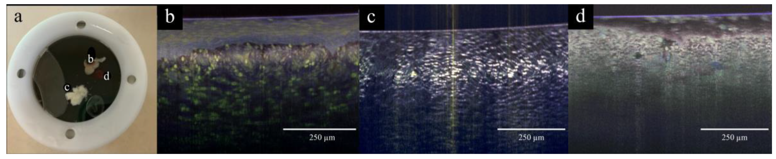
2.3. Study Design and Experimental Strategy
2.4. Human Tissue Sample Collection
2.5. Histology
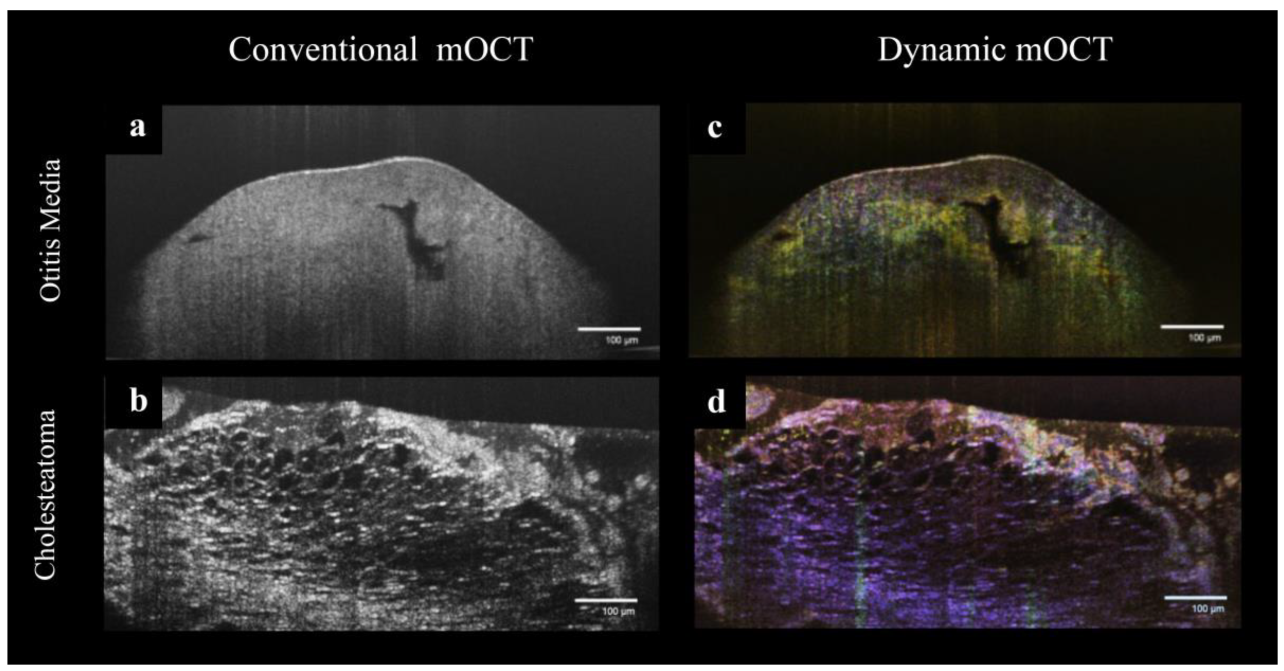
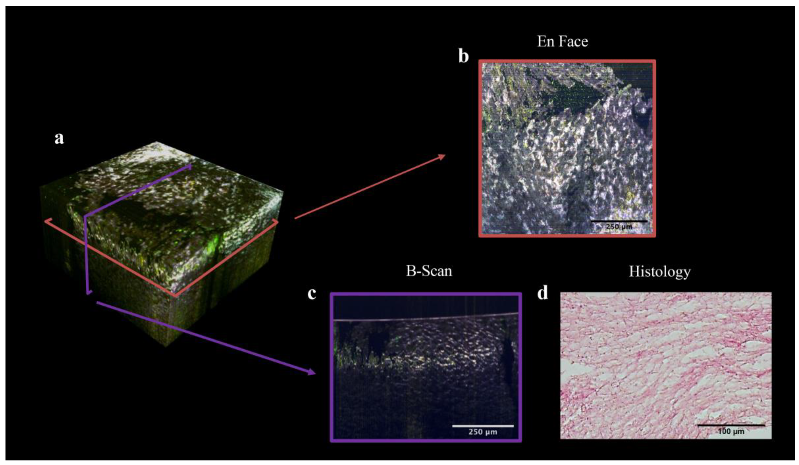
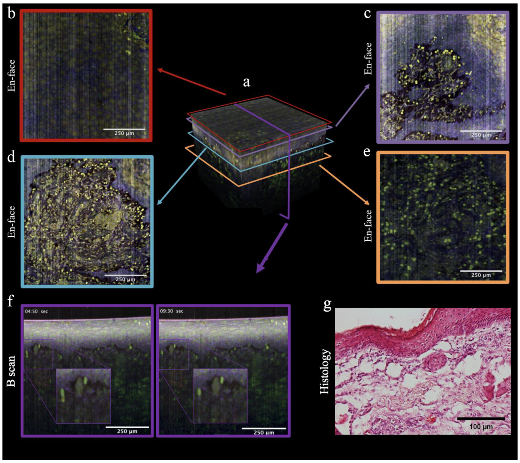
3. Results
4. Discussion
5. Conclusions
Author Contributions
Funding
Institutional Review Board Statement
Informed Consent Statement
Data Availability Statement
Acknowledgments
Conflicts of Interest
References
- Arguedas, A.; Kvaerner, K.; Liese, J.; Schilder, A.G.M.; Pelton, S.I. Otitis media across nine countries: Disease burden and management. Int. J. Pediatr. Otorhinolaryngol. 2010, 74, 1419–1424. [Google Scholar] [CrossRef] [PubMed]
- Schilder, A.G.M.; Chonmaitree, T.; Cripps, A.W.; Rosenfeld, R.M.; Casselbrant, M.L.; Haggard, M.P.; Venekamp, R.P. Otitis media. Nat. Rev. Dis. Prim. 2016, 2, 16063. [Google Scholar] [CrossRef] [PubMed]
- Ahmed, S.; Shapiro, N.L.; Bhattacharyya, N. Incremental health care utilization and costs for acute otitis media in children. Laryngoscope 2014, 124, 301–305. [Google Scholar] [CrossRef] [PubMed]
- Mittal, R.; Parrish, J.M.; Soni, M.; Mittal, J.; Mathee, K. Microbial otitis media: Recent advancements in treatment, current challenges and opportunities. J. Med. Microbiol. 2018, 67, 1417–1425. [Google Scholar] [CrossRef]
- Haile, L.M.; Kamenov, K.; Briant, P.S.; Orji, A.U.; Steinmetz, J.D.; Abdoli, A.; Abdollahi, M.; Abu-Gharbieh, E.; Afshin, A.; Ahmed, H.; et al. Hearing loss prevalence and years lived with disability, 1990–2019: Findings from the Global Burden of Disease Study 2019. Lancet 2021, 397, 996–1009. [Google Scholar] [CrossRef] [PubMed]
- Kuo, C.-L.; Shiao, A.-S.; Yung, M.; Sakagami, M.; Sudhoff, H.; Wang, C.-H.; Hsu, C.-H.; Lien, C.-F. Updates and knowledge gaps in cholesteatoma research. BioMed Res. Int. 2015, 2015, 854024. [Google Scholar] [CrossRef]
- Jung, J.Y.; Lee, D.H.; Wang, E.W.; Nason, R.; Sinnwell, T.M.; Vogel, J.P.; Chole, R.A. P. aeruginosa infection increases morbidity in experimental cholesteatomas. Laryngoscope 2011, 121, 2449–2454. [Google Scholar] [CrossRef] [PubMed]
- Chole, R.A.; Gagnon, P.M.; Vogel, J.P. Inactivation of specific Pseudomonas aeruginosa biofilm factors does not alter virulence in infected cholesteatomas. Otol. Neurotol. 2014, 35, 1585–1591. [Google Scholar] [CrossRef]
- AWMF. 017-006l_S1_Cholesteatom_2014-06. Available online: https://register.awmf.org/assets/guidelines/017-006l_S1_Cholesteatom_2014-06.pdf (accessed on 15 March 2023).
- AWMF. 017-074l_S2k_Chronisch-Mesotympanale-Otitis-Media_2020-11_01. Available online: https://register.awmf.org/assets/guidelines/017-074l_S2k_Chronisch-mesotympanale-Otitis-media_2020-11_01.pdf (accessed on 15 March 2023).
- Mishiro, Y.; Sakagami, M.; Kitahara, T.; Kondoh, K.; Okumura, S. The investigation of the recurrence rate of cholesteatoma using Kaplan-Meier survival analysis. Otol. Neurotol. 2008, 29, 803–806. [Google Scholar] [CrossRef]
- Aumann, S.; Donner, S.; Fischer, J.; Müller, F. High Resolution Imaging in Microscopy and Ophthalmology: New Frontiers in Biomedical Optics; Springer: Cham, Switzerland, 2019; ISBN 9783030166373. [Google Scholar]
- Fujimoto, J.G.; Pitris, C.; Boppart, S.A.; Brezinski, M.E. Optical coherence tomography: An emerging technology for biomedical imaging and optical biopsy. Neoplasia 2000, 2, 9–25. [Google Scholar] [CrossRef]
- Qin, J.; An, L. Optical Coherence Tomography for Ophthalmology Imaging. Adv. Exp. Med. Biol. 2021, 3233, 197–216. [Google Scholar] [CrossRef] [PubMed]
- Wan, B.; Ganier, C.; Du-Harpur, X.; Harun, N.; Watt, F.M.; Patalay, R.; Lynch, M.D. Applications and future directions for optical coherence tomography in dermatology. Br. J. Dermatol. 2021, 184, 1014–1022. [Google Scholar] [CrossRef] [PubMed]
- Münter, M.; vom Endt, M.; Pieper, M.; Casper, M.; Ahrens, M.; Kohlfaerber, T.; Rahmanzadeh, R.; König, P.; Hüttmann, G.; Schulz-Hildebrandt, H. Dynamic contrast in scanning microscopic OCT. Opt. Lett. 2020, 45, 4766–4769. [Google Scholar] [CrossRef] [PubMed]
- Apelian, C.; Harms, F.; Thouvenin, O.; Boccara, A.C. Dynamic full field optical coherence tomography: Subcellular metabolic contrast revealed in tissues by interferometric signals temporal analysis. Biomed. Opt. Express 2016, 7, 1511–1524. [Google Scholar] [CrossRef]
- Musial, G.; Kohlfaerber, T.; Ahrens, M.; Schulz-Hildebrandt, H.; Steven, P.; Hüttmann, G. Dynamic Contrast Microscopic Optical Coherence Tomography As a Novel Method for Assessing Corneal Epithelium During Exposure to Benzalkonium Chloride. Transl. Vis. Sci. Technol. 2022, 11, 28. [Google Scholar] [CrossRef]
- Leung, H.M.; Wang, M.L.; Osman, H.; Abouei, E.; MacAulay, C.; Follen, M.; Gardecki, J.A.; Tearney, G.J. Imaging intracellular motion with dynamic micro-optical coherence tomography. Biomed. Opt. Express 2020, 11, 2768–2778. [Google Scholar] [CrossRef]
- Kohlfaerber, T.; Pieper, M.; Münter, M.; Holzhausen, C.; Ahrens, M.; Idel, C.; Bruchhage, K.-L.; Leichtle, A.; König, P.; Hüttmann, G.; et al. Dynamic microscopic optical coherence tomography to visualize the morphological and functional micro-anatomy of the airways. Biomed. Opt. Express 2022, 13, 3211–3223. [Google Scholar] [CrossRef]
- Münter, M.; Pieper, M.; Kohlfaerber, T.; Bodenstorfer, E.; Ahrens, M.; Winter, C.; Huber, R.; König, P.; Hüttmann, G.; Schulz-Hildebrandt, H. Microscopic optical coherence tomography (mOCT) at 600 kHz for 4D volumetric imaging and dynamic contrast. Biomed. Opt. Express 2021, 12, 6024–6039. [Google Scholar] [CrossRef]
- Pieper, M.; Schulz-Hildebrandt, H.; Mall, M.A.; Hüttmann, G.; König, P. Intravital microscopic optical coherence tomography imaging to assess mucus-mobilizing interventions for muco-obstructive lung disease in mice. Am. J. Physiol. Lung Cell. Mol. Physiol. 2020, 318, L518–L524. [Google Scholar] [CrossRef]
- Thouvenin, O.; Boccara, C.; Fink, M.; Sahel, J.; Pâques, M.; Grieve, K. Cell Motility as Contrast Agent in Retinal Explant Imaging With Full-Field Optical Coherence Tomography. Investig. Ophthalmol. Vis. Sci. 2017, 58, 4605–4615. [Google Scholar] [CrossRef]
- Li, Z.; Sun, H.; Turek, J.; Jalal, S.; Childress, M.; Nolte, D.D. Doppler fluctuation spectroscopy of intracellular dynamics in living tissue. J. Opt. Soc. Am. A Opt. Image Sci. Vis. 2019, 36, 665–677. [Google Scholar] [CrossRef]
- Si, Y.; Chen, Y.B.; Chen, S.J.; Zheng, Y.Q.; Liu, X.; Liu, Y.; Jiang, H.L.; Xu, G.; Li, Z.H.; Huang, Q.H.; et al. TLR4 drives the pathogenesis of acquired cholesteatoma by promoting local inflammation and bone destruction. Sci. Rep. 2015, 5, 16683. [Google Scholar] [CrossRef]
- Schürmann, M.; Oppel, F.; Shao, S.; Volland-Thurn, V.; Kaltschmidt, C.; Kaltschmidt, B.; Scholtz, L.-U.; Sudhoff, H. Chronic inflammation of middle ear cholesteatoma promotes its recurrence via a paracrine mechanism. Cell Commun. Signal. 2021, 19, 25. [Google Scholar] [CrossRef]
- Leichtle, A.; Klenke, C.; Ebmeyer, J.; Daerr, M.; Bruchhage, K.-L.; Hoffmann, A.S.; Ryan, A.F.; Wollenberg, B.; Sudhoff, H. NOD-Like Receptor Signaling in Cholesteatoma. BioMed Res. Int. 2015, 2015, 408169. [Google Scholar] [CrossRef] [PubMed]
- Leichtle, A.; Leffers, D.; Daerr, M.G.; Draf, C.; Kurabi, A.; Ryan, A.F.; Rupp, J.; Bruchhage, K.-L. Immunmodulation im Cholesteatom. Laryngorhinootologie 2022, 101, 310–319. [Google Scholar] [CrossRef]
- Klenke, C.; Janowski, S.; Borck, D.; Widera, D.; Ebmeyer, J.; Kalinowski, J.; Leichtle, A.; Hofestädt, R.; Upile, T.; Kaltschmidt, C.; et al. Identification of novel cholesteatoma-related gene expression signatures using full-genome microarrays. PLoS ONE 2012, 7, e52718. [Google Scholar] [CrossRef] [PubMed]
- Early, S.; Saad, M.A.; Mallidi, S.; Mansour, A.; Seist, R.; Hasan, T.; Stankovic, K.M. A Fluorescent Photoimmunoconjugate for Imaging of Cholesteatoma. Sci. Rep. 2022, 12, 19905. [Google Scholar] [CrossRef]
- Djalilian, H.R.; Rubinstein, M.; Wu, E.C.; Naemi, K.; Zardouz, S.; Karimi, K.; Wong, B.J.F. Optical coherence tomography of cholesteatoma. Otol. Neurotol. 2010, 31, 932–935. [Google Scholar] [CrossRef]
- Locke, A.K.; Zaki, F.R.; Fitzgerald, S.T.; Sudhir, K.; Monroy, G.L.; Choi, H.; Won, J.; Mahadevan-Jansen, A.; Boppart, S.A. Differentiation of otitis media-causing bacteria and biofilms via Raman spectroscopy and optical coherence tomography. Front. Cell. Infect. Microbiol. 2022, 12, 869761. [Google Scholar] [CrossRef] [PubMed]
- Monroy, G.L.; Won, J.; Shi, J.; Hill, M.C.; Porter, R.G.; Novak, M.A.; Hong, W.; Khampang, P.; Kerschner, J.E.; Spillman, D.R.; et al. Automated classification of otitis media with OCT: Augmenting pediatric image datasets with gold-standard animal model data. Biomed. Opt. Express 2022, 13, 3601–3614. [Google Scholar] [CrossRef] [PubMed]
- Lui, C.G.; Kim, W.; Dewey, J.B.; Macías-Escrivá, F.D.; Ratnayake, K.; Oghalai, J.S.; Applegate, B.E. In vivo functional imaging of the human middle ear with a hand-held optical coherence tomography device. Biomed. Opt. Express 2021, 12, 5196–5213. [Google Scholar] [CrossRef]
- Yung, M.; Tono, T.; Olszewska, E.; Yamamoto, Y.; Sudhoff, H.; Sakagami, M.; Mulder, J.; Kojima, H.; İncesulu, A.; Trabalzini, F.; et al. EAONO/JOS Joint Consensus Statements on the Definitions, Classification and Staging of Middle Ear Cholesteatoma. J. Int. Adv. Otol. 2017, 13, 1–8. [Google Scholar] [CrossRef] [PubMed]
- Maniu, A.; Harabagiu, O.; Perde Schrepler, M.; Cătană, A.; Fănuţă, B.; Mogoantă, C.A. Molecular biology of cholesteatoma. Rom. J. Morphol. Embryol. 2014, 55, 7–13. [Google Scholar] [PubMed]
- Schürmann, M.; Goon, P.; Sudhoff, H. Review of potential medical treatments for middle ear cholesteatoma. Cell Commun. Signal. 2022, 20, 148. [Google Scholar] [CrossRef] [PubMed]
- Kretschmer, S.; Pieper, M.; Hüttmann, G.; Bölke, T.; Wollenberg, B.; Marsh, L.M.; Garn, H.; König, P. Autofluorescence multiphoton microscopy for visualization of tissue morphology and cellular dynamics in murine and human airways. Lab. Investig. 2016, 96, 918–931. [Google Scholar] [CrossRef]
- Germain, R.N.; Miller, M.J.; Dustin, M.L.; Nussenzweig, M.C. Dynamic imaging of the immune system: Progress, pitfalls and promise. Nat. Rev. Immunol. 2006, 6, 497–507. [Google Scholar] [CrossRef] [PubMed]
- Schulz-Hildebrandt, H.; Pieper, M.; Stehmar, C.; Ahrens, M.; Idel, C.; Wollenberg, B.; König, P.; Hüttmann, G. Novel endoscope with increased depth of field for imaging human nasal tissue by microscopic optical coherence tomography. Biomed. Opt. Express 2018, 9, 636–647. [Google Scholar] [CrossRef]


Disclaimer/Publisher’s Note: The statements, opinions and data contained in all publications are solely those of the individual author(s) and contributor(s) and not of MDPI and/or the editor(s). MDPI and/or the editor(s) disclaim responsibility for any injury to people or property resulting from any ideas, methods, instructions or products referred to in the content. |
© 2023 by the authors. Licensee MDPI, Basel, Switzerland. This article is an open access article distributed under the terms and conditions of the Creative Commons Attribution (CC BY) license (https://creativecommons.org/licenses/by/4.0/).
Share and Cite
Leichtle, A.; Penxova, Z.; Kempin, T.; Leffers, D.; Ahrens, M.; König, P.; Brinkmann, R.; Hüttmann, G.; Bruchhage, K.-L.; Schulz-Hildebrandt, H. Dynamic Microscopic Optical Coherence Tomography as a New Diagnostic Tool for Otitis Media. Photonics 2023, 10, 685. https://doi.org/10.3390/photonics10060685
Leichtle A, Penxova Z, Kempin T, Leffers D, Ahrens M, König P, Brinkmann R, Hüttmann G, Bruchhage K-L, Schulz-Hildebrandt H. Dynamic Microscopic Optical Coherence Tomography as a New Diagnostic Tool for Otitis Media. Photonics. 2023; 10(6):685. https://doi.org/10.3390/photonics10060685
Chicago/Turabian StyleLeichtle, Anke, Zuzana Penxova, Thorge Kempin, David Leffers, Martin Ahrens, Peter König, Ralf Brinkmann, Gereon Hüttmann, Karl-Ludwig Bruchhage, and Hinnerk Schulz-Hildebrandt. 2023. "Dynamic Microscopic Optical Coherence Tomography as a New Diagnostic Tool for Otitis Media" Photonics 10, no. 6: 685. https://doi.org/10.3390/photonics10060685
APA StyleLeichtle, A., Penxova, Z., Kempin, T., Leffers, D., Ahrens, M., König, P., Brinkmann, R., Hüttmann, G., Bruchhage, K.-L., & Schulz-Hildebrandt, H. (2023). Dynamic Microscopic Optical Coherence Tomography as a New Diagnostic Tool for Otitis Media. Photonics, 10(6), 685. https://doi.org/10.3390/photonics10060685