Review

Optical Diagnostics in Herpetic Keratitis

by
Federico Di Staso
1,2,
Daria Rullo
1,
Mariachiara Di Pippo
1,
Silvio Di Staso
3 and
Solmaz Abdolrahimzadeh
1,4,*
1
Ophthalmology Unit, Neurosciences, Mental Health, and Sensory Organs (NESMOS) Department, Sapienza University of Rome, Via di Grottarossa 1035/1039, 00189 Rome, Italy
2
Connecticut Uveitis Foundation, 1043 Farmington Avenue, West Hartford, CT 06107, USA
3
Ophthalmic Clinic, Department of Life, Health and Environmental Sciences, University of L’Aquila, 67100 L’Aquila, Italy
4
St. Andrea Hospital, Via di Grottarossa 1035/1039, 00189 Rome, Italy
*
Author to whom correspondence should be addressed.
Photonics 2023, 10(4), 349; https://doi.org/10.3390/photonics10040349
Submission received: 18 February 2023 / Revised: 12 March 2023 / Accepted: 20 March 2023 / Published: 23 March 2023
(This article belongs to the Special Issue Optical Diagnostics)

Abstract

:
One of the leading global causes of vision impairment due to anterior segment disease is herpes simplex keratitis (HSK). The routine clinical method in the diagnosis of HSK is examination of the eye using slit lamp biomicroscopy; nevertheless, this is a subjective examination and can potentially lead to an erroneous diagnosis. Optical devices such as in vivo confocal microscopy and anterior segment optical coherence tomography are among the additional diagnostic tools that provide a valuable resource in the diagnosis and management of the condition. In research settings, these technologies have already enhanced our understanding of the microscopic causes of numerous common in vivo observations. This review aims to highlight the multiple emerging clinical and research applications for optical imaging devices in HSK.

1. Introduction

Herpes simplex keratitis (HSK) is a leading global cause of vision impairment [1]. The estimated prevalence of HSV (herpes simplex virus) type 1 (HSV-1) was 47.8%, and HSV type 2 (HSV-2) was 11.9% in 2015–2016 [2]. The worldwide incidence is 1.5 million per year [3]. In most cases, HSV-1 is responsible for unilateral epithelial and stromal keratitis with a predilection for patients with atopy due to concomitant immune dysregulation [4]. With a lower frequency, HSV-1 can affect any structure of the eye, potentially causing conjunctivitis, blepharitis, or uveitis [5].
Most primary HSV-1 infections are asymptomatic, but some patients may present follicular conjunctivitis as well as mild upper respiratory infection. After the primary contact, the virus may travel via nerve axons to establish a permanent latent infection in the trigeminal ganglion, which will act as a reservoir for future reactivations along any branch of the trigeminal nerve [6]. Local and environmental stress, such as direct corneal injuries, surgery, immunocompromised states, or even sunlight exposure, are all risk factors for reactivation of the disease [6,7]. The most common manifestation of HSK is epithelial keratitis, where the corneal basal epithelium becomes infected, and the virus can actively replicate and spread [8]. The pathognomonic signs of an active HSK are dendritic lesions that can be examined at the slit lamp and stained with fluorescein dye [9]. HSV can also affect the deeper layers of the cornea with different clinical manifestations, such as stromal keratitis, where the middle layer of the cornea is involved and is often accompanied by corneal edema and neovascularization [10], as well as endothelial keratitis that can cause anterior chamber inflammatory reaction [11]. HSK diagnosis is largely based on slit lamp clinical evaluation and patient anamnesis [12]. When the clinical spectrum of findings is not classic, diagnosis can become a challenge. The major diagnostic complications arise in cases with concomitant infective processes, pre-existing corneal lesions, or atypical/rare manifestations [5,13]. In such difficult cases or in suspected neonatal herpetic infection, the currently available laboratory tests to confirm the diagnosis are impression cytology, cytology on conjunctival or vesicular scrapings, culture, polymerase chain reaction (PCR), and enzyme-linked immunosorbent assay (ELISA) for the presence of HSV [14]. Complementary methods, such as optical devices, may be effective in challenging clinical pictures. Optical coherence tomography (OCT) is a diagnostic method that enables noninvasive, non-contact, in vivo imaging of the posterior ocular structures [15] where tomographic images are generated through the measurement of the echo time delay of the light beam from the structures and tissues encountered. In vivo confocal microscopy (IVCM) is a non-invasive tool for detecting morphological abnormalities in corneal layers [16] developed over 30 years ago and still used in a variety of research settings and clinical applications [17]. IVCM technology is based on a light beam that passes through an opening and is then focused on a small target area of the cornea. The reflected light is detected by a second opening to eliminate the out-of-focus light. Both illumination and detection pathways share the same focal plane, explaining the term “confocal” [18].
We aim to explore studies conducted with advanced imaging optical tools and methods of diagnosing HSK and discuss their clinical significance.

2. Materials and Methods

We conducted a PubMed database literature search (all fields) through 20 January 2023, with the following keywords: (“Herpes” OR “Herpetic” OR “Herpetic simplex” OR “HSV”) AND (“Keratitis”or “Cornea”) AND (“Diagnosis” OR “Diagnostic” OR “Detection”) AND (“Anterior segment optical coherence tomography” OR “AS-OCT”) AND “In vivo confocal microscopy”). We included articles published in English. The articles could be prospective or retrospective studies with the specific aim of evaluating the diagnostic potential of optical techniques to detect HSK.

3. Results

3.1. Herpetic Keratitis Pathophysiology

HSV is a linear double-stranded DNA virus classified as an α-member of the Herpesviridae family [9]. Primary infection is caused by HSV spread after direct contact with mucous membranes of the host. In the specific setting of ocular infections, the virus is transported in a retrograde fashion via sensory neurons to establish latency in trigeminal ganglia; once there, it can remain asymptomatic until reactivation [19].
HSV keratitis is one of the possible manifestations of virus reactivation and can affect the cornea at different sites, such as epithelial, stromal, and endothelial levels [20]. The most common clinical findings in epithelial keratitis include geographic ulcers and dendritic keratitis. Corneal complications and consequences of epithelial herpetic eye disease range from simple epitheliopathy to neurotrophic or meta-herpetic ulcers. Long-standing infections may lead to irreversible corneal damage and blindness [21].

3.2. Characteristics and Diagnostic Challenges

HSV keratitis is primarily diagnosed by clinical examination of the slit lamp [19].
Common manifestations include irritation, redness, watery eye, discharge, pain, and photophobia. In most cases, the acute phase starts to decrease after the first 2 weeks [19]. The most common and recurrent subtype, epithelial keratitis, appears as scattered and granular spots that produce punctuate lesions, but these rapidly merge into dendritic lesions [22]. Initial corneal epithelial changes of HSV-1 may include a large number of clear vesicles with ulcerative features [23]. After 5–7 days, on slit lamp examination, epithelial keratitis presents as a typical dendritic lesion with a terminal bulb, swollen borders, and intraepithelial cell infiltration [20]. The dendritic lesions can be enhanced by topical fluorescein dye staining [9], as shown in Figure 1.
Atypical epithelial lesions can be more difficult to identify, and PCR may be used to confirm HSV infection [24]. New methods such as immunofluorescence antibody assay (IFA) and tear collection are available and have been used to assist in the identification of epithelial lesions [25,26].
Stromal keratitis may appear as opaque or whitened lesions due to stromal infiltration [27]. Likewise, the necrotizing type of stromal keratitis appears as gray, white, or opaque lesions, but there is associated necrosis and ulceration [27,28]. Edema and abscess may be visible as well [19]. In immune-mediated stromal keratitis, necrosis or ulceration is habitually absent, but there is stromal infiltration [29]. Another less common form of corneal HSV infection is the disciform lesion; this particular subtype is a primary endotheliitis that presents as a ground-glass disk-shaped lesion with surrounding stromal edema [29].
A typical dendritic epithelial lesion on slit lamp examination is pathognomonic for keratitis; however, atypical presentations can make diagnosis problematic [20]. Many factors can negatively affect diagnosis capabilities, such as rare forms, duration of illness, associated systemic diseases, previous or concurrent medication use, and corneal transplantation [24]. Misdiagnosis is not uncommon, especially because other pathogens can initially present with similar lesions, such as amebic and fungal infections [30,31].
Interestingly, in a study specifically designed to focus on the diagnosis of atypical HSV lesions, Koizumi et al. [24] defined atypical lesions as “a condition characterized by the presence of unusually shaped defects or ulcers in the cornea, and in which characteristic HSV keratitis findings, such as dendritic or geographical ulcers with terminal bulbs and epithelial infiltrations, are not evident.” Very little agreement (p = 0.22) between PCR results and clinical diagnosis was found. PCR is considered to be highly sensitive; however, significant variability has been observed in various studies between PCR HSV detection rate and clinical diagnosis [30]. PCR is more efficient in identifying patients with typical lesions or patients without a history of antiviral medication use (p = 0.022); on the other hand, it is less reliable in patients with atypical lesions or in patients who had previous or current antiviral medication use (p = 0.968) [32].

3.3. Anterior Segment Optical Coherence Tomography

Anterior segment OCT (AS-OCT) is an imaging modality capable of producing two or three-dimensional imaging of a target tissue by creating a merged B-scan image from multiple axial scans [33]. AS-OCT was developed to study the anterior structures of the eye and can be classified into time-domain and frequency-domain OCT. In time-domain OCT, a moving reference mirror is used to acquire serial images depending on tissue reflectivity to infrared light, which in turn matches tissue depth [34]. Hence, time-domain OCT is limited by the reference mirror speed and prone to motion artifacts. Frequency-domain OCT (FD-OCT) can be further classified into Fourier-domain OCT, also known as spectral-domain OCT (SD-OCT) and swept-source OCT (SS-OCT). SD-OCT produces variable light wavelengths causing an interference pattern between the target tissue and the reference arm [35]. In this case, a dispersive spectrometer is needed to measure the interference [36]. This allows the FD-OCT to achieve a high scanning speed up to about 100 times faster than TD-OCT. Furthermore, the FD-OCT allows a substantial artifact rate reduction [37].
The most recent frequency-domain OCT is the SS-OCT, which uses a “swept laser”, defined as a narrowband light source that sweeps across a wide-ranging optical bandwidth [38]. Instead of a spectrometer, SS-OCT utilizes a semiconductor camera and two photodiode detectors [34]. These factors combined allow a significant image acquisition speed improvement and a reasonable resolution enhancement. Today, AS-OCT technology is becoming a cornerstone in refractive surgery, such as cataract and keratomileusis, as well as in evaluating the iridocorneal angle in glaucoma [39]. In corneal transplantation, AS-OCT can be determinant in planning endothelial keratoplasty by exploring the status of the anterior segment layers in detail and guiding the surgical choices [40]. Furthermore, AS-OCT can assess and track subtle corneal changes that can help to evaluate the severity of diseases, such as its ability to discriminate malignant ocular surface squamous neoplasia from benign lesions [40].

3.4. Clinical Application of AS-OCT in HSV Infection

Slit lamp biomicroscopy is routinely used for the clinical examination, diagnosis, and follow-up of HSV keratitis. The examination, however, has disadvantages and limitations, creating difficulties in accurately quantifying corneal changes in vivo, such as in corneal edema. For this reason, the use of advanced methods, such as AS-OCT, can help the clinician to identify typical signs of keratitis with more confidence and to follow the outcome of the chosen treatment. Soliman et al. [41] studied 42 patients with herpetic keratitis of varying severity, assessing the main HSV keratitis features with AS-OCT. They identified herpetic corneal infiltrates as lentiform or spindle-shaped, localized or diffuse, hyper-reflective areas. The use of AS-OCT helped distinguish active infiltrates from scarring outcomes. However, no pathognomonic features of herpetic keratitis have been identified, making the use of imaging more functional in the follow-up compared to the diagnostic framework. Accordingly, Young Ming Park et al. [42] investigated the AS-OCT features of herpetic epithelial keratitis, reporting hyper-reflective lesions in the subepithelial area, and in some patients, corneal epithelial irregularity. Another interesting aspect to consider with AS-OCT is the evaluation of corneal thickness. Hixson et al. [43] monitored two cases of disciform keratitis using AS-OCT imaging by studying the trend of corneal edema. The authors located and measured the area of greatest corneal thickness until complete resolution of the corneal edema. This enabled the accurate localization of corneal edema as well as providing an objective parameter to evaluate the treatment efficacy in reducing the associated increased corneal thickness. Similarly, Louise et al. reported two patients with HSV stromal keratitis and epithelial ulceration diagnosed and followed up using pachymetry and epithelial thickness mapping with AS-OCT [44]. The clinical characteristics of eyes in studies using AS-OCT are shown in Table 1.

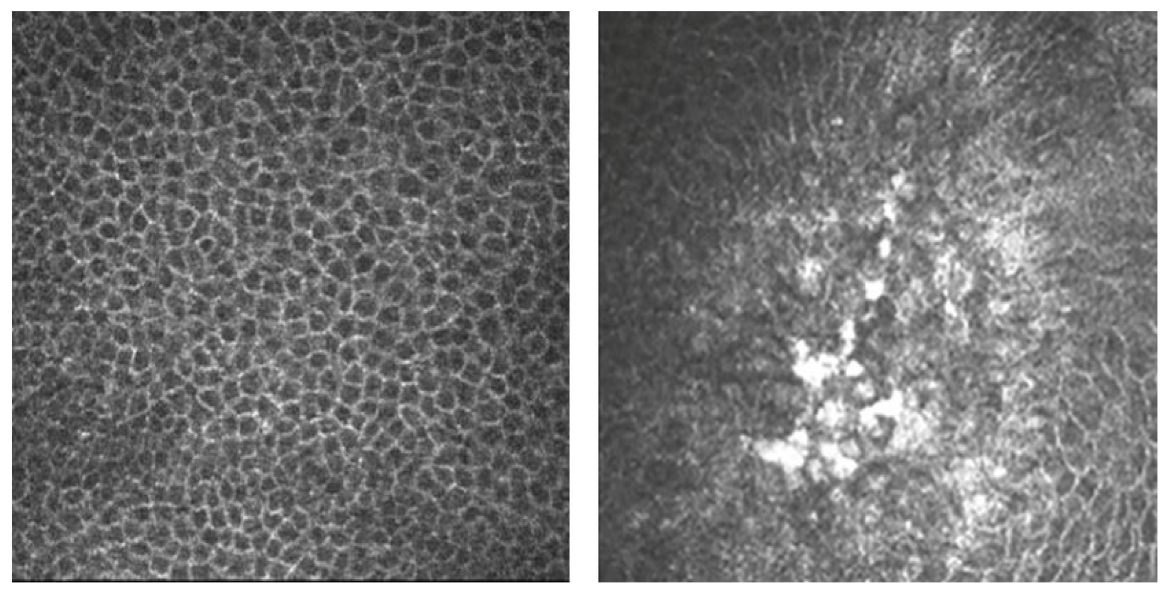
3.5. In Vivo Confocal Microscopy

IVCM is a non-invasive optical device that, compared with conventional microscopy, provides high-resolution en face imaging of the entire cornea, and enables exploring and differentiating every single layer during the same examination [17]. The first confocal microscope was invented by Minsky in 1955 [45]. The original prototype was the “double focusing stage scanning microscope” which allowed observation of specimens mounted onto a stage electrically moved by a tuning fork. This solution provided an adequate field of observation with the movement of the stage with respect to the optical system. More than 10 years later, Petran et al. developed the first tandem scanning confocal microscope (TSCM) [46]. The new design allowed the simultaneous scanning of multiple points on a fixed specimen using a rotating Nipkow disc. Lemp et al., almost 20 years later, used the TSCM to examine ocular tissue, obtaining the first full-thickness image of a human cornea ex vivo [47]. Subsequently, the clinical version of this microscope was developed by the Tandem Scanning Corporation (Reston, VA, USA) and upgraded by the Advanced Scanning Corporation (New Orleans, LA, USA) but is no longer in production [48]. In parallel, Svishchev, in 1969, developed the slit scanning confocal microscope (SSCM) consisting of an oscillating two-sided mirror for the simultaneous scanning and de-scanning of the sample [49]. Nowadays, SSCM commercially available devices include: Confoscan P4 (Tomey Corporation, Cambridge, MA, USA), Confoscan 4 (Nidek Technologies, Gamagori, Japan), and Microphthal (Helmut Hund, Wetzlar, Germany).
The most recent and diffuse IVCM device available on the market is the Heidelberg Retina Tomograph Rostock Corneal Module (RCM), a laser scanner that utilizes a 670 nm red wavelength Helium Neon diode laser source. Each acquired image represents 400 μm × 400 μm of the cornea with an optical lateral resolution as low as 2 μm and an axial resolution as low as 4 μm [18]. There are three image-acquisition modes available with the RCM. The “section” mode allows acquisition of a single image at a time so that the cornea can be scanned manually in the X–Y–Z axes and images can be collected by pressing a foot pedal when desired. The Z-axis can be scanned with a depth range from 0 to 1500 μm. The “volume” mode enables the acquisition of 40 consecutive images on focal planes approximately 2 μm apart. The “sequence” mode allows the acquisition of 100 images resulting in a 3 to 100 s movie sequence. During each acquisition, the lens can be either manually adjusted or remain stationary.

3.6. In Vivo Confocal Microscopy Clinical Applications

The normal human cornea consists of five layers: epithelium, Bowman’s layer, stroma, Descemet’s membrane, and endothelium. With the only exception of Descemet’s membrane, all these layers can be observed by IVCM and the resolution allows imaging of corneal cells and their nuclei [18].
The corneal epithelium consists of superficial epithelial cells, wing cells, and basal epithelial cells. Superficial epithelial cells are 20–30 µm in extent and approximately 5 µm thick [50]; they have a typical polygonal shape with various sizes and reflectivities and visible cell nuclei surrounded by a dark band [51]. Wing cells can be recognized by bright borders and a bright cell nucleus but not surrounded by the dark ring [50]. Epithelial cells are typically 10–15 µm in diameter and organized in a regular mosaic with dark cell bodies and bright cell borders [52]. The sub-basal corneal nerves are situated underneath the basal epithelium. The nerve bundles consist of straight and beaded fibers arranged to form a whorl-like pattern [53]. Bowman’s layer appears as a bright homogenous layer [51]. Corneal stroma IVCM can identify stromal keratocytes as well-defined bright, oval-round objects with varying orientations in the anterior stroma [54]. The mid-stroma population is more regular and oval-shaped, in contrast with posterior stromal keratocytes that appear more spindle-shaped [55]. HSV diagnosis with current clinical diagnostic tools often depends greatly on subjective judgment after examination with a slit lamp; however, IVCM corneal imaging can offer reduced bias and lower subjective variability, producing high-resolution images of the affected area [56].
In 2015, Yokugawa et al. examined 4 patients affected by HSV keratitis with IVCM [57]. The authors studied the most common presentation of HSK: the dendritic lesion of infectious epithelial keratitis, which may occur as a primary HSV infection but is more commonly associated with the reactivation of a latent virus. Dendritic lesions are characterized by linear lesions with branching and swollen terminal bulbs, all of which can be well visualized with slit lamp biomicroscopy and fluorescein staining. The IVCM epithelial changes of dendritic lesions, as well as other corneal layers, were examined using a promising mapping technique. As a result, large images of the dendritic lesions were obtained and, in all cases, they appeared as hyperreflective irregular epithelial cells, surrounded by elongated epithelial cells [57]. Figure 2 shows IVCM images of swollen and necrotic epithelial cells during active herpes simplex keratitis.
The authors, using histopathologic sections of HSV-infected specimens as a term of comparison, hypothesized that these cells may represent HSV-infected cells, such as multinucleated giant cells and cells distorted by intranuclear inclusions. The frequently observed hyperreflective deposits within the dendritic lesion, especially at the branching point, probably represent infected necrotic abnormal cells. Furthermore, the authors found Langerhans cells, a type of inflammatory cell, as dendritic structures within the epithelium and at the level of the nerve plexus [57]. Figure 3 shows IVCM images of Langerhans cells as dendritic inflammatory cells during active herpes simplex keratitis.
These results are consistent with previous studies, showing the importance of confocal imaging in detecting cellular-level changes [58,59]. In another prospective cross-sectional study conducted on 21 patients, Mocan et al. reported a detailed IVCM observation of HSV keratitis [60]. The confocal images obtained from the corneal sections showed an extensive inflammatory response observed as a heavy infiltration of the anterior stroma adjacent to Bowman’s layer, many dendritic cells concentrated in the basal epithelial layers, and an endothelial layer altered by keratic precipitates. Figure 4 shows IVCM images of inflammatory cells observed at the stromal level during active herpetic keratitis.
Another main finding was the loss of the sub-basal nerves in HSV-infected corneas as well as a significant loss of endothelial cells. In particular, the sub-basal nerve plexus was absent in 47.6% of cases and reduced in the remaining 42.9%, providing evidence and support for sub-basal nerve damage in HSV keratitis. Furthermore, intraepithelial inflammatory cell infiltration was observed in 52.4% of all cases as hyperreflective dendritiform and small round cells [60]. Dendritiform cells in the basal epithelium are believed to be antigen-presenting cells including either dendritic cells or Langerhans cells [61]. Rosenberg et al., in another IVCM study, demonstrated the persistence of dendritiform cells in the basal epithelium after the resolution of HSV keratitis in 62.5% of corneas [59]. However, dendritiform cells do not appear in all cases of active HSV keratitis, suggesting that these cells may not play a main role in the immunopathogenesis of herpetic keratitis. Nonetheless, imaging of a very small and central corneal section must be considered as a potential sampling limitation. In another prospective cross-sectional study on 31 eyes with HSV keratitis, Hamrah et al. analyzed the innervation alterations in HSV keratitis compared to healthy controls [62]. The authors showed that in HSV-affected eyes, mean nerve density, total nerve number, main nerve trunks, and number of branches were significantly lower as compared to controls. Changes in nerve density and morphology were noted within days after the onset of the disease. Interestingly, the nerve alterations analysis between acute and chronic HSV keratitis showed no statistical difference, probably indicating that the changes noted in patients with active HSV keratitis are induced during the acute phase and may persist for a long time. From a purely diagnostic point of view, Wang et al. assessed the real IVCM capabilities by measuring specificity and sensitivity in detecting various infectious keratitis entities, including Acanthamoeba keratitis, fungal keratitis, bacterial keratitis, and HSV viral keratitis [63]. The confocal images from each eye were reviewed by two experienced confocal microscopists in a masked and independent fashion. The sensitivity and specificity of HSV keratitis were found to be 100% (95% CI 46.3–100%) and 93.2% (95% CI 80.3–98.2%), respectively. Despite this excellent outcome, the result is likely biased due to a small sample size: only 4 patients out of 46 were affected by HSV keratitis; in addition, 3 false-positive cases were reported. The clinical characteristics of eyes in studies analyzing the cornea using IVCM are shown in Table 2.

4. Discussion

Currently, among the available diagnostic techniques for HSV, PCR is the most diffuse diagnostic tool since it offers the shortest detection time. PCR has been adopted as an HSV detection technique over the past few decades, and at the moment, numerous studies exist about the evaluation of its efficacy [56]. PCR can be performed on various specimens, such as corneal scrapings, tear samples, and corneal impression membranes, with a slight sensitivity variability between methods [64,65]. One of the possible flaws of using PCR in clinical settings is its inability to discriminate between viable and non-viable viral genetic material [66,67,68], bearing potential implications in the clinical application of PCR, in particular evaluating treatment regimen efficacy. Furthermore, PCR results may be affected by previous treatment with anti-HSV medication [69]. The clinical use of optical devices can help in overcoming such limitations, but the utility of AS-OCT, despite its imaging abilities, remains severely affected by the lack of identifiable pathognomonic features for HSV keratitis. This imaging method can visualize several morphological patterns in HSV-infected eyes, but none of these are specific; hence, AS-OCT can be used to provide additional information over the course of the disease, but it is not recommended as a primary diagnostic approach for HSV keratitis.
However, in several other clinical fields, AS-OCT has shown great diagnostic capabilities, and one of the most promising future applications is AS-OCT angiography, which has already proven distinct advantages over traditional fluorescein angiography and indocyanine green angiography, making it a rising alternative for clinical practitioners [70]. On the other hand, IVCM allows the detection of immune cells and structures in the healthy and diseased cornea. The distribution, density, and quality of these elements can be determined in a non-invasive fashion. Serial imaging of the same tissue over time allows clinicians to follow the progression of the disease and monitor the effects of therapy [71]. Therefore, IVCM offers a non-invasive alternative increasingly used in clinical settings for the management of HSV keratitis. One major IVCM shortcoming is its limited resolution [72], since viral particles are very difficult to detect, and the diagnostic criteria for HSV keratitis can only rely on corneal histology changes. Despite the undeniable utility of IVCM, a definitive diagnosis via other techniques is still necessary, also considering the limited diffusion of this device due to its expensive and time-consuming nature. In the near future, advances in optical and digital imaging technology may lead to better visualization of viral particles, giving optical devices the opportunity to reveal their full diagnostic potential.

Author Contributions

Conceptualization, F.D.S., D.R. and S.A.; methodology, F.D.S., D.R., M.D.P., S.D.S. and S.A.; software, F.D.S., M.D.P. and S.D.S.; validation, F.D.S., D.R., M.D.P., S.D.S. and S.A.; formal analysis, F.D.S., D.R., M.D.P., S.D.S. and S.A.; investigation, F.D.S., D.R., M.D.P. and S.D.S.; data curation, D.R., M.D.P. and S.D.S.; writing—original draft preparation, F.D.S., D.R., M.D.P., S.D.S. and S.A.; writing—review and editing, F.D.S. and S.A.; supervision, S.A. All authors have read and agreed to the published version of the manuscript.

Funding

This research received no external funding.

Institutional Review Board Statement

Not applicable.

Informed Consent Statement

Not applicable.

Data Availability Statement

Not applicable.

Conflicts of Interest

The authors declare no conflict of interest.

References

  1. Liesegang, T.J. Herpes Simplex Virus Epidemiology and Ocular Importance. Cornea 2001, 20, 1–13. [Google Scholar] [CrossRef] [PubMed]
  2. McQuillan, G.; Kruszon-Moran, D.; Flagg, E.W.; Paulose-Ram, R. Prevalence of Herpes Simplex Virus Type 1 and Type 2 in Persons Aged 14–49: United States, 2015–2016; NCHS Data Brief: Hyattsville, MD, USA, 2018; pp. 1–8. [Google Scholar]
  3. Farooq, A.V.; Shukla, D. Herpes Simplex Epithelial and Stromal Keratitis: An Epidemiologic Update. Surv. Ophthalmol. 2012, 57, 448–462. [Google Scholar] [CrossRef] [PubMed] [Green Version]
  4. Borkar, D.S.; Gonzales, J.A.; Tham, V.M.; Esterberg, E.; Vinoya, A.C.; Parker, J.V.; Uchida, A.; Acharya, N.R. Association between Atopy and Herpetic Eye Disease: Results from the Pacific Ocular Inflammation Study. JAMA Ophthalmol. 2014, 132, 326–331. [Google Scholar] [CrossRef] [PubMed] [Green Version]
  5. Gonzalez-Gonzalez, L.A.; Molina-Prat, N.; Doctor, P.; Tauber, J.; Sainz de la Maza, M.T.; Foster, C.S. Clinical Features and Presentation of Infectious Scleritis from Herpes Viruses: A Report of 35 Cases. Ophthalmology 2012, 119, 1460–1464. [Google Scholar] [CrossRef] [PubMed]
  6. Roozbahani, M.; Hammersmith, K.M. Management of Herpes Simplex Virus Epithelial Keratitis. Curr. Opin. Ophthalmol. 2018, 29, 360–364. [Google Scholar] [CrossRef]
  7. Arshad, S.; Petsoglou, C.; Lee, T.; Al-Tamimi, A.; Carnt, N.A. 20 Years since the Herpetic Eye Disease Study: Lessons, Developments and Applications to Clinical Practice. Clin. Exp. Optom. 2021, 104, 396–405. [Google Scholar] [CrossRef] [PubMed]
  8. Rowe, A.M.; St Leger, A.J.; Jeon, S.; Dhaliwal, D.K.; Knickelbein, J.E.; Hendricks, R.L. Herpes Keratitis. Prog. Retin. Eye Res. 2013, 32, 88–101. [Google Scholar] [CrossRef] [PubMed]
  9. Liesegang, T.J. Classification of Herpes Simplex Virus Keratitis and Anterior Uveitis. Cornea 1999, 18, 127–143. [Google Scholar] [CrossRef] [PubMed]
  10. Giménez, F.; Suryawanshi, A.; Rouse, B.T. Pathogenesis of Herpes Stromal Keratitis—A Focus on Corneal Neovascularization. Prog. Retin. Eye Res. 2013, 33, 1–9. [Google Scholar] [CrossRef] [PubMed] [Green Version]
  11. Suzuki, T.; Ohashi, Y. Corneal Endotheliitis. Semin. Ophthalmol. 2008, 23, 235–240. [Google Scholar] [CrossRef]
  12. Azher, T.N.; Yin, X.-T.; Tajfirouz, D.; Huang, A.J.; Stuart, P.M. Herpes Simplex Keratitis: Challenges in Diagnosis and Clinical Management. Clin. Ophthalmol. 2017, 11, 185–191. [Google Scholar] [CrossRef] [PubMed] [Green Version]
  13. Carnt, N.; Robaei, D.; Minassian, D.C.; Dart, J.K.G. Acanthamoeba Keratitis in 194 Patients: Risk Factors for Bad Outcomes and Severe Inflammatory Complications. Br. J. Ophthalmol. 2018, 102, 1431–1435. [Google Scholar] [CrossRef] [PubMed]
  14. Subhan, S.; Jose, R.J.; Duggirala, A.; Hari, R.; Krishna, P.; Reddy, S.; Sharma, S. Diagnosis of Herpes Simplex Virus-1 Keratitis: Comparison of Giemsa Stain, Immunofluorescence Assay and Polymerase Chain Reaction. Curr. Eye Res. 2004, 29, 209–213. [Google Scholar] [CrossRef] [PubMed]
  15. Huang, D.; Swanson, E.A.; Lin, C.P.; Schuman, J.S.; Stinson, W.G.; Chang, W.; Hee, M.R.; Flotte, T.; Gregory, K.; Puliafito, C.A.; et al. Optical Coherence Tomography. Science 1991, 254, 1178–1181. [Google Scholar] [CrossRef] [PubMed] [Green Version]
  16. Stachs, O.; Guthoff, R.F.; Aumann, S. In Vivo Confocal Scanning Laser Microscopy. In High Resolution Imaging in Microscopy and Ophthalmology: New Frontiers in Biomedical Optics; Bille, J.F., Ed.; Springer: Cham, Switzerland, 2019; ISBN 978-3-030-16637-3. [Google Scholar]
  17. Petroll, W.M.; Robertson, D.M. In Vivo Confocal Microscopy of the Cornea: New Developments in Image Acquisition, Reconstruction, and Analysis Using the HRT-Rostock Corneal Module. Ocul. Surf. 2015, 13, 187–203. [Google Scholar] [CrossRef] [Green Version]
  18. Patel, D.V.; McGhee, C.N.J. Contemporary in Vivo Confocal Microscopy of the Living Human Cornea Using White Light and Laser Scanning Techniques: A Major Review. Clin. Exp. Ophthalmol. 2007, 35, 71–88. [Google Scholar] [CrossRef]
  19. Darougar, S.; Wishart, M.S.; Viswalingam, N.D. Epidemiological and Clinical Features of Primary Herpes Simplex Virus Ocular Infection. Br. J. Ophthalmol. 1985, 69, 2–6. [Google Scholar] [CrossRef] [Green Version]
  20. Shoji, J.; Sakimoto, T.; Inada, N.; Kamei, Y.; Matsubara, M.; Takamura, E.; Sawa, M. A Diagnostic Method for Herpes Simplex Keratitis by Simultaneous Measurement of Viral DNA and Virus-Specific Secretory IgA in Tears: An Evaluation. Jpn. J. Ophthalmol. 2016, 60, 294–301. [Google Scholar] [CrossRef]
  21. Austin, A.; Lietman, T.; Rose-Nussbaumer, J. Update on the Management of Infectious Keratitis. Ophthalmology 2017, 124, 1678–1689. [Google Scholar] [CrossRef]
  22. Chang, E.J.; Dreyer, E.B. Herpesvirus Infections of the Anterior Segment. Int. Ophthalmol. Clin. 1996, 36, 17–28. [Google Scholar] [CrossRef]
  23. Tabery, H.M. Epithelial Changes in Early Primary Herpes Simplex Virus Keratitis. Photomicrographic Observations in a Case of Human Infection. Acta Ophthalmol. Scand. 2000, 78, 706–709. [Google Scholar] [CrossRef] [PubMed]
  24. Koizumi, N.; Nishida, K.; Adachi, W.; Tei, M.; Honma, Y.; Dota, A.; Sotozono, C.; Yokoi, N.; Yamamoto, S.; Kinoshita, S. Detection of Herpes Simplex Virus DNA in Atypical Epithelial Keratitis Using Polymerase Chain Reaction. Br. J. Ophthalmol. 1999, 83, 957–960. [Google Scholar] [CrossRef] [PubMed]
  25. El-Aal, A.M.A.; Sayed, M.E.; Mohammed, E.; Ahmed, M.; Fathy, M. Evaluation of Herpes Simplex Detection in Corneal Scrapings by Three Molecular Methods. Curr. Microbiol. 2006, 52, 379–382. [Google Scholar] [CrossRef] [PubMed]
  26. Satpathy, G.; Mishra, A.K.; Tandon, R.; Sharma, M.K.; Sharma, A.; Nayak, N.; Titiyal, J.S.; Sharma, N. Evaluation of Tear Samples for Herpes Simplex Virus 1 (HSV) Detection in Suspected Cases of Viral Keratitis Using PCR Assay and Conventional Laboratory Diagnostic Tools. Br. J. Ophthalmol. 2011, 95, 415–418. [Google Scholar] [CrossRef] [Green Version]
  27. Reidy, J.J.; American Academy of Ophthalmology. External Disease and Cornea; American Academy of Ophthalmology: San Francisco, CA, USA, 2011. [Google Scholar]
  28. Kaye, S.; Choudhary, A. Herpes Simplex Keratitis. Prog. Retin. Eye Res. 2006, 25, 355–380. [Google Scholar] [CrossRef]
  29. Olsen, T.W.; Hardten, D.R.; Meiusi, R.S.; Holland, E.J. Linear Endotheliitis. Am. J. Ophthalmol. 1994, 117, 468–474. [Google Scholar] [CrossRef]
  30. McGilligan, V.; Moore, J.E.; Tallouzi, M.; Atkinson, S.D.; O’Neill, H.; Feeney, S.; Novitskaya, E.S.; Sharma, A.; Shah, S.; Jackson, J.A.; et al. A Comparison of the Clinical and Molecular Diagnosis of Herpes Simplex Keratitis. Open J. Ophthalmol. 2014, 4, 65–74. [Google Scholar] [CrossRef] [Green Version]
  31. Wilhelmus, K.R. Antiviral Treatment and Other Therapeutic Interventions for Herpes Simplex Virus Epithelial Keratitis. Cochrane Database Syst. Rev. 2015, 1, CD002898. [Google Scholar] [CrossRef]
  32. Kowalski, R.P.; Gordon, Y.J.; Romanowski, E.G.; Araullo-Cruz, T.; Kinchington, P.R. A Comparison of Enzyme Immunoassay and Polymerase Chain Reaction with the Clinical Examination for Diagnosing Ocular Herpetic Disease. Ophthalmology 1993, 100, 530–533. [Google Scholar] [CrossRef]
  33. Ramos, J.L.B.; Li, Y.; Huang, D. Clinical and Research Applications of Anterior Segment Optical Coherence Tomography—A Review. Clin. Exp. Ophthalmol. 2009, 37, 81–89. [Google Scholar] [CrossRef] [Green Version]
  34. Han, S.B.; Liu, Y.-C.; Noriega, K.M.; Mehta, J.S. Applications of Anterior Segment Optical Coherence Tomography in Cornea and Ocular Surface Diseases. J. Ophthalmol. 2016, 2016, 4971572. [Google Scholar] [CrossRef] [PubMed] [Green Version]
  35. van Velthoven, M.E.J.; Faber, D.J.; Verbraak, F.D.; van Leeuwen, T.G.; de Smet, M.D. Recent Developments in Optical Coherence Tomography for Imaging the Retina. Prog. Retin. Eye Res. 2007, 26, 57–77. [Google Scholar] [CrossRef] [PubMed]
  36. Jancevski, M.; Foster, C.S. Anterior Segment Optical Coherence Tomography. Semin. Ophthalmol. 2010, 25, 317–323. [Google Scholar] [CrossRef] [PubMed]
  37. Wang, C.; Xia, X.; Tian, B.; Zhou, S. Comparison of Fourier-Domain and Time-Domain Optical Coherence Tomography in the Measurement of Thinnest Corneal Thickness in Keratoconus. J. Ophthalmol. 2015, 2015, 402925. [Google Scholar] [CrossRef] [Green Version]
  38. Choma, M.; Sarunic, M.; Yang, C.; Izatt, J. Sensitivity Advantage of Swept Source and Fourier Domain Optical Coherence Tomography. Opt. Express 2003, 11, 2183. [Google Scholar] [CrossRef] [Green Version]
  39. Wang, S.B.; Cornish, E.E.; Grigg, J.R.; McCluskey, P.J. Anterior Segment Optical Coherence Tomography and Its Clinical Applications. Clin. Exp. Optom. 2019, 102, 195–207. [Google Scholar] [CrossRef] [Green Version]
  40. Gupta, N.; Varshney, A.; Ramappa, M.; Basu, S.; Romano, V.; Acharya, M.; Gaur, A.; Kapur, N.; Singh, A.; Shah, G.; et al. Role of AS-OCT in Managing Corneal Disorders. Diagnostics 2022, 12, 918. [Google Scholar] [CrossRef]
  41. Soliman, W.; Nassr, M.A.; Abdelazeem, K.; Al-Hussaini, A.K. Appearance of Herpes Simplex Keratitis on Anterior Segment Optical Coherence Tomography. Int. Ophthalmol. 2019, 39, 2923–2928. [Google Scholar] [CrossRef]
  42. Park, Y.M.; Lee, J.S.; Yoo, J.-M.; Park, J.M.; Seo, S.-W.; Chung, I.-Y.; Kim, S.J. Comparison of Anterior Segment Optical Coherence Tomography Findings in Acanthamoeba Keratitis and Herpetic Epithelial Keratitis. Int. J. Ophthalmol. 2018, 11, 1416–1420. [Google Scholar] [CrossRef]
  43. Hixson, A.; Blanc, S.; Sowka, J. Monitoring Keratitis Resolution with Optical Coherence Tomography. Optom. Vis. Sci. 2014, 91, S40–S45. [Google Scholar] [CrossRef]
  44. Lu, L.; Palioura, S. Management of Stromal Herpes Simplex Virus Keratitis With Epithelial Ulceration Using Optical Coherence Tomography-Generated Corneal Thickness Maps. Cornea 2020, 39, 1566–1570. [Google Scholar] [CrossRef] [PubMed]
  45. Minsky, M. Memoir on Inventing the Confocal Scanning Microscope. Scanning 1988, 10, 128–138. [Google Scholar] [CrossRef]
  46. Petráň, M.; Hadravský, M.; Egger, M.D.; Galambos, R. Tandem-Scanning Reflected-Light Microscope*. J. Opt. Soc. Am. 1968, 58, 661. [Google Scholar] [CrossRef]
  47. Lemp, M.A.; Dilly, P.N.; Boyde, A. Tandem-Scanning (Confocal) Microscopy of the Full-Thickness Cornea. Cornea 1985, 4, 205–209. [Google Scholar] [CrossRef]
  48. McLaren, J.W.; Nau, C.B.; Kitzmann, A.S.; Bourne, W.M. Keratocyte Density: Comparison of Two Confocal Microscopes. Eye Contact Lens. 2005, 31, 28–33. [Google Scholar] [CrossRef] [PubMed]
  49. Svishchev, G. Microscope for the Study of Transparent Light-Scattering Objects in Incident Light. Opt. Spectrosc. 1969, 30, 188–191. [Google Scholar]
  50. Masters, B.R.; Thaer, A.A. In-Vivo Real-Time Confocal Microscopy of the Human Cornea. In Proceedings of the Ophthalmic Technologies III, Los Angeles, CA, USA, 24 June 1993; Volume 1877, pp. 100–121. [Google Scholar]
  51. Mustonen, R.K.; McDonald, M.B.; Srivannaboon, S.; Tan, A.L.; Doubrava, M.W.; Kim, C.K. Normal Human Corneal Cell Populations Evaluated by in Vivo Scanning Slit Confocal Microscopy. Cornea 1998, 17, 485–492. [Google Scholar] [CrossRef]
  52. Harrison, D.A.; Joos, C.; Ambrósio, R. Morphology of Corneal Basal Epithelial Cells by in Vivo Slit-Scanning Confocal Microscopy. Cornea 2003, 22, 246–248. [Google Scholar] [CrossRef]
  53. Matsuda, H. Electron Microscopic Study on the Corneal Nerve with Special Reference to Its Endings. Jpn. J. Ophthalmol. 1968, 12, 163–173. [Google Scholar]
  54. Popper, M.; Morgado, A.M.; Quadrado, M.J.; Best, J.A. van Corneal Cell Density Measurement in Vivo by Scanning Slit Confocal Microscopy: Method and Validation. ORE 2004, 36, 270–276. [Google Scholar] [CrossRef]
  55. Hollingsworth, J.; Perez-Gomez, I.; Mutalib, H.A.; Efron, N. A Population Study of the Normal Cornea Using an in Vivo, Slit-Scanning Confocal Microscope. Optom. Vis. Sci. 2001, 78, 706–711. [Google Scholar] [CrossRef] [PubMed] [Green Version]
  56. Poon, S.H.L.; Wong, W.H.L.; Lo, A.C.Y.; Yuan, H.; Chen, C.-F.; Jhanji, V.; Chan, Y.K.; Shih, K.C. A Systematic Review on Advances in Diagnostics for Herpes Simplex Keratitis. Surv. Ophthalmol. 2021, 66, 514–530. [Google Scholar] [CrossRef] [PubMed]
  57. Yokogawa, H.; Kobayashi, A.; Mori, N.; Sugiyama, K. Mapping of Dendritic Lesions in Patients with Herpes Simplex Keratitis Using in Vivo Confocal Microscopy. Clin. Ophthalmol. 2015, 9, 1771–1777. [Google Scholar] [CrossRef] [Green Version]
  58. Nagasato, D.; Araki-Sasaki, K.; Kojima, T.; Ideta, R.; Dogru, M. Morphological Changes of Corneal Subepithelial Nerve Plexus in Different Types of Herpetic Keratitis. Jpn. J. Ophthalmol. 2011, 55, 444–450. [Google Scholar] [CrossRef]
  59. Rosenberg, M.E.; Tervo, T.M.T.; Müller, L.J.; Moilanen, J.A.O.; Vesaluoma, M.H. In Vivo Confocal Microscopy after Herpes Keratitis. Cornea 2002, 21, 265–269. [Google Scholar] [CrossRef]
  60. Mocan, M.C.; Irkec, M.; Mikropoulos, D.G.; Bozkurt, B.; Orhan, M.; Konstas, A.G.P. In Vivo Confocal Microscopic Evaluation of the Inflammatory Response in Non-Epithelial Herpes Simplex Keratitis. Curr. Eye Res. 2012, 37, 1099–1106. [Google Scholar] [CrossRef] [PubMed]
  61. Zhivov, A.; Stave, J.; Vollmar, B.; Guthoff, R. In Vivo Confocal Microscopic Evaluation of Langerhans Cell Density and Distribution in the Corneal Epithelium of Healthy Volunteers and Contact Lens Wearers. Cornea 2007, 26, 47–54. [Google Scholar] [CrossRef] [PubMed]
  62. Hamrah, P.; Cruzat, A.; Dastjerdi, M.H.; Zheng, L.; Shahatit, B.M.; Bayhan, H.A.; Dana, R.; Pavan-Langston, D. Corneal Sensation and Subbasal Nerve Alterations in Patients with Herpes Simplex Keratitis: An in Vivo Confocal Microscopy Study. Ophthalmology 2010, 117, 1930–1936. [Google Scholar] [CrossRef] [Green Version]
  63. Wang, Y.E.; Tepelus, T.C.; Vickers, L.A.; Baghdasaryan, E.; Gui, W.; Huang, P.; Irvine, J.A.; Sadda, S.; Hsu, H.Y.; Lee, O.L. Role of in Vivo Confocal Microscopy in the Diagnosis of Infectious Keratitis. Int. Ophthalmol. 2019, 39, 2865–2874. [Google Scholar] [CrossRef]
  64. Fox, P.D.; Khaw, P.T.; McBride, B.W.; McGill, J.I.; Ward, K.A. Tear and Serum Antibody Levels in Ocular Herpetic Infection: Diagnostic Precision of Secretory IgA. Br. J. Ophthalmol. 1986, 70, 584–588. [Google Scholar] [CrossRef] [Green Version]
  65. Ma, J.-X.; Wang, L.-N.; Zhou, R.-X.; Yu, Y.; Du, T.-X. Real-Time Polymerase Chain Reaction for the Diagnosis of Necrotizing Herpes Stromal Keratitis. Int. J. Ophthalmol. 2016, 9, 682–686. [Google Scholar] [CrossRef]
  66. Graiver, D.A.; Saunders, S.E.; Topliff, C.L.; Kelling, C.L.; Bartelt-Hunt, S.L. Ethidium Monoazide Does Not Inhibit RT-PCR Amplification of Nonviable Avian Influenza RNA. J. Virol. Methods 2010, 164, 51–54. [Google Scholar] [CrossRef] [PubMed]
  67. Lee, N.; Chan, P.K.S.; Hui, D.S.C.; Rainer, T.H.; Wong, E.; Choi, K.-W.; Lui, G.C.Y.; Wong, B.C.K.; Wong, R.Y.K.; Lam, W.-Y.; et al. Viral Loads and Duration of Viral Shedding in Adult Patients Hospitalized with Influenza. J. Infect. Dis. 2009, 200, 492–500. [Google Scholar] [CrossRef] [PubMed] [Green Version]
  68. Mahlum, C.E.; Haugerud, S.; Shivers, J.L.; Rossow, K.D.; Goyal, S.M.; Collins, J.E.; Faaberg, K.S. Detection of Bovine Viral Diarrhea Virus by TaqMan Reverse Transcription Polymerase Chain Reaction. J. Vet. Diagn. Investig. 2002, 14, 120–125. [Google Scholar] [CrossRef] [PubMed]
  69. Inoue, Y.; Shimomura, Y.; Fukuda, M.; Miyazaki, D.; Ohashi, Y.; Sasaki, H.; Tagawa, Y.; Shiota, H.; Inada, N.; Okamoto, S.; et al. Multicentre Clinical Study of the Herpes Simplex Virus Immunochromatographic Assay Kit for the Diagnosis of Herpetic Epithelial Keratitis. Br. J. Ophthalmol. 2013, 97, 1108–1112. [Google Scholar] [CrossRef] [PubMed]
  70. Wen, Y.; Jiang, D.; Tang, K.; Chen, W. Current Clinical Applications of Anterior Segment Optical Coherence Tomography Angiography: A Review. Graefes Arch. Clin. Exp. Ophthalmol. 2023. [Google Scholar] [CrossRef]
  71. Patel, D.V.; Zhang, J.; McGhee, C.N.J. In Vivo Confocal Microscopy of the Inflamed Anterior Segment: A Review of Clinical and Research Applications. Clin. Exp. Ophthalmol. 2019, 47, 334–345. [Google Scholar] [CrossRef] [Green Version]
  72. Kumar, R.L.; Cruzat, A.; Hamrah, P. Current State of in Vivo Confocal Microscopy in Management of Microbial Keratitis. Semin. Ophthalmol. 2010, 25, 166–170. [Google Scholar] [CrossRef] [PubMed] [Green Version]
Figure 1. Corneal dendritic epithelial ulcers stained with fluorescein dye at slit lamp examination with a cobalt blue filter.
Photonics 10 00349 g001
Figure 2. In vivo confocal microscopy of a normal human cornea observed at the basal epithelium (left); swollen and necrotic epithelial cells during active herpes simplex keratitis (right).
Photonics 10 00349 g002
Figure 3. In vivo confocal microscopic images of a normal eye at the level of deep epithelium to sub-basal nerve plexus (left). Langerhans cells as dendritic inflammatory cells at the same level during active herpes simplex keratitis (right).
Photonics 10 00349 g003
Figure 4. In vivo confocal microscopy of a normal human cornea at the stromal level (left); inflammatory cells observed at the same level during active herpes keratitis (right).
Photonics 10 00349 g004
Table 1. Clinical characteristics of eyes in studies analyzing the cornea using anterior segment-OCT (AS-OCT).
AuthorsN EyesAS OCT Finding
Soliman et al. [41]11Stromal haze without epithelial defect
14Sub-epithelial haze, diffuse stromal infiltrates, corneal epithelium cystic spaces
10Hyper-reflective stroma associated with epithelial defects
3Stromal condensation with intact epithelium
3Stromal condensation and thinning with overlying epithelial defect
1Localized stromal and endothelial thickening.Epithelial heaping at the edge of epithelial defect
Young Ming Park et al. [42]3Subepithelial highly reflective lesions, corneal epithelial irregularity
Hixson et al. [43]2To locate and assess corneal thickness to manage the follow up and treatment of corneal edema
Louise et al. [44]2Pachymetric and corneal epithelial maps to assess stromal inflammation for diagnostic purposes and treatment follow up
Table 2. Clinical characteristics of eyes in studies analyzing the cornea using in vivo confocal microscopy (ICVM).
AuthorsN EyesICVM
Yokugawa et al. [57]4Hyperreflective irregular epithelial cells surrounded by elongated epithelial cells. Langerhans cells within the epithelium and nerve plexus
Rosenberg et al. [59]10Highly reflective dendritic structures in the basal epithelium
Mocan et al. [60]11Infiltration of basal epithelial layers with inflammatory cells
20Stromal inflammation evidence: round-shaped, with hyperreflective nuclei, smaller than keratocytes, surrounded by microdeposits
18Keratic precipitates (KPs): in 11 eyes, small round KPs; in 9, globular KPs; in 8, dendritiform KPs
17Endothelial blebs as hyporeflective dark spaces between endothelial cells
19Alteration in the sub-basal plexus: attenuated in 9 eyes, not visible in 10 eyes
Hamrah et al. [62]32Lower mean nerve density, total nerve number, main nerve trunks, and number of branches
Disclaimer/Publisher’s Note: The statements, opinions and data contained in all publications are solely those of the individual author(s) and contributor(s) and not of MDPI and/or the editor(s). MDPI and/or the editor(s) disclaim responsibility for any injury to people or property resulting from any ideas, methods, instructions or products referred to in the content.

