Review

Blue Light and Eye Damage: A Review on the Impact of Digital Device Emissions

by
Vladimiro Hipólito
1,2 and
João M. P. Coelho
1,2,*
1
Departamento de Física, Faculdade de Ciências, Universidade de Lisboa, 1749-016 Lisboa, Portugal
2
Instituto de Biofísica e Engenharia Biomédica, Faculdade de Ciências, Universidade de Lisboa, 1749-016 Lisboa, Portugal
*
Author to whom correspondence should be addressed.
Photonics 2023, 10(5), 560; https://doi.org/10.3390/photonics10050560
Submission received: 17 February 2023 / Revised: 27 April 2023 / Accepted: 8 May 2023 / Published: 11 May 2023
(This article belongs to the Special Issue Optics of the Eye: From Visual Optics to Clinical Application)

Abstract

:
The pandemic and lockdown caused by COVID-19 accelerated digitalization. Personal digital devices, emitting high-energy light, namely in the blue wavelength, have raised concerns about possible harmful effects on users’ eyes. Scientific research history has shown a relationship between exposure to blue light and changes in ocular structures. The main goal of this review is to examine frequent and prolonged exposure to blue radiation from computers, tablets and smartphones and its consequences on vision and ocular structures. Bibliographic research was carried out on changes induced by blue light in ocular structures, the cornea, the crystalline lens and the retina based on the following scientific databases: BioOne Complete™; Google Scholar™; Paperity™; PubMed™; and ScienceOpen™. The most significant studies on blue light and ocular damage were selected and reviewed. The most relevant bibliographic data were analyzed and summarized and some gaps in the theme of blue light from digital devices were identified. The experimental need to acquire additional new data is suggested. The hypothesis that continued use of digital devices enriched with blue light may interfere with the biological tissues of the cornea, crystalline lens, or retina is not clarified in the available scientific evidence. Therefore, additional studies are needed to answer this problem.

1. Introduction

Eye exposure to artificial blue radiation has increased with the frequent use of digital devices. COVID-19 and mandatory confinement accelerated this trend, modifying work, studying and socialization habits. Smartphone users have increased worldwide by 70%, while laptop computer users have increased by 40% since the beginning of the pandemic [1]. Bahkir et al. reported that 94% of users increased their average screen time from 4.8 to 8.6 h a day during lockdown [2]. The Global Digital Report 2023 [3] reveals that users spend more time using devices online. Research reveals that internet users spend around 7 h a day on all devices. The time spent online has increased and the daily average has grown by around 4 min per day (+1.0%) compared to 2021.
The duality of blue radiation means that this light can have both a negative and positive impact on human eyes. Thus, according to the UNE EN/IEC 62471, the standard classification for photobiological safety, visible light to the human visual system is between 380 nm and 780 nm [4] and blue radiation is contained between the range of 380–495 nm, depending on the reference documents [5]. This radiation is relevant for adequate vision performance and for some physiological processes’ balance. Human vision and daily biological rhythms evolve with sunlight, the greatest natural blue radiation source with the shortest wavelength and highest frequency within the visible spectrum. When sunlight passes through the atmosphere, it causes an oscillation of the particles that scatter the light, proportionally to the speed of acceleration of these particles. In this interaction called scattering, light is completely absorbed and then re-emitted. The light is scattered by particles much smaller than its wavelength (Rayleigh’s phenomenon). When passing through the atmosphere, the particles oscillate much more towards blue frequencies than towards red frequencies, and this is the reason for the blue perception of the sky [6].
Digital devices that use LED technology, as well as other sources of artificial light, expose their users to digital artificial blue radiation daily. This can be potentially harmful to the human eye due to the proximity of the ultraviolet spectrum, namely to the retina, due to its higher-energy wavelengths and high potential to alter ocular tissues [7]. Light is detected by the human eye, which sends the information received by the brain through the visual pathways. Specialized photoreceptors of the retina cells, cones and rods, responsible for part of the formation of images, respond to different wavelengths. The S, M and L cones contain proteins with photosensitive photopigments (Figure 1) of maximum sensitivity in the blue (S cones), green (M cones) and red (L cones) regions, corresponding to peaks of around 420 nm, 530 nm and 560 nm, respectively [8].
In the retinal photoexcitation process, the electrical properties of photopigments are altered, triggering a biochemical process that informs the brain about light. At the end of retinal phototransduction, visual sensations occur, which are produced by impulses that reach regions of the primary visual cortex; however, these sensations are secondary to the primary functions of photoreceptors (cones and rods) that carry light information to retinal ganglion cells and the lateral geniculate body of the thalamus [8].
Rods, another type of photoreceptor cell in the retina, are responsible for black-and-white (or monochromatic) vision and are sensitive to light wavelengths of around 500 nm [9]. In the posterior eye segment, light radiation must be refracted by the transparent ocular structures. To reach the retina, blue light can functionally interfere with the cornea and lens, in addition to the ocular surface (tear film), aqueous and vitreous humor [10]. The cornea is located at the anterior chamber, and this is the first surface that light encounters in the human eyes. Corneal epithelial cells have an oxidative increase motivated by light, which triggers inflammation of this structure and may cause cell apoptosis, ocular inflammation and xerophthalmia [11].
The crystalline lens, a suspended structure in the posterior chamber just after the iris, absorbs wavelengths in the visible range of up to 420 nm, and the light reaching the retina is reduced by the pupil. Miosis (pupillary constriction) increases when the eye is exposed to blue light [12,13]. The crystalline lens has protective action on the retina, but this fact promotes a transparency decrease, changing its appearance and color and inducing cataract formation [14]. Sunlight exposure is considered a risk factor for cataracts due to ultraviolet radiation exposure. Several studies have shown that blue light can also induce the production of oxygen reactive species in lens epithelial cells, which can lead to early cataract development [15].
Light interaction on different ocular surfaces can directly interfere with various tissues, but blue radiation exposure also interferes with the circadian cycle and refractive development. Thus, exposure to environment light, with a high blue light concentration, may also have an advantage against the development and progression of myopia [16]. There is a correlation between myopia low incidence, short-term exposure to near vision and outdoor activities [17]. Regarding digital devices and outdoor activities, Rucker et al. suggested that sunlight is much richer in short-wavelength light than most artificial sources. They add that blue light reduces the eye’s axial length through the mechanism of dopamine release in the retina, which is more favorable for controlling the reactive growth of myopia or astigmatism [18]. The eye’s axial growth and consequent myopia progression are slower during the summer months when children and teenagers spend more time participating in outdoor activities [19]. This information is a theoretical basis for the hypothesized correlation between light exposure and the occurrence and development of myopia. Other experimental works have shown a potential link between light luminance and myopia. Models of myopia experimented in birds found that chicks exposed to high-intensity light (15,000 lx) had greater resistance to myopia development and exhibited slower myopia progression than chicks exposed to low-intensity light (500 lx) and that exposure to bright light can suppress the development of myopia [20,21].
This review summarizes the most recent evidence on frequent exposure to artificial blue light beyond daylight hours, major ocular changes and impacts on visual health.

1.1. What Is the Blue Light Emission from Digital Devices Impact?

While there is evidence that digital devices can increase eyestrain for long-term users, there is currently not enough evidence to say that blue light from digital devices contributes to the development of eye diseases, such as age-related macular degeneration. On the other hand, there are no publications on the effect of long-term exposure to blue light and consequent eye diseases in humans. There are only data on the effects of visible blue light irradiation on rat and monkey retinas. Based on the available evidence, the Association of Optometrists (AOP) concludes that there is insufficient evidence to support the claim that exposure to visible blue light from digital devices leads to eye pathologies and damage to eye health [22]. However, blue light is not all harmful. As schematized in Figure 2, it can be essentially divided into two ranges: blue-violet light (380–455 nm) and turquoise light (455–495 nm), and these can affect the ocular tissues quite differently [23]. Turquoise light is essential for synchronizing our biological rhythms (the circadian cycle). It helps to maintain and regulate memory, cognition, mood and hormonal balance [24]. It urges a scientific commitment to develop solutions to the potential risk of blue light exposure and it is necessary to know which wavelengths (Figure 2) help guide technological decisions and research.
Retinal damage, changes in the lens and cornea, dry-eye syndrome, digital eye fatigue, aging, sleep disorders and circadian rhythm are the most investigated topics. The present review has blue light as its central object. Recent concerns about this radiation impact on visual health and environment due to increased exposure to artificial sources of blue light (mainly from digital devices) are also relevant.

1.1.1. Retinal Impact

Animal models have shown that phototoxicity results in human degenerative pathologies, such as age-related macular degeneration (AMD). A recent model of blue LED-induced phototoxicity in rats was developed, causing damage to the outer layers of the retina [26]. Progressive reduction in retinal thickness was noted in these in vivo studies. The photoreceptor layer was the most affected, and in electroretinogram evaluation, a transient reduction in the amplitude of waves a and b was observed [27]. The progressive reduction in the cones and the involvement of retinal pigment epithelium cells around the injury perfectly circumscribes the damage to the outer layer of the retina [28].
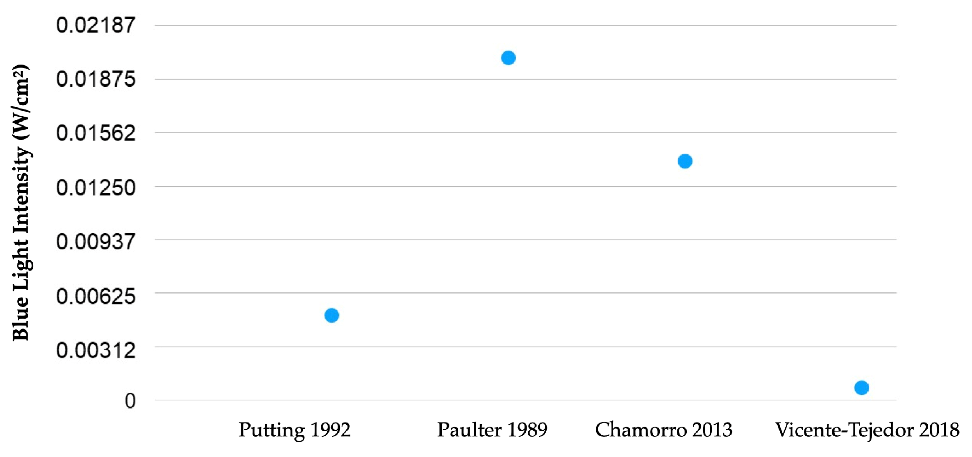
Many models of phototoxicity use white light due to its similarity to sunlight when studying the effect of blue light on the retina and retinal pigment epithelium (RPE) [27,29,30,31,32]. Knowing that lipofuscin accumulation intensifies aging in RPE, the relationship between phototoxicity and AMD supports the hypothesis of the potential risk of retinal phototoxicity in the elderly [33,34]. However, six of the eight most significant epidemiological studies found no correlation between AMD and light exposure over the users’ lifetime [35,36,37,38,39,40,41,42]. Figure 3 shows the blue light intensities’ radiance used in each referenced laboratory research study [43,44,45,46]. Only studies that used comparable units were included. Values represent the intensity used in each research study, which one can compare with the intensities produced by personal digital devices, such as those that can be found in the study by Gringas et al. (iPhone 5s, Kindle Paperwhite and iPad Air) [47]. Naturally, due to the fast evolution of these devices, future tests will always require updates on their emission spectra and intensity. Furthermore, the values can be useful for further studies about damage induced by blue light, and they are easily measured. Blue light irradiance (W/cm2) can be compared with peak spectral radiance (nm) or the ability of each source to stimulate different photopigments of the retina of a human eye, i.e., the capacity of each source to stimulate different photopigments to the S, M and L cones, rods and intrinsically photosensitive retinal ganglion cells [47]. No such comparison was found in the literature search considered in this article.
Compared to natural outdoor light, artificial light is often lower and has a different spectral distribution than sunlight. Outdoor light illuminance can reach values of up to 130,000 lux, depending on location, climate and elevation. Countries located on the equator receive a daily average of 7.7 kW/m2 of solar radiation between September and December [48]. In the CIE 62471 risk classification, irradiance is an important radiometric parameter to assess the level of harmful radiation produced by a light source [49]. CIE photobiological safety data, which compare the effective irradiance values of some common office light sources used for general lighting (LED, incandescent and halogen lamps) and digital devices (laptop and smartphone), indicate that the blue-light-effective radiance of general lighting was at least 20 times that of LED (cold-white, LW W5AP) and, in the case of the two electronic devices, 200,000 times less [49].
Damage induced by digital devices can be of a photothermal nature, which results from the increase in temperature induced by light in the retina, or of a photochemical nature induced by ambient light [50,51,52]. Photochemical damage to the retina is produced at wavelengths in the visible blue light range [53]. Many new devices have a greater intensity of light in blue wavelengths than conventional light sources [33,46]. Exposure to blue light from some of these light sources, more specifically from light-emitting diodes (LEDs), can damage the retina and produce other potentially harmful physiological changes [33,45,49,54,55,56,57]. Thus, the aim of this review is to explore the state of the art on whether exposure to blue light emitted by computers, tablets and smartphones can, when used frequently and for prolonged periods, can be harmful to the retina.

1.1.2. Impact on Crystalline Lens

The lens is composed of structural proteins, enzymes and metabolites that absorb the most energetic visible light. These substances produce yellow pigments that gradually cause the crystalline lens to opacify and turn yellow. This radiation absorption by the lens blocks and protects the retina from potential damage from blue light exposure [37]. Some studies have shown that blue light can induce the production of reactive oxygen species (ROS) in the mitochondria of lens epithelial cells, which can anticipate cataract occurrence [13,15].
The lens blocks most UV radiation between 300 nm and 420 nm and light transmission decreases with aging, with medical–surgical intervention being the only effective solution to treat cataracts, which is one of the main causes of blindness in the world [14,58,59,60]. In around 1980, specialists realized that the intraocular lens (IOL) could not only provide major optical power (in diopters), but it could also filter short light waves, reducing the risks of retinal damage. Consequently, most IOLs used in cataract surgery have incorporated UV-blocking filters since 1986 [61]. Blue light loss is permanent for pseudophakes with blue-blocking IOLs, and despite the lack of evidence, blue light hazard is often discussed [62].
The hypothesis of phototoxicity by exposure to light has increased concern and interest in blocking, in addition to ultraviolet radiation, part of visible light, by causing or accelerating age-related macular degeneration (AMD) [63,64,65,66,67,68,69,70,71,72].
Carotenoids found in the lens, such as lutein and zeaxanthin, are effective in absorbing blue light due to their antioxidant characteristics [73]. In the oxidative stress of the lens, antioxidants provide greater protection [74].

1.1.3. Impact on the Cornea and Ocular Surface

Some articles have shown that after exposure to blue radiation, the survival rate of corneal epithelial cells decreases [75]. The oxidative increase in corneal epithelial cells triggers inflammation and causes oxidative damage and, consequently, cell apoptosis and formation of xerophthalmia [11].
The cornea, aqueous humor and vitreous are the ocular refractive media, permeable to wavelengths between 300 nm and 400 nm. UVA radiation causes damage to the basal layer of keratinocytes, which is responsible for the occurrence of most skin tumors. Sunburn, photokeratitis, cataracts and retinal damage are common consequences of UV-B exposure rays (290 nm to 320 nm) [76]. Digital device users with digital eye strain (DES) experience symptoms such as eye irritation, burning, tiredness and redness, dryness, blurred vision and double vision. During digital devices’ prolonged use, a significant proportion of users (40–60%) experience visual or ocular symptoms. Most blue-light-blocking lenses used to reduce DES symptoms have not shown to be effective in reducing visual symptomatology, such as reading a task on a computer for 30 min [77].
Other studies have investigated the relationship between the use of digital monitors and changes in the ocular surface. Tear film break-up time (BUT), tear film volume (tear film meniscus and Schirmer’s test) and the lipid layer of the tear film state were quantified. Cardona et al. verified several changes in the tear film. Twenty-five healthy young adults were exposed to 20 min of video games. The lacrimal meniscus decreased, the time and area of tear rupture increased, and the interference patterns of the lipid layer were also altered after the game [78]. Another article studied changes in tear film over an 8-h workday in computer users [79]. The results did not demonstrate significant changes in Schirmer’s test, but BUT (tear break-up time test) decreased after screen exposure [80]. Reading on a computer, the conjunctival surface presents greater hyperemia compared to reading on a smartphone. When using a smartphone, a smaller extent of the exposed ocular surface was identified, due to convergence, in comparison with a computer [81,82]. Most results suggest that there is a relationship between deterioration in tear film quality and the use of digital devices.

2. Materials and Methods

Articles were compiled by searching the terms “blue light”, “eye damage”, “retinal damage”, “lens damage” and “corneal damage” in BioOne Complete™, Google Scholar™, Paperity™, PubMed™ and ScienceOpen™. The latest searches were conducted on 1 February 2023. Each article was reviewed and the most relevant to the hypothesis that blue light from personal digital devices exposure is harmful to users’ eyes were selected. The main keywords used were “digital devices”, “blue light”, “retina”, “crystalline lens”, cornea” and “damage”, with several combinations between them. The selected articles were reviewed and included for a new narrative review and to verify if the information was pertinent enough to discuss the association between blue light, digital devices and the consequent changes in ocular structures. References within articles were also reviewed and included. An assessment of the need for additional data was also made after verifying the articles’ data, to check the hypothesis that blue light present in digital devices could damage the cornea, crystalline lens and retina.

3. Results

The strategy used in the literature and epidemiological data source search was carried out according to the Preferred Reporting Items for Systematic Reviews and Meta-Analyses (PRISMA) procedures. The review process, as well as the steps for implementing the systematic review, included answering the clinical question to perform a detailed and comprehensive literature search. Several electronic scientific databases, such as BioOne Complete™, Google Scholar™, Paperity™, PubMed™ and ScienceOpen™, MEDLINE/PubMed, Web of Science and Scopus, were used with the terminology “blue light” and “eye damage” to identify all potential publications with relevant information on the impact of blue light on ocular structures. No range of dates for conducting and publishing the studies was defined, and for each publication or article found, the list of references was reviewed to find additional data to gather all the most relevant information. Initially, 4237 articles were identified, reviewed and extracted based on the author’s name, title, year of study and publication format (poster, academic thesis, dissertation or scientific publication). At the screening process conclusion, 95 articles that had an association between blue light, vision and ocular structures, namely the ocular surface, cornea, lens and retina, were used.
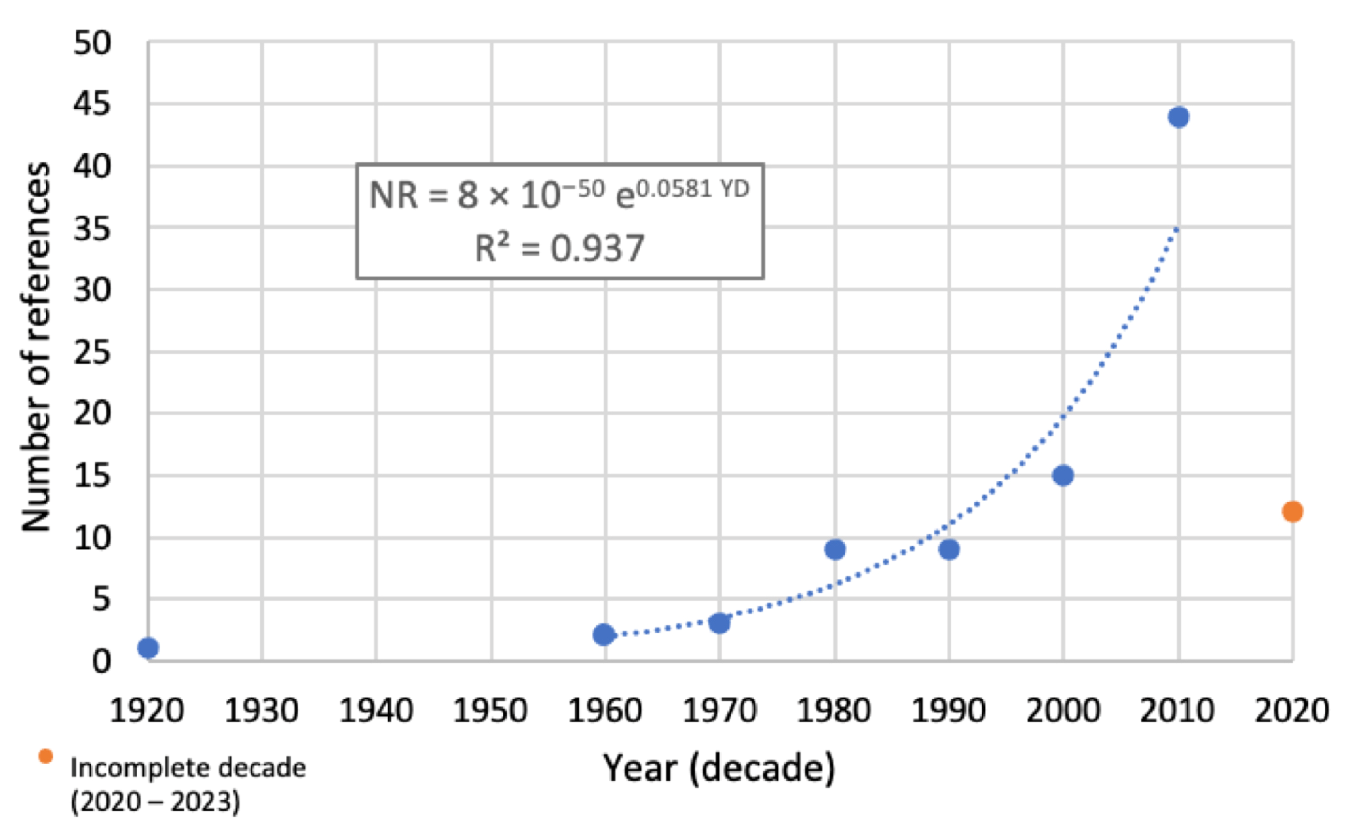
The studies covered a period between 1920 and 2023, and the number of references found per decade is shown in Figure 4. In the plot, we can see that there has been an exponential global increase in the number of studies, although this trend has diminished in recent years. Nevertheless, no conclusion can be made because this period is influenced by the COVID-19 pandemic, which might influence advances in the field.
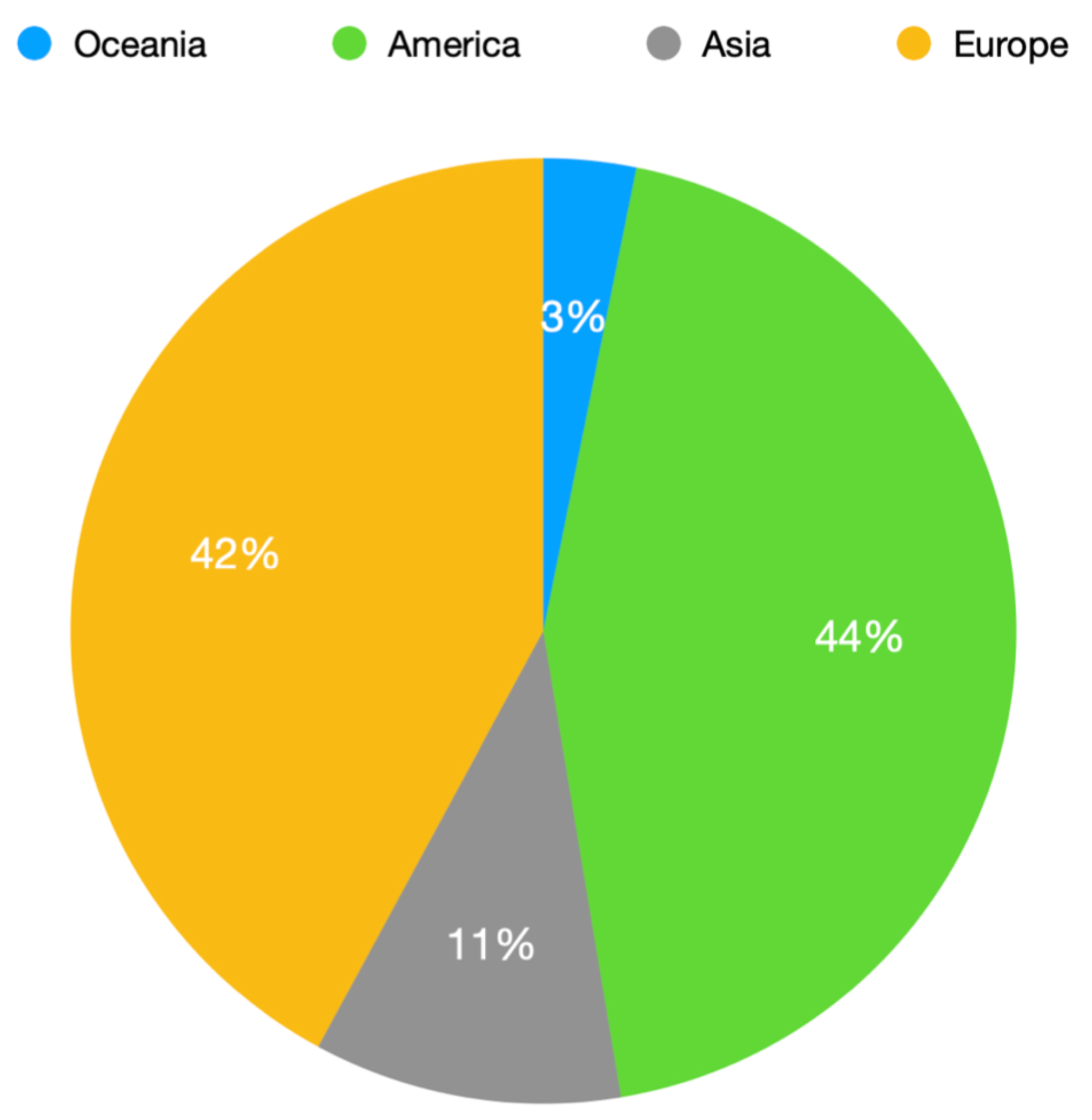
The plot in Figure 5 shows that there is an asymmetrical distribution of references by the first author’s affiliation, highlighting the importance of America and Europe, which represent more than 87% of all selected references. The relevance of these data is still to be evaluate, but at least it might indicate the degree of concern regarding this subject in different regions. Although not the scope of this article, future studies correlating this data with epidemiological parameters (e.g., time in front of digital devices, internet usage, etc.) could give a broader overview of the problem by geographical area. For example, and overall, the “ranking” presented in Figure 5 has some direct relation with the time spent using internet in those regions [3], the internet being one of the most likely causes for spending time using digital devices such as smartphones and tablets.
The literature about blue light presents several different parameters to characterize the effect of light sources. Some examples include illuminance (lux), luminance (cd/m2) and irradiance (W/m2). This fact makes it difficult to compare experimental conditions between different studies. Information about the potential effects of overexposure to artificial blue light is identified and additional experimental research is needed to obtain new information.
A study of rats evaluated the retinas after exposure to light emitted by LED tablet screens. Rats were divided into three groups: the first had a selective short-wavelength absorption filter, the second had no filter, and the third control group was not exposed to the tablet. The retinal structure results revealed that upon exposure to light from the tablet screen, there was a significant decrease in the entire thickness of the retina due to the reduction in the number of cells in the outer nuclear and inner nuclear layers [7].
In another study with rats, two types of retinal damage were reported with light wavelengths of 380 and 470 nm. The temporal sequence of retinal changes was followed for up to 2 months by fundoscopy and histology. For both types of damage, retina changes became more visible after 3 days. Histology showed that 380 nm specifically damaged photoreceptor cells. The 470 nm wavelength caused damage to the photoreceptor layer and the retinal pigment epithelium [83].
To validate the contribution of digital screens to dry-eye disease, a study was carried out to analyze the differences in ocular surface parameters, tear film and visual fatigue after reading from different digital monitors. Thirty-one healthy people, between 20 and 26 years, were enrolled in this clinical study. The results showed significant differences in ocular surface disease index, such as tear meniscus height, Schirmer’s test, BUT test, osmolarity and bulbar redness [82].
Considering the mentioned evidence and the complex interactions between the different variables of exposure to blue light, it is of great interest to continue studies in order to determine whether exposure to blue light from digital devices can cause damage to ocular structures.

4. Discussion

Faster technological advances generate more concerns regarding security levels in an increasingly digital world. The risks of blue light emitted by digital devices are, for now, possibilities, but it remains a controversial topic and still scientifically unsolved. This review aimed to summarize the safety of available artificial blue light to provide new matches identified in the existing literature.
Research over the past few decades has shown that exposing the eyes to blue light can cause damage under certain conditions. Blue light emissions from digital devices screens are much lower than the level known to cause photochemical damage to the retina. According to the CIE, the laptop and smartphone monitors studied do not represent any risk to users’ eyes [4]. However, the fact that digital device users are looking directly at a light source for extended periods of time can potentially endanger their eyes compared to general lighting users.
A summary of the main conclusions found in the literature is presented next for discussion, where the possible blue light threats by digital devices are considered.

4.1. Retinal Damage from Blue Light

It is essential to study blue light to assess the cause of actual damage to the retina, as demonstrated in several studies [55,84]. Additional research in the late 1980s and 1990s identified photoreceptors and the retinal pigment epithelium (RPE) as primary targets for damage induced by blue light [65,66,67,84,85].
The historical chronology of population studies with damage by association between blue light and the retina has been consistent with exposure to blue light damaging the retina [50,72]. Studies, with references dated in the 2000s, support the relationship between this radiation and age-related macular degeneration (AMD) [86]. The European Eye Study found a relationship between exposure to sunlight and neovascular AMD [87]. The contribution of blue light was not always considered in these studies, but some authors suggest that blue light may be responsible for these effects [88].

4.2. Blue Light Exposure by Digital Devices

The digitization trend and current artificial light sources have increased our eyes’ exposure to artificial blue radiation [89]. A 2020 survey describes that American Academy of Pediatrics suggested specific limits for screen time, without specifying universal daily screen time limits [90]. The use of digital devices grew sharply in 2020, but digitalization driven by the COVID-19 lockdowns has accelerated significantly [91]. Professionally, many people are exposed to screens for nearly 8 h a day [92]. Thus, the concern with digital device use has increased according to the proportion of blue light emitted, which is greater than conventional sources of incandescent and fluorescent lighting [37].

4.3. Relationship between Digital Devices and Ocular Structures

There is an association between negative effects on human health, well-being and digital device use. High exposure to blue light triggers visual discomfort in the ocular surface of the cornea (digital eye strain), disruption of circadian rhythms, increased insulin resistance, increased affective disorders and even increased incidence of cancer pathologies. Therefore, it is possible that the effects of overexposure to blue light on the retina by using these devices may contribute to a higher incidence of pathological changes in the retina, such as AMD [44,93,94]. Blue light from digital device exposure must be approached in population studies. Others have already attempted to assess the harmful effects on the retina and other ocular structures. The available studies have taken a laboratorial approach that does not reflect real life and does not clarify the probability of an ocular injury occurring. The exact nature of blue light exposure in terms of intensity and spectral power, duration and repetition would allow for better variable control.
RPE cell layers may represent a model for studying blue-light-induced retinal damage. Cells after exposure to blue light show damage according to some laboratory research studies [23,87]. However, RPE cell dysfunction may play a secondary role in AMD pathology, which is significant because population studies on the effects of blue light on the retina have used AMD as a marker of retinal damage [50,85,86]. Age-related macular degeneration (AMD) is an eye disease that gradually causes loss of central vision. Retinal pigment epithelial cells and photoreceptor exposure to blue light is indicated as a factor of this pathology progression, but the use of devices that block this radiation reduces these effects on retinal cells and delays the onset and progression of AMD [95].
In the population studies examined in this review [50,54,84,85,87], the source of blue light was the sun and when studying the effects of exposure to blue light on retinal cells, it is relevant to also study variable parameters such as its intensity [88]. The brightness of blue sunlight is about 50 times greater than that of digital devices [34]. The intensity of blue light required to produce retinal damage is even greater (38 to 3261 times) than that produced by digital devices [37,50,57,85]. When intensity towards the limit of the visible spectrum increases in blue light wavelengths, which are potentially harmful to the retina, it becomes problematic to assume that the effects observed in sunlight can also occur proportionately with digital devices [31]. The duration of each instance of blue light exposure is another important variable that must be quantified because exposure is not continuous throughout the day. During the daily cycle, there are periods of high exposure, such as during work or school hours, but also periods of non-exposure, such as during sleep.
There is also evidence that some of the harmful effects caused by blue light on the retinal functional process may be reversible after interruption of exposure to blue light for a certain period [88,89,90,91]. However, repeated exposure to blue light was not addressed or only marginally addressed in the laboratory research studies reviewed here, which used only one or a few instances of exposure to blue light [8,22,54,56].
Population studies suggest that continuous exposure to blue light from digital devices is important for producing effects on the retina. However, it takes years of exposure to see effects on the retina [84,85,86,87,88,89,90]. Blue light intensity from digital devices is below the level at which retinal damage occurs or results in increased AMD occurrence, according to studies of laboratory data indicating that a prolonged period of exposure is required to observe or rule out effects [8,33,57,82]. Furthermore, previous works suggest that this type of laboratory research is easily achievable and that, for further studies, damage in RPE cell layers will be used as a starting point for blue-light-induced damage (easy to measure).
Thus, supported by the data found in the literature, to obtain a better understanding of the different experiments carried out, we propose to create two large groups of users with the following:
  • high daily use of digital devices;
  • little or no exposure to digital devices daily.
  • We also propose to create the following guidelines:
  • duration of daily exposure administration set to at least 1 year if no blue light damage occurs earlier;
  • the lowest light intensity set to approximately the average of that emitted by personal digital devices;
  • if a relationship is found between blue light exposure and RPE cell layer damage at each intensity tested, this relationship can be extrapolated to the light levels of all digital devices;
  • after extrapolation, if the duration of exposure required to damage the RPE cell layers is within the range that a person might experience over a lifetime using these devices, this may indicate that their use may lead to retinal damage;
  • otherwise, it suggests that the use of such devices is not harmful to the retina.
The devices vary by category (computer, tablet and smartphone) and spectral emissions of each device require the development of new studies that indicate new paths and provide new data for human eyes.

5. Conclusions

The objective of this review was to evaluate the state of the art on photobiological safety in the use of emitting blue light digital equipment. Of all the artificial light sources, the only ones we looked directly at are digital devices. Therefore, as we spend more time using digital devices, it is essential to understand how the blue light they emit can affect our health and well-being. The data and evidence collected so far regarding the effect of blue light exposure from personal digital devices does not show enough evidence to refute the hypothesis that the use of these devices can produce retinal damage throughout life. The reviewed literature suggests that blue light is not significant in altering ocular health and that existing forms of blue light blocking do not protect against digital eye strain or age-related macular degeneration [91,93].
Selective research is needed on the impact of blue light exposure and how quickly lighting technologies, including digital devices, can negatively affect frequent exposure. From this review study, some suggestions for further investigation are as follows:
  • investigate and relate the impacts on the eye health of shift workers more exposed to artificial lighting;
  • the effects of blue light on circadian rhythms and ocular health at different stages of life;
  • interventions in the investigation of light exposure and the inhibition of myopia in children may also be of great interest;
  • the impact of the retina overexposure to blue wavelengths from common white light sources;
  • how to mitigate health and environmental lighting impacts while maintaining the obvious benefits of using artificial light at night;
  • impact of long-term use of digital devices on the cornea, lens and retina;
  • a multidisciplinary approach that develops progress in research on safety in the use of artificial blue light and digital devices and their implications for users.
In this review, contributions and scientific perspectives on blue light and the complex interaction of factors implicit in this seemingly simple and everyday phenomenon are brought. It will be interesting to carry out new observational studies that quantify exposure to light and measure the main indices of ocular health. References from different contexts have been brought together to provide an interdisciplinary narrative to deepen understanding of this interesting subject from a vision science perspective. Despite the growing number of studies, the development and interest in blue light research is very significant on the American and European continents.
There are several challenges to conducting research in the field of blue light safety. In the future, it is necessary to consider ocular structures and exposure to artificial blue light in humans. Until now, studies have provided information about the potential long-term effects of repeated exposure to artificial blue light, and further experimental research is important to providing more information in this area. It would also be worthwhile to validate an approach with a protocol that measures the characteristics of light sources. We believe that a multidisciplinary approach on the safety of blue light from digital devices can bring advances in research and new data on the implications of blue light and its impact on ocular structures.
There are some gaps in this matter that could be solved with relevant data in future studies on this topic, namely to integrate data from contemporary digital devices, check the effectiveness of blue-light-blocking ophthalmic lenses, create a comparative database of ocular anatomical structures in humans and human retinal bioelectricity after saturation with blue radiation from digital devices.

Author Contributions

Conceptualization, J.M.P.C.; methodology, V.H.; formal analysis, V.H. and J.M.P.C.; investigation, V.H. and J.M.P.C.; writing—original draft preparation, V.H.; writing—review and editing, V.H. and J.M.P.C.; supervision, V.H. and J.M.P.C.; project administration, V.H. and J.M.P.C.; funding acquisition, J.M.P.C. All authors have read and agreed to the published version of the manuscript.

Funding

This research was funded by Fundação para a Ciência e a Tecnologia (FCT), project reference UIDB/00645/2020.

Data Availability Statement

As a review article, the data is presented in the references mentioned in the text.

Conflicts of Interest

The authors declare no conflict of interest.

References

  1. Watson, A. In-Home Media Consumption Due to the Coronavirus Outbreak among Internet Users Worldwide as of March 2020, by Country. Available online: https://www.statista.com/statistics/1106498/home-media-consumption-coronavirus-worldwide-by-country/ (accessed on 31 January 2023).
  2. Bahkir, F.A.; Grandee, S.S. Impact of the COVID-19 lockdown on digital device-related ocular health. Indian J. Ophthalmol. 2020, 68, 2378–2383. [Google Scholar] [CrossRef] [PubMed]
  3. Digital 2023: Global Overview Report. Available online: https://datareportal.com/reports/digital-2023-global-overview-report (accessed on 5 April 2023).
  4. CIE 2019. CIE Position Statement on the Blue Light Hazard. 2019. Available online: http://cie.co.at/publications/position-statement-blue-light-hazard-april-23-2019 (accessed on 5 April 2023).
  5. Renard, G.; Leid, J. Les dangers de la lumière bleue: La vérité! [The dangers of blue light: True story!]. J. Fr. Ophtalmol. 2016, 39, 483–488. [Google Scholar] [CrossRef] [PubMed]
  6. Nasrallah, M. Why is the sky blue? Sci. Am. 2003, 289, 103. [Google Scholar]
  7. Sanchez-Ramos, C.; Bonnin-Arias, C.; Blázquez-Sánchez, V.; Aguirre-Vilacoro, V.; Cobo, T.; García-Suarez, O.; Perez-Carrasco, M.J.; Alvarez-Peregrina, C.; Vega, J.A. Retinal Protection from LED-Backlit Screen Lights by Short Wavelength Absorption Filters. Cells 2021, 10, 3248. [Google Scholar] [CrossRef]
  8. Jaadane, I.; Villalpando Rodriguez, G.E.; Boulenguez, P.; Chahory, S.; Carré, S.; Savoldelli, M.; Jonet, L.; Behar-Cohen, F.; Martinsons, C.; Torriglia, A. Effects of white light-emitting diode (LED) exposure on retinal pigment epithelium in vivo. J. Cell Mol. Med. 2017, 21, 3453–3466. [Google Scholar] [CrossRef] [PubMed]
  9. Jameson, D.; Hurvich, L.M. Handbook of Sensory Physiology: Visual Psychophysics; Springer: Berlin/Heidelberg, Germany, 1973; Volume 7/4. [Google Scholar]
  10. Widomska, J.; Witold, K.S. Why has Nature Chosen Lutein and Zeaxanthin to Protect the Retina? J. Clin. Exp. Ophthalmol. 2014, 5, 326. [Google Scholar] [CrossRef] [PubMed]
  11. Zheng, Q.; Ren, Y.; Reinach, P.S.; Xiao, B.; Lu, H.; Zhu, Y.; Qu, J.; Chen, W. Reactive oxygen species activated NLRP3 inflammasomes initiate inflammation in hyperosmolarity stressed human corneal epithelial cells and environment-induced dry eye patients. Exp. Eye Res. 2015, 134, 133–140. [Google Scholar] [CrossRef]
  12. Boettner, E.A.; Wolter, J.R. Transmission of the Ocular Media. Investig. Ophthalmol. Vis. Sci. 1962, 1, 776–783. [Google Scholar]
  13. Norren, D.V.; Vos, J.J. Spectral transmission of the human ocular media. Vis. Res. 1974, 14, 1237–1244. [Google Scholar] [CrossRef]
  14. Mellerio, J. Yellowing of the human lens: Nuclear and cortical contributions. Vis. Res. 1987, 27, 1581–1587. [Google Scholar] [CrossRef]
  15. Babizhayev, M.A. Mitochondria induce oxidative stress, generation of reactive oxygen species and redox state unbalance of the eye lens leading to human cataract formation: Disruption of redox lens organization by phospholipid hydroperoxides as a common basis for cataract disease. Cell Biochem. Funct. 2011, 29, 183–206. [Google Scholar] [CrossRef] [PubMed]
  16. Norton, T.T.; Siegwart, J.T., Jr. Light levels, refractive development, and myopia–a speculative review. Exp. Eye Res. 2013, 114, 48–57. [Google Scholar] [CrossRef]
  17. Ramamurthy, D.; Chua, S.Y.L.; Saw, S. A review of environmental risk factors for myopia during early life, childhood and adolescence. Clin. Exp. Optom. 2015, 98, 497–506. [Google Scholar] [CrossRef] [PubMed]
  18. Rucker, F.J.; Britton, S.; Spatcher, M.; Hanowsky, S. Blue Light Protects Against Temporal Frequency Sensitive Refractive Changes. Investig. Ophthalmol. Vis. Sci. 2015, 56, 6121–6131. [Google Scholar] [CrossRef]
  19. Fulk, G.W.; Cyert, L.A.; Parker, D.A. Seasonal Variation in Myopia Progression and Ocular Elongation. Optom. Vis. Sci. 2002, 79, 46–51. [Google Scholar] [CrossRef]
  20. Ashby, R.; Ohlendorf, A.; Schaeffel, F. The Effect of Ambient Illuminance on the Development of Deprivation Myopia in Chicks. Investig. Ophthalmol. Vis. Sci. 2009, 50, 5348–5354. [Google Scholar] [CrossRef] [PubMed]
  21. Stone, R.A.; Cohen, Y.; McGlinn, A.M.; Davison, S.; Casavant, S.; Shaffer, J.; Khurana, T.S.; Pardue, M.T.; Iuvone, P.M. Development of Experimental Myopia in Chicks in a Natural Environment. Investig. Ophthalmol. Vis. Sci. 2016, 57, 4779–4789. [Google Scholar] [CrossRef]
  22. McClean, A.; Derbyshire, L. Understanding Blue Light. Available online: https://www.aop.org.uk/our-voice/policy/position-statements/2023/01/03/visible-blue-light (accessed on 25 March 2023).
  23. Arnault, E.; Barrau, C.; Nanteau, C.; Gondouin, P.; Bigot, K.; Viénot, F.; Gutman, E.; Fontaine, V.; Villette, T.; Cohen-Tannoudji, D.; et al. Phototoxic Action Spectrum on a Retinal Pigment Epithelium Model of Age-Related Macular Degeneration Exposed to Sunlight Normalized Conditions. PLoS ONE 2013, 8, e71398. [Google Scholar] [CrossRef]
  24. Wahl, S.; Engelhardt, M.; Schaupp, P.; Lappe, C.; Ivanov, I.V. The inner clock—Blue light sets the human rhythm. J. Biophotonics 2019, 12, e201900102. [Google Scholar] [CrossRef]
  25. Verhoeven, G.J. The reflection of two fields—Electromagnetic radiation and its role in (aerial). AARGnews 2017, 55, 13–18. [Google Scholar] [CrossRef]
  26. Youssef, P.N.; Sheibani, N.; Albert, D.M. Retinal light toxicity. Eye 2011, 25, 1–14. [Google Scholar] [CrossRef] [PubMed]
  27. Behar-Cohen, F.; Martinsons, C.; Viénot, F.; Zissis, G.; Barlier-Salsi, A.; Cesarini, J.; Enouf, O.; Garcia, M.; Picaud, S.; Attia, D. Light-emitting diodes (LED) for domestic lighting: Any risks for the eye? Prog. Retin. Eye Res. 2011, 30, 239–257. [Google Scholar] [CrossRef] [PubMed]
  28. Valiente-Soriano, F.J.; Ortín-Martínez, A.; Di Pierdomenico, J.; García-Ayuso, D.; Gallego-Ortega, A.; de Imperial-Ollero, J.A.M.; Jiménez-López, M.; Villegas-Pérez, M.P.; Wheeler, L.A.; Vidal-Sanz, M. Topical Brimonidine or Intravitreal BDNF, CNTF, or bFGF Protect Cones Against Phototoxicity. Transl. Vis. Sci. Technol. 2019, 8, 36. [Google Scholar] [CrossRef] [PubMed]
  29. Jaadane, I.; Boulenguez, P.; Chahory, S.; Carré, S.; Savoldelli, M.; Jonet, L.; Behar-Cohen, F.; Martinsons, C.; Torriglia, A. Retinal damage induced by commercial light emitting diodes (LEDs). Free Radic. Biol. Med. 2015, 84, 373–384. [Google Scholar] [CrossRef] [PubMed]
  30. Lin, C.-H.; Wu, M.-R.; Huang, W.-J.; Chow, D.S.-L.; Hsiao, G.; Cheng, Y.-W. Low-Luminance Blue Light-Enhanced Phototoxicity in A2E-Laden RPE Cell Cultures and Rats. Int. J. Mol. Sci. 2019, 20, 1799. [Google Scholar] [CrossRef] [PubMed]
  31. Krigel, A.; Berdugo, M.; Picard, E.; Levy-Boukris, R.; Jaadane, I.; Jonet, L.; Dernigoghossian, M.; Andrieu-Soler, C.; Torriglia, A.; Behar-Cohen, F. Light-induced retinal damage using different light sources, protocols and rat strains reveals LED phototoxicity. Neuroscience 2016, 339, 296–307. [Google Scholar] [CrossRef] [PubMed]
  32. Xia, H.; Hu, Q.; Li, L.; Tang, X.; Zou, J.; Huang, L.; Li, X. Protective effects of autophagy against blue light-induced retinal degeneration in aged mice. Sci. China Life Sci. 2019, 62, 244–256. [Google Scholar] [CrossRef]
  33. Nakamura, M.; Yako, T.; Kuse, Y.; Inoue, Y.; Nishinaka, A.; Nakamura, S.; Shimazawa, M.; Hara, H. Exposure to excessive blue LED light damages retinal pigment epithelium and photoreceptors of pigmented mice. Exp. Eye Res. 2018, 177, 1–11. [Google Scholar] [CrossRef]
  34. Feeney-Burns, L.; Berman, E.R.; Rothman, H. Lipofuscin of Human Retinal Pigment Epithelium. Am. J. Ophthalmol. 1980, 90, 783–791. [Google Scholar] [CrossRef]
  35. Weiter, J.J.; Delori, F.C.; Wing, G.L.; Fitch, K.A. Retinal pigment epithelial lipofuscin and melanin and choroidal melanin in human eyes. Investig. Ophthalmol. Vis. Sci. 1986, 27, 145–152. [Google Scholar]
  36. Feeney-Burns, L.; Hilderbrand, E.S.; Eldridge, S. Aging human RPE: Morphometric analysis of macular, equatorial, and peripheral cells. Investig. Ophthalmol. Vis. Sci. 1984, 25, 195–200. [Google Scholar]
  37. Taylor, H.R.; West, S.; Muñoz, B.; Rosenthal, F.S.; Bressler, S.B.; Bressler, N.M. The Long-term Effects of Visible Light on the Eye. Arch. Ophthalmol. 1992, 110, 99–104. [Google Scholar] [CrossRef] [PubMed]
  38. The Eye Disease Case-Control Study Group. Risk Factors for Neovascular Age-Related Macular Degeneration. Arch. Ophthalmol. 1992, 110, 1701–1708. [Google Scholar] [CrossRef]
  39. Hirvelä, H.; Luukinen, H.; Läärä, E.; Laatikainen, L. Risk Factors of Age-related Maculopathy in a Population 70 Years of Age or Older. Ophthalmology 1996, 103, 871–877. [Google Scholar] [CrossRef] [PubMed]
  40. Darzins, P.; Mitchell, P.; Heller, R. Sun Exposure and Age-related Macular Degeneration. An Australian case-control study. Ophthalmology 1997, 104, 770–776. [Google Scholar] [CrossRef]
  41. McCarty, C.A.; Mukesh, B.N.; Fu, C.L.; Mitchell, P.; Wang, J.J.; Taylor, H.R. Risk Factors for Age-Related Maculopathy: The Visual Impairment Project. Arch. Ophthalmol. 2001, 119, 1455–1462. [Google Scholar] [CrossRef]
  42. Delcourt, C.; Carrière, I.; Ponton-Sanchez, A.; Fourrey, S.; Lacroux, A.; Papoz, L.; POLA Study Group. Light Exposure and the Risk of Age-Related Macular Degeneration: The Pathologies Oculaires Liées à l’Age (POLA) study. Arch. Ophthalmol. 2001, 119, 1463–1468. [Google Scholar] [CrossRef]
  43. Putting, B.J.; Zweypfenning, R.C.; Vrensen, G.F.; Oosterhuis, J.A.; Van Best, J.A. Blood-retinal barrier dysfunction at the pigment epithelium induced by blue light. Investig. Ophthalmol. Vis. Sci. 1992, 33, 3385–3393. [Google Scholar]
  44. Pautler, E.L.; Morita, M.; Beezley, D. Reversible and irreversible blue light damage to the isolated, mammalian pigment epithelium. Prog. Clin. Biol. Res. 1989, 314, 555–567. [Google Scholar]
  45. Chamorro, E.; Bonnin-Arias, C.; Pérez-Carrasco, M.J.; de Luna, J.M.; Vázquez, D.; Sánchez-Ramos, C. Effects of Light-emitting Diode Radiations on Human Retinal Pigment Epithelial Cells In Vitro. Photochem. Photobiol. 2013, 89, 468–473. [Google Scholar] [CrossRef]
  46. Vicente-Tejedor, J.; Marchena, M.; Ramírez, L.; García-Ayuso, D.; Gómez-Vicente, V.; Sánchez-Ramos, C.; De La Villa, P.; Germain, F. Removal of the blue component of light significantly decreases retinal damage after high intensity exposure. PLoS ONE 2018, 13, e0194218. [Google Scholar] [CrossRef] [PubMed]
  47. Egringras, P.; Emiddleton, B.; Skene, D.J.; Revell, V.L. Bigger, Brighter, Bluer-Better? Current Light-Emitting Devices—Adverse Sleep Properties and Preventative Strategies. Front. Public Health 2015, 3, 233. [Google Scholar] [CrossRef]
  48. Pazikadin, A.R.; Rifai, D.; Ali, K.; Mamat, N.H.; Khamsah, N. Design and Implementation of Fuzzy Compensation Scheme for Temperature and Solar Irradiance Wireless Sensor Network (WSN) on Solar Photovoltaic (PV) System. Sensors 2020, 20, 6744. [Google Scholar] [CrossRef]
  49. Udovicic, L.; Janßen, M. Photobiological Safety of Common Office Light Sources. In Proceedings of the 29th CIE Session, Washington, DC, USA, 14–22 June 2019; CIE: Vienna, Austria, 2019; pp. 1256–1261. [Google Scholar] [CrossRef]
  50. Tomany, S.C.; Cruickshanks, K.J.; Klein, R.; Klein, B.E.K.; Knudtson, M.D. Sunlight and the 10-Year Incidence of Age-Related Maculopathy: The Beaver Dam Eye Study. Arch. Ophthalmol. 2004, 122, 750–757, Erratum in Arch. Ophthalmol. 2005, 123, 362. [Google Scholar] [CrossRef]
  51. Rozanowska, M.B. Light-Induced Damage to the Retina: Current Understanding of the Mechanisms and Unresolved Questions: A Symposium-in-Print. Photochem. Photobiol. 2012, 88, 1303–1308. [Google Scholar] [CrossRef] [PubMed]
  52. Sciences Online: American Society for Photobiology. Available online: http://photobiology.info/Rozanowska.html (accessed on 20 March 2022).
  53. Noell, W.K.; Walker, V.S.; Kang, B.S.; Berman, S. Retinal damage by light in rats. Investig. Ophthalmol. 1966, 5, 450–473. [Google Scholar]
  54. O’Hagan, J.B.; Khazova, M.; Price, L.L.A. Low-energy light bulbs, computers, tablets and the blue light hazard. Eye 2016, 30, 230–233. [Google Scholar] [CrossRef]
  55. Tosini, G.; Ferguson, I.; Tsubota, K. Effects of blue light on the circadian system and eye physiology. Mol. Vis. 2016, 22, 61–72. [Google Scholar]
  56. Nash, T.R.; Chow, E.S.; Law, A.D.; Fu, S.D.; Fuszara, E.; Bilska, A.; Bebas, P.; Kretzschmar, D.; Giebultowicz, J.M. Daily blue-light exposure shortens lifespan and causes brain neurodegeneration in Drosophila. NPJ Aging Mech. Dis. 2019, 5, 8. [Google Scholar] [CrossRef]
  57. Xie, C.; Li, X.; Tong, J.; Gu, Y.; Shen, Y. Effects of white light-emitting diode (LED) light exposure with different Correlated Color Temperatures (CCTs) on human lens epithelial cells in culture. Photochem. Photobiol. 2014, 90, 853–859. [Google Scholar] [CrossRef]
  58. Weale, R.A. Age and the transmittance of the human crystalline lens. J. Physiol. 1988, 395, 577–587. [Google Scholar] [CrossRef] [PubMed]
  59. Xu, J.; Pokorny, J.; Smith, V.C. Optical density of the human lens. J. Opt. Soc. Am. A 1997, 14, 953–960. [Google Scholar] [CrossRef] [PubMed]
  60. Resnikoff, S.; Pascolini, D.; Etya’Ale, D.; Kocur, I.; Pararajasegaram, R.; Pokharel, G.P.; Mariotti, S.P. Global data on visual impairment in the year 2002. Bull. World Health Organ. 2004, 82, 844–851. [Google Scholar]
  61. Mainster, M.A. The Spectra, Classification, and Rationale of Ultraviolet-Protective Intraocular Lenses. Am. J. Ophthalmol. 1986, 102, 727–732. [Google Scholar] [CrossRef]
  62. Van Der Hoeve, J. Eye Lesions Produced by Light Rich in Ultraviolet Rays. Senile Cataract, Senile Degeneration of Macula. Am. J. Ophthalmol. 1920, 3, 178–194. [Google Scholar] [CrossRef]
  63. Tso, M.O.; La Piana, F.G. The human fovea after sungazing. Trans. Sect. Ophthalmol. Am. Acad. Ophthalmol. Otolaryngol. 1975, 79, OP788-95. [Google Scholar] [PubMed]
  64. Mainster, M.A.; Findl, O.; Dick, H.B.; Desmettre, T.; Ledesma-Gil, G.; Curcio, C.A.; Turner, P.L. The Blue Light Hazard Versus Blue Light Hype. Am. J. Ophthalmol. 2022, 240, 51–57. [Google Scholar] [CrossRef]
  65. Tso, M.O. Pathogenetic Factors of Aging Macular Degeneration. Ophthalmology 1985, 92, 628–635. [Google Scholar] [CrossRef]
  66. Marshall, J. Radiation and the ageing eye. Ophthalmic Physiol. Opt. 1985, 5, 241–263. [Google Scholar] [CrossRef]
  67. Mainster, M.A. Light and macular degeneration: A biophysical and clinical perspective. Eye 1987, 1, 304–310. [Google Scholar] [CrossRef]
  68. Mainster, M.A. Violet and blue light blocking intraocular lenses: Photoprotection versus photoreception. Br. J. Ophthalmol. 2006, 90, 784–792. [Google Scholar] [CrossRef] [PubMed]
  69. Marshall, J.D. The ageing retina: Physiology or pathology. Eye 1987, 1, 282–295. [Google Scholar] [CrossRef] [PubMed]
  70. Remé, C.; Reinboth, J.; Clausen, M.; Hafezi, F. Light damage revisited: Converging evidence, diverging views? Graefes Arch. Clin. Exp. Ophthalmol. 1996, 234, 2–11. [Google Scholar] [CrossRef]
  71. Boulton, M.; Różanowska, M.; Różanowski, B. Retinal photodamage. J. Photochem. Photobiol. B Biol. 2001, 64, 144–161. [Google Scholar] [CrossRef] [PubMed]
  72. Margrain, T.H.; Boulton, M.; Marshall, J.; Sliney, D.H. Do blue light filters confer protection against age-related macular degeneration? Prog. Retin. Eye Res. 2004, 23, 523–531. [Google Scholar] [CrossRef]
  73. Bernstein, P.S.; Khachik, F.; Carvalho, L.S.; Muir, G.J.; Zhao, D.-Y.; Katz, N.B. Identification and Quantitation of Carotenoids and their Metabolites in the Tissues of the Human Eye. Exp. Eye Res. 2001, 72, 215–223. [Google Scholar] [CrossRef]
  74. Gao, S.; Qin, T.; Liu, Z.; Caceres, M.A.; Ronchi, C.F.; Chen, C.-Y.O.; Yeum, K.-J.; Taylor, A.; Blumberg, J.B.; Liu, Y.; et al. Lutein and zeaxanthin supplementation reduces H2O2-induced oxidative damage in human lens epithelial cells. Mol. Vis. 2011, 17, 3180–3190. [Google Scholar]
  75. Lee, H.S.; Cui, L.; Li, Y.; Choi, J.S.; Choi, J.-H.; Li, Z.; Kim, G.E.; Choi, W.; Yoon, K.C. Correction: Influence of Light Emitting Diode-Derived Blue Light Overexposure on Mouse Ocular Surface. PLoS ONE 2016, 11, e0167671. [Google Scholar] [CrossRef]
  76. Yam, J.C.S.; Kwok, A.K.H. Ultraviolet light and ocular diseases. Int. Ophthalmol. 2014, 34, 383–400. [Google Scholar] [CrossRef] [PubMed]
  77. Vera, J.; Redondo, B.; Ortega-Sanchez, A.; Molina-Molina, A.; Molina, R.; Rosenfield, M.; Jiménez, R. Blue-blocking filters do not alleviate signs and symptoms of digital eye strain. Clin. Exp. Optom. 2023, 106, 85–90. [Google Scholar] [CrossRef]
  78. Cardona, G.; García, C.; Serés, C.; Vilaseca, M.; Gispets, J. Blink Rate, Blink Amplitude, and Tear Film Integrity during Dynamic Visual Display Terminal Tasks. Curr. Eye Res. 2011, 36, 190–197. [Google Scholar] [CrossRef] [PubMed]
  79. Portello, J.K.; Rosenfield, M.; Bababekova, Y.; Estrada, J.M.; Leon, A. Computer-related visual symptoms in office workers. Ophthalmic Physiol. Opt. 2012, 32, 375–382. [Google Scholar] [CrossRef]
  80. Akkaya, S.; Atakan, T.; Acikalin, B.; Aksoy, S.; Ozkurt, Y. Effects of long-term computer use on eye dryness. North. Clin. Istanb. 2018, 5, 319–322. [Google Scholar] [CrossRef] [PubMed]
  81. Rosenfield, M. Computer vision syndrome: A review of ocular causes and potential treatments. Ophthalmic Physiol. Opt. 2011, 31, 502–515. [Google Scholar] [CrossRef] [PubMed]
  82. Talens-Estarelles, C.; Sanchis-Jurado, V.; Esteve-Taboada, J.J.; Pons, M.; García-Lázaro, S. How Do Different Digital Displays Affect the Ocular Surface? Optom. Vis. Sci. 2020, 97, 1070–1079. [Google Scholar] [CrossRef]
  83. Busch, E.M.; Gorgels, T.G.; van Norren, D. Temporal sequence of changes in rat retina after UV-A and blue light exposure. Vis. Res. 1999, 39, 1233–1247. [Google Scholar] [CrossRef]
  84. Dorey, C.K.; Delori, F.C.; Akeo, K. Growth of cultured RPE and endothelial cells is inhibited by blue light but not green or red light. Curr. Eye Res. 1990, 9, 549–559. [Google Scholar] [CrossRef]
  85. Hunter, J.J.; Morgan, J.I.; Merigan, W.H.; Sliney, D.H.; Sparrow, J.R.; Williams, D.R. The susceptibility of the retina to photochemical damage from visible light. Prog. Retin. Eye Res. 2012, 31, 28–42. [Google Scholar] [CrossRef]
  86. Schick, T.; Ersoy, L.; Lechanteur, Y.T.E.; Saksens, N.T.M.; Hoyng, C.B.; den Hollander, A.I.; Kirchhof, B.; Fauser, S. History of Sunlight Exposure is a Risk Factor for Age-related Macular Degeneration. Retina 2016, 36, 787–790. [Google Scholar] [CrossRef]
  87. Fletcher, A.E.; Bentham, G.C.; Agnew, M.; Young, I.S.; Augood, C.; Chakravarthy, U.; de Jong, P.T.; Rahu, M.; Seland, J.; Soubrane, G.; et al. Sunlight Exposure, Antioxidants, and Age-Related Macular Degeneration. Arch. Ophthalmol. 2008, 126, 1396–1403. [Google Scholar] [CrossRef]
  88. Salvatori, A.; Menon, S.; Zwysen, W. The Effect of Computer Use on Job Quality; OECD Social, Employment and Migration Working Papers Paris; OECD: Paris, France, 2018; No. 200. [Google Scholar] [CrossRef]
  89. Nagata, J.M.; Magid, H.S.A.; Gabriel, K.P. Screen Time for Children and Adolescents During the Coronavirus Disease 2019 Pandemic. Obesity 2020, 28, 1582–1583. [Google Scholar] [CrossRef] [PubMed]
  90. Majumdar, P.; Biswas, A.; Sahu, S. COVID-19 pandemic and lockdown: Cause of sleep disruption, depression, somatic pain, and increased screen exposure of office workers and students of India. Chronobiol. Int. 2020, 37, 1191–1200. [Google Scholar] [CrossRef] [PubMed]
  91. Hefner, B. Warding off the blues. Rev. Optom. 2018, 155, 71–78. [Google Scholar]
  92. Spaide, R. Etiology of late-age-related macular disease. In Age-Related Macular Degeneration: A Comprehensive Textbook; Alfaro, D.V., Quiroz-Mercado, H., Liggett, P.E., Mieler, W.F., Eds.; Lippincott Williams & Wilkins: Philadelphia, PA, USA, 2006; pp. 23–39. [Google Scholar]
  93. Kozlowski, M.R. The ARPE-19 Cell Line: Mortality Status and Utility in Macular Degeneration Research. Curr. Eye Res. 2015, 40, 501–509. [Google Scholar] [CrossRef] [PubMed]
  94. Abdouh, M.; Lu, M.; Chen, Y.; Goyeneche, A.; Burnier, J.V.; Burnier, M.N. Filtering blue light mitigates the deleterious effects induced by the oxidative stress in human retinal pigment epithelial cells. Exp. Eye Res. 2022, 217, 108978. [Google Scholar] [CrossRef]
  95. Gordon, A. Digital eye strain, blue light, and contact lens wear. Contact Lens Spectr. 2018, 1, 27–32. [Google Scholar]
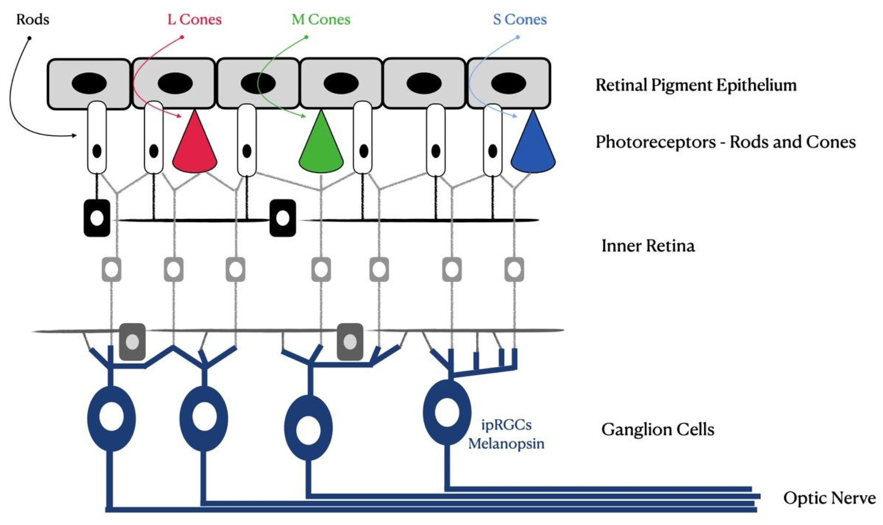
Figure 1. Schematic of the human retina. Photoreceptors, rods, cones and newly discovered intrinsically photosensitive retinal ganglion cells (ipRGCs) are shown.
Photonics 10 00560 g001
Figure 2. Schematic representing the different wavelength ranges for electromagnetic radiation (top), visible light (middle) and blue light emitted by digital devices (bottom)—adapted from [25].
Photonics 10 00560 g002
Figure 3. Irradiance values from several blue light source studies (not digital light sources). The light sources used were a xenon arc lamp (Putting et al. [43] and Paulter et al. [44]), a LED (Chamorro et al. [45]) and a fluorescent emission lamp (Vicente-Tejedor et al. [46]).
Photonics 10 00560 g003
Figure 4. Plot indicating the number of references selected for the present review and their publication year (per decade). The fit regards the data between the decades between the 1960s until the 2010s. The corresponding equation and its R squared value are also present. NR: number of references; YD: year (decades).
Photonics 10 00560 g004
Figure 5. Plot indicating the number of references selected for the present review regarding their first author’s affiliation country.
Photonics 10 00560 g005
Disclaimer/Publisher’s Note: The statements, opinions and data contained in all publications are solely those of the individual author(s) and contributor(s) and not of MDPI and/or the editor(s). MDPI and/or the editor(s) disclaim responsibility for any injury to people or property resulting from any ideas, methods, instructions or products referred to in the content.