Article

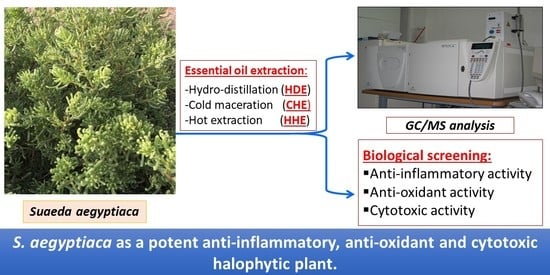
GC-MS Analysis and Bioactivities of the Essential Oil of Suaeda aegyptiaca

by
Elham Amin
1,2,*,†,
Ahlam Elwekeel
2,†,
Nasrah F. Alshariedh
3,
Mohamed Sadek Abdel-Bakky
4,5 and
Marwa H. A. Hassan
2
1
Department of Medicinal Chemistry and Pharmacognosy, College of Pharmacy, Qassim University, Buraydah 51452, Saudi Arabia
2
Department of Pharmacognosy, Faculty of Pharmacy, Beni-Suef University, Beni-Suef 62514, Egypt
3
College of Pharmacy, Qassim University, Buraydah 51452, Saudi Arabia
4
Department of Pharmacology and Toxicology, College of Pharmacy, Qassim University, Buraydah 51452, Saudi Arabia
5
Department of Pharmacology and Toxicology, Faculty of Pharmacy, Al-Azhar University, Cairo 11751, Egypt
*
Author to whom correspondence should be addressed.
These authors contributed equally to this work.
Separations 2022, 9(12), 439; https://doi.org/10.3390/separations9120439
Submission received: 17 November 2022 / Revised: 1 December 2022 / Accepted: 8 December 2022 / Published: 13 December 2022

Abstract

:
Suaeda aegyptiaca is a halophytic plant widely growing in northeast Africa and Asia. The current study reports on the GC-MS analysis of S. aegyptiaca essential oil. The essential oil was prepared using three different methods: cold n-hexane extraction (CHE), hot n-hexane extraction (HHE), and hydro-distillation extraction (HDE). The GC-MS analysis detected twenty-eight compounds in both CHE (97.28%) and HHE (97.35%) and twenty compounds in HDE (98.65%). 2-methyloctacosane (48.72%); 11-decyldocosane (29.20%); and 1, 2-benzenedicarboxylic acid diisooctyl ester (57.87%) were the main constituents in CHE, HHE, and HDE, respectively. Free radical scavenging activity testing using 2,2-diphenyl-1-picrylhydrazyl (DPPH) revealed the notable anti-oxidant potential of HDE (IC50 0.358 mg/mL) compared to ascorbic acid (IC50 0.264 mg/mL). Moreover, in vitro anti-inflammatory activity testing using COX-1 and COX-2 showed the notable activity of HDE (IC50 5.50 µg/mL and 2.59 µg/mL, respectively). The observed anti-inflammatory activity of HDE was further confirmed by the characteristic decrease in TNF-α levels in RAW264.7 to 572.20 Pg/mL compared to the decrease of 442.80 Pg/mL caused by the positive control (Celecoxib®). On the other hand, a cytotoxic activity investigation indicated that CHE was the most potent against the Caco-2 and HCT-116 cell lines (IC50 8.11 and 11.18 µg/mL, respectively), and it was closely followed by HHE (IC50 12.42 µg/mL) against the Caco-2 cell line. Collectively, S. aegyptiaca essential oil prepared by the hydro-distillation method exhibited notable anti-oxidant and anti-inflammatory activities, while the same essential oil prepared by solvent extraction methods displayed a characteristic cytotoxic activity against the Caco-2 and HCT-116 cell lines. These results confirmed that different extraction methods greatly influence the biological potential of an essential oil, which, in turn, is attributable to the different constituents in each extract Moreover, S. aegyptiaca was noted as a promising halophytic plant for more phytochemical and biological investigations.

Graphical Abstract

1. Introduction

Halophytes are widely distributed plants that can tolerate high soil salinity. Approximately 2500 halophyte species are classified under variable plant families, most commonly, Chenopodiaceae, Zygophyllaceae, Aizoaceae, Poaceae, and Tamaricaceae [1]. The plants’ ability to adapt to high salinity is attributed to the genes that operate several activities such as osmotic pressure regulation, loss of water through stomata, restoration of ions, management of metabolites, and improvement of anti-oxidant methods [2]. Plants grown in these tough conditions undergo oxidative stress resulting from increased levels of reactive oxygen species (ROS), which may lead to damaged proteins, enzymes, and cell membranes, and it may end in plant death [3]. Halophytic plants that tolerate salinity and aridity generate anti-oxidant defense systems through the use of enzymatic anti-oxidants such as catalases, superoxide dismutases, and peroxidases, together with nonenzymatic anti-oxidants, e.g., flavonoids, carotenoids, ascorbates, phenols, and other phytonutrients [4,5].
Inflammation and cancer conditions are characterized by increased levels of ROS, which in turn may cause damage to supermolecules, such as proteins, glucose, fats, and DNA. This might result in the destruction of structural proteins and enzymes, damage to cellular membranes, and gene modifications, and it may also activate pro-oncogenic signaling [6]. Therefore, tumor initiation, development, and progression may be directly attributed to the development of oxidative stress [7]. On the other hand, inflammation is considered as the body‘s response to any kind of injury, such as an infection or irritation, and this response is related to many diseases such as rheumatoid arthritis, diabetes, and cancer [8]. During inflammation, many mediators are produced through macrophages such as cytokines, NO, and prostaglandins, and furthermore, increased levels of ROS are typically recorded [4]. Accordingly, anti-oxidant nutrients have been reported to play a critical role in the management of cancer and inflammation.
Essential (volatile) oils are a concentrated hydrophobic mixture of volatile constituents. They represent a diverse group of natural products that are widely employed for medicinal uses, as well as for use in the food and pharmaceuticals industries.
Several methods are typically adopted for the extraction of essential oils, such as solvent extraction, steam and hydro-distillation, microwave-assisted extraction, head space extraction, and liquid CO2 extraction. The oil composition will vary according to the extraction method used, and the selected extraction method depends on several variables such as sample size, amount of essential oil, objective of the study, etc., [9]. Although the head space and liquid CO2 extraction methods are the most rapid and reliable techniques, the small size of the plant material, high cost, and incomplete recovery of volatiles (in the head space method) limit their widespread use. On the other hand, the hydro-distillation and solvent extraction methods are commonly employed due to their availability, reliability, and cost effectiveness [10,11], and these techniques are commonly applicable for samples with low concentrations of essential oils [12].
The genus Suaeda (family Chenopodiaceae) constitutes approximately 100 species and includes perennial succulent halophytic shrubs, which tolerate high salt environments and are widely distributed on land and in coastal salt marshes [13].
Several Suaeda species, e.g., S. vermiculata, S. esteroa, and S. maritima, are used as food for livestock in Saudi Arabia and India, while S. monoica has been reported to be used in the manufacture of paper. Furthermore, the seeds of some species, e.g., S. aralocaspica, S. vermiculata, S. vera, S. fruticosa, S. moquinii, and S. sals, have been employed to produce edible oils [14,15,16,17]. The Suaeda species has been reported to have variable biological activities as an anti-oxidant [13,18] and anti-inflammatory [18,19,20], with cytotoxic activity [20,21,22]. Phytochemical investigations of this genus have revealed that phenolics, terpenoids, and alkaloids are its major secondary metabolites [23]. S. aegyptiaca was traditionally used for the treatment of stomach pain and topically for wound and skin infections [24]. Investigations of the physical characters of the plant have revealed a characteristic aromatic odor that addressed the presence of volatile constituents. In reviewing the literature, no records were found that discussed the phytochemical content or bioactivities of the volatile oil extracted from this halophytic species.
In this context, the present research investigated the phytochemical content and biological potential of the volatile oil from S. aegyptiaca prepared using three different methods: cold maceration, hot extraction using soxhlet, and hydro-distillation. Interestingly, this is the first study that discusses the chemical profiling and bioactivities of S. aegyptiaca essential oil.

2. Materials and Methods

2.1. Sample Preparation

The aerial parts of Suaeda aegyptiaca were collected between September and October of 2021 from the Qassim region of Saudi Arabia. The identity of the plant was verified by Ibrahim Aldakhil, a botanical expert in the Qassim region. A voucher sample with the number QPP-110 was saved at the College of Pharmacy of Qassim University (KSA).

2.2. Extraction of Essential Oil

Three different methods of extraction (cold maceration, hot extraction using Soxhlet, and hydro-distillation) were applied to extract the essential oil of the plant as follows:

2.2.1. Cold n-Hexane Extraction (CHE)

Dried ground aerial plant parts (100 g) were macerated in 300 mL n-hexane twice (6 h each, at room temperature), with frequent shaking at 100 rpm using an orbital shaker (Cole-Parmer™ Stuart™ Orbital Shaker, Thermo Scientific, Waltham, MA, USA). The n-hexane extracts were pooled, and the solvent was dried using a vacuum to produce 380 mg of dry residue.

2.2.2. Hot n-Hexane Extraction (HHE)

A total of 100 g of the dried plant material was extracted twice (6 h each, 70 °C) with 300 mL n-hexane using a soxhlet apparatus, and then the n-hexane extracts were pooled and dried under a vacuum to yield 690 mg of dry residue.

2.2.3. Hydro-distillation Extraction (HDE)

The powdered plant material (100 g) was mixed with distilled water (300 mL) in a rounded bottom flask and connected to a Clevenger apparatus and then heated for 12 h at 100 °C. The water of the extraction was collected and extracted with ethyl acetate and then dried under a vacuum to yield 540 mg of dry residue.

2.3. GC-MS Analysis of the Different Extracts

We analyzed 1 μL of the different extracts using a GC-MS apparatus, adopting the following parameters: a TRACE GC Ultra Gas Chromatograph (Thermo Scientific, Waltham, MA, USA) attached to a thermo mass spectrometer detector (ISQ Single Quadrupole Mass Spectrometer, Thermo Scientific, Waltham, MA, USA). The samples were injected into a TR-5 MS column with the following specifications: 30 m x 0.32 mm i.d. and 0.25 μm film thickness. Helium gas was used at a flow rate of 1.0 mL/min, with a 1:10 split ratio. The oven temperature program was set as follows: 60 °C for 1 min, then increasing to 240 °C at a rate of 4.0 °C/min. The injector and detector temperatures were adjusted at 210 °C. Electron ionization (EI) at 70 eV and a mass range of m/z 40–450 were used to obtain the mass spectra. The phytoconstituents of the essential oil were identified using the AMDIS software (www.amdis.net accessed on 13 November 2021) retention indices (relative to the n-alkanes C8-C22) and mass spectrum matching (authentic standards when available; Wiley spectral library collection and NSIT library database).

2.4. Biological Evaluation

2.4.1. Anti-Oxidant Activity

The anti-oxidant potential of the different samples of S. aegyptiaca essential oil were evaluated by DPPH assay according to [25]. Briefly, 100 µL of each oil sample (1000–250 µg/mL), along with the positive control (ascorbic acid), were mixed with 3.9 mL of DPPH in methanol (0.1 mM) and held at room temperature, protected from light, for 30 min. Afterwards, the absorbance was measured at 517 nm. In the control experiment, 100 μL of methanol was used instead of the extract. The free radical scavenging activity was calculated employing the equation:
Scavenging effect (%) = [absorbance of control − absorbance of sample/absorbance of control)] × 100

2.4.2. Anti-Inflammatory Activity

The anti-inflammatory potentials of the S. aegyptiaca essential oil extracts were determined through measuring their inhibitory effects against the two cyclooxygenase enzyme isoforms COX-1 and COX-2. Furthermore, the TNF-α level was measured in murine macrophage (RAW264.7) cell line stimulated by lipopolysaccharide (LPS).

COX-1 and COX-2 Inhibition Assay

The essential oil extracts in serial dilutions (100, 10, 1, 0.1, and 0.01 ug/mL) were dissolved in DMSO and evaluated for their inhibition of prostaglandin synthesis utilizing the COX inhibitor screening assay kits (BioVision, Inc., Mountain View, CA, USA) COX-1 (ovine) and COX-2 (human recombinant). The experimental protocol was performed according to the supplier’s instructions. The cyclooxygenase enzyme (COX) drives the biosynthetic production of prostaglandin (PGH2) from the arachidonic acid. The PGF resulting from the reduction in PGH2 via stannous chloride was measured by enzyme linked immunoassay (ELISA). This immunoassay depends on the challenge between a PG-specific antibody and a PG-acetyl cholinesterase conjugate. The complex (antibody-PG) was mixed with Ellman’s reagent containing an acetylcholinesterase substrate. Twenty µL of the samples, the enzyme (10 µL), and heme (10 µL) were added to 160 µL of the reaction buffer solution (0.1 M Tris-HCl, pH 8 containing 2 mM phenol and 5 mM ethylenediamine tetra acetate (EDTA)), and then the mixture was incubated at 37 °C for 10 min in a water bath. Afterwards, arachidonic acid (10 µL, final concentration in reaction mixture of 100 mM) was added to start the COX reactions. Two minutes later, the COX reactions were stopped via the addition of 30 µL saturated stannous chloride, followed by incubation at room temperature for 5 min. Then, ELISA was used for assessing the PGF level. Following transfer to a 96-well plate, the samples were incubated for 18 h at room temperature, and then the plate was washed to remove any unbound reagent and 200 µL of Ellman’s reagent (containing an acetylcholinesterase substrate) was added and incubated at room temperature for 60 to 90 min. The resulting color was measured spectrophotometrically using a microplate reader at 410 nm. The IC50 values of COX-1 and COX-2 inhibition were calculated by comparing the sample-treated incubations to the control incubations and Indomethacin® and Celecoxib®, respectively, were used as the reference standard drugs [26].

Measurement of TNF-α in an LPS-stimulated murine macrophage cell line (RAW264.7)

The production of TNF-α was determined using the RAW264.7 cell line, which was kindly supplied by VACSERA, Egypt. The cells’ viability assays were completed using an MTT assay to determine the safe, non-toxic dose of the samples prior to TNF-α measurement [27]. After determining the safe doses of the three extracts, the cells were cultured in DMEM (Dulbecco’s modified Eagle’s medium) supplemented with 10% FBS and 100 U/mL penicillin and streptomycin at 37 °C in a humidified atmosphere with 5% CO2. The inflammation of the cells was induced as follows: in 6-well plates, cells (5 × 10⁵ cells/well) were plated and incubated for 24 h, then 1600 µL of the growth media and 200 µL of the extracts (prepared in different concentrations) were mixed. Two hours later, 200 μL LPS (1 μg/mL) from Escherichia coli was added to the medium and incubated for 24 h. Afterwards, centrifugation at 2000× g was performed for 10 min, and the supernatant was kept at −79 °C [28]. An Ab181421 Human TNF-α Simple Step ELISA® Kit was utilized to quantify the TNF-α in the cells as follows: in a 96-well plate, the supernatant (50 µL) was mixed with the antibody solution (50 µL), and then the plate was sealed and incubated for one hour at room temperature on a plate shaker adjusted to 400 rpm. Then, 100 μL of the TMB substrate solution was added and the plate was incubated for 10 min in dark conditions on a plate shaker at 400 rpm. Blue color production indicated TMB oxidation by peroxidase enzymes. Afterwards, a stop solution (100 μL) was added, the plate was shaken on a plate shaker for 1 min to mix all the content, and the OD was recorded at 450 nm. The TNF-α concentration was calculated using a standard curve. Celecoxib® was utilized as standard drug.

2.4.3. Cytotoxic Evaluation of the Different Extracts Using an MTT Assay

The cytotoxicity of the essential oil extracts prepared from S. aegyptiaca was screened against two colon cancer cell lines, HCT-116 and Caco-2, using an MTT assay as follows:
  • Cell preparation
The HCT-116 and Caco-2 cell lines were obtained from the American Type Culture Collection and maintained in DMEM (Dulbecco’s Modified Eagle’s Medium, Invitrogen/Life Technologies) supplemented with 10% FBS (Hyclone, South logan, UT, USA), 10 µg/mL of insulin (Sigma. Saint Louis, MO, USA), and a 1% penicillin-streptomycin antibiotic mixture.
  • MTT assay
The cytotoxic activity of the three volatile oil extracts were tested using the two cell lines, HCT-116 and Caco-2, adopting the MTT (3-(4,5-dimethylthiazol-2-yl)-2,5- diphenyltetrazolium bromide, Sigma, Chem, St. Louis, MO, USA) assay method described by [27]. The cells were cultured in 96-well plates (1×10⁵ per well), the volatile oil samples (100 ppm) were added in triplicates, and the cells were incubated for 48 h. Staurosporine and 0.5% DMSO were used as positive and negative controls, respectively. The MTT was prepared in 3 mL of the medium or in a balanced salt solution without phenol red and serum. Then, the MTT (10% of the cell culture medium volume) was added, and the mixture was incubated for 2–4 h according to the type and maximum density of the cells. Afterwards, the resulting formazan crystals were dissolved in the MTT solubilization solution [Sigma] using an amount equal to the original volume of culture medium, and the absorbance was measured at 570 nm.
Statistical analysis:
In this research, the results are expressed as means ± standard errors of the mean (SEM) and statistically analyzed adopting Graph Pad Prism 6 software (GraphPad Software, San Diego, CA, USA). For comparison of the results from all essential oil extracts, a one-way analysis of variance (ANOVA) test followed by a Tukey–Kramer post ANOVA test were used, and p values of less than 0.05 were considered statistically significant.

3. Results

3.1. GC-MS Analysis of the Different Extracts of S. aegyptiaca

The GC-MS analysis revealed a difference between the extracts from the three methods of volatile oil preparation. A total of forty-three compounds were identified in the three different extracts, where twenty-eight compounds were recognized in both CHE and HHE, while in HDE, twenty compounds were identified (Table 1). Among the identified compounds, 2-methyloctacosane (35) (48.72%) and 1,2-benzenedicarboxylic acid diisooctyl ester (19) (19.28%) were the most prevailing in CHE, in addition11-decyldocosane (34) (29.20%) and 1, 2-benzenedicarboxylic acid diisooctyl ester (19) (13.12%) appeared in HHE, while 1, 2-benzenedicarboxylic acid diisooctyl ester (19) (57.87%) and 11-decyldocosane (34) (13.38%) appeared in HDE. The structures of the major identified metabolites are illustrated in (Figure 1).

3.2. Biological Evaluation

3.2.1. Anti-Oxidant Activity

The anti-oxidant activity levels of the three oil samples prepared from S. aegyptiaca were evaluated using the DPPH method. DPPH is a stable free radical that is typically utilized to evaluate the radical scavenging activities of anti-oxidants. The results shown in (Table 2) reveal the significant variation between the three extracts at p < 0.05. HDE exhibited the most potent anti-oxidant potential among the tested extracts, with an IC50 value of 0.36 mg/mL, which was non-significant compared to the standard drug (ascorbic acid) at p < 0.05.

3.2.2. Cytotoxic Activity

The cytotoxic activity levels of the three S. aegyptiaca volatile oil samples were evaluated against two colon cancer cell lines, Caco-2 and HCT-116, using MTT (Table 2). The results indicated significant differences between the three essential oil extracts at p < 0.05. CHE exhibited the most characteristic activity against both Caco-2 and HCT-116, with IC50 values of 8.11 and 11.18 µg/mL, respectively, while HHE displayed good activity against Caco-2 (IC50 of 12.42 µg/mL) and weak activity against HCT-116 (IC50 of 32.51µg/mL). On the other hand, HDE expressed the lowest activity against both cell lines.

3.2.3. Anti-Inflammatory Evaluation

The anti-inflammatory potential of CHE, HHE, and HDE was evaluated through the inhibition of COX-1, COX-2, and TNF-α. The COX-1 and COX-2 inhibitory activity results (Table 3) revealed significant differences between HDE, CHE, and HHE, while, considering the COX-1 and TNF-α results, there was a non-significant difference between HHE and CHE at p < 0.05. HDE recorded the highest activity against COX-1 and COX-2, with IC50 values of 5.50 µg/mL and 2.59 µg/mL, respectively, compared to HHE and CHE. The TNF-α levels were measured in LPS-stimulated RAW264.7 cells, where the results (Table 3) indicated that HDE displayed a non-significant decrease in TNF-α level to 572.20 Pg/mL compared to Celecoxib® (442.80 Pg/mL) at p < 0.05.

4. Discussion

It is noteworthy that this is the first study discussing the GC-MS analysis, in addition to the evaluation of the anti-oxidant, anti-inflammatory, and cytotoxic activities, of the volatile oil from the halophytic plant S. aegyptiaca. As was previously stated, different extraction techniques greatly influence the essential oil’s composition [11], and steam distillation and solvent extraction are the most widely used conventional techniques for the essential oil’s extraction [10,11]. Previously, through the preparation of S. vermiculata essential oil using the standard distillation technique, the yield of the essential oil was low. Therefore, ethyl acetate was used for the volatile oil’s extraction from the distillated water [29]. Herein, three different essential oil extraction techniques—CHE, HHE, and HDE—were adopted, and the extracts were compared to analyze their chemical contents, as well as their biological activities. The GC-MS analysis results revealed differences between the three extracts. A total of forty-three compounds were identified in the three extracts. HDE exhibited a lower number of identified compounds (20 compounds) compared to the 28 compounds identified in each of the other two extracts (CHE and HHE). Notably, 1,2-benzenedicarboxylic acid diisooctyl ester was recognized as the major component in HDE, and it was also recorded in high proportions in both CHE and HHE. However, 2-methyloctacosane (35, the major constituent) was detected only in CHE. 11-decyldocosane (34) was detected in a substantial amount in HHE and in an appreciable amount in HDE. Previous investigations of other Suaeda species using GC-MS analysis indicated that palmitic acid was the main component in S. fruticose volatile oil [30], while the analysis of S. vermiculata essential oil resulted in the identification of camphor, borneol, and α-terpineol as the main components [29]. Interestingly, 1, 2-benzenedicarboxylic acid diisooctyl ester (19), which was detected in high concentrations in all three extracts, was reported to have antibacterial [31], anti-inflammatory [32], and cytotoxic activities on the MCF-7, HepG-2, and HCT-116 cell lines [33]. This compound was also noted to inhibit melanogenesis [34]. Moreover, the cyclic alkane 2-methyloctacosane (35), detected only in CHE as its major constituent, was reported as an antimicrobial agent against A. flavus, C. albicans, B. subtilis, E. coli, P. aeruginosa, and S. aureus [35], while the alkane 11-decyldocosane (33), detected in both HHE and HDE, was described to have in silico anticholinestrase potential [36].
The alkene compounds neophytadiene (4) and hentriacontane (22), detected in all three extracts, were stated to possess anti-oxidant and anti-inflammatory potential in [37,38,39]. Another compound detected in all extracts, identified as hexahydrofarnesyl acetone (5), is a sesquiterpene compound that was previously noted for its potent antimicrobial and anti-inflammatory activity [40,41]. Additionally, phytol acetate (8), 4,8,12,16-tetramethylheptadecan-4-olide (15), isochiapin b (29), octadecane, 3-ethyl-5-(2-ethylbutyl) (38), and 6,7-Epoxypregn-4-ene-9,11,18-triol-3,20-dione, 11,18-diacetate (42) were detected in all samples in minor amounts. Two sterols were detected in HHE and HDE and were identified as Stigmasterol (17) and Stigmast-5-en-3-ol, (3α) (21).
The anti-oxidant potential of the three oil samples prepared from S. aegyptiaca was evaluated, and HDE exhibited an IC50 value of 0.358 mg/mL, followed by HHE with an IC50 value of 1.560 mg/mL. These results, together with the GC-MS results, acknowledged the presence of many compounds that were previously reported for their anti-oxidant activity, such as Neophytadiene (4) [37] and 1-heptatriacotanol (18) [42]. A DPPH assay was employed to assess the anti-oxidant efficacy of many Suaeda species. The acetone and ethanol extracts of S. maritima were stated to possess 80% and 92% inhibition, respectively, against DPPH [16]. Another study estimated the anti-oxidant potential of a 95% ethanol extract of S. maritima that exhibited an IC50 value of 91.70 mg/mL [43]. In addition, different fractions of S. asparagoides were assessed for their anti-oxidant activity, and the n-butanol fraction was the most active [18]. The acetone extracts of four Tunisian species (S. fruticosa, S. mollis, S. pruinose, and S. maritima) were evaluated for their anti-oxidant potential, and S. mollis was the most potent, with an IC50 value of 2.5 µg/mL [13]. Further, the S. heterophylla extract displayed good activity in comparison to ascorbic acid [44], and the polysaccharide fraction of S. fruticosa exhibited 69.5% inhibition at 10 mg/mL [19]. Accordingly, our results indicated that S. aegyptiaca essential oil prepared by different methods exhibits potent anti-oxidant potential when evaluated using a DPPH assay.
Additionally, the anti-inflammatory testing results were in great accordance with the anti-oxidant results of the three oil extracts, where HDE displayed the highest activity against COX-1 and COX-2 (IC50 values of 5.50 and 2.59 µg/mL, respectively). Furthermore, it decreased the TNF-α level to 572.20 Pg/mL. Previously, the anti-inflammatory activity of different S. asparagoides extracts was evaluated and the results demonstrated the potential of chloroform extract to inhibit inducible NO synthase production and the mRNA expression of pro-inflammatory cytokines (MCP-1), while the n-hexane fraction showed inhibitory activity on the expression of (GM-CSF) mRNA in LPS-stimulated RAW264.7 cells [18]. Similarly, S. fruticosa methanol extract was noted for its anti-inflammatory potential [20], and the anti-inflammatory activity of the polysaccharide fraction from S. fruticosa in a rat foot swelling model was previously recorded [19].
Finally, the oil’s cytotoxic activity against two cell lines, HCT-116 and Caco-2, was evaluated for the first time, and the results noted significant differences between the three extracts at p < 0.05. CHE displayed a potent activity against Caco-2 and HCT-116, with IC50 values of 8.11 and 11.18 µg/mL, respectively, followed by HHE, with IC50 values of 12.42 and 32.51 µg/mL, respectively. In contrast, HDE expressed the lowest activity against both cell lines. Previous studies have reported that the S. fruticosa n-hexane extract exhibited high cytotoxic activity against three cell lines (HepG2, HCT-116, and MCF-7) [45], while the methanol and dichloromethane extracts showed moderate activity against the MDA-MB-231, MCF-7, and DU-145 cell lines [22]. Another study stated that the cytotoxic potential of the S. fruticosa dichloromethane extract against DLD-1 and HT-29 cell lines, with IC50 values of 10 and 12 µg/mL, respectively [20]. Similarly, the S. maritima methanol extract was noted for its characteristic cytotoxic activity against variable cell lines [21].
Generally, this research demonstrated that 2-methyloctacosane, 11-decyldocosane, and 1, 2-benzenedicarboxylic acid diisooctyl ester were the primary detected compounds in the three extracts. The bioactivity testing results acknowledged HDE for the highest anti-oxidant and anti-inflammatory activities among the three oil extracts, while CHE was nominated for having the most characteristic cytotoxic effect on the two cell lines (Caco-2 and HCT-116), followed by HHE, which displayed good activity against the Caco-2 cell line. These results imply the effectiveness of HDE as an anti-oxidant and anti-inflammatory and of CHE as a cytotoxic agent against both the HCT-116 and Caco-2 cell lines.
The current study highlighted the effect of the three different extraction methods on the phytochemical contents, as well as on the biological potential of S. aegyptiaca essential oil, with the objective of addressing a simple, easy, and cost-effective technique for the preparation of S. aegyptiaca essential oil. Moreover, it endorsed S. aegyptiaca as a promising anti-oxidant, anti-inflammatory, and cytotoxic halophytic plant. As per the presented results, future investigations of the metabolite contents, in addition to screening the biological potential, of different extracts of this plant are highly recommended. This may lead to reaching novel lead compound(s) with significant bioactivities, which is the cornerstone of the drug discovery process.

Author Contributions

Conceptualization, E.A.; investigation, E.A. and N.F.A.; methodology, A.E. and M.H.A.H.; software, M.S.A.-B.; writing—original draft preparation, E.A., A.E. and M.H.A.H.; writing—review and editing, E.A., M.S.A.-B., A.E. and M.H.A.H.; supervision, E.A. All authors have read and agreed to the published version of the manuscript.

Funding

This research received no external funding.

Institutional Review Board Statement

Not applicable.

Informed Consent Statement

Not applicable.

Data Availability Statement

Not applicable.

Acknowledgments

The researchers would like to thank the Deanship of Scientific Research, Qassim University, for funding the publication of this project.

Conflicts of Interest

The authors declare that they have no conflict of interest to disclose.

References

  1. Boscaiu, M. Anatomical Adaptations of Halophytes. A Review of Classic Literature and Recent Findings. An. Stiintifice Ale Univ. Al. I. Cuza Din Iasi 2017, 63, 59–61. [Google Scholar]
  2. Abogadallah, G.M. Insights into the significance of antioxidative defense under salt stress. Plant Signal. Behav. 2010, 5, 369–374. [Google Scholar] [CrossRef] [PubMed] [Green Version]
  3. Saleh, I.A.; Usman, K.; Abu-Dieyeh, M.H. Halophytes as important sources of antioxidants and anti-cholinesterase compounds. In Handbook of Halophytes: From Molecules to Ecosystems towards Biosaline Agriculture; Springer: Berlin/Heidelberg, Germany, 2020; pp. 1–22. [Google Scholar] [CrossRef]
  4. Hussain, T.; Tan, B.; Yin, Y.; Blachier, F.; Tossou, M.C.; Rahu, N. Oxidative stress and inflammation: What polyphenols can do for us? Oxidative Med. Cell. Longev. 2016, 2016, 7432797. [Google Scholar] [CrossRef] [PubMed] [Green Version]
  5. Amin, E.; Abdel-Bakky, M.S.; Mohammed, H.A.; Chigrupati, S.; Qureshi, K.A.; Hassan, M.H. Phytochemical Analysis and Evaluation of the Antioxidant and Antimicrobial Activities of Five Halophytes from Qassim Flora. Pol. J. Environ. Stud. 2022, 31, 3005–3012. [Google Scholar] [CrossRef]
  6. Luo, M.; Zhou, L.; Huang, Z.; Li, B.; Nice, E.C.; Xu, J.; Huang, C. Antioxidant Therapy in Cancer: Rationale and Progress. Antioxidants 2022, 11, 1128. [Google Scholar] [CrossRef]
  7. Canli, Ö.; Nicolas, A.M.; Gupta, J.; Finkelmeier, F.; Goncharova, O.; Pesic, M.; Neumann, T.; Horst, D.; Löwer, M.; Sahin, U. Myeloid cell-derived reactive oxygen species induce epithelial mutagenesis. Cancer Cell 2017, 32, 869–883.e865. [Google Scholar] [CrossRef] [Green Version]
  8. Laksmitawati, D.R.; Prasanti, A.P.; Larasinta, N.; Syauta, G.A.; Hilda, R.; Ramadaniati, H.U.; Widyastuti, A.; Karami, N.; Afni, M.; Rihibiha, D.D. Anti-inflammatory potential of gandarusa (Gendarussa vulgaris Nees) and soursoup (Annona muricata L) extracts in LPS stimulated-macrophage cell (RAW264. 7). J. Nat. Remedies 2016, 16, 73–81. [Google Scholar] [CrossRef] [Green Version]
  9. Rassem, H.H.; Nour, A.H.; Yunus, R.M. Techniques for extraction of essential oils from plants: A review. Aust. J. Basic Appl. Sci. 2016, 10, 117–127. [Google Scholar]
  10. Charles, D.J.; Simon, J.E. Comparison of extraction methods for the rapid determination of essential oil content and composition of basil. J. Am. Soc. Hortic. Sci. 1990, 115, 458–462. [Google Scholar] [CrossRef] [Green Version]
  11. Okonkwo, C.; Ohaeri, O. Comparative study of steam distillation and soxhlet for the extraction of botanical oils. Asian J. Biol. Sci. 2020, 13, 62–69. Available online: https://scialert.net/abstract/?doi=ajbs.2020.62.69 (accessed on 13 November 2021). [CrossRef] [Green Version]
  12. Zhang, C.-Y.; Guo, M. Comparing three different extraction techniques on essential oil profiles of cultivated and wild lotus (Nelumbo nucifera) flower. Life 2020, 10, 209. [Google Scholar] [CrossRef] [PubMed]
  13. Oueslati, S.; Trabelsi, N.; Boulaaba, M.; Legault, J.; Abdelly, C.; Ksouri, R. Evaluation of antioxidant activities of the edible and medicinal Suaeda species and related phenolic compounds. Ind. Crops Prod. 2012, 36, 513–518. [Google Scholar] [CrossRef]
  14. Vizetto-Duarte, C.; Figueiredo, F.; Rodrigues, M.J.; Polo, C.; Rešek, E.; Custódio, L. Sustainable valorization of halophytes from the mediterranean area: A comprehensive evaluation of their fatty acid profile and implications for human and animal nutrition. Sustainability 2019, 11, 2197. [Google Scholar] [CrossRef] [Green Version]
  15. Mohammed, H.A.; Al-Omar, M.S.; El-Readi, M.Z.; Alhowail, A.H.; Aldubayan, M.A.; Abdellatif, A.A. Formulation of ethyl cellulose microparticles incorporated pheophytin a isolated from suaeda vermiculata for antioxidant and cytotoxic activities. Molecules 2019, 24, 1501. [Google Scholar] [CrossRef] [Green Version]
  16. Patra, J.; Dhal, N.; Thatoi, H. In vitro bioactivity and phytochemical screening of Suaeda maritima (Dumort): A mangrove associate from Bhitarkanika, India. Asian Pac. J. Trop. Med. 2011, 4, 727–734. [Google Scholar] [CrossRef] [Green Version]
  17. Panta, S.; Flowers, T.; Lane, P.; Doyle, R.; Haros, G.; Shabala, S. Halophyte agriculture: Success stories. Environ. Exp. Bot. 2014, 107, 71–83. [Google Scholar] [CrossRef]
  18. Park, J.M.; Kim, S.D.; Lee, W.M.; Cho, J.Y.; Park, H.J.; Kim, T.W.; Choe, N.-H.; Kim, S.K.; Rhee, M.H. In vitro anti-oxidative and anti-inflammatory effects of solvent-extracted fractions from Suaeda asparagoides. Die Pharm.-Int. J. Pharm. Sci. 2007, 62, 453–458. [Google Scholar] [CrossRef]
  19. Mzoughi, Z.; Abdelhamid, A.; Rihouey, C.; Le Cerf, D.; Bouraoui, A.; Majdoub, H. Optimized extraction of pectin-like polysaccharide from Suaeda fruticosa leaves: Characterization, antioxidant, anti-inflammatory and analgesic activities. Carbohydr. Polym. 2018, 185, 127–137. [Google Scholar] [CrossRef]
  20. Oueslati, S.; Ksouri, R.; Falleh, H.; Pichette, A.; Abdelly, C.; Legault, J. Phenolic content, antioxidant, anti-inflammatory and anticancer activities of the edible halophyte Suaeda fruticosa Forssk. Food Chem. 2012, 132, 943–947. [Google Scholar] [CrossRef]
  21. Abd El-Latif, R.R.; Mansour, R.M.; Sharaf, M.; Farag, A. Three new flavonol glycosides from Suaeda maritima. J. Asian Nat. Prod. Res. 2014, 16, 434–439. [Google Scholar] [CrossRef]
  22. Saleem, H.; Khurshid, U.; Sarfraz, M.; Tousif, M.I.; Alamri, A.; Anwar, S.; Alamri, A.; Ahmad, I.; Abdallah, H.H.; Mahomoodally, F.M. A comprehensive phytochemical, biological, toxicological and molecular docking evaluation of Suaeda fruticosa (L.) Forssk.: An edible halophyte medicinal plant. Food Chem. Toxicol. 2021, 154, 112348. [Google Scholar] [CrossRef] [PubMed]
  23. Wang, X.; Shao, X.; Zhang, W.; Sun, T.; Ding, Y.; Lin, Z.; Li, Y. Genus Suaeda: Advances in Phytology, Chemistry, Pharmacology and Clinical Application (1895–2021). Pharmacol. Res. 2022, 179, 106203. [Google Scholar] [CrossRef] [PubMed]
  24. AL-Alamiry, A.A.N. Comparative study of antibacterial effect of ethanolic leaf extracts Suaeda aegyptiaca plant and some antibiotics in the growth of pathogenic bacteria. Univ. Thi-Qar J. Med. 2015, 9, 102–112. [Google Scholar]
  25. Ao, C.; Li, A.; Elzaawely, A.A.; Xuan, T.D.; Tawata, S. Evaluation of antioxidant and antibacterial activities of Ficus microcarpa L. fil. extract. Food Control 2008, 19, 940–948. [Google Scholar] [CrossRef]
  26. Ahmed, E.M.; Kassab, A.E.; El-Malah, A.A.; Hassan, M.S. Synthesis and biological evaluation of pyridazinone derivatives as selective COX-2 inhibitors and potential anti-inflammatory agents. Eur. J. Med. Chem. 2019, 171, 25–37. [Google Scholar] [CrossRef]
  27. Mosmann, T. Rapid colorimetric assay for cellular growth and survival: Application to proliferation and cytotoxicity assays. J. Immunol. Methods 1983, 65, 55–63. [Google Scholar] [CrossRef]
  28. Dewi, K.; Widyarto, B.; Erawijantari, P.P.; Widowati, W. In vitro study of Myristica fragrans seed (Nutmeg) ethanolic extract and quercetin compound as anti-inflammatory agent. Int. J. Res. Med Sci. 2015, 3, 2303–2310. [Google Scholar] [CrossRef] [Green Version]
  29. Mohammed, H.A.; Al-Omar, M.S.; Aly, M.S.; Hegazy, M.M. Essential oil constituents and biological activities of the halophytic plants, Suaeda vermiculata Forssk and Salsola cyclophylla Bakera growing in Saudi Arabia. J. Essent. Oil Bear. Plants 2019, 22, 82–93. [Google Scholar] [CrossRef]
  30. Saïdana, D.; Mahjoub, S.; Boussaada, O.; Chriaa, J.; Mahjoub, M.A.; Chéraif, I.; Daami, M.; Mighri, Z.; Helal, A.N. Antibacterial and antifungal activities of the essential oils of two Saltcedar species from Tunisia. l and antifungal activities of the essential oils of two Saltcedar species from Tunisia. J. Am. Oil Chem. Soc. 2008, 85, 817–826. [Google Scholar] [CrossRef]
  31. FADIPE, L.A.; Haruna, A.; Mohammed, I. Antibacterial activity of 1, 2-benzenedicarboxylic acid, dioctyl ester isolated from the ethyl acetate soluble sub-portion of the unripe fruits of Nauclea latifolia. Int. J. Pure Appl. Biosci. 2014, 2, 223–230. [Google Scholar]
  32. Joghee, S.; Kalarikkal, S.P.; Sundaram, G.M.; Kumar, T.D.A.; Chidambaram, S.B. Chemical profiling and in-vitro anti-inflammatory activity of bioactive fraction (s) from Trichodesma indicum (L.) R. Br. against LPS induced inflammation in RAW 264.7 murine macrophage cells. J. Ethnopharmacol. 2021, 279, 114235. [Google Scholar] [CrossRef]
  33. Youssif, K.A.; Elshamy, A.M.; Rabeh, M.A.; Gabr, N.; Afifi, W.M.; Salem, M.A.; Albohy, A.; Abdelmohsen, U.R.; Haggag, E.G. Cytotoxic potential of green synthesized silver nanoparticles of Lampranthus coccineus extracts, metabolic profiling and molecular docking study. ChemistrySelect 2020, 5, 12278–12286. [Google Scholar] [CrossRef]
  34. Nguyen, D.; Nguyen, D.H.; Hwa-La, L.; Lee, H.-B.; Shin, J.-H.; Kim, E.-K. Inhibition of melanogenesis by dioctyl phthalate isolated from Nigella glandulifera Freyn. J. Microbiol. Biotechnol. 2007, 17, 1585–1590. [Google Scholar] [PubMed]
  35. Pelo, S.P.; Adebo, O.A.; Green, E. Chemotaxonomic profiling of fungal endophytes of Solanum mauritianum (alien weed) using gas chromatography high resolution time-of-flight mass spectrometry (GC-HRTOF-MS). Metabolomics 2021, 17, 1–13. [Google Scholar] [CrossRef]
  36. Kebbi, S.; Noman, L.; Demirtas, I.; Bensouici, C.; Adem, S.; Benayache, S.; Benayache, F.; Seghiri, R.; Gok, M. In vitro Antioxidant and Anticholinesterase Activities of Senecio massaicus Essential Oil and Its Molecular Docking Studies as a Potential Inhibitor of COVID-19 and Alzheimer’s Diseases. J. Biol. Act. Prod. Nat. 2021, 11, 380–394. [Google Scholar] [CrossRef]
  37. Bhardwaj, M.; Sali, V.K.; Mani, S.; Vasanthi, H.R. Neophytadiene from Turbinaria ornata suppresses LPS-induced inflammatory response in RAW 264.7 macrophages and Sprague Dawley rats. Inflammation 2020, 43, 937–950. [Google Scholar] [CrossRef]
  38. Kim, S.J.; Chung, W.S.; Kim, S.S.; Ko, S.G.; Um, J.Y. Antiinflammatory effect of Oldenlandia diffusa and its constituent, hentriacontane, through suppression of caspase-1 activation in mouse peritoneal macrophages. Phytother. Res. 2011, 25, 1537–1546. [Google Scholar] [CrossRef]
  39. Khajuria, V.; Gupta, S.; Sharma, N.; Kumar, A.; Lone, N.A.; Khullar, M.; Dutt, P.; Sharma, P.R.; Bhagat, A.; Ahmed, Z. Anti-inflammatory potential of hentriacontane in LPS stimulated RAW 264.7 cells and mice model. Biomed. Pharmacother. 2017, 92, 175–186. [Google Scholar] [CrossRef]
  40. Avoseh, O.N.; Mtunzi, F.M.; Ogunwande, I.A.; Ascrizzi, R.; Guido, F. Albizia lebbeck and Albizia zygia volatile oils exhibit anti-nociceptive and anti-inflammatory properties in pain models. J. Ethnopharmacol. 2021, 268, 113676. [Google Scholar] [CrossRef]
  41. Wei, G.; Kong, L.; Zhang, J.; Ma, C.; Wu, X.; Li, X.; Jiang, H. Essential oil composition and antibacterial activity of Lindera nacusua (D. Don) Merr. Nat. Prod. Res. 2016, 30, 2704–2706. [Google Scholar] [CrossRef] [PubMed]
  42. Mohammed, Y.H.; Ghaidaa, J.M.; Imad, H.H. Analysis of bioactive chemical compounds of Nigella sativa using gas chromatography-mass spectrometry. J. Pharmacogn. Phytother. 2016, 8, 8–24. [Google Scholar] [CrossRef] [Green Version]
  43. Ravikumar, S.; Gnanadesigan, M.; Inbaneson, S.J.; Kalaiarasi, A. Hepatoprotective and antioxidant properties of Suaeda maritima (L.) Dumort ethanolic extract on concanavalin—A induced hepatotoxicity in rats. Indian J. Pharmacol. Exp. Ther. 2011, 49, 455–460. [Google Scholar]
  44. Ali, I.; Bibi, S.; Hussain, H.; Bano, F.; Ali, S.; Khan, S.W.; Ahmad, V.U.; Al-Harrasi, A. Biological activities of Suaeda heterophylla and Bergenia stracheyi. Asian Pac. J. Trop. Dis. 2014, 4, S885–S889. [Google Scholar] [CrossRef]
  45. Saleh, K.A.; Albinhassan, T.H.; Al-Ghazzawi, A.M.; Mohaya, A.; Shati, A.A.; Ayoub, H.J.; Abdallah, Q.M. Anticancer property of hexane extract of Suaeda fruticose plant leaves against different cancer cell lines. Trop. J. Pharm. Res. 2020, 19, 129–136. [Google Scholar] [CrossRef]
Figure 1. Structures of the major metabolites identified in the S. aegyptiaca essential oil extracts.
Separations 09 00439 g001
Table 1. GC-MS analysis of the S. aegyptiaca essential oil extracts.
Compound NameRTMolecular
Formula
% Area
Cold n-Hexane Extraction
(CHE)
Hot n-Hexane Extraction
(HHE)
Hydro-Distillation Extraction
(HDE)
1.2-allyl-5-t-butylhydroquinone14.40C13H18O20.44NDND
2.2(4H)-Benzofuranone, 5,6,7,7a-tetrahydro-4,4,7a-trimethyl-, (R)14.76C11H16O2ND0.570.48
3.9-eicosene, (e)-19.19C20H401.220.61ND
4.Neophytadiene19.93C20H383.435.703.40
5.Hexahydrofarnesyl acetone20.00C18H36O1.761.140.42
6.Cyclopropanenonanoic acid, 2-[(2-butylcyclopropyl) methyl]-, methyl ester20.32C21H38O2ND0.840.47
7.Dibutyl phthalate20.33C16H22O40.88NDND
8.Phytol, acetate20.58C22H42O20.510.930.54
9.7,9-Di-tert-butyl-1-oxaspiro (4,5)deca-6,9-diene-2,8-dione21.13C17H24O3ND0.66ND
10.Cis-11-Eicosenoic acid21.17C20H38O20.83NDND
11.Heptacos-1-ene22.25C21H44O1.681.22ND
12.Phytol23.99C20H40O0.840.74ND
13.Nonacos-1-ene25.05C29H581.09NDND
14.E-8-Methyl-9-tetradecen-1-ol acetate25.61C17H32O2ND1.16ND
15.4,8,12,16-Tetramethylheptadecan-4-olide27.12C21H40O20.930.700.26
16.1-nonadecene27.63C19H381.080.75ND
17.Stigmasterol28.50C29H48OND6.353.52
18.1-heptatriacotanol29.00C37H76OND4.342.22
19.1,2-benzenedicarboxylic acid diisooctyl ester29.50C24H38O419.2813.1257.87
20.9-octadecenoic acid (z)30.03C18H34O20.63NDND
21.Stigmast-5-en-3-ol, (3α)31.08C29H50OND3.582.01
22.Hentriacontane31.23C31H643.403.401.62
23.Cholestanol31.46C27H48OND1.15ND
24.Undec-10-ynoic acid, octadecyl ester31.56C29H54O2ND1.753.37
25.Octacosanol31.74C28H58OND6.672.08
26.Tert-Hexadecanethiol32.27C16H34S0.46NDND
27.Dotriacontane32.32C32H660.49NDND
28.2,2-dideutero octadecanal32.72C18H34D2O1.651.37ND
29.Isochiapin b32.92C19H22O61.260.710.38
30.17-pentatriacontene33.05C35H70ND0.68ND
31.3′,4′,7-trimethylquercetin33.16C18H16O71.09ND0.54
32.Heptacosane33.25C27H560.44NDND
33.Docosane33.30C22H460.42NDND
34.11-decyldocosane33.47C32H66ND29.2013.38
35.2-methyloctacosane33.51C29H6048.72NDND
36.E-10,13,13-Trimethyl-11-tetradecen-1-ol acetate33.90C19H36O20.58NDND
37.Hexadecenoic acid, phenylmethyl ester34.08C23H38O2NDND1.19
38.Octadecane, 3-ethyl-5-(2-ethylbutyl)34.47C26H540.800.730.37
39.Cis-1-Chloro-9-octadecene34.78C18H35ClNDND3.05
40.Triacontyl acetate34.82C32H64O2ND7.05ND
41.Ethanol, 2-(9-octadecenyloxy)-, (Z)34.94C20H40O22.431.44ND
42.6,7-Epoxypregn-4-ene-9,11,18-triol-3,20-dione, 11,18-diacetate35.12C25H32O80.600.791.03
43.9,12,15-octadecatrienoic acid, 2,3-bis[(trimethylsilyl)oxy]propyl ester, (z,z,z)35.37C27H52O4Si20.34NDND
Total percentage of identified components97.28%97.35%98.65%
ND: not detected.
Table 2. Antioxidant and cytotoxic activities of the S. aegyptiaca essential oil extracts.
ExtractsCytotoxicity (IC50 µg/mL)Antioxidant Activity
HCT-116Caco2DPPH (IC50 mg/mL)
HHE32.51 ± 1.812.42 ± 0.631.56 ± 0.57
CHE11.18 ± 0.62 a 8.11 ± 0.41a3.39 ± 0.44 a
HDE46.21 ± 2.57 a,b32.35 ± 1.65 a,b 0.36 ± 0.17 a,b
Staurosporine5.25 ± 0.29 a,b,c 3.69 ± 0.19 a,b,c---
Ascorbic acid------0.26 ± 0.17 a,b
Results in the table are expressed as means ± SEMs, and they were processed adopting one way ANOVA and Tukey–Kramer as the post-ANOVA test. The values with different letters were statistically different at p ≤ 0.05. a compared to HHE, b compared to CHE, and c compared to HDE.
Table 3. Anti-inflammatory activity of the S. aegyptiaca essential oil extracts.
ExtractsCOX-1COX-2TNF-α (LPS-Stimulated Raw264.7 Cells)
(IC50 µg/mL)Pg/mL
HHE28.88 ± 1.4615.62 ± 0.69868.80 ± 3.0
CHE24.41 ± 1.247.25 ± 0.32 a836.20 ± 25.1
HDE5.50 ± 0.28 a,b2.59 ± 0.11 a,b572.20 ± 17.4 a,b
Indomethacin®0.14 ± 0.01 a,b,c0.71 ± 0.03 a,b,c---
Celecoxib®15.05 ± 0.76 a,b,c,d1.11 ± 0.05 a,b,c442.80 ± 14.6 a,b
Control------1755 ± 117 a,b,c,e
Results in the table are expressed as means ± SEMs, and they were processed adopting one-way ANOVA and Tukey–Kramer as the post-ANOVA test. The values with different letters were statistically different at p ≤ 0.05. a compared to HHE, b compared to CHE, c compared to HDE, d compared to indomethacin, and e compared to celecoxib.
Publisher’s Note: MDPI stays neutral with regard to jurisdictional claims in published maps and institutional affiliations.

