Article

Target Discovery of Flavonoids from Elymus nutans Griseb Using Medium- and High-Pressure Liquid Chromatography Combined with Online High-Performance Liquid Chromatography–1,1-diphenyl-2-picrylhydrazyl Detection

1
Minmetals Salt Lake Co., Ltd., Xining 810008, China
2
China Minmetals Co., Ltd., Beijing 100010, China
3
School of Chemical Engineering and Environment, University of Petroleum, Beijing 100010, China
*
Authors to whom correspondence should be addressed.
Separations 2022, 9(12), 437; https://doi.org/10.3390/separations9120437
Submission received: 16 November 2022 / Revised: 3 December 2022 / Accepted: 6 December 2022 / Published: 13 December 2022

Abstract

:
Forage-based nutrients constitute the main forage value of forage grass. Elymus nutans Griseb possesses a wide ecological adaptability, enhanced crude protein content, good palatability, and excellent genes. Herein, employing medium- (MPLC) and high-pressure liquid chromatography (HPLC), along with online HPLC-DPPH (OHD)-based identification, two primary radical scavenging compounds were extracted and identified from the methanolic extract of Elymus nutans Griseb. With a starting material of 300 g of Elymus nutans Griseb, 5.95 g of the target DPPH suppressors fraction (Fr6) was separated following one cycle of MCI GEL® CHP20P medium pressure liquid chromatography. A Kromasil 100-5-Phenyl column was subsequently employed for further purification of the fraction, which yielded 432.16 mg of Fr62 (7.26% recovery) and 489.01 mg of Fr63 (8.22% recovery). The target compounds were then assessed based on their structure and purity, and two compounds (salcolin A and tricin) were extracted with > 95% purity. This newly designed procedure was highly effective for the targeted flavonoids, and high-purity radical scavenger extraction from forage extracts. This methodology can potentially provide a scientific basis for their quality evaluation.

1. Introduction

Oxidative stress is a physiological event whereby excess stress results in the large accumulation of free radicals and reactive oxygen species, which in turn, diminishes the antioxidant defense capacity of the body [1,2]. Oxidative stress often causes biomolecular damage, as well as metabolic and physiological dysregulation, which subsequently tips the delicate balance between in immune function and inflammatory response in animals [3,4,5]. Oxidative stress and the inflammatory responses are integrated and amplified in specific cell types to enhance disease progression. Inflammatory diseases can decrease the production performance, reproductive performance, life span and quality of livestock products. This can not only result in huge economic losses to farms, but also endanger the health of consumers [6,7]. Therefore, early regulation of livestock and poultry diets are necessary for intervention, not only to alleviate antibiotics usage to combat drug residues and bacterial resistance challenges while reducing antibiotics costs, but also to protect the health of the animal organism [8,9].
Elymus nutans Griseb. (E. nutans) is a perennial, tufted and self-pollinated plant belonging to the Elymus L. genus of the Poaceae family. It inhabits the grasslands, thickets, river banks, mountain slopes, and forest margins, of the Inner Mongolia Autonomous Region, Gansu Province, Hebei Province, Shaanxi Province, Qinghai Province, Sichuan Province, Xinjiang Uygur Autonomous Region and Tibet Autonomous Region. E. nutans possesses broad-ranging ecological adaptability, cold and drought tolerance, elevated crude protein content, good palatability, and excellent genes that are lacking in other wheat crops, such as disease and insect resistance [10]. It is widely used in the improvement of alpine degraded pastures and the construction of artificial grasses, and it is an excellent source of forage. High quality forage is the key to enhancing animal husbandry, and the nutritional quality of forage will not only impact the growth and development of livestock, but also modulate the yield and quality of products. Hence, forage quality evaluation is of particular importance to the animal husbandry industry [11,12]. E. nutans, is a well-established high quality forage grass, which is rich in flavonoids. Flavonoids are essential nutrients that directly scavenge free radicals, and augment antioxidant enzyme activity. Using E. nutans in animal feed can ensure adequate flavonoids supplementation in animals, which can be beneficial to their health and the quality of their meat products.
Antioxidant active substances are generally assessed using two procedures, namely, traditional and online modes [13,14]. The traditional involves hydroxyl radical scavenging and the superoxide anion method. In contrast, the on-line mode involves HPLC-DPPH, with the added benefits of automation, stabilization, and rapidity. In addition, this mode simultaneously analyzes and evaluates the antioxidant activity of a single component in traditional Chinese medicine [15,16]. Subsequently, the potential antioxidant components are separated using preparative chromatography, which can efficiently achieve the preparative separation of antioxidant active substances within samples.
Quality forage is an indispensable feed material for cattle, sheep and other herbivores, and is of great importance to the development of animal husbandry [11,17]. The antioxidant active substance enrichment in forage grasses is critical for sustaining the normal physiological growth requirements of livestock and poultry [18]. It is well reported that E. nutans is a high-quality forage grass preferred by horses, cattle and sheep. Currently, most studies on E. nutans have examined genetic diversity and strategies to enhance yield. This study, examining the E. nutans quality using antioxidant active substances as indicators, is the first report of its kind. Herein, an HPLC-1,1-diphenyl-2-picrylhydrazyl (HPLC-DPPH) on-line analytical procedure was developed for the rapid selection of antioxidant active components from complex chemical compositions. After on-line analysis of the crude ethanol based E. nutans extract, the main components were quickly identified as having certain antioxidant activities. The main component was enriched using MCI GEL® CHP20P medium-pressure liquid chromatography (MPLC), and was detected in fraction Fr6 after liquid chromatography analysis. The Kromasil 100-5-Phenyl column was selected for further preparative separation of the two potential antioxidant active components in Fr6 after the analysis of Fr6 using different columns. Finally, using HPLC-DPPH accompanied by the medium- and high-pressure liquid chromatography method, the two potential antioxidant flavonoid main components were successfully identified and separated from E. nutans, providing a scientific basis for their quality evaluation.

2. Materials and Methods

2.1. Apparatus and Reagents

LC-10AD and LC-16 HPLC apparatuses were utilized to generate an online HPLC-DPPH identification system. An NU3000 UV-Vis detector, two NP7000 prep-HPLC pumps, a 5 mL manual administrator, and an LC workstation were employed in a preparative MPLC workstation (Hanbon Science & Technology Co., Ltd., Jiangsu, China). Samples were degassed with DGU-20A3R (Shimadzu Instruments Co., Ltd., Beijing, China) prior to HPLC analyses via an LC-16A equipment with attached autosampler and column thermostat. The Waters QDa electrospray ionization (ESI) mass spectrometer (Waters Instruments Co., Ltd., Milford, MA, USA) was used to acquire the ESI-MS data. The 1H and 13C NMR spectra were measured via the Bruker Avance 600 MHz (Bruker, Karlsruhe, Germany) using the DMSO-d6 solvent. An UV absorbance reading was acquired via a Readmax 1900 microplate reader (Shanghai Shanpu Biotechnology Co., Ltd. Shanghai, China).
MCI GEL® CHP20P (120 μm) separation materials were collected from Mitsubishi Chemical Corporation (Japan). Odyssil C18 column (4.6 × 250 mm, 5 μm) was from Agela Technologies (Tianjin, China). Two Kromasil 100-5-Phenyl columns (4.6 × 250 mm, 5 μm and 20 × 250 mm, 5 μm) were supplied by Nouryon Kromasil Corporation (Sweden). Ethanol, preparative acetonitrile (ACN), and HPLC grade ACN were acquired from Kelon Chemical Reagent Factory (Chengdu, China). DPPH was obtained from Sigma Aldrich (Berlin, Germany). Moore water purification station was used to obtain ultrapure water from deionized water for use in HPLC (Chongqing, China).

2.2. Sample Preparation, DPPH Suppressor Identification, and Active Fraction Enrichment

The medicinal material of E. nutans was acquired in July 2020 from the Goluo Tibetan Autonomous Prefecture, Qinghai Province (4200 m, N 34°28′, 17″, E 100°14′, 41″) and was subsequently identified as E. nutans by Lijuan Mei of the Northwest Institute of Plateau Biology, Chinese Academy of Sciences. The acquired samples were housed in the Key Laboratory of Adaptation and Evolution of Plateau Biota, Chinese Academy of Sciences under the number No. 20200709.
The sample underwent air drying in a cool place prior to being crushed into powder, then 300 g of E. nutans was accumulated and isolated two times with methanol (4.0 L for 24 h per isolation). The resulting extract (8 L) underwent filtration, and was further concentrated via evaporation under reduced pressure to a 500 mL volume. Next, this was combined with silica gel (10 g), followed by drying at 40 °C. Subsequently, the sample was ground, then sifted to achieve a 48.0 g weight (12.67% yield). DPPH suppressors were then identified within the E. nutans extract, using the online HPLC-DPPH technique. The methanolic extract was separated on the Odyssil C18 (4.6 × 250 mm, 5 μm) column. A gradient elution was performed using a linear rise in ACN (mobile phase B) concentration in 0.1% v/v formic acid aqueous solution (HCOOH, mobile phase A) from 5 to 95% B over a 60 min period. In this case, the eluent flow rate was 1.0 mL/min, and the administration volume was 1 μL. Post-column derivatization, a methanolic solution of DPPH (50 μg/mL) was applied at a flow rate of 0.8 mL/min, and the absorbance was monitored at 517 nm.
The dried silica gel mixture was then placed into a medium pressure chromatography tower (49 × 100 mm), attached to a medium pressure chromatography column (49 × 460 mm) with 1.2 L MCI GEL CHP20P stationary phase. The elution step was performed with ACN/water for 0–120 min (5–95% ACN) at a flow rate of 57.0 mL/min and a detection wavelength of 254 nm. Following one experimental operation, the process yielded a 5.95 g sample after concentration of the condensed target fraction (Fr6), with recovery of 15.66%.

2.3. Identification and Isolation of Potential DPPH Suppressors Based on High-Pressure Liquid Chromatography Separation

To further separate Fr6, 20 mg of the sample was suspended in methanol/water solution (1.0 mL, 9:1 v/v), and prior to filtration, a 0.45 μm organic filter membrane (20.0 mg/mL). DPPH suppressors were identified in the targeted fraction Fr6 via the online HPLC-DPPH approach. HPLC was conducted as follows: Using the Odyssil C18 analytical column and an elution gradient, ACN (mobile phase B) was dropped linearly from 5% to 95% B into 0.1% v/v formic acid in water (mobile phase A), over 30 min. The flow rate and injection volume were 1.0 mL/min, 10 μL, respectively. It was detected at 254 nm. The column oven temperature was 30 °C. To conduct post-column derivatization, a 25 μg/mL solution of 100% DPPH anhydrous ethanol was used. The DPPH flow rate was 0.8 mL/min. The absorption was measured, at 517 nm.
The fraction analysis indicated that the Fr6 fraction possessed active compounds. For Fr6 (5.95 g), preparative separation was performed on a preparative Kromasil 100-5-Phenyl column (20 × 250 mm, 5 μm). The 0.1% v/v formic acid in water served as mobile phase A, while the acetonitrile solution (analytical purity) served as mobile phase B. Elution conditions were determined by an isocratic elution with 35% acetonitrile for 20 min. The flow rate was 19.0 mL/min, the detection wavelength was 254 nm. The preparation was performed at room temperature. Lastly, Fr62 and Fr63 were acquired and condensed to yield 432.16 and 489.01 mg respectively with a net recovery rate of 15.48%.

2.4. Purity and Activity Evaluation of the Extracted DPPH Suppressors

The Odyssil C18 analytical column was used to assess the purity of the isolated DPPH inhibitors. The mobile phase A was formic acid in water at 0.1% v/v, while the mobile phase B was acetonitrile. The analytical column was employed with gradient elution for 60 min in 5–95% acetonitrile, with a flow rate of 1.0 mL/min and an injection volume of 1 μL, respectively. The wavelength of detection was recorded at 254 nm.

3. Results and Discussion

3.1. Sample Pretreatment, DPPH Suppressor Identification, and MCI GEL® CHP20P Medium Pressure Liquid Chromatography Enrichment

DPPH is a stably present free radical with the potential to chemically react with the active hydrogen of antioxidant substances, thereby altering the chemical structure of the antioxidant substances. The two HPLCs were attached in series by a three-way valve and a polyether ether ketone (PEEK) tube approximately 18 m in length and 0.25 μm in radius. Owing to the presence of free radical suppressors within the sample which interacted with DPPH, the absorption value at 517 nm is reduced to an inverted peak. The chromatograms of the two HPLC analyses were compared, which led to the rapid identification of free radical suppressors in the samples [19,20].
Owing to the extremely limited information on E. nutans based DPPH suppressors, the condensed E. nutans methanol extract (500.0 mL) was identified using the online HPLC-DPPH system with a Odyssil C18 analytical column (4.6 × 250 mm, 5 μm), and the identification chromatogram is presented in Figure 1B (Figure 1A depicts the actual online HPLC-DPPH system). As illustrated in Figure 1B, the main DPPH suppressor peaks (marked with hearts in Figure 1B) were evident at retention durations between 30–32.5 min of E. nutans methanol extract.
There was a considerable amount of chlorophyll in the methanol E. nutans extract. These substances can interfere with the subsequent separation and can easily contaminate the column. Hence, chlorophyll elimination was performed before chromatographic evaluations. It is well established that the MCI GEL® CHP20P filler for medium pressure chromatographic separation is composed of styrene-divinylbenzene copolymer matrix, which stabilizes the spherical structure and properties in extreme acid-base solutions and organic solvents, which in turn, guarantees target compound repeatability during purification. This approach allows for process development and separation optimization, to yield enhanced resolution and product recovery. Dry sample loading was performed by placing 48.0 g of the combined sample to a small medium pressure column (49 × 100 mm) and connecting it to a medium pressure column (49 × 460 mm) equipped with 1.2 L of MCI GEL® CHP20P for dry sample loading preparation (Figure 1C). Figure 1D depicts the chromatogram for extract separation using MCI GEL® CHP20P and methanol–water eluent. After one cycle, Fr1, Fr2, Fr3, Fr4, Fr5, and Fr6 were obtained by collecting, condensing, and weighing the desired component.
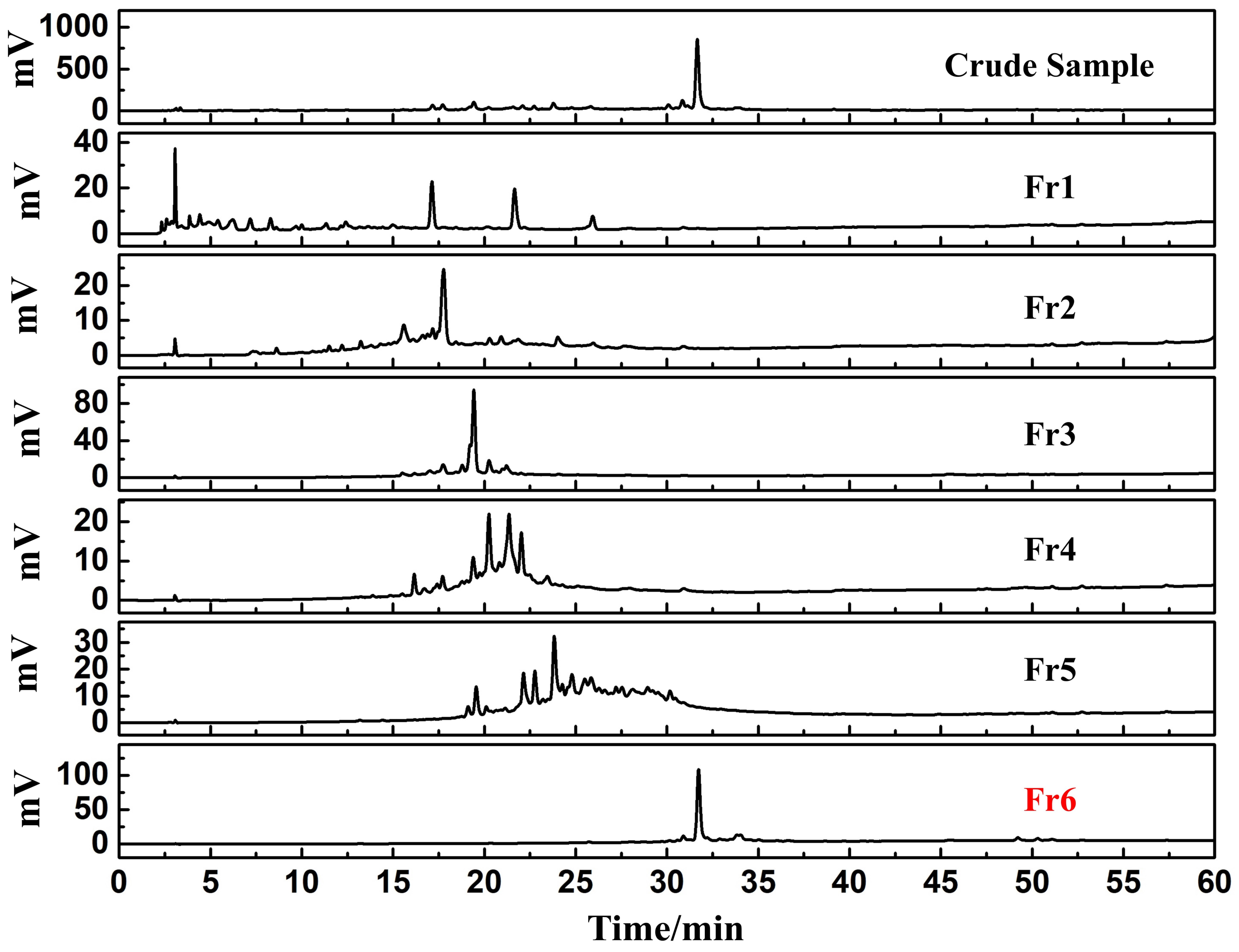
To further analyze the sample, these fractions were resuspended in a methanol/water mixture (9:1 v/v, 1.0 mL, 20.0 mg/mL) prior to filtration through a 0.45 μM organic filter membrane. Subsequently, the accumulated fractions Fr1-Fr6 were assessed via a Odyssil C18 analytical column under the identical parameters as in Figure 1B. The chromatogram of the analysis of the crude sample of E. nutans and fractions Fr1-Fr6 is shown in Figure 2. As shown in Figure 2, the main DPPH inhibitors of the E. nutans crude sample were efficiently enriched in fraction Fr6. A solution for further chromatographic separation was obtained by resuspending Fr6 in 60.0 mL of water/methanol (1:9 v/v), followed by filtering through a 0.45 μm filter.

3.2. Identification and Extraction of Candidate DPPH Suppressors Based on High-Pressure Liquid Chromatography Separation

The target fraction Fr6 was initially assessed by a Odyssil C18 column (4.6 × 250 mm, 5 μm). As depicted in Figure 3A, a primary peak was evident with asymmetric peak shape for Fr6 on the Odyssil C18 column. Hence, its separation on the Odyssil C18 column did not achieve good results. Subsequently, we chose to analyze it on a Kromasil 100-5-Phenyl column. The analytical chromatogram is illustrated in Figure 3B. Based on our results, Fr6 was able to achieve a good separation on the Kromasil 100-5-Phenyl column, with four symmetrical peaks obtained (peaks 1–4). By comparison, the Kromasil 100-5-Phenyl column was chosen for further Fr6 separation and purification in order to improve the separation efficiency.
An online HPLC-DPPH approach was employed to quickly screen for free radical suppressors in Fr6. The E. nutans Fr6 sample was identified with the online HPLC-DPPH system using the Kromasil 100-5-Phenyl column and the recognition chromatogram is displayed in Figure 3C,D. DPPH suppressors peaks (negative peaks I–II in Figure 3D corresponds to the red heart in Figure 3C) were strongly visible in the E. nutans target fraction Fr6.
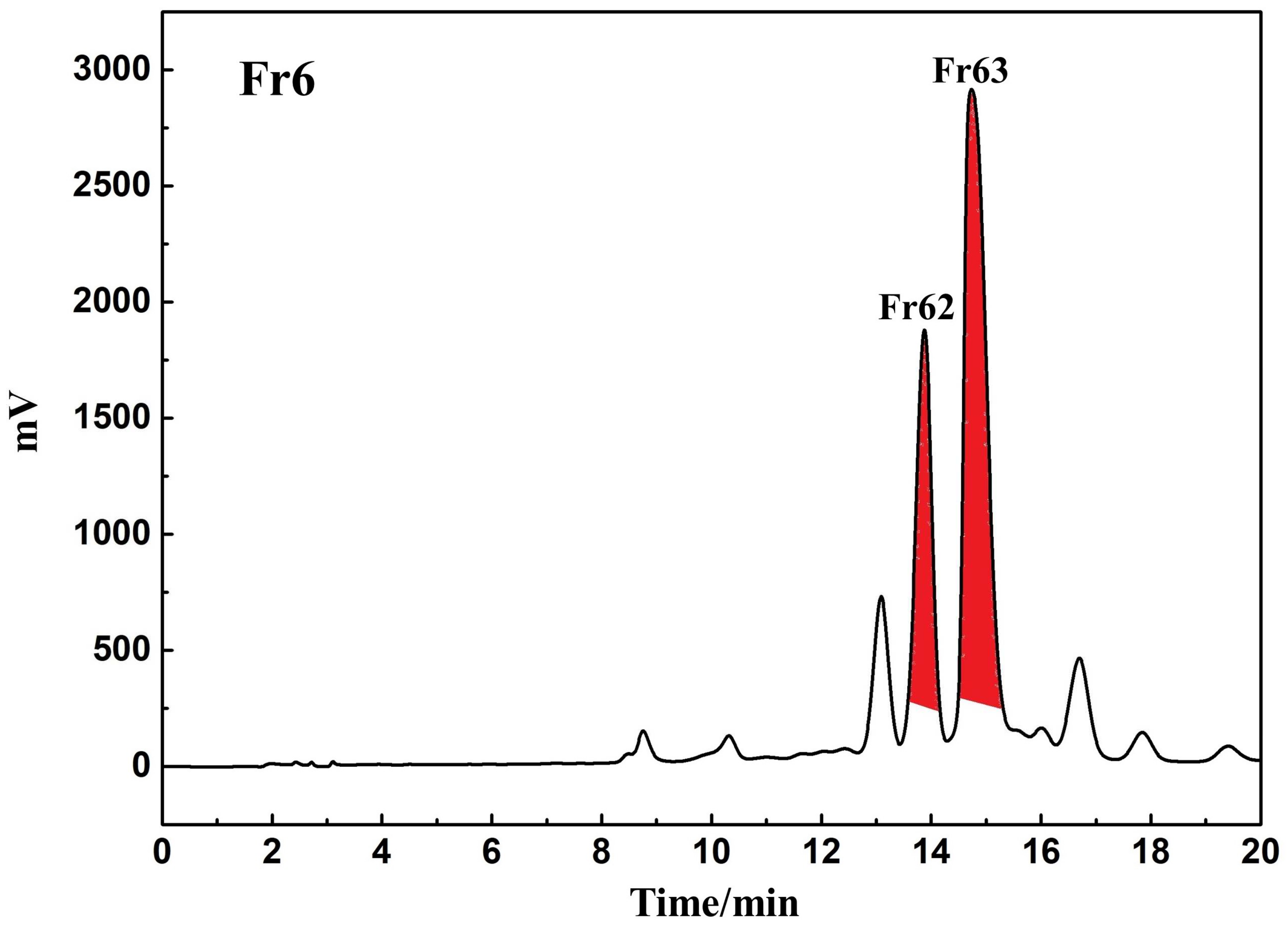
Based on the aforementioned analytical findings, a Kromasil 100-5-Phenyl preparative column (20 × 250 mm, 5 μm) was selected for additional preparation to enhance the DPPH suppressor purity. Under the condition of isocratic elution of acetonitrile-0.1% v/v formic acid in water (0–20 min, 35% acetonitrile), the DPPH suppressor peaks showed sufficient resolution, as indicated in Figure 3C,D. Hence, subsequent examinations were made under this setting. Following linear amplification, the DPPH inhibitor peaks were separated and purified on the Kromasil 100-5-Phenyl preparative column under optimized conditions. The result is presented in Figure 4. It can be seen from the figure that good baseline separation of Fr62, Fr63 can be achieved on the Kromasil 100-5-Phenyl column, and the peak shapes revealed good symmetry. This explains the good complementary selectivity of Odyssil C18 and Kromasil 100-5-Phenyl columns, which can be used in combination to improve separation efficiency and monomer purity in natural product separation and purification work. Fr62 and Fr63 were accrued and condensed after passing through 75 replicates, yielding 432.16 and 489.01 mg, respectively, and providing a net recovery of 15.48%.

3.3. Purity, Activity and Structural Characterization of the Isolated DPPH Suppressors

The extracted DPPH suppressors, Fr62 and Fr63 activities, and purity were re-evaluated using the online HPLC- DPPH system with a Odyssil C18 analytical column. As depicted in Figure 5, all compounds possessed purities above 95%.
The resulting ESI-MS, 1H NMR and 13C NMR spectra were compared against the published literature to delineate the structure of the target DPPH suppressors Fr62 and Fr63 (Figure 6). The structural identification of Fr62 and Fr63 from the supplementary information are presented in Figures S1–S6 (Supplementary Materials). The two DPPH suppressors Fr62 and Fr63 match the data for salcolin A and tricin, respectively, based on all spectrum data [21,22].
Compound Fr62 (salcolin A, 432.16 mg, yellow powder, ESI-MS m/z: 525.39 [M-H], calc. for C27H26O11 m/z 526.1475): 1H NMR (600 MHz, DMSO-d6) 7.31 (2H, s, H-2’, 6’), 6.93 (1H, d, J = 1.8 Hz, H-2’’), 6.75 (1H, dd, J = 8.1, 1.8 Hz, H-6’’), 6.70 (1H, d, J = 8.1 Hz, H-5’’), 6.69 (1H, s, H-3), 6.56 (1H, d, J = 2.0 Hz, H-8), 6.21 (1H, d, J = 2.0 Hz, H-6), 5.17 (1H, m, H-7’’), 4.35 (1H, ddd, J = 5.2, 5.1, 3.7 Hz, H-8’’), 3.88 (6H, s, 3’, 5’-OCH3), 3.75 (3H, s, 3’’-OCH3), 3.49 (1H, dd, J = 11.5, 5.2 Hz, H-9’’a), 3.44 (1H, dd, J = 11.5, 5.1 Hz, H-9’’b); 13C NMR (151 MHz, DMSO- d6) 181.8 (C-4), 164.7 (C-7), 163.0 (C-2), 161.4 (C-5), 157.4 (C-9), 153.0 (C-3’, 5’), 147.0 (C-3’’), 145.4 (C-4’’), 139.5 (C-4’), 133.2 (C-1’’), 125.2 (C-1’), 119.4 (C-6’’), 114.7 (C-5’’), 111.0 (C-2’’), 104.8 (C-3), 104.3 (C-10), 103.7 (C-2’, 6’), 99.0 (C-6), 94.4 (C-8), 86.5 (C-8’’), 72.2 (C-7’’), 60.2 (C-9’’), 56.4 (C-3’, 5’-OCH3), 55.5 (C-3’’-OCH3). According to our ESI-MS, 1H NMR and 13C NMR data, as well as in the literature review, the compound was identified as salcolin A [21].
Compound Fr63 (tricin, 489.01 mg, yellow powder, ESI-MS m/z: 329.21 [M-H], calc. for C17H14O7 m/z 330.0740): 1H NMR (600 MHz, DMSO-d6) 3.88 (6H, s, 2OCH3), 6.20 (1H, d, J = 2.0 Hz, H-6), 6.56 (1H, d, J = 2.0 Hz, H-8), 6.98 (1H, s, H-3), 7.32 (2H, s, H-2’, 6’); 13C NMR (151 MHz, DMSO-d6) 181.8 (C-4), 164.3 (C-7), 163∙6 (C-2), 161.4 (C-9), 157.4 (C-5), 148.2 (C-3’, 5’), 139.9 (C-4’), 120.4 (C-1’), 104.4 (C-2’, 6’), 103.7 (C-10), 103.6 (C-3), 98.9 (C-6), 94.2 (C-8), 56.4 (OCH3). According to our ESI-MS, 1H NMR and 13C NMR data, as well as in the literature review, the compound was identified as tricin, by a comparison with the compound in the literature [22].
Sacolin A and tricin participate in a myriad of biological activities, namely, anti-inflammatory, antioxidant, antiviral, antitumor and immunomodulatory, especially anti-inflammatory effects. Both compounds are flavonoids, which are commonly found in graminaceous plants, such as bamboo, barley, rice, wheat, and maize, and are considered to be healthy and harmless nutrients. The present study demonstrated that sacolin A and tricin are abundant in E. nutans, thus, providing insight into the extraction and purification of sacolin A and tricin.

4. Conclusions

Flavonoids are widely available in a variety of forage grasses. Owing to their numerous biological functions, the application of flavonoids can significantly improve animal production performance, enhance animal body resistance to disease and improve animal body immune function [23]. Flavonoids also possess weak estrogenic activity, which can markedly enhance animal growth, the egg-laying performance of poultry, promote reproductive system development, and improve fertility. Moreover, flavonoids augment the immune function of T cells, natural killer cells, and killer cells. Lastly, owing to their excellent antioxidant activity, flavonoids scavenge free radicals and reduce their production.
Herein, we employed a rapid and efficient method based on a combination of medium- and high-pressure chromatography, and online HPLC-DPPH analysis was designed to screen, isolate and purify flavonoids from E. nutans. By pre-processing the sample using MCI GEL® CHP20P MPLC, the process yielded 5.95 g of the target fraction Fr6. Further, Kromasil 100-5-Phenyl column was used for the separation and purification of target fraction Fr6, yielding 432.16 mg of Fr62 (7.26% recovery) and 489.01 mg of Fr63 (8.22% recovery). The reassessment of purity and radical scavenging activity showed that all compounds are active with >95% purity. In all, two flavonoids were isolated with satisfactory purity from the target sample, which indicated that the combination of medium- and high-pressure chromatography and online HPLC-DPPH method was efficient for preparative separation of the flavonoids from E. nutans. Moreover, this procedure is significant for the isolation and purification of flavonoids from other forage grasses.

Supplementary Materials

The following supporting information can be downloaded at: https://www.mdpi.com/article/10.3390/separations9120437/s1, Figure S1: ESI mass spectrum of salcolin A; Figure S2: 1H NMR Spectrum (600 MHz) of salcolin A (in DMSO-d6); Figure S3: 13C NMR Spectrum (151 MHz) of salcolin A (in DMSO-d6). Figure S4: ESI mass spectrum of tricin; Figure S5: 1H NMR Spectrum (600 MHz) of tricin (in DMSO-d6); Figure S6: 13C NMR Spectrum (151 MHz) of tricin (in DMSO-d6).

Author Contributions

Writing—original draft, methodology, data curation, formal analysis, investigation L.S.; writing—review and editing, conceptualization, project administration and funding acquisition Z.H.; data curation, formal analysis Y.H. All authors have read and agreed to the published version of the manuscript.

Funding

This research was funded by the Fund of Technical and Economic Research Institute of China Minmetals Salt Lake Co., Ltd. [2022].

Institutional Review Board Statement

Not applicable.

Informed Consent Statement

Not applicable.

Data Availability Statement

All data generated or analyzed during this study are included in this published article (and its Supplementary Materials).

Conflicts of Interest

The authors declare no conflict of interest.

References

  1. Sies, H. Oxidative stress: A concept in redox biology and medicine. Redox. Biol. 2015, 4, 180–183. [Google Scholar] [CrossRef] [PubMed] [Green Version]
  2. Forman, H.J.; Zhang, H. Targeting oxidative stress in disease: Promise and limitations of antioxidant therapy. Nat. Rev. Drug. Discov. 2021, 20, 689–709. [Google Scholar] [CrossRef] [PubMed]
  3. Senoner, T.; Dichtl, W. Oxidative Stress in Cardiovascular Diseases: Still a Therapeutic Target? Nutrients 2019, 11, 2090. [Google Scholar] [CrossRef] [PubMed] [Green Version]
  4. Ighodaro, O.M. Molecular pathways associated with oxidative stress in diabetes mellitus. Biomed. Pharmacother. 2018, 108, 656–662. [Google Scholar] [CrossRef] [PubMed]
  5. Poprac, P.; Jomova, K.; Simunkova, M.; Kollar, V.; Rhodes, C.J.; Valko, M. Targeting Free Radicals in Oxidative Stress-Related Human Diseases. Trends. Pharmacol. Sci. 2017, 38, 592–607. [Google Scholar] [CrossRef] [PubMed]
  6. Tan, B.L.; Norhaizan, M.E. Effect of High-Fat Diets on Oxidative Stress, Cellular Inflammatory Response and Cognitive Function. Nutrients 2019, 11, 2579. [Google Scholar] [CrossRef] [Green Version]
  7. Dutta, S.; Sengupta, P.; Slama, P.; Roychoudhury, S. Oxidative Stress, Testicular Inflammatory Pathways, and Male Reproduction. Int. J. Mol Sci. 2021, 22, 10043. [Google Scholar] [CrossRef]
  8. McGarry, T.; Biniecka, M.; Veale, D.J.; Fearon, U. Hypoxia, oxidative stress and inflammation. Free. Radic. Biol. Med. 2018, 125, 15–24. [Google Scholar] [CrossRef]
  9. Mavangira, V.; Sordillo, L.M. Role of lipid mediators in the regulation of oxidative stress and inflammatory responses in dairy cattle. Res. Vet. Sci. 2018, 116, 4–14. [Google Scholar] [CrossRef]
  10. Yang, Y.; He, T.; Lu, S.; Huang, R.; Wang, Z. Records of Tibetan Medicine; Qinghai Publishing House: Qinghai, China, 1991; pp. 450–451. [Google Scholar]
  11. Capstaff, N.M.; Miller, A.J. Improving the Yield and Nutritional Quality of Forage Crops. Front. Plant. Sci. 2018, 9, 535. [Google Scholar] [CrossRef]
  12. Manzanares, C.; Yates, S.; Ruckle, M.; Nay, M.; Studer, B. TILLING in forage grasses for gene discovery and breeding improvement. N. Biotechnol. 2016, 33, 594–603. [Google Scholar] [CrossRef] [Green Version]
  13. Macáková, K.; Afonso, R.; Saso, L.; Mladěnka, P. The influence of alkaloids on oxidative stress and related signaling pathways. Free. Radic. Biol. Med. 2019, 134, 429–444. [Google Scholar] [CrossRef]
  14. Dawa, Y.; Du, Y.; Wang, Q.; Chen, C.; Zou, D.; Qi, D.; Ma, J.; Dang, J. Targeted isolation of 1,1-diphenyl-2-picrylhydrazyl inhibitors from Saxifraga atrata using medium- and high- pressure liquid chromatography combined with online high performance liquid chromatography-1,1-diphenyl-2- picrylhydrazyl detection. J. Chromatogr. A 2021, 1635, 461690. [Google Scholar] [CrossRef]
  15. Ionita, P. The Chemistry of DPPH· Free Radical and Congeners. Int. J. Mol. Sci. 2021, 22, 1545. [Google Scholar] [CrossRef]
  16. Gulcin, İ. Antioxidants and antioxidant methods: An updated overview. Arch. Toxicol. 2020, 94, 651–715. [Google Scholar] [CrossRef] [Green Version]
  17. Zhou, M.; Wei, L.; Sun, Z.; Gao, L.; Meng, Y.; Tang, Y.; Wu, Y. Production and transcriptional regulation of proanthocyanidin biosynthesis in forage legumes. Appl. Microbiol. Biotechnol. 2015, 99, 3797–3806. [Google Scholar] [CrossRef]
  18. Lee, M.R. Forage polyphenol oxidase and ruminant livestock nutrition. Front. Plant. Sci. 2014, 5, 694. [Google Scholar] [CrossRef]
  19. Cui, Y.; Shen, N.; Yuan, X.; Dang, J.; Shao, Y.; Mei, L.; Tao, Y.; Wang, Q.; Liu, Z. Two-dimensional chromatography based on on-line HPLC-DPPH bioactivity-guided assay for the preparative isolation of analogue antioxidant compound from Arenaria kansuensis. J. Chromatogr. B. 2017, 1046, 81–86. [Google Scholar] [CrossRef]
  20. Fang, Y.; Dawa, Y.; Wang, Q.; Lv, Y.; Yu, W.; Li, G.; Dang, J. Targeted isolation of 1,1-diphenyl-2-picrylhydrazyl inhibitors from Saxifraga atrata and their antioxidant activities. J. Sep. Sci. 2022, 45, 2435–2445. [Google Scholar] [CrossRef]
  21. Jung, Y.J.; Park, J.H.; Cho, J.G.; Seo, K.H.; Lee, D.S.; Kim, Y.C.; Kang, H.C.; Song, M.C.; Baek, N.I. Lignan and flavonoids from the stems of Zea mays and their anti-inflammatory and neuroprotective activities. Arch. Pharm. Res. 2015, 38, 178–185. [Google Scholar] [CrossRef]
  22. Zhang, X.Q.; Jiang, W.W.; Wang, Y.; Li, Y.L.; Ye, W.C. A new phenylpropanoic acid derivatives from the roots of Ficus stenophylla. Yao. Xue. Xue. Bao. 2008, 43, 281–283. [Google Scholar] [PubMed]
  23. Gholami, A.; De Geyter, N.; Pollier, J.; Goormachtig, S.; Goossens, A. Natural product biosynthesis in Medicago species. Nat. Prod. Rep. 2014, 31, 356–380. [Google Scholar] [CrossRef] [PubMed]
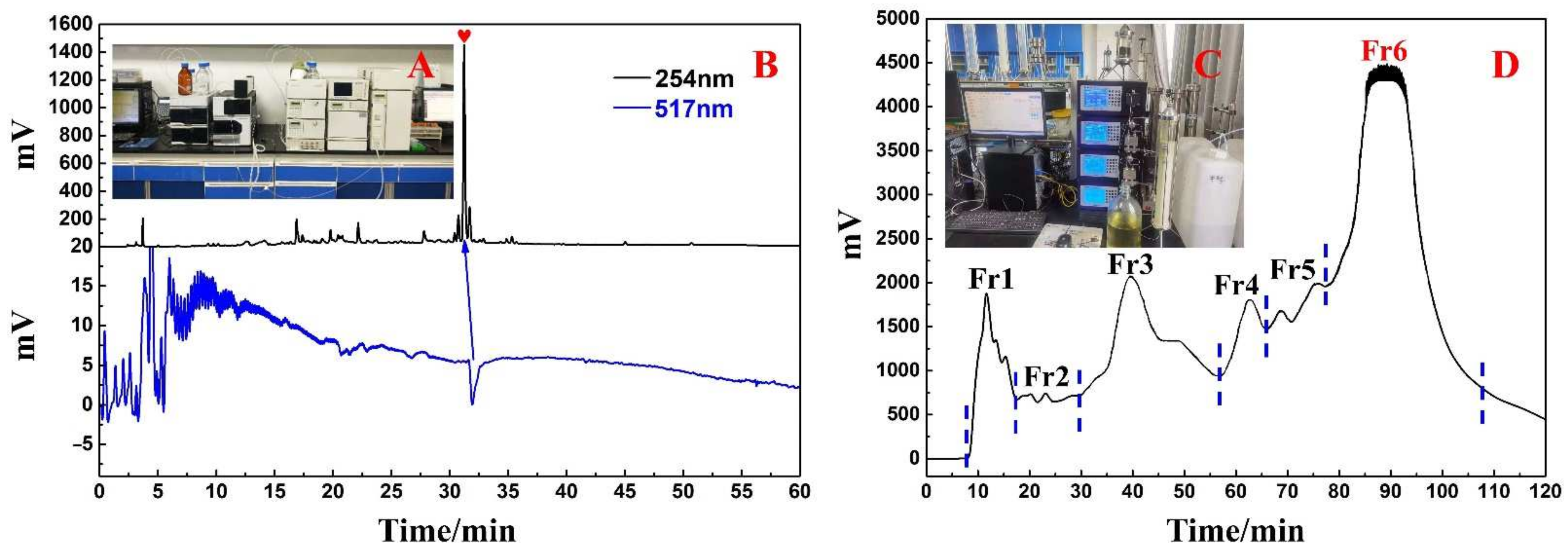
Figure 1. The actual online HPLC-DPPH system (A) and the analytical chromatogram on a Odyssil C18 analytical column and DPPH inhibitory activity profile of the E. nutans crude sample (B). The actual micro gel chp20p medium pressure liquid chromatography system (C) and E. nutans amorphous silica gel mixture separation chromatogram (D).
Separations 09 00437 g001
Figure 2. HPLC analysis of the E. nutans crude extract and Fr1-Fr6 on the Odyssil C18 analytical column.
Separations 09 00437 g002
Figure 3. HPLC analysis of E. nutans Fr6 on the Odyssil C18 analytical column (A) and on a Kromasil 100-5-Phenyl analytical column (B). The analytical chromatogram (C) and DPPH radical scavenging profile (D) of the E. nutans target Fr6 sample on the Kromasil 100-5-Phenyl analytical column analytical column. The numbers 1, 2, 3 and, 4 represent four different peaks. Red hearts indicate peaks with antioxidant activity, and I and II correspond to corresponding inverted peaks, indicated by arrows.
Separations 09 00437 g003
Figure 4. The preparative chromatogram of the E. nutans target Fr6 sample on a Kromasil 100-5-Phenyl preparative column. The red part represents the target component (Fr62 and Fr63).
Separations 09 00437 g004
Figure 5. Purity and DPPH inhibitory activity of the target fraction activity verification chromatogram of the isolated Fr62 (A,B), and Fr63 (C,D) fractions on the Odyssil C18 analytical column. Red hearts indicate peaks with antioxidant activity, indicated by arrows.
Separations 09 00437 g005
Figure 6. Chemical structures of the isolated DPPH inhibitors. (A,B) represent the chemical structure of Fr62 and Fr63, respectively.
Separations 09 00437 g006
Publisher’s Note: MDPI stays neutral with regard to jurisdictional claims in published maps and institutional affiliations.