Article

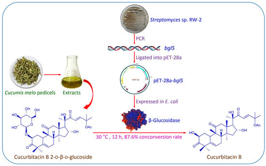
Improvement of Cucurbitacin B Content in Cucumis melo Pedicel Extracts by Biotransformation Using Recombinant β-Glucosidase

1
College of Pharmaceutical Science, Zhejiang University of Technology, Hangzhou 310014, China
2
Zhoushan Institute for Food and Drug Control, Zhoushan 316021, China
*
Author to whom correspondence should be addressed.
Separations 2021, 8(9), 138; https://doi.org/10.3390/separations8090138
Submission received: 22 July 2021 / Revised: 24 August 2021 / Accepted: 26 August 2021 / Published: 1 September 2021

Abstract

:
For the efficient biotransformation of cucurbitacin B 2-o-β-d-glucoside (CuBg) to cucurbitacin B (CuB) in Cucumis melo pedicel extracts, the β-glucosidase gene bglS—consisting of 1344 bp (447 amino acids) from Streptomyces sp. RW-2—was cloned and expressed in Escherichia coli BL21(DE3). The activity of recombinant β-glucosidase with p-nitrophenyl-β-d-glucoside (pNPG) as a substrate was 3.48 U/mL in a culture. Using the recombinant β-glucosidase for the biotransformation of C. melo pedicel extracts, CuBg was converted into CuB with a conversion rate of 87.6% when the concentration of CuBg was 0.973 g/L in a reaction mixtures. The concentration of CuB in C. melo pedicel extracts was improved from 13.6 to 20.2 g/L after biotransformation. The present study provides high-efficiency technology for the production of CuB from its glycoside by biotransformation.

Graphical Abstract

1. Introduction

Cucurbitacin B (CuB) has been reported to show various biological activities, such as anti-inflammatory, anticancer, antimicrobial, antidiabetic, antiobesity, and antihyperglycemic effects [1,2,3,4,5]. In particular, the anticancer activity of CuB has attracted much attention because of its significant cytotoxic effects in more than 100 cancer cell lines [6]. Though CuB is not used worldwide as a drug at present, it has been considered to be the main active ingredient in extracts of Cucumis melo L. pedicel, which have been used as a traditional herbal medicine (HuLuSu Pian) for the digestive system and hepatic diseases for more than 20 years in China [7]. The content of CuB in C. melo pedicels ranges from 8 to 14 mg/g [8,9]. In fact, the glycoside of CuB, cucurbitacin B 2-o-β-d-glucoside (CuBg), is also abundant in C. melo pedicels. Our analysis showed that the contents of CuB and CuBg in C. melo pedicels from Bozhou (Anhui, China) were 13.6 and 9.73 mg/g, respectively. Due to the relatively low bioavailability of glycosides [10], CuBg is not considered to be an active ingredient in extracts of C. melo pedicel. If CuBg in C. melo pedicel is converted into CuB, the content of CuB can be markedly increased, which will then greatly increase the extraction yield of CuB. The conversion of CuBg to CuB can be accomplished through acid hydrolysis, but this method is not efficient. For example, when extracts from C. melo pedicels were treated by 0.1 mol/L acetic acid, the content of CuB was only increased by 7.46% [11].
Biotransformation by a microbial enzyme has high specificity and efficiency; it has been applied in the production of many natural drugs. In our previous study, a process for the biotransformation of CuBg to CuB using crude enzyme from Streptomyces sp. RW-2 was developed [12]. When C. melo pedicel extracts were used as the substrate, CuBg was specifically converted into CuB with an almost 100% conversion rate, and the concentration of CuB in C. melo pedicel extracts doubled after biotransformation. However, with this method, the concentration of substrate is low, resulting in a low productivity. Using purified β-glucosidase from Streptomyces sp. RW-2 may improve biotransformation efficiency. However, the low production of β-glucosidase from Streptomyces may impede the application of this method. Using recombinant Escherichia coli to express heterologous enzymes has numerous advantages; in particular, it can increase enzyme production [13]. This method has been applied in the biotransformation of many natural products [14,15,16,17,18], resulting in improved biotransformation efficiency.
In the present study, the β-glucosidase gene bglS from Streptomyces sp. RW-2 was cloned and expressed in Escherichia coli BL21(DE3). Using the recombinant β-glucosidase for the biotransformation of C. melo pedicel extracts, the productivity of CuB was significantly improved. The biotransformation scheme of CuBg to CuB by β-glucosidase is illustrated in Figure 1.

2. Materials and Methods

2.1. Chemicals and Enzymes

Standard CuB was purchased from Sigma-Aldrich (St. Louis, MO, USA). CuBg (97.4% purity) was separated from C. melo pedicels by HPLC. DNA isolation, PCR, and plasmid isolation kits, as well as restriction enzymes, were obtained from Sangon Biotech (Shanghai, China). All other chemicals were of HPLC, analytical, or biochemical grade and obtained from local suppliers.

2.2. Strains and Plasmids

Streptomyces sp. RW-2, which was isolated from an enrichment culture of C. melo pedicels, was deposited at the China Center for Type Culture Collection (CCTCC, no. M2013330). E. coli BL21(DE3) cells from Tsingke Biological Technology (Beijing, China) were used for enzyme expression. The pET-28a (+) plasmid (Sangon Biotech, Shanghai, China) was used for cloning.

2.3. Construction of Recombinant E. coli

The degenerate primers (5′-AC(G/C)CT(G/C)T(A/T)CCACTGGGACCT-3′ and 5′-(G/C)GCCCA(C/T)TCGAAGTTGTC-3′) were designed based on the conserved sequences of β-glucosidase genes from known Streptomyces strains retrieved from NCBI (http://www.ncbi.nlm.nih.gov). Using the degenerate primers, a partial sequence of the β-glucosidase gene was amplified from the genomic DNA of Streptomyces sp. RW-2 via polymerase chain reaction (PCR), and compared by NCBI BLAST to obtain a known β-glucosidase gene sequence from a Streptomyces strain. According to this sequence, two primers (5′-tgggtcgcggatccGAATTCATGGCGATCGACGAGCGCGGC-3′ and 5′-tggtggtggtggtggtgCTCGAGTCAGCCGGTCCGGGGC-3′), which included EcoRI and XhoI restriction sites (underlined) and homologous arm sequences (lower case), were designed. The full-length β-glucosidase gene was amplified from genomic DNA of Streptomyces sp. RW-2, ligated into the pET-28a (+) expression vector, sequenced, and introduced into E. coli BL21(DE3) by the standard method, thus generating recombinant E. coli BL21-bglS.

2.4. Expression of Recombinant β-Glucosidase

Pre-cultures of E. coli BL21-bglS were prepared in a Luria-Bertani (LB) medium containing 50 μg/mL of kanamycin and cultured overnight in a shaking incubator at 37 °C and 200 r/min. Aliquots (2.5 mL) of the pre-culture were inoculated into fresh LB containing kanamycin. β-Glucosidase expression was evaluated by culturing at 37 °C and 200 r/min until an OD600 of 0.6 was reached; then, 0.5 mmol/L IPTG was added to induce enzyme expression at 25 °C and 200 r/min for a further 10 h.

2.5. Enzyme Essay

BL21-bglS cells were harvested from 50 mL cultures by centrifugation at 8000× g for 5 min, washed twice with deionized water, resuspended in 5 mL phosphate-buffered saline (PBS; 0.2 mol/L, pH 6.0), and then disrupted by sonication. The cell lysates were centrifuged at 8000× g for 10 min. Then, the supernatant fraction was used for sodium dodecyl sulfate polyacrylamide gel electrophoresis (SDS-PAGE) and a β-glucosidase activity assay.
The expression of the recombinant β-glucosidase was confirmed by SDS-PAGE using 12% (w/v) polyacrylamide gels [19].
The β-glucosidase activity was assayed using p-nitrophenyl-β-d-glucoside (pNPG) as a substrate [20]. The enzymatic reaction mixtures (100 μL) containing 50 μL of the enzyme preparation and 50 μL of pNPG (5 mmol/L in PBS) were incubated for 15 min at 30 °C in a 96-well microtiter plate. After the addition of 50 μL of 0.4 mol/L Na2CO3 to the reaction, the amount of p-nitrophenol (pNP) released was measured at 410 nm on a microplate reader. One enzyme unit (U) was defined as the amount of enzyme that released 1 μmol pNP per minute in the abovementioned conditions.

2.6. Biotransformation Process

CuBg, dissolved in 1 mL of methanol or 1 mL C. melo pedicel extracts, was mixed with 1 mL of cell lysates and 8 mL of PBS in a 50 mL flask. Biotransformation was performed at 30 °C and 200 r/min for 16 h. After biotransformation, the mixtures were extracted twice with 10 mL of ethyl acetate. After the evaporation of the solvent, the residue was dissolved in 1 mL of methanol, filtered, and analyzed by high performance liquid chromatography (HPLC).
The preparation of C. melo pedicel extracts has previously been described [12]. Extracts from C. melo pedicels (10 g) were dissolved in 10 mL of ethanol, resulting in concentrations of CuBg and CuB of 9.73 and 13.6 g/L, respectively.

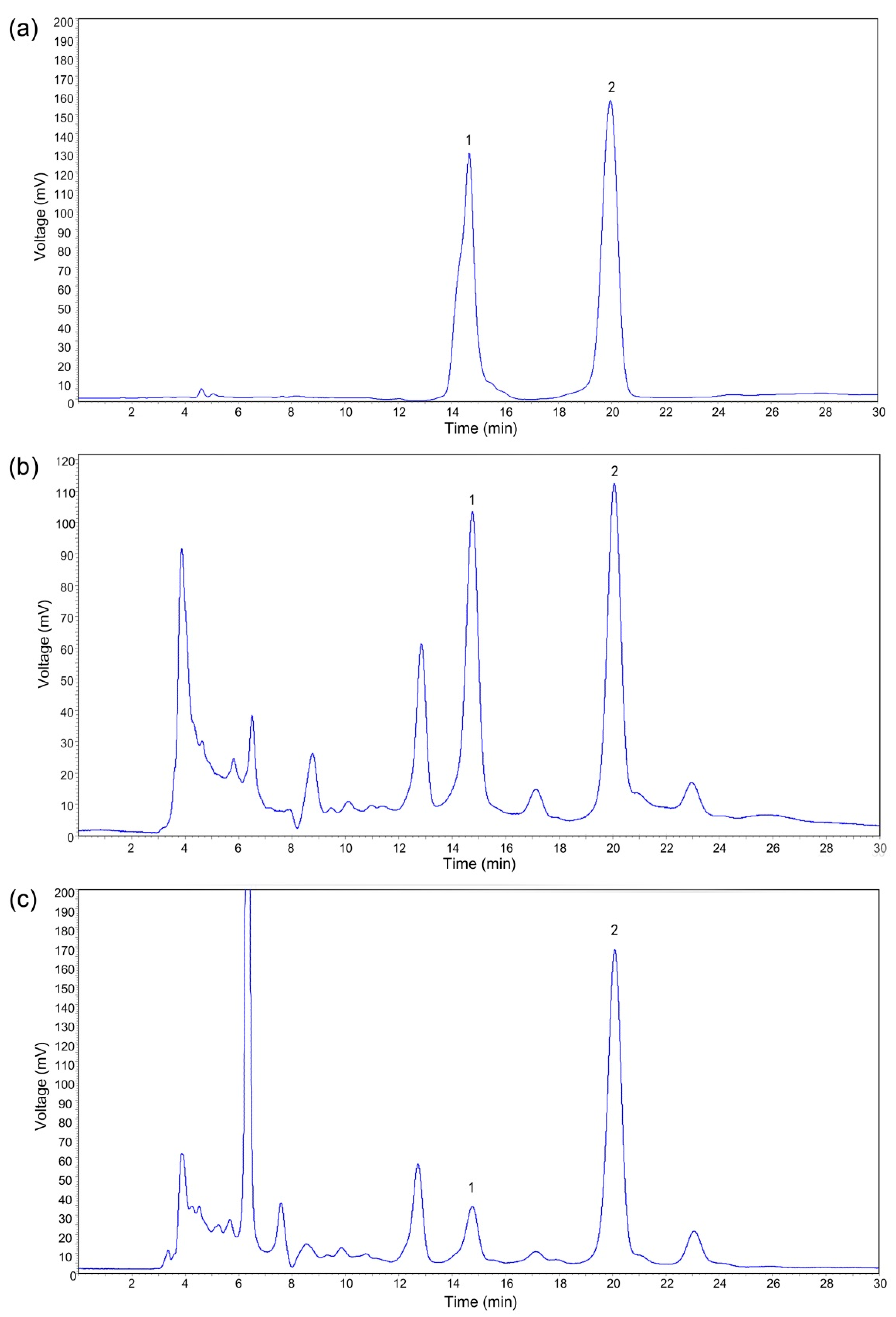
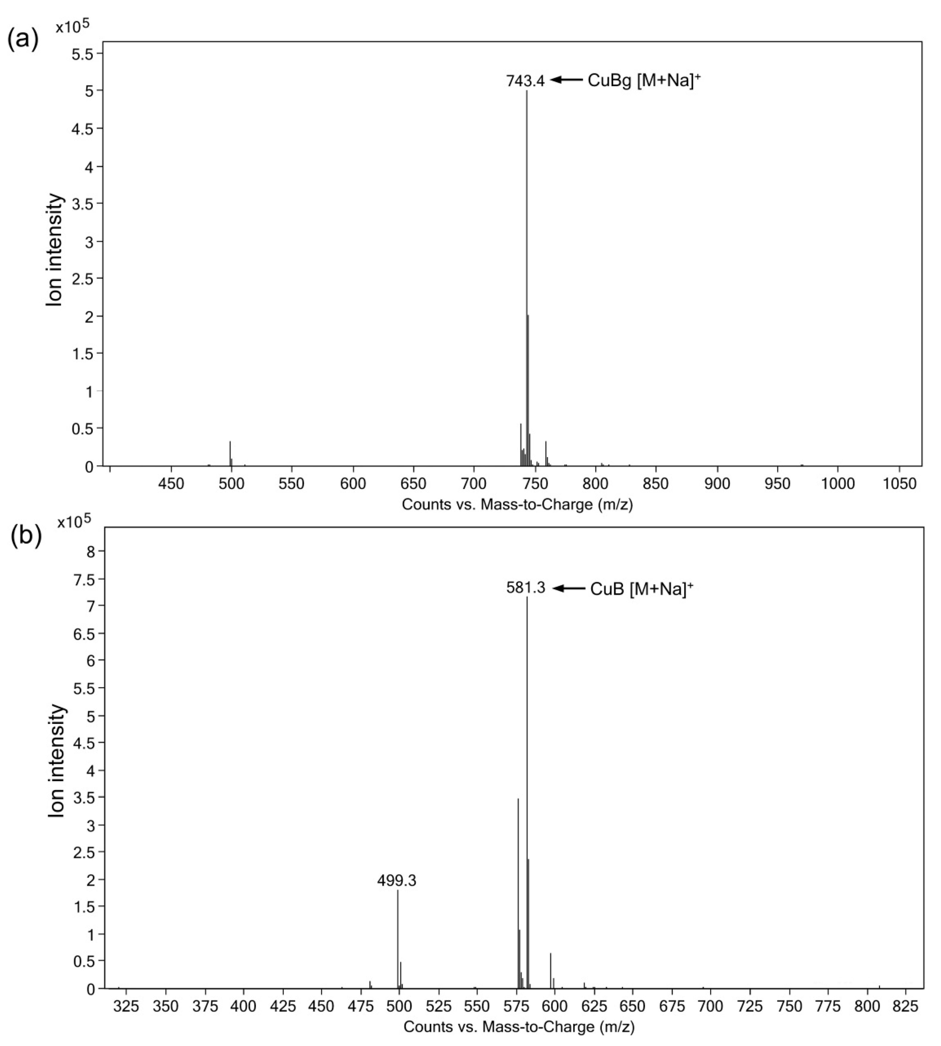
2.7. Analysis of Biotransformation Products

The quantification of CuB and CuBg was performed by HPLC on a Shimadzu SPD-20A (Kyoto, Japan) equipped with a Zorbax Eclipse XDB-C18 column (5 µm, 4.6 × 150 mm). The mobile phase was methanol-water solution (60:40, v/v), at a flow rate of 1 mL/min. The constituents were detected at 228 nm.
The presence of CuBg and CuB in C. melo pedicel extracts after biotransformation was confirmed by liquid chromatography–electrospray ionization-mass spectrometry (LC–ESI-MS) on an LCQTM Deca XP plus mass spectrometer (Thermo-Fisher, Waltham, MA, USA). The capillary temperature was 350 °C, and the capillary voltage was 4.0 kV.

3. Results and Discussion

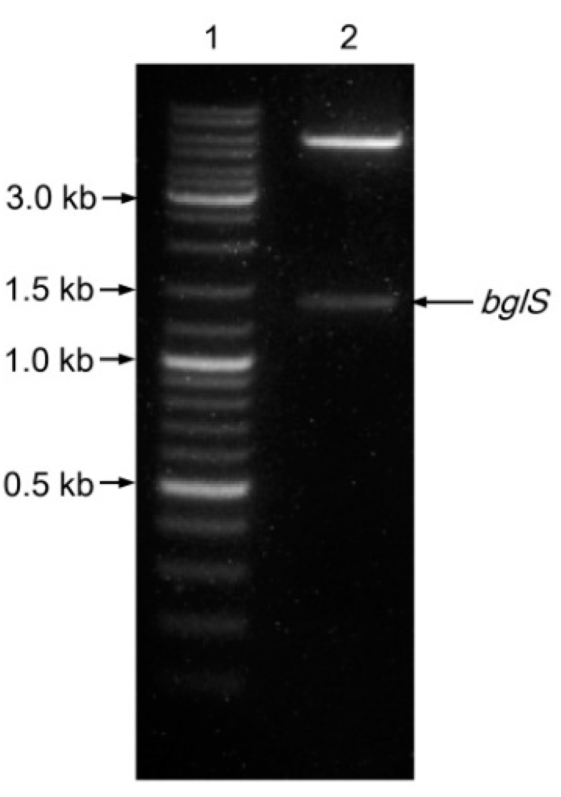
3.1. Cloning of bglS and Sequence Analysis

An 836 bp nucleotide sequence was cloned from genomic DNA of Streptomyces RW-2 by PCR with degenerate primers. The NCBI BLAST analysis of this sequence showed that it had 99.4% similarity to a β-glucosidase gene from Streptomyces nigra strain 452 (GenBank accession no. AWE50172). According to this β-glucosidase gene sequence, two primers were designed to clone the full-length β-glucosidase gene from the genomic DNA of Streptomyces sp. RW-2, and a sequence of approximately 1400 bp was obtained by PCR (Figure 2). An analysis of the sequenced DNA by DNAMAN 10.0 (http://dnaman.software.informer.com/6.0) showed that it contained a 1344 bp open reading frame encoding a protein of 447 amino acids. The nucleotide sequence of bglS was submitted to GeneBank (accession no. MW916284).
The NCBI BLAST analysis of bglS showed the highest nucleotide sequence similarity (99.63%) to a β-glucosidase gene from S. nigra strain 452 but less than 87.35% similarity to that from other Streptomyces strains. The similarity of the 16S rRNA sequences between the Streptomyces sp. RW-2 strain (GenBank accession no. KJ472430.1) and the S. nigra strain 452 (GenBank accession no. CP029043) was 99.72%. The high similarity of the 16S rRNA sequences and β-glucosidase genes between the two strains suggested that strain RW-2 may be S. nigra.

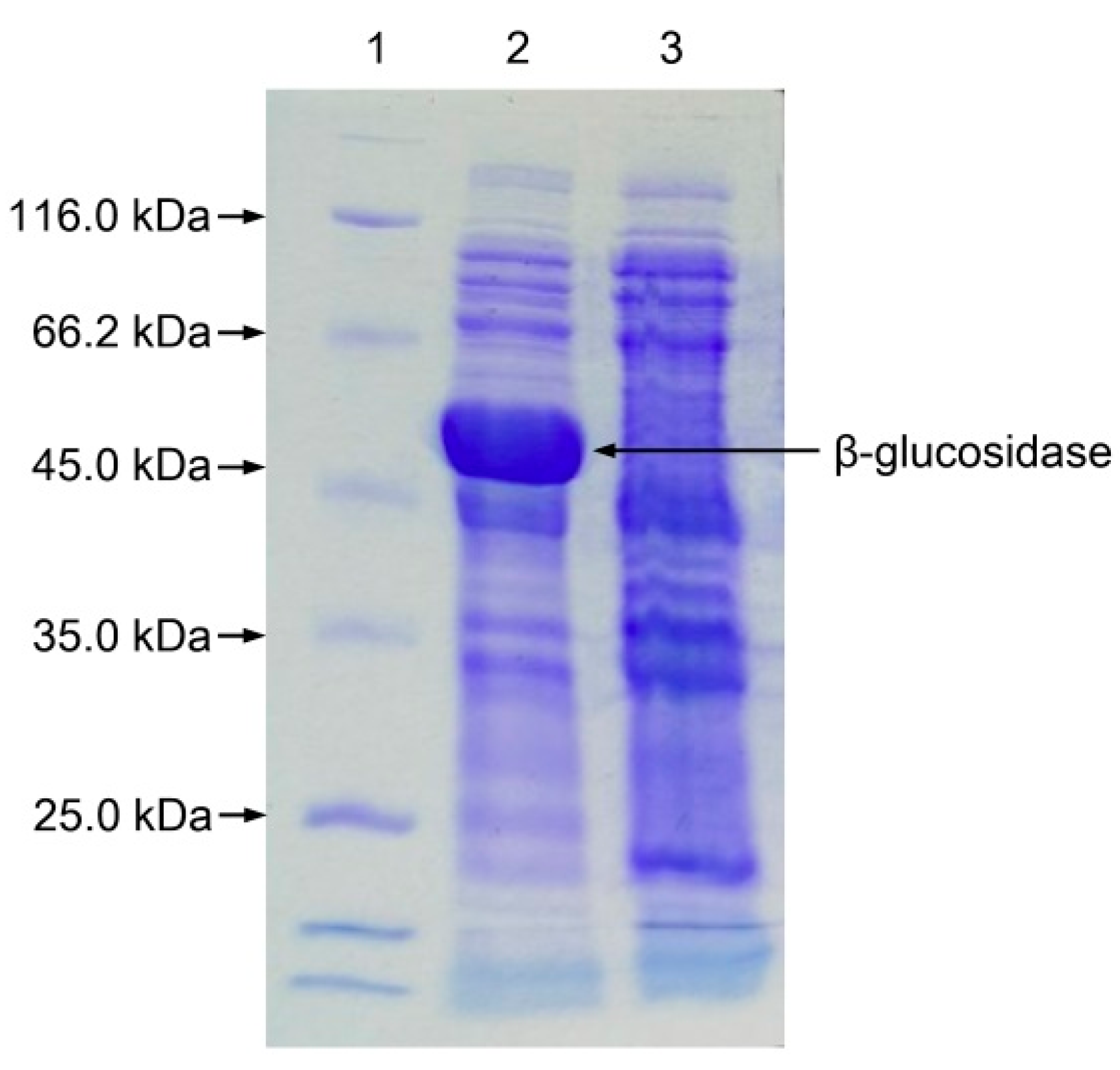
3.2. Expression of bglS in E. coli

The β-glucosidase encoded by bglS was successfully expressed in E. coli BL21(DE3) following induction at 25 °C for 10 h with 0.5 mmol/L IPTG. SDS-PAGE analysis revealed that the expressed protein appeared as a single band with a molecular mass of approximately 50 kDa (Figure 3), consistent with the predicted value of 49.5 kDa based on its 447 amino acids (http://web.expasy.org/compute_pi/). An analysis of the amino acid sequence of this β-glucosidase indicated that it was most similar (99.55%) to the β-glucosidase of two S. nigra strains (GenBank accession nos. WP_164492287 and AWE50172), followed by that of Streptomyces sp. JHA19 (97.32%, GenBank accession no. WP_199835400) and Streptomyces sp. M7 (96.88%, GenBank accession no. WP_181890503.1); additionally, it had less than 93.74% similarity with other Streptomyces strains. An analysis of the amino acid sequence using the SMART program (http://smart.embl.de/) showed that it contained a typical glycoside hydrolase 1 family (GH1) domain, indicating it belongs to the GH1 family.
SDS-PAGE analysis indicated that most of the β-glucosidase was in the soluble fraction, so the crude cell extracts were expected to exhibit enzymatic activity. The activity of recombinant β-glucosidase against pNPG was 3.48 U/mL in the culture.

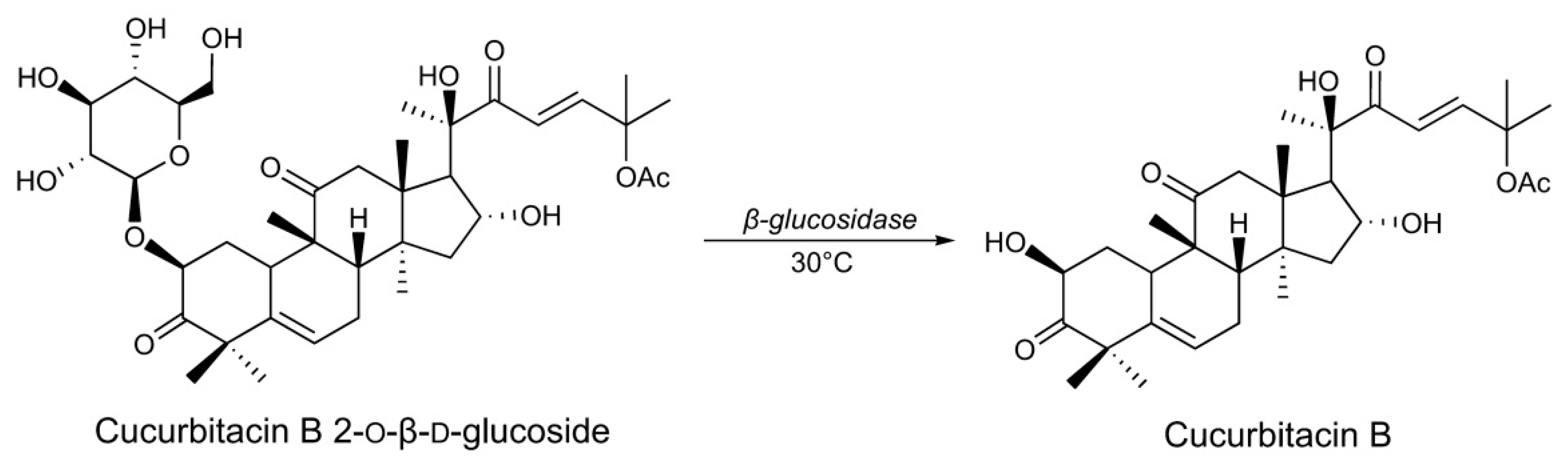
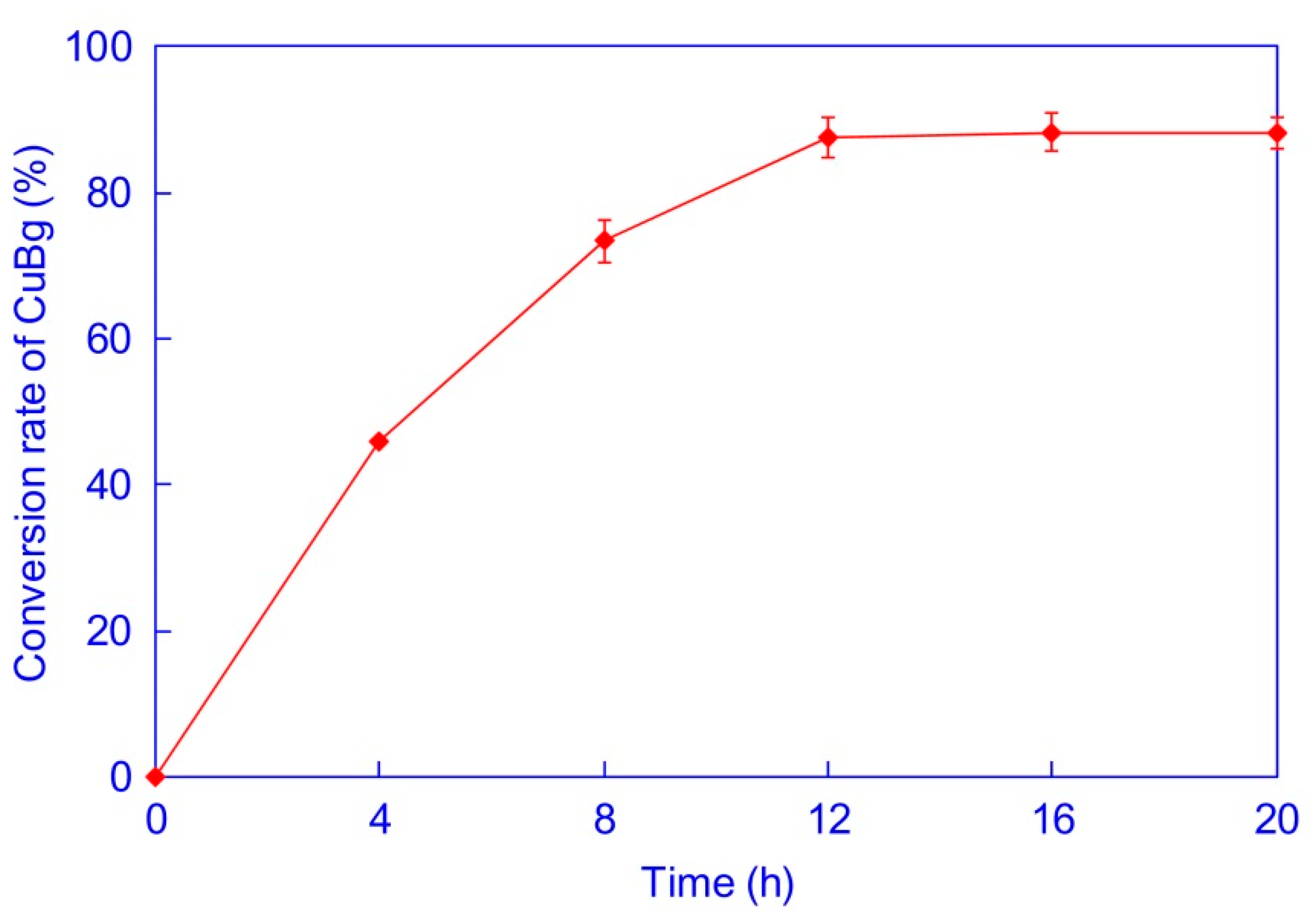
3.3. Biotranformation of CuBg by Recombinant β-glucosidase

The biotransformation of CuBg to CuB using the recombinant β-glucosidase was carried out with an enzymatic activity of 3.48 U/mL in the reaction system. Conversion rates at different CuBg concentrations are shown in Figure 4. At 0.25 g/L of CuBg in the reaction mixture, the conversion rate was as high as 98.1%, indicating that CuBg was specifically converted into CuB. Indeed, no other product was detected at 228 nm by HPLC. During biotransformation by the crude enzyme from Streptomyces sp. RW-2, 0.25 g/L of CuBg in the reaction system resulted in a conversion rate of 92.6% [12]. However, during biotransformation with the recombinant β-glucosidase, 0.75 g/L of CuBg resulted in a conversion rate of 93.2%. These conversion rates were nearly equal, but the substrate concentration was increased three-fold, resulting in a three-fold increase in productivity. These results indicate that using recombinant β-glucosidase from Streptomyces sp. RW-2 significantly improved biotransformation efficiency.

3.4. Biotransformation of C. melo Pedicel Extracts by Recombinant β-Glucosidase

Extracts (1 mL) of C. melo pedicels in a 10 mL reaction system—with concentrations of CuBg and CuB of 0.973 and 1.36 g/L, respectively—were treated with the recombinant β-glucosidase with an activity of 3.48 U/mL. Biotransformation was carried out at 30 °C and monitored for 20 h by HPLC (Figure 5). The enzyme hydrolyzed the greatest amount of CuBg to CuB within 12 h. After biotransformation, the concentration of CuB in the reaction mixture was improved to 2.02 g/L, which represented an increase of 48.5% compared to the absence of biotransformation. The conversion rate of CuBg reached 87.6%. When the concentration of CuBg from C. melo pedicel extracts added to the reaction mixture was 0.973 g/L, the conversion rate was nearly equal to that of the biotransformation of pure CuBg at a concentration of 1.0 g/L. This indicated that the existing CuB from C. melo pedicel extracts did not inhibit the enzymatic reaction.
After biotransformation, the concentration of CuB in C. melo pedicel extracts improved from 13.6 to 20.2 g/L. The HPLC analysis of C. melo pedicel extracts after biotransformation for 12 h using the recombinant β-glucosidase is shown in Figure 6. ESI-MS spectra of CuBg and CuB in C. melo pedicel extracts after biotransformation are shown in Figure 7.

4. Conclusions

A gene encoding β-glucosidase for the biotransformation of CuBg into CuB was cloned from Streptomyces sp. RW-2. This enzyme was expressed in E. coli BL21(DE3) in a soluble form with an activity of 3.48 U/mL with pNPG as substrate in a culture. The recombinant β-glucosidase efficiently converted CuBg into CuB in a PBS system. When using the recombinant β-glucosidase for the biotransformation of C. melo pedicel extracts, CuBg was converted into CuB with a conversion rate of 87.6% at a concentration of 0.973 g/L of CuBg in the reaction mixture. The concentration of CuB in C. melo pedicel extracts was improved from 13.6 to 20.2 g/L after biotransformation. The present study provides high-efficiency technology for the production of CuB from its glycoside by biotransformation that also has potential applications in the pharmaceutical industry for increasing the productivity of CuB from C. melo pedicels.

5. Patents

Chinese Patent CN202110607500.1 resulted from the work reported in this manuscript.

Author Contributions

Conceptualization, J.M.; methodology, J.M. and X.W.; validation, X.C. and Y.W.; formal analysis, S.Z. and Z.H.; investigation, X.W. and S.Z.; resources, Z.H. and Y.W.; writing—original draft preparation, X.W.; writing—review and editing, J.M.; visualization, X.W. and S.Z.; supervision, J.M. and X.C.; project administration, X.C.; funding acquisition, X.C. All authors have read and agreed to the published version of the manuscript.

Funding

This research was funded by the Zhejiang Province Public Welfare Technology Application Research Project (Grant No. LGN19H300001).

Institutional Review Board Statement

Not applicable.

Informed Consent Statement

Not applicable.

Data Availability Statement

The data within the article are available from the corresponding author upon request.

Conflicts of Interest

The authors declare no conflict of interest.

References

  1. Chen, J.C.; Chiu, M.H.; Nie, R.L.; Cordell, G.A.; Qiu, S.X. Cucurbitacins and cucurbitane glycosides: Structures and biological activities. Nat. Prod. Rep. 2005, 22, 386–399. [Google Scholar] [CrossRef] [PubMed]
  2. Miró, M. Cucurbitacins and their pharmacological effects. Phytother. Res. 1995, 9, 159–168. [Google Scholar] [CrossRef]
  3. Aeri, V.; Kaushik, U.; Mir, S. Cucurbitacins—An insight into medicinal leads from nature. Pharmacogn. Rev. 2015, 9, 12–18. [Google Scholar] [CrossRef] [PubMed] [Green Version]
  4. Cai, Y.; Fang, X.; He, C.; Li, P.; Xiao, F.; Wang, Y.; Chen, M. Cucurbitacins: A systematic review of the phytochemistry and anticancer activity. Am. J. Chin. Med. 2015, 43, 1331–1350. [Google Scholar] [CrossRef] [PubMed]
  5. Garg, S.; Kaul, S.; Wadhwa, R. Cucurbitacin B and cancer intervention: Chemistry, biology and mechanisms (Review). Int. J. Oncol. 2017, 52, 19–37. [Google Scholar] [CrossRef]
  6. Hussain, H.; Green, I.R.; Saleem, M.; Khattak, K.F.; Irshad, M.; Ali, M. Cucurbitacins as anticancer agents: A patent review. Recent Pat. Anti-Cancer Drug Discov. 2019, 14, 133–143. [Google Scholar] [CrossRef]
  7. Piao, X.-M.; Gao, F.; Zhu, J.-X.; Wang, L.-J.; Zhao, X.; Li, X.; Sheng, M.M.; Zhang, Y. Cucurbitacin B inhibits tumor angiogenesis by triggering the mitochondrial signaling pathway in endothelial cells. Int. J. Mol. Med. 2018, 42, 1018–1025. [Google Scholar] [CrossRef] [PubMed] [Green Version]
  8. Shan, W.G.; Tang, L.; Shan, H.F.; Zhan, Z.J. A Method for Extraction and Separation of Cucurbitacin B. CN101759746A, 30 June 2010. [Google Scholar]
  9. Su, L.H. A Method for Extraction of Cucurbitacin B. CN101974055A, 16 February 2011. [Google Scholar]
  10. Chao, P.-D.L.; Hsiu, S.-L.; Hou, Y.-C. Bioavailability, metabolism, and pharmacokinetics of glycosides in Chinese herbs. ACS Symp. Ser. 2006, 925, 212–223. [Google Scholar] [CrossRef]
  11. Xu, X.; Tang, L.; Shan, H.F.; Wang, Z.Q.; Shan, W.G. Study on extraction of cucurbitacin B from the pedicel of Cucumis melo L. by acid hydrolysis. Adv. Mater. Res. 2013, 704, 61–65. [Google Scholar] [CrossRef]
  12. Mei, J.; Li, S.; Jin, H.; Tang, L.; Yi, Y.; Wang, H.; Ying, G. A biotransformation process for the production of cucurbitacin B from its glycoside using a selected Streptomyces sp. Bioprocess. Biosyst. Eng. 2016, 39, 1435–1440. [Google Scholar] [CrossRef] [PubMed]
  13. Rosano, G.L.; Ceccarelli, E.A. Recombinant protein expression in Escherichia coli: Advances and challenges. Front. Microbiol. 2014, 5, 172. [Google Scholar] [CrossRef] [PubMed] [Green Version]
  14. Ruan, C.-C.; Zhang, H.; Zhang, L.-X.; Liu, Z.; Sun, G.-Z.; Lei, J.; Qin, Y.-X.; Zheng, Y.-N.; Li, X.; Pan, H.-Y. Biotransformation of ginsenoside Rf to Rh1 by recombinant β-glucosidase. Molecules 2009, 14, 2043–2048. [Google Scholar] [CrossRef] [PubMed]
  15. Cardillo, A.B.; Perassolo, M.; Sartuqui, M.; Talou, J.R.; Giulietti, A.M. Production of tropane alkaloids by biotransformation using recombinant Escherichia coli whole cells. Biochem. Eng. J. 2017, 125, 180–189. [Google Scholar] [CrossRef]
  16. Park, H.L.; Lee, J.C.; Lee, K.; Lee, J.M.; Nam, H.J.; Bhoo, S.H.; Lee, T.H.; Lee, S.-W.; Cho, M.-H. Biochemical characterization of a flavonoid o-methyltransferase from Perilla leaves and its application in 7-methoxyflavonoid production. Molecules 2020, 25, 4455. [Google Scholar] [CrossRef] [PubMed]
  17. Siddiqi, M.Z.; Medjebouri, S.; Liu, Q.; Park, H.Y.; Kim, G.-R.; Im, W.-T. Efficient production of various minor ginsenosides from PPD- and PPT-type major ginsenosides using a single recombinant BglFc isolated from Flavobacterium chilense. Biotechnol. Bioprocess. Eng. 2021, 26, 232–246. [Google Scholar] [CrossRef]
  18. Koko, M.; Sami, R.; Muhoza, B.; Khojah, E.; Mansour, A. Promising pathway of thermostable mannitol dehydrogenase (MtDH) from Caldicellulosiruptor hydrothermalis 108 for d-mannitol synthesis. Separations 2021, 8, 76. [Google Scholar] [CrossRef]
  19. Brunelle, J.L.; Green, R. One-dimensional SDS-polyacrylamide gel electrophoresis (1D SDS-PAGE). Methods Enzymol. 2014, 541, 151–159. [Google Scholar] [CrossRef] [PubMed]
  20. Cruz Rodríguez, A.; Sánchez Esperanza, F.A.; Pérez-Campos, E.; Hernández-Huerta, M.T.; Pérez-Campos Mayoral, L.; Matias-Cervantes, C.A.; Martínez Barras, A.; Mayoral-Andrade, G.; Santos Pineda, L.Á.; Díaz Barrita, A.J.; et al. Aggregation and molecular properties of β-glucosidase isoform II in chayote (Sechium edule). Molecules 2020, 25, 1699. [Google Scholar] [CrossRef] [PubMed] [Green Version]
Figure 1. Biotransformation of CuBg to CuB by β-glucosidase.
Separations 08 00138 g001
Figure 2. Electrophoretogram of PCR product corresponding to a β-glucosidase gene from Streptomyces sp. RW-2. 1, DNA marker; 2, PCR product.
Separations 08 00138 g002
Figure 3. SDS-PAGE analysis of the recombinant β-glucosidase expressed in E. coli BL21(DE3). Lane 1, molecular weight marker; lane 2, total protein of E. coli BL21(DE3) harboring the recombinant blgS in pET28a (+); lane 3, total protein of E. coli BL21(DE3) harboring empty pET28a (+) as a negative control.
Separations 08 00138 g003
Figure 4. Conversion rate of CuBg to CuB by biotransformation using the recombinant β-glucosidase at different substrate concentrations.
Separations 08 00138 g004
Figure 5. Time course of biotransformation of C. melo pedicel extracts using recombinant β-glucosidase.
Separations 08 00138 g005
Figure 6. HPLC–UV chromatograms of cucurbitacin B 2-o-β-d-glucoside (CuBg) and cucurbitacin B (CuB). (a) Standard CuBg and CuB dissolved in methanol. (b) C. melo pedicel extracts without biotransformation. (c) C. melo pedicel extracts after biotransformation by the recombinant β-glucosidase. 1, CuBg; 2, CuB.
Separations 08 00138 g006
Figure 7. LC–ESI-MS spectra of CuBg and CuB in C. melo pedicel extracts after biotransformation. (a) Peak 1 in Figure 6, corresponding to [M + Na] + at m/z 743.4, indicates that the molecular weight of the compound is 720.4 Da (CuBg). (b) Peak 2 in Figure 6, corresponding to [M + Na] + at m/z 581.3, indicates that the molecular weight of the compound is 558.3 Da (CuB).
Separations 08 00138 g007
Publisher’s Note: MDPI stays neutral with regard to jurisdictional claims in published maps and institutional affiliations.

