Article

Enantioselective Study on the Biodegradation of Verapamil and Cytalopram by Chiral Capillary Electrophoresis

by
Yolanda Martín-Biosca
1,*,
Laura Escuder-Gilabert
1,
Salvador Sagrado
1,2 and
María José Medina-Hernández
1
1
Departamento de Química Analítica, Universitat de València, 46100 Burjassot, Spain
2
Instituto Interuniversitario de Investigación de Reconocimiento Molecular y Desarrollo Tecnológico (IDM), Universitat Politècnica de València, Universitat de València, 46100 Burjassot, Spain
*
Author to whom correspondence should be addressed.
Separations 2021, 8(3), 29; https://doi.org/10.3390/separations8030029
Submission received: 8 February 2021 / Revised: 3 March 2021 / Accepted: 5 March 2021 / Published: 8 March 2021
(This article belongs to the Special Issue Enantiomeric Separations)

Abstract

:
Many of the currently available drugs are chiral compounds that are marketed as racemates or, to a lesser extent, in the form of one of the enantiomers since a pair of enantiomers may have different toxicological and ecotoxicological properties compared to each other. The evaluation of enantioselectivity in biodegradation processes is essential for environmental risk assessment. The objective of this research is to study the enantioselectivity in the biodegradation of two common chiral drugs, citalopram and verapamil, using highly sulphated-γ-cyclodextrin (HS-γ-CD) as chiral selector in Capillary Electrophoresis. Biodegradation experiments were performed in batch mode using a minimal salt medium inoculated with an activated sludge and supplemented with the corresponding enantiomeric mixture. The cultures were incubated at 20 °C for 28 days. Abiotic degradation of verapamil and citalopram enantiomers was also assessed. The concentration of the enantiomers of verapamil and citalopram were monitored using 0.7% and 0.1% m/v HS-γ-CD solutions as chiral selector, respectively. Separations were carried out using the complete filling technique. The results of biodegradability tests indicate that citalopram could be considered potentially persistent while verapamil is presumed to be a non-persistent compound. No evidence of enantioselectivity was observed in any of the biodegradation processes.

1. Introduction

In recent years, the presence of pharmaceuticals in the environment (mainly in watercourses) has become a major problem due to their potential adverse effects, not only on the environment but also on human health [1,2]. Drugs, their metabolites, and their degradation products could reach the environment through different routes such as household and industrial wastewater, hospital effluents, or from agricultural and livestock activities. The poor removal rates in Wastewater Treatment Plants (WWTP) are the main factor responsible for the presence of these compounds in surface waters [3].
Many of the currently available drugs are chiral compounds that are marketed as racemates or, to a lesser extent, in the form of one of the enantiomers [4]. Different processes that these compounds undergo before reaching the environment, for instance metabolism in humans and animals, or microbial biodegradation during the wastewater treatment process, can be enantioselective and lead to an enantiomeric fraction (EF) in environmental compartments different from the original one. Since a pair of enantiomers may have different toxicological and ecotoxicological properties compared to each other the evaluation of the EF during the biodegradation process is essential for environmental risk assessment [5]. This evaluation can be carried out either by the direct EF quantification in influents and effluents of WWTP [6,7,8,9] or through in vitro biodegradability assays.
The OECD ready biodegradability tests (RBTs) are well recognised as a screening approach in the first step of persistence assessment [10]. In these tests, a volume of water introduced in a reactor is spiked with the test substance at a high concentration as a sole carbon source, inoculated with unadapted microorganisms and incubated under defined laboratory conditions. The subsequent separation and determination of the individual enantiomers of the original compound (or its metabolites) in the test solution by analytical techniques allow the estimation of EF. The use of chiral liquid chromatography has been described for the enantioselective assessment of the biodegradation of drugs from different therapeutic families [5,8,9,11,12,13,14,15,16,17]; however, few references on the application of capillary electrophoresis (CE) have been found in the literature [18,19].
In CE, enantioseparations are usually performed by including chiral selectors in the background electrolyte, in the so-called electrokinetic chromatography (EKC) mode [20,21,22,23]. In chiral EKC, the host-guest complexation between the chiral selector, which constitutes a “pseudo-stationary phase”, and the enantiomers is responsible for the enantiodiferentiation enabling the enantioseparation of a chiral compound. Cyclodextrins (CDs) are the most widely used chiral selectors due to their good enantiorecognition capacity, good water solubility, UV transparency, and the wide variety of neutral, cationic, and anionic CDs with different functional groups [21]. Partial (PFT) and complete (CFT) filling techniques are useful alternatives to conventional EKC [24,25,26]. In EKC-PFT and EKC-CFT the capillary is filled (partially or totally, respectively) with a chiral selector solution prior to analyte injection, whereas the BGE remains free of chiral selector during separation resulting in a drastic decrease in selector consumption and thus in cost. [20]. An additional advantage of these techniques is the possibility of using a mass spectrometer (MS) as detector, since the chiral selector does not arrive to the MS if the separation conditions are correctly chosen. The combination of EKC-CFT in normal polarity mode and sulfated CDs has proven to be an excellent choice for the enantioseparation of basic compounds at neutral or basic pHs [27,28]. Under these conditions, cationic or neutral compounds move through the anionic chiral selector plug that migrates in the opposite direction, allowing interaction and enantioseparation. As an alternative to classical trial-and-error method development strategies our research group has proposed the use of Quantitative-structure property relationships (QSPRs) to describe and predict the enantioseparation of a given compound. We have modelled for the first time the ability of the highly sulfated β cyclodextrin (HS-β-CD) [27] and highly sulfated γ cyclodextrin (HS-γ-CD) [28] as chiral selectors in EKC-CFT. These models allow to anticipate enantioresolution possibilities of new molecules from a few structural and topological variables.
Verapamil (VER) and citalopram (CIT) are widely used chiral drugs. CIT is a selective serotonin reuptake inhibitor used to treat depression by improving the energy level and feelings of well-being. Although S-CIT (escitalopram) has been reported to be two times more active than racemic R,S-CIT and approximately 300 times more potent than its distomer, R-CIT, both the racemic form and the pure enantiomer S-CIT are currently marketed for therapeutic use [29,30]. VER is a member of the calcium channel blocker class, which acts by relaxing the muscles of the heart and blood vessels. VER is used for the treatment of cardiovascular diseases such as hypertension, angina, and arrhythmia. Although the S-VER is more active (about 20 times) than the R- VER, it is commercialised as a racemate [31].
Not much information on the biodegradation of VER and CIT has been found in the literature. Partial biodegradation for racemic VER was reported from the Zahn–Wellens test for inherent biodegradability [32]. Evans et al. studied CIT biodegradation in different simulating microcosms [11]. The biodegradation was >90% and enantioselective (in favour of S-(+)-CIT) in an activated sludge microcosm while in a receiving water simulating microcosm, CIT was found to be only mildly biodegraded (32%) in a not enantioselective way. In other study, complete non-enantioselective biodegradation was observed for CIT [33]. In all these studies, liquid chromatography was used to perform the chiral analysis.
To our knowledge, the use of CE to the evaluation of the biodegradation of VER and CIT has not been reported in the literature. Thus, the main aim of this work is to prove the usefulness of CE in enantioselective biodegradability studies of chiral drugs, and to provide additional information on the possible enantioselective biodegradation of VER and CIT. Experiments are performed in similar conditions to those recommended in OECD test using an activated sludge inoculum from a local WWTP. Two electrophoretic methods based on the combination of EKC-CFT in normal polarity mode with HS-γ-CDs are developed for monitoring the concentration of the enantiomers of both compounds over time during the assays.

2. Materials and Methods

2.1. Instrumentation

A Hewlett-Packard HP 3DCE capillary electrophoresis system (Hewlett-Packard, Waldbronn, Germany) equipped with a diode array detector (DAD) and HP 3DCE Chemstation software and a Beckman P/ACE MDQ Capillary Electrophoresis System equipped with a DAD (Beckman Coulter, Fullterton, CA, USA), and 32Karat software version 8.0 were used throughout. Fused-silica capillaries with 50 µm inner diameter (i.d.) and 375 µm outer diameter (o.d.), and an effective length of 50 cm (60.2 cm total length for the Beckman instrument and 58.5 cm for the HP3DCE system) were employed (Beckman Coulter, Fullterton, CA, USA). Electrophoretic solutions and samples were filtered through 0.45 µm pore size nylon membranes (Micron Separation, Westboro, MA, USA) and degassed in an ultrasonic bath (JP Selecta, Barcelona, Spain) prior to use. A Crison Micro pH 2000 pH-meter from Crison Instruments (Barcelona, Spain) was employed to adjust the pH of buffer solutions.
For the biodegradability assays, a shaking incubator with temperature control (WiseCube® Wis-30, Witeg Labortechnik GmbH, Wetheim, Germany) was used. Microbial growth was monitored by measuring the optical density of the cultures at 600 nm (OD600) with an Epoch 2 microplate reader (Biotek Instruments Inc., Vermont, USA) and 96-well plates with UV transparent flat bottom (Corning, Kennebunk, ME, USA). Samples were stored at −80 °C in an ultra-low temperature freezer (U570 Premium, New Brunswick Scientific, Herts, UK) until analysis. Prior to injection into the electrophoretic system, samples were centrifuged (Hettich Lab Technology, EBA 20, Tuttlingen, Germany) and filtered through disposable 0.22 μm polyethersulphone syringe filters (Frisenette, Knebel, Denmark).

2.2. Chemicals and Solutions

All reagents were of analytical grade. Sodium hydroxide, sodium acetate, sodium dihydrogen phosphate monohydrated, benzoic acid, boric acid, acetanilide, and methanol (MeOH) (®Multisolvent, HPLC grade) were from Scharlau S.L. (Barcelona, Spain); HS-γ-CD (20% m/v) aqueous solutions were purchased from Beckman Coulter (Fullterton, CA, USA). Verapamil hydrochloride and ranitidine were from Sigma (St. Louis, MO, USA). Citalopram hydrobromide was from Tokyo Chemical Industry (Tokyo, Japan). Ultra Clear TWF UV deionized water (SG Water, Barsbüttel, Germany) was used to prepare solutions.
The minimal salts medium (MSM) solution used in the biodegradation assays was prepared with the following composition per litre [13,14]: 2.1 g Na2HPO4 (Panreac Química, S.A., Barcelona, Spain); 1.4 g KH2PO4 (Panreac); 0.2 g MgSO4·7H2O (Scharlau Chemie S.A., Barcelona, Spain); 0.5 g (NH4)2SO4 (Scharlau), and 10 mL of a trace elements solution with the following composition per litre: 2.0 g NaOH (Scharlau); 12.0 g Na2EDTA2·2H2O (Scharlau); 1.4 g FeSO4·2H2O (Panreac); 1.3 g CaCl2·2H2O (Panreac); 10 g Na2SO4 (Panreac); 0.7 g ZnSO4·7H2O (Merck, Darmstadt, Germany); 0.3 g MnSO4·H2O (Panreac); 0.1 g CuSO4·5H2O (Merck); 0.1 g Na2MoO4·2H2O (Merck); 0.5 mL H2SO4 (98%) (Scharlau).
Activated sludge was kindly donated by General de Análisis Materiales y Servicios, S.L (GAMASER, S.L., Valencia, Spain), a laboratory and inspection entity of the Group “Aguas de Valencia”. It was obtained from the aerated tanks of a municipal WWTP (Quart Benàger, Valencia, Spain), which receives domestic, agricultural, livestock and industrial wastewater from eight Valencian towns with a surface area of 164,171 ha and a population of approximately 1 million. Its average flow rate is 30,318 m3/day. Activated sludge was collected in plastic flasks and stored at 4 °C until usage. The storage of the activated sludge before usage was inferior to 24 h.
Background electrolytes (BGE) containing 50 mM phosphate at pH 7.0 and, 20 mM borate at pH 9.0 were prepared by dissolving the appropriate amount of NaH2PO4·H2O and H3BO3 in water, respectively, and adjusting the pH with 1 M NaOH. Stock standard solutions of 1000 mg L−1 of VER, CIT and benzoic acid were prepared by dissolving the adequate amount of each compound in few drops of methanol and bringing to final volume with MSM. Stock standard solutions of 1000 mg L−1 of ranitidine and acetanilide, used as internal standards in the quantification of CIT and VER, respectively, were also prepared in the same way.
To prepare the calibration standards, intermediate stock solutions of 100 mg L−1 of VER, CIT and benzoic acid were prepared by dilution of the corresponding 1000 mg L−1 stock solution in the MSM. For VER, calibration solutions in the 10–200 mg L−1 range (for racemic drug) were prepared by dilution of the adequate stock solution in the MSM. For CIT and benzoic acid, calibration solutions were prepared in the same way in the 50–300 mg L−1 range (for racemic drug).

2.3. EKC-CFT Methods

For the enantioselective biodegradation study, the separation of the VER enantiomers was carried out using the following experimental conditions: BGE, 50 mM phosphate buffer at pH 7.0; capillary temperature, 40 °C; voltage, 20 kV; detection wavelength, 220 nm. The conditions for CIT were: BGE, 50 mM phosphate buffer at pH 7.0/MeOH 95/5 (v/v); 25 °C; 20 kV; 240 nm. For both compounds, previously to the sample injection (50 mbar, 3 s), the capillary was filled with a HS-γ-CD solution (0.7% and 0.1% m/v for VER and CIT, respectively) prepared in the corresponding BGE, by rinsing it at 1000 mbar for 1 min. Injection of samples and standards was performed hydrodynamically at 50 mbar for 3 s from microvials containing 190 µL of the corresponding solution and 10 µL of an internal standard solution used to correct for hydrodynamic injection variability. The experimental conditions for benzoic acid were: BGE, 20 mM borate buffer at pH 9.0; 25 °C; 25 kV; 220 nm.

2.4. Biodegradability Assays

The biodegradation assays were performed in batch mode using 100 mL conical flasks containing 25 mL of MSM. Stock standard solutions of VER and CIT were added to the flasks to obtain initial concentrations of 100 mg L−1 and 200 mg L−1, respectively. In the VER assay, sample mixtures were inoculated with 1 mL of the activated sludge, whereas for CIT two different volumes were used (1 or 10 mL) to study the effect on the biodegradation. Abiotic assays, without inoculation, were included for both compounds. For VER, assays under dark conditions, to study the influence of light on the degradation processes, and with acetate as an additional carbon source (added to the MSM at an initial concentration of 100 mg L−1) were also performed. All experiments were done in duplicate and conducted aerobically at 20 °C with constant shaking (150 rpm) under natural light cycles (except for the VER assay under dark conditions). Assays were monitored for 28 days; during this period, sample aliquots of 500 µL were taken at regular intervals and frozen al −80 °C until EKC analysis (described in Section 2.3). Aliquots of 200 µL were also taken to control the microbial growth during the CIT biotic assays by measuring the OD600 of the cultures. Prior to EKC analysis, the samples were centrifugated at 6000 rpm for 15 min and the supernatant was filtered. A similar procedure was used for the biodegradability assays of benzoic acid at 200 mg L−1, used as a reference substance.

3. Results and Discussion

3.1. Chiral Electrophoretic Analysis

Table 1 shows the structures of VER and CIT, the octanol-water partition coefficients (log P) and the dissociation constants (pKa) in aqueous medium. As both compounds are basic the use of EKC-CFT in normal polarity mode with sulfated CDs as chiral selectors could be a good choice for the separation of their enantiomers, as reported above [27,28]. Some references about the chiral separation of VER [34,35] and CIT [30,36,37,38,39] with sulfated-CD in EKC have been reported, but none of them use normal polarity.
In a previous paper, the ability of HS-γ-CD as chiral selector in EKC-CFT was modelled for the first time. For a set of 33 compounds, a discriminant partial least squares (DPLS1) variable selection process revealed that four C* parameters based on the atoms/groups directly attached to the chiral carbon (C*X (heteroatoms), C*A (aromatic rings), C*XH and C*a (aliphatic chain)), and one molecular topological parameter, Arom, (aromatic atom count) were the biggest determinants for enantioresolution [28]. These parameters enable the estimation of a novel enantioresolution topological index, Tfav, capable of anticipating the enantioresolution possibilities of new molecules using the HS-γ-CD as chiral selector by means of a software-free protocol. Tfav is calculated with the criteria: C*X presence (+); C*A presence (+); C*XH absence (+); C*a absence (+) and Arom ≥ 10 (+), and counting the positive outputs. If Tfav ≥ 3 (i.e., at least three positive contributions in the molecular structure to enantioresolution) full enantioresolution is anticipated. For Tfav ≤ 1, there is no enantioresolution or it is very difficult, and for Tfav = 2, anticipated enantioresolution is unclear.
The protocol was applied to anticipate the enantioresolution of VER. From visual inspection of the 2D structure and counting selected atoms/groups: C*X = 0 (−); C*A = 1 (+); C*XH = 0 (+); C*a = 1 (−) and Arom = 12 (+). Counting the positive outputs, Tfav = 2, thus anticipated enantioresolution of VER using the HS-γ-CD as chiral selector is unclear. Additionally, this compound has a higher molecular size than those of the training set used to construct the model being outside the threshold limits established for the topological parameter Atom count [28], and therefore, this anticipation should be taken with caution. Despite this fact, different experimental conditions were assayed to prove the usefulness of the HS-γ-CD for the separation of the VER enantiomers in EKC-CF in normal polarity. From the results obtained, the use of 50 mM phosphate buffer at pH 7.0 as BGE, 0.7% m/v HS-γ-CD solution as chiral selector, 40 °C and 40 kV, which provided a good separation (Rs = 2.1) in less than 7 min, was chosen for further studies.
CIT was one of the compounds included in the training set to model the enantioresolution with HS-γ-CD in EKC-CF [28]. An excellent separation was obtained (Rs = 8.1) in less than 7 min using a 0.1% HS-γ-CD, a 50 mM phosphate buffer at pH 7.0 as BGE, 20 kV and 25 °C as experimental conditions. However, the electrophoretic peaks were broad and asymmetric, especially that of second eluted enantiomer, which could make the quantification in the biodegradability test samples difficult. This fact could be due to a strong interaction between the E2 enantiomer and the HS-γ-CD. In the separation conditions, the enantiomers of CIT move towards the cathode while the plug of the anionic HS-γ-CD migrates in the opposite direction pushing the stronger complexed analyte enantiomer towards the anode, which results in the broadening and deformation of the corresponding peak With the aim to improve the peak shape while maintaining adequate Rs values, the effect of the following variables was studied: Applied voltage, HS-γ-CD concentration, and the addition of MeOH to the CD and BGE solutions.
Increasing the separation voltage from 20 to 25 kV decreased migration times and resolution (Rs ~ 2) but did not significantly improve the separation efficiency of the electrophoretic peaks. HS-γ-CD concentrations of 0.05 and 0.10% (m/v), and MeOH between 0 and 15% (v/v) were tested. Figure 1 shows that excellent Rs values were obtained for the HS-γ-CD, and MeOH concentrations assayed. In general, for a given proportion of MeOH, similar Rs values and no significant differences in the migration time and in the peak efficiency were obtained for both HS-γ-CD concentration tested. A more significant effect of %MeOH on the enantioseparation was observed. The increase of %MeOH in HS-γ-CD and BGE solutions led to a small increase in migration times but to a significant decrease in Rs values. As for the efficiency of the electrophoretic peaks, a considerable improvement in the width and symmetry of the peaks was observed when using methanol in HS-γ-CD and BGE solutions, with this improvement being slightly greater for a MeOH concentration of 5% (v/v). From the results, the use of 0.1% m/v HS-γ-CD as chiral selector, 50 mM phosphate buffer at pH 7.0/MeOH 95/5 (v/v) as BGE, 25 °C and 20 kV, was selected for further studies.
To establish the quantification limit of the EKC-CFT methods, standard solutions of VER and CIT prepared at different concentrations in MSM were injected into the electrophoretic systems. It was observed that concentrations below 10 and 50 mg L−1 of racemic VER and CIT, respectively (5 and 25 mg L−1 of the corresponding enantiomers) did not provide adequate peak area for quantification, i.e., they provide signal to noise ratio values below 10. The noise was measured from chromatograms of blank samples in the same regions as the peaks of VER and CIT enantiomers. So, the peak areas at these concentration levels were considered as the critical signal for quantification. The limits of detection obtained for racemic VER and CIT, calculated according to the 3s criterion from the signal to noise ratio values of the blank samples, were 3 and 15 mg L−1, respectively (1.5 and 7.5 mg L−1 of the corresponding enantiomers).
The internal standard calibration method was used to avoid the results being affected by the variability of hydrodynamic injection. For this purpose, acetanilide and ranitidine were chosen as internal standards in the VER and CIT EKC-CFT methods, respectively (see Section 2.3). The calibration curves of each enantiomer were obtained by injection of standard solutions containing racemic VER and CIT in the 10–200 mg L−1 and 50–300 mg L−1 range, respectively (5 concentration levels) (see Section 2.2). The ratio of the peak area of the analyte (A) to the peak area for the internal standard (Ai.s.) was used as the dependent variable to construct the calibration curves. Table 2 shows the calibration statistics obtained for the enantiomers of VER and CIT. Satisfactory results were obtained (threshold value, R2 > 0.99) for the enantiomers of both compounds, except in session 5 where the R2 for the E1 of VER was lower than the threshold value.
Inter-day precision (n = 5 replicates) was evaluated by injecting solutions containing 120 mg L−1 and 200 mg L−1 of racemic VER and CIT, respectively. The precision, expressed as coefficient of variation, was 3.4% and 2.7% for the enantiomers of VER (E1 and E2, respectively), and 5.5% and 5.1% for the CIT enantiomers (E1 and E2, respectively).
For VER, the analysis of the biodegradability test samples was carried out in seven different working sessions. For each working session, independent calibration standards were prepared and injected to obtain the corresponding calibration curve. Table 2 shows, according to the 95% confidence intervals, that there are statistical differences between the slope values obtained in some of the working sessions (for instance, in session 7), indicating the convenience of using a daily calibration curve for the analysis of the corresponding samples. The analysis of the CIT samples was conducted in a single working session.

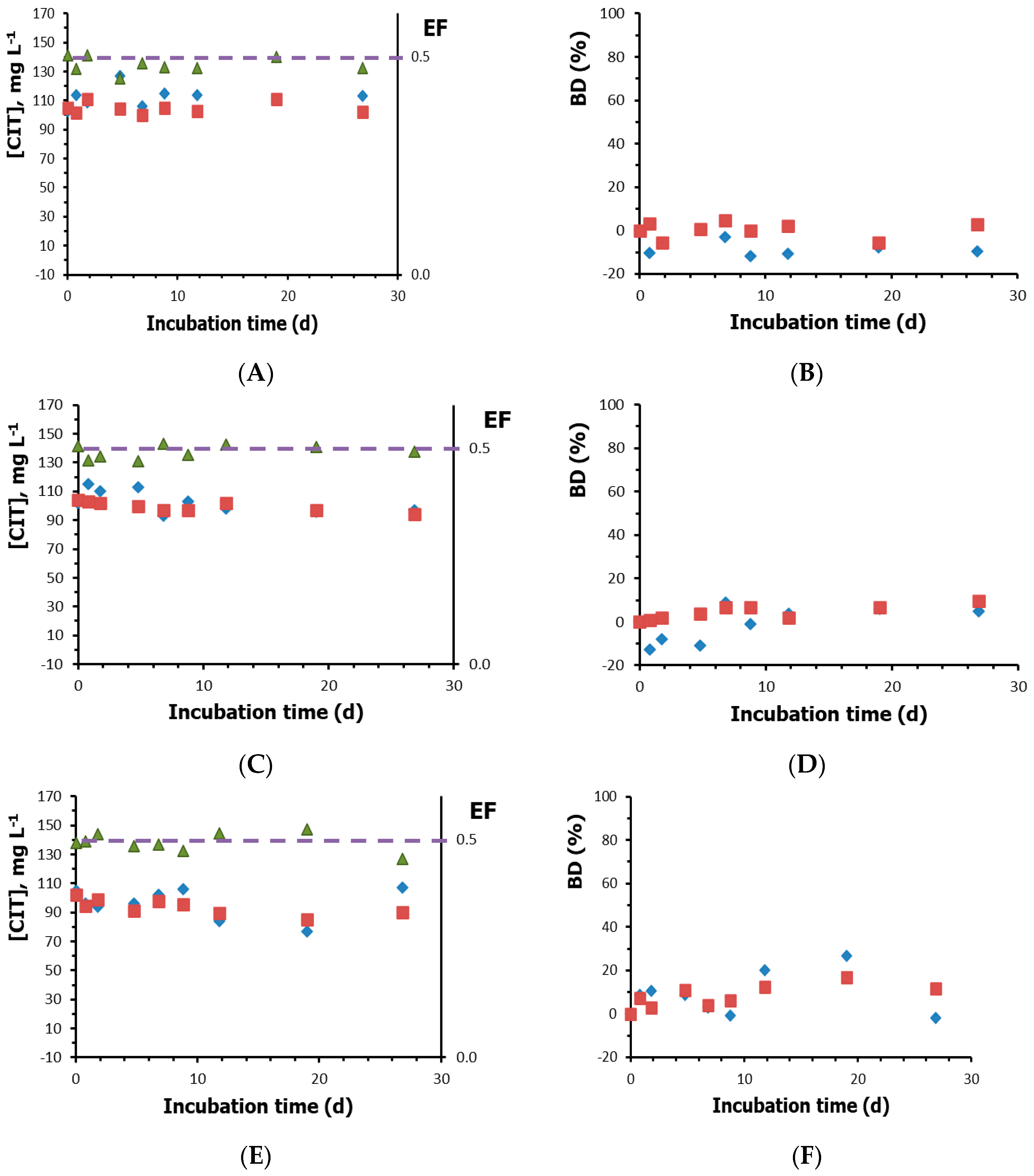
3.2. Biodegradation Results

The biodegradability assays for the enantiomers of VER and CIT were performed in batch mode by duplicate (Section 2.4). Sample aliquots at different times for the biotic and abiotic assays were analysed in the EKC-CF conditions previously chosen. According to OECD [10], benzoic acid was used as reference substance to ensure that the microbial community in the test system is active. After 48 h of incubation, a complete degradation of benzoic acid (200 mg L−1) was achieved in biotic assays (using 1 and 10 mL of activated sludge), while the degradation was only 8% in the abiotic assay, which indicated the adequacy of the inoculum and, therefore, the validity of the biodegradability assays.
Additionally, to control the biomass growth, the optical density at 600 nm (OD600) during the CIT biotic assays was monitored. OD600 measures the solution turbidity and can be considered proportional to biomass growth [40]. The OD600 values (results not shown) indicated a biomass growth along the biotic assays. For both biotic assays (inoculated with 1 and 10 mL of activated sludge), the maximum biomass growth was observed approximately at t = 5 days, followed by a stationary/decline growth phase.
Figure 2A,B shows two electropherograms corresponding to t = 0 and 15 days for the biotic and abiotic assays of VER. The peak areas of VER enantiomers decreased after 15 days of incubation, indicating that a degradation process has occurred. No differences are observed between enantiomers, so the degradation of VER does not seem to be enantioselective. Figure 2C,D shos the electropherograms of CIT for samples taken at t = 0 and at the end of the assay, t = 28 days, respectively. In this case, no variation was observed after the incubation period, suggesting the absence of degradation.
Figure 3 and Figure 4 show the variation of the concentration (S, mean of two replicates) of the CIT and VER enantiomers during the different assays together the values of the enantiomeric fraction (EF). The initial concentration of each enantiomer (S0) was 50 mg L−1 for VER and 100 mg L−1 for CIT. EF at each incubation time was calculated according to:
E F = S E 1 S E 1 + S E 2
where SE1 and SE2 refer to the estimated concentration of enantiomers at a given incubation time. The horizontal line at EF = 0.5 in Figure 3 and Figure 4 represents no enantioselectivity. The biodegradation, BD, was also calculated for the enantiomers of CIT (Figure 3) and VER (Figure 5) from the concentrations obtained over time:
% B D = S 0 S S 0 × 100
Figure 3 shows that in no case is there a significant decrease in the concentrations of CIT enantiomers, although the concentration for the assay performed with more inoculum seems to present a slight decrease (Figure 3E). The results for the abiotic assay indicate the absence of physico-chemical degradation (Figure 3A). The values of biodegradation confirm these results since no significant biodegradation was obtained in any case (BD < 20%). Therefore, citalopram can be considered as a potentially persistent compound, and it would be necessary to perform more assays (inherent or simulation assays) to confirm its biodegradability. On the other hand, the EF values slightly vary around the initial value of 0.5 (corresponding to a racemic mixture), which indicates the absence of enantioselectivity as can be expected because of the lack of degradation. The results obtained agree with those reported by Evans et al. [11] who found that CIT was mildly biodegraded in a not enantioselective way in a microcosm with a low microbial content (as in the tests performed in this work).
Unlike CIT, for VER there is a decrease in the concentration over time, not only in the biotic assays (Figure 4A,C,D) but also in the abiotic one (Figure 4B) indicating the contribution of both microbiological and physico-chemical processes to the VER degradation. Moreover, no difference of behaviour can be appreciated between both enantiomers in any case (EF ~ 0.5).
Figure 5 shows the biodegradation, BD, for the VER enantiomers in the four assays carried out. In the first stage of the assay, after five days of incubation, the physicho-chemical degradation measured in the abiotic assay was no significant (BD < 13%), whereas the degradation in the biotic assays (both in presence and absence of light) was around 45%, which indicates that light has not an important influence. In the case of the assay carried out supplemented with acetate, the BD was slightly lower than in the other biotic assays. This can be due to a preference of the microorganism for the acetate as a carbon source, as acetate has a simpler chemical structure and therefore it can be more easily biodegraded. From day 5, the physico-chemical degradation begun to be significant, and after 28 days of incubation the values of BD reached in all the assays were similar (between 63 and 69%) even for the abiotic assay. It can be concluded that VER is a non-persistent compound, which could be degraded in the environment either by biological or by physico-chemical processes. Besides, non-enantioselective behaviour was observed in the conditions of this study.

4. Conclusions

In this work, two electrophoretic methods, based on the combination of the electrokinetic chromatography (EKC) mode and the complete filling technique (CFT), and the use of the highly sulfated γ cyclodextrin (HS-γ-CD) as chiral selector, were proposed for the determination of the enantiomers of verapamil (VER) and citalopram (CIT). For both compounds, the EKC-CFT method provide good enantioresolution in less than 7 min.
An enantioselective biodegradability test of VER and CIT was established using an inoculum (activated sludge from a waste water treatment plant) whose activity was verified using benzoic acid as reference compound. VER and CIT enantiomers were monitored using the two EKC-CFT methods developed. As a result of this test, CIT can be considered as a potentially persistent compound since no significant degradation was observed, while VER showed both physico-chemical and microbial degradation, being the predominant biodegradation process during the first stage of the assay. No evidence of enantioselectivity was observed in any of the biodegradation processes.
The results obtained in this work correspond to ready biodegradability test obtained in a laboratory scale compatible with the OECD recommendations (whose analyte and microorganisms amounts differ from those in the environment). The data can be considered as indicative of its potential degradation according to OECD guidelines; however, further tests should be undertaken under other experimental conditions to increase knowledge on the degradation of these compounds in the environment.

Author Contributions

Supervision, Y.M.-B., L.E.-G., S.S. and M.J.M.-H.; Writing-original draft, Y.M.-B.; Writing-review & editing, Y.M.-B., L.E.-G., S.S. and M.J.M.-H. All authors have read and agreed to the published version of the manuscript.

Funding

Spanish Ministerio de Economía y Competitividad (MINECO) and the European Regional Development Fund (ERDF) (Project CTQ2015-70904-R, MINECO/FEDER, UE).

Institutional Review Board Statement

Not applicable.

Informed Consent Statement

Not applicable.

Data Availability Statement

Data is contained within the article.

Conflicts of Interest

The authors declare no conflict of interest.

References

  1. Hernando, M.D.; Mezcua, M.; Fernández-Alba, A.R.; Barceló, D. Environmental risk assessment of pharmaceutical residues in wastewater effluents, surface waters and sediments. Talanta 2006, 69, 370–376. [Google Scholar] [CrossRef]
  2. Hernando, M.D.; Fernández-Alba, A.R.; Tauler, R.; Barceló, D. Toxicity assays applied to wastewater treatment. Talanta 2005, 65, 358–366. [Google Scholar] [CrossRef]
  3. Kümmerer, K. The presence of pharmaceuticals in the environment due to human use- present knowledge and future changes. J. Environ. Manag. 2009, 90, 2354–2366. [Google Scholar] [CrossRef] [PubMed]
  4. Ribeiro, A.R.; Santos, L.H.M.L.M.; Maia, A.S.; Delerue-Matos, C.; Castro, P.M.L.; Tiritan, M.E. Enantiomeric fraction evaluation of pharmaceuticals in environmental matrices by liquid chromatography-tandem mass spectrometry. J. Chromatogr. A 2014, 1363, 226–235. [Google Scholar] [CrossRef] [Green Version]
  5. Escuder-Gilabert, L.; Martín-Biosca, Y.; Pérez-Baeza, M.; Sagrado, S.; Medina-Hernández, M.J. Direct chromatographic study of the enantioselective biodegradation of ibuprofen and ketoprofen by an activated sludge. J. Chromatogr. A 2018, 1568, 140–148. [Google Scholar] [CrossRef] [PubMed]
  6. Vazquez-Roig, P.; Kasprzyk-Hordem, B.; Blasco, C.; Picó, Y. Stereoisomeric profiling of drugs of abuse and pharmaceuticals in wastewaters of Valencia (Spain). Sci. Total Environ. 2014, 494–495, 49–57. [Google Scholar] [CrossRef] [PubMed] [Green Version]
  7. Kasprzyk-Hordem, B.; Baker, D.R. Enantiomeric profiling of chiral drugs in wastewater and receiving waters. Environ. Sci. Technol. 2012, 46, 1681–1691. [Google Scholar] [CrossRef] [PubMed] [Green Version]
  8. Evans, S.E.; Bagnall, J.; Kasprzyk-Hordem, B. Enantioselective degradation of amphetamine-like environmental micropollutants (amphetamine, methamphetamine, MDMA and MDA) in urban water. Environ. Pollut. 2016, 215, 154–163. [Google Scholar] [CrossRef]
  9. Ribeiro, A.R.; Maia, A.S.; Moreira, I.S.; Afonso, C.M.; Castro, P.M.L.; Tiritan, M.E. Enantioselective quantification of fluoxetine and norfluoxetine by HPLC in wastewater effluents. Chemosphere 2014, 95, 589–596. [Google Scholar] [CrossRef]
  10. Ready Biodegradability Test. OECD Guideline for testing of chemicals. OECD 1992, 71, 1–9. [Google Scholar]
  11. Evans, S.; Bagnall, J.; Kasprzyk-Hordern, B. Enantiomeric profiling of a chemically diverse mixture of chiral pharmaceuticals in urban water. Environ. Pollut. 2017, 230, 368–377. [Google Scholar] [CrossRef]
  12. Bagnall, J.; Malia, L.; Lubben, A.; Kasprzyk-Hordem, B. Stereoselective biodegradation of amphetamine and methamphetamine in river microcosms. Water Res. 2013, 47, 5708–5718. [Google Scholar] [CrossRef] [Green Version]
  13. Ribeiro, A.R.; Afonso, C.M.; Castro, P.M.L.; Tiritan, M.E. Enantioselective HPLC analysis and biodegradation of atenolol, metoprolol and fluoxetine. Environ. Chem. Lett. 2013, 11, 83–90. [Google Scholar] [CrossRef]
  14. Ribeiro, A.R.; Afonso, C.M.; Castro, P.M.L.; Tiritan, M.E. Enantioselective biodegradation of pharmaceuticals, alprenolol and propranolol, by an activated sludge inoculum. Ecotoxicol. Environ. Saf. 2013, 87, 108–114. [Google Scholar] [CrossRef]
  15. Li, Z.; Gomez, E.; Fenet, H.; Chiron, S. Chiral signature of venlafaxine as a marker of biological attenuation processes. Chemosphere 2013, 90, 1933–1938. [Google Scholar] [CrossRef]
  16. Moreira, I.S.; Ribeiro, A.R.; Afonso, C.M.; Castro, P.M.L.; Tiritan, M.E. Enantioselective biodegradation of fluoxetine by the bacterial strain Labrys portucalensis F11. Chemosphere 2014, 111, 103–111. [Google Scholar] [CrossRef] [PubMed]
  17. Escuder-Gilabert, L.; Martín-Biosca, Y.; Pérez-Baeza, M.; Sagrado, S.; Medina-Hernández, M.J. Trimeprazine is enantioselectively degraded by an activated sludge in ready biodegradability test conditions. Water Res. 2018, 141, 57–64. [Google Scholar] [CrossRef]
  18. Klein, C.; Schneider, R.J.; Meyer, M.T.; Aga, D.S. Enantiomeric separation of metolachlor and its metabolites using LC–MS and CZE. Chemosphere 2006, 62, 1591–1599. [Google Scholar] [CrossRef] [PubMed]
  19. Chu, B.L.; Guo, B.Y.; Peng, Z.; Wang, Z.; Guo, G.; Lin, J.M. Studies on degradation of imazalil enantiomers in soil using capillary electrophoresis. J. Sep. Sci. 2007, 30, 923–929. [Google Scholar] [CrossRef] [PubMed]
  20. Chankvetadze, B.; Blaschke, G. Enantioseparations in capillary electromigration techniques: Recent developments and future trends. J. Chromatogr. A 2001, 906, 309–363. [Google Scholar] [CrossRef]
  21. Cserháti, T. New applications of cyclodextrins in electrically driven chromatographic systems: A review. Biomed. Chromatogr. 2008, 22, 563–571. [Google Scholar] [CrossRef] [PubMed]
  22. Fanali, S.; Chankvetadze, B. Some thoughts about enantioseparations in capillary electrophoresis. Electrophoresis 2019, 40, 2420–2437. [Google Scholar] [CrossRef] [PubMed]
  23. Bernardo-Bermejo, S.; Sanchez-Lopez, E.; Castro-Puyana, M.; Marina, M.L. Chiral capillary electrophoresis. TrAC Trends Anal. Chem. 2020, 124, 115807. [Google Scholar] [CrossRef]
  24. Amini, A.; Paulsen-Sörman, U.; Westerlund, D. Principle and applications of the partial filling technique in capillary electrophoresis. Chromatographia 1999, 50, 497–506. [Google Scholar] [CrossRef]
  25. Asensi-Bernardi, L.; Martín-Biosca, Y.; Sagrado, S.; Medina-Hernández, M.J. Characterizing the interaction between enantiomers of eight psychoactive drugs and highly sulfated-β-cyclodextrin by counter-current capillary electrophoresis. Biomed. Chromatogr. 2014, 28, 120–126. [Google Scholar] [CrossRef]
  26. Ruzicka, M.; Koval, D.; Vavra, J.; Reyes-Gutierrez, P.E.; Teply, F.; Kasicka, V. Interactions of helquats with chiral acidic aromatic analytes investigated by partial-filling affinity capillary electrophoresis. J. Chromatogr. A 2016, 1467, 417–426. [Google Scholar] [CrossRef] [PubMed]
  27. Asensi-Bernardi, L.; Martín-Biosca, Y.; Escuder-Gilabert, L.; Sagrado, S.; Medina-Hernández, M.J. Modeling the chiral resolution ability of highly sulfated β-cyclodextrin for basic compounds in electrokinetic chromatography. J. Chromatogr. A 2013, 1308, 152–160. [Google Scholar] [CrossRef] [PubMed]
  28. Escuder-Gilabert, L.; Martín-Biosca, Y.; Medina-Hernández, M.J.; Sagrado, S. Enantioresolution in electrokinetic chromatography-complete filling technique using sulfated gamma-cyclodextrin. Software-free topological anticipation. J. Chromatogr. A 2016, 1467, 391–399. [Google Scholar] [CrossRef] [PubMed]
  29. Baumann, P.; Zullino, D.F.; Eap, C.B. Enantiomers’ potential in psychopharmacology—A critical analysis with special emphasis on the antidepressant escitalopram. Eur. Neuropsychopharmacol. 2002, 2, 433–444. [Google Scholar] [CrossRef]
  30. Budău, M.; Hancu, G.; Muntean, D.L.; Papp, L.A.; Cârje, A.G.; Garaj, V. Enantioseparation of citalopram enantiomers by capillary electrophoresis: Method development through experimental design and computational modeling. Chirality 2020, 32, 1119–1128. [Google Scholar] [CrossRef]
  31. Hamidi, S.; Jouyban, A. Capillary electrophoresis with UV detection, on-line stacking and off-line dispersive liquid-liquid microextraction for determination of verapamil enantiomers in plasma. Anal. Methods 2015, 7, 5820. [Google Scholar] [CrossRef]
  32. Trautwein, C.; Kümmerer, K.; Metzger, J.W. Aerobic biodegradability of the calcium antagonist verapamil and identification of a microbial dead-end transformation product studies by LC-MS/MS. Chemosphere 2008, 72, 442–450. [Google Scholar] [CrossRef] [PubMed]
  33. Mechelke, J.; Rust, D.; Jaeger, A.; Hollender, J. Enantiomeric Fractionation during Biotransformation of Chiral Pharmaceuticals in Recirculating Water-Sediment Test Flumes. Environ. Sci. Technol. 2020, 54, 7291–7301. [Google Scholar] [CrossRef] [PubMed]
  34. Valimaña-Traverso, J.; Morante-Zarcero, S.; Pérez-Quintanilla, D.; García, M.A.; Sierra, I.; Marina, M.L. Periodic mesoporous organosilica materials as sorbents for solid-phase extraction of drugs prior to simultaneous enantiomeric separation by capillary electrophoresis. J. Chromatogr. A 2018, 1566, 135–145. [Google Scholar] [CrossRef] [PubMed]
  35. Matthijs, N.; Hemelryck, S.V.; Maftouh, M.; Massart, D.L.; Heyden, Y.V. Electrophoretic separation strategy for chiral pharmaceuticals using highly-sulfated and neutral cyclodextrins based dual selector systems. Anal. Chim. Acta 2004, 525, 247–263. [Google Scholar] [CrossRef]
  36. Andersen, S.; Halvorsen, T.G.; Pedersen-Bjergaard, S.; Rasmussen, K.E.; Tanum, L.; Refsum, H. Stereospecific determination of citalopram and desmethylcitalopram by capillary electrophoresis and liquid-phase microextraction. J. Pharm. Biomed. Anal. 2003, 33, 263–273. [Google Scholar] [CrossRef]
  37. Mandrioli, R.; Fanali, S.; Pucci, V.; Raggi, M.A. Enantiomeric separation of citalopram and its metabolites by capillary electrophoresis. Electrophoresis 2003, 24, 2608–2616. [Google Scholar] [CrossRef]
  38. Deng, X.; De Wolf, J.; Vervoort, R.; Pamperin, D.; Adams, E.; Schepdael, A.V. Development and validation of a capillary electrophoresis method for the determination of escitalopram and sensitive quantification of its enantiomeric impurity in formulations. Electrophoresis 2012, 33, 1648–1651. [Google Scholar] [CrossRef] [PubMed]
  39. Wang, Y.; Zhang, S.; Breitbach, Z.S.; Petersen, H.; Ellegaard, P.; Armstrong, D.W. Enantioseparation of citalopram analogues with sulfated beta-cyclodextrin by capillary electrophoresis. Electrophoresis 2016, 37, 841–848. [Google Scholar] [CrossRef] [PubMed]
  40. Shuler, M.L.; Kargi, F. (Eds.) Bioprocess Engineering: Basic Concepts, 2nd ed.; Prentice Hall: Upper Saddle River, NJ, USA, 2002. [Google Scholar]
Figure 1. Effect of the methanol concentration in the background electrolyte (BGE) on the resolution at two concentration of HS-γ-CD: () 0.1 and () 0.05 (%, m/v). Other separation conditions: BGE, 50 mM phosphate buffer at pH 7.0; 20 kV; 25 °C.
Separations 08 00029 g001
Figure 2. Electropherograms of samples containing racemic mixtures of (A,B) verapamil (VER) and (C, D) citalopram (CIT) taken at t = 0 (A,C), t = 15 (B) and t = 28 (D) days of incubation. Experimental conditions detailed in Section 2.3. Initial racemate concentrations were 100 and 200 mg L−1 for VER and CIT, respectively. I.S., internal standard: acetanilide (A,B) and ranitidine (C,D) for VER and CIT, respectively; E1 and E2, first and second eluted enantiomer, respectively.
Separations 08 00029 g002
Figure 3. Estimated concentrations (S), biodegradation (BD), and enantiomeric fraction (EF) obtained for citalopram (CIT) in incubated samples during the biodegradability assays: (A,B), abiotic assay; (C,D), biotic assay inoculated with 1 mL of activated sludge; (E,F), biotic assay inoculated with 10 mL of activated sludge. S, BD and EF values correspond to the mean of two replicates. Dashed line, EF = 0.5, represents no enantioselectivity. Symbols: () E1-CIT; () E2-CIT; () EF.
Separations 08 00029 g003
Figure 4. Estimated concentrations (S) and enantiomeric fraction (EF) obtained for verapamil (VER) in incubated samples during the biodegradability assays: (A), biotic assay; (B), abiotic assay; (C), biotic assay in darkness; (D) biotic assay supplemented with acetate. S and F values correspond to the mean of two replicates. Dashed line, EF = 0.5, represents no enantioselectivity. Symbols: () E1-VER; () E2-VER; () EF.
Separations 08 00029 g004
Figure 5. Biodegradation (BD, mean of two replicates) obtained for the enantiomers of verapamil (VER) in incubated samples during the biodegradability assays: (), biotic assay; (), abiotic assay; (), biotic assay in darkness; () biotic assay supplemented with acetate. A: E1-VER; B: E2-VER.
Separations 08 00029 g005
Table 1. Structure, acidity constant of conjugated acid (pKa), and logarithm of the partition coefficient in the n-octanol/water system (logP) of compounds studied.
CompoundStructurepKalogP
Verapamil Separations 08 00029 i0018.93.79
Citalopram Separations 08 00029 i0029.782.51
Acetanilide Separations 08 00029 i0030.51.16
Ranitidine Separations 08 00029 i0047.81.23
Table 2. Calibration statistics a for verapamil and citalopram along working sessions. Regression curve equation (A/Ai.s. = b0 + b1 S), obtained using the internal standard method.
Compound
Interval b, mg/L−1
E1E2
Working
Session
b1 ± tsb0 ± tsR2b1 ± tsb0 ± tsR2
Verapamil
10–200
10.023 ± 0.0030.0 ± 0.20.9930.023 ± 0.0040.0 ± 0.30.996
20.024 ± 0.0040.0 ± 0.30.99980.024 ± 0.002−0.01 ± 0.120.998
30.026 ± 0.002−0.09 ± 0.110.9990.026 ± 0.002−0.08 ± 0.100.999
40.026 ± 0.003−0.1 ± 0.20.9960.026 ± 0.003−0.1 ± 0.20.996
50.021 ± 0.0050.2 ± 0.40.980.023 ± 0.0030.18 ± 0.180.99
60.020 ± 0.002−0.01 ± 0.090.9930.020 ± 0.0020.01 ± 0.090.994
70.017 ± 0.002 −0.01 ± 0.080.9930.017 ± 0.002−0.02 ± 0.080.994
Citalopram
5–300
-0.029 ± 0.007−0.1 ± 0.70.9910.016 ± 0.0030.3 ± 0.30.997
E1 and E2 refer to the first and second eluted enantiomers, respectively. R2 is the coefficient of determination. a A = peak area of the enantiomer; Ai.s. = peak area of the internal standard; S = enantiomer concentration in the calibration standards; b0 = intercept; b1 = slope; ts = confidence interval at the 95% probability level; R2 = coefficient of determination. b Racemate concentration.
Publisher’s Note: MDPI stays neutral with regard to jurisdictional claims in published maps and institutional affiliations.

