Article

To Develop the Method for UHPLC-HRMS to Determine the Antibacterial Potential of a Central American Medicinal Plant

New Jersey Center for Science, Technology and Mathematics—Kean University, Union, NJ 07083, USA
*
Author to whom correspondence should be addressed.
Separations 2019, 6(3), 37; https://doi.org/10.3390/separations6030037
Submission received: 5 June 2019 / Revised: 7 July 2019 / Accepted: 15 July 2019 / Published: 29 July 2019
(This article belongs to the Special Issue Trends in Modern High Performance Liquid Chromatography Separations)

Abstract

:
The development of antibiotic resistance by microbials has long been acknowledged. The major challenge worldwide is to develop novel, natural, and potent antibiotics against the multidrug resistant bacteria. In this study, our aim was to develop the method for a highly sensitive instrument, ultra-high performance liquid chromatograph-high resolution mass spectrometer (UHPLC-HRMS), to evaluate the antibacterial property of a natural product. Aechmea magdalenae (Andre) Andre ex Baker, a plant belonging to the family Bromeliaceae, a native of Central America was used in this study. Based on the available literature, it was hypothesized that Aechmea magdalenae has antibacterial activity. In addition, the profiling done on A. magdalenae using gas chromatography-mass spectrometry (GC-MS) also revealed the presence of medicinally important chemical compounds, such as acetic acid. Minimum inhibitory concentration (MIC) of dried Aechmea plant extract was determined for the first time using 96-well plate assay, followed by determination of antibacterial potential using LC-MS. The reason being that other dried methanolic plant extracts, such as Vismia macrophylla, lined up for antibacterial testing have dark extracts, for which determining the antibacterial potential and reading the results with the naked eye would be challenging. To overcome the situation of dark plant extracts, a generalized novel LC-MS method was developed that was used for the plant A. magdalenae, and would be used further for other plants. A blue indicator called resazurin was added to the wells; resazurin, upon incubation with the living cells, got reduced to resorufin (which was pink), while it remained blue with bacterial growth inhibition. The mass difference created due to reduction of resazurin to resorufin was detected by using LTQ Orbitrap Discovery in positive ion mode to determine the antibacterial activity of the plant extract. The sample preparation for LC-MS assay included centrifugation of the samples taken from 96-well plate, followed by filtration of the supernatant, before exposing them to C-18 column. The results obtained from full scan LC-MS spectrum consistently demonstrated the presence of resorufin from wells with bacterial growth, and resazurin from wells with inhibition through peaks of relevant masses.

1. Introduction

Medicines obtained from traditional plants are called phytomedicines. The phytomedicines have long been used after several generations of trial and error [1]. The medicinal plants contain certain chemical compounds responsible for their antimicrobial activity [2]. Aechmea magdalenae (Andre) Andre ex Baker, is a plant falling under the family Bromeliaceae, which is a native of Central America. Although the literature availability for the antibacterial activity of this species has been limited, the documented research revealed the use of A. magdalenae as caustic for wounds [3]. Therefore, it was hypothesized that plant A. magdalenae has antibacterial properties. The ethnobotanical approach has also proved to be advantageous while finding new drug candidates.
GC-MS profiling confirmed acetic acid as a high probability (>90%) chemical compound, out of many others, present in the rhizome of A. magdalenae [4]. The antibacterial potential of acetic acid was confirmed in our laboratory using agar disc diffusion assay performed on gram negative bacteria [4]. The agar well diffusion assay was performed to evaluate the antibacterial activity while a 96-well plate assay determined the MIC of the rhizome extract of A. magdalenae [4]. Both the agar well diffusion assay and the 96-well plate assay included gram-negative Escherichia coli (E. coli) and gram-positive Staphylococcus aureus (S. aureus) bacteria. In this study, we aimed to develop a highly rapid and sensitive ultra-high performance liquid chromatograph-high resolution mass spectrometer (UHPLC-HRMS) method to detect the accurate mass of indicator resazurin and its reduced form, resorufin.
There was a significant mass difference between indicator resazurin and its reduced form, resorufin (Figure 1). This difference in mass between resazurin’s intact and reduced forms was detected by mass spectrometer in positive ion mode. The MIC of plant A. magdalenae was determined previously using a 96-well plate assay [4]. In the present study, a novel rapid LC-MS method was developed that could determine the antibacterial potential of medicinal plants using the mass difference of resazurin and resorufin. The Aechmea plant extract was not too dark, so the MIC was read with naked eye from the 96-well plate before determining the antibacterial potential by using mass spectrometer.
But there are several other medicinal plants in our lab with dark extracts for which the antibacterial activity has yet to be determined. For those plants, determination of antibacterial potential using a 96-well plate would be challenging. In that situation, LC-MS method would determine the antibacterial activity of dark medicinal plant extracts by using the property of mass difference created after reduction of resazurin to resorufin instead of colorimetric analysis. For the plant, A. magdalena, the MIC was already determined before using the 96-well plate assay. But for future dark plant extracts, LC-MS method could be used directly.

2. Materials and Methods

2.1. The Instrument

The instrument used for the present study was the LTQ Orbitrap Discovery by Thermo-Fischer scientific. The instrument was equipped with Accela auto sampler. Auto sampler has the sample compartments that can hold sample vials or the 96-well plates. LTQ Orbitrap can use ionization techniques such as electrospray ionization (ESI) and atmospheric pressure chemical ionization (APCI), but the instrument used in this study used ESI as source of ionization. Ions are transferred through square quadrupole lenses to the ion trap that is optimized for axial ion ejection into the curved linear trap. Linear ion trap is a unique system which isolates, prepares, and stores ions for the Orbitrap MS. Orbitrap mass analyzer is the heart of the entire system, which applies electric fields to keep the ions confined and directed. Orbitrap works on the principle of an axial, central, spindle shaped electrode; the stable ion trajectories rotate around, as well as oscillate along, the electrode. The image current of the oscillating ions is detected by the outer electrode of the Orbitrap, which with the help of Fast Fourier Transformation (FFT), gets frequencies and therefore, mass-to-charge ratios are obtained. These mass-to-charge ratios were analyzed using Excalibur software (Thermo-Fisher Scientific, https://tools.thermofisher.com/content/sfs/manuals).

2.2. LC- MS Parameters and Assay Conditions

This research involved the usage of only HPLC-grade solvents. The reverse phase UHPLC was performed using acetonitrile (ACN) with 0.1% formic acid as organic solvent, UHPLC water, and 0.1% formic acid as aqueous solvent (Table 1). The column used was Acquity C-18, 1.7 µm (2.1 × 50 mm) by Waters. Injection volume used was 5.0 µL with a flow rate of 700 µL/min. All the results obtained were with ESI as the source of ionization, in positive mode. Capillary temperature was kept at 350 °C and capillary voltage was 5.96 kV. A full scan MS was performed with a mass scan range between 50 and 500 m/z.
To perform LC-MS assay, 0.1% resazurin solution was freshly prepared using HPLC grade water. Resazurin indicator is blue which, when reduced, gets converted to a pink-colored chemical called resorufin. In solution, both resazurin and resorufin co-exist. In the living systems, with the bacterial growth, blue indicator resazurin (m/z = 230.0448) gets reduced to its pink resorufin (m/z = 214.0499). The different masses of resazurin and resorufin created due to the reduction of resazurin to resorufin were used to determine antibacterial activity using mass-spectrometer. In the 96-well plate assay, the plant extract was added along with bacteria and resazurin to the assigned wells. With bacterial growth, resazurin got reduced to resorufin while resazurin remained as is with bacterial inhibition. Since the mass of resazurin was different than resorufin, a rapid and very sensitive 3.5 min gradient method was developed to detect both resazurin and resorufin.
The gradient conditions used are shown below.

2.3. Plant Extract and Inoculum Preparation

Six grams of the powdered rhizome of plant A. magdalena was mixed with 60 mL HPLC grade methanol. The sample was shaken for 72 h at the speed of 150 rpm by changing the solvent every 24 h [4]. E. coli (ATCC 25922), a gram negative, and S. aureus, a gram-positive bacterium, were used as test organisms. The inoculum was prepared by using sterile bacterial growth medium. The pure colonies were picked from the nutrient agar plates using sterile loops. The loops with bacterial cells were swirled in the culture tubes containing 10 mL sterile LB broth (Teknova, Hollister, CA, USA). The culture tubes were placed in the incubator for 24 h at 37 °C. The bacterial cultures were adjusted to 0.5 McFarland standard before the experiment.

2.4. Antibacterial Activity Testing

2.4.1. MIC Determination Using 96-Well Plate Assay

The MIC is the lowest concentration of antimicrobial agent that completely inhibits the growth of microorganisms in the wells [5]. MIC of the A. magdalenae was determined using 96-well plate assay. For each bacterium, the rows were assigned for media control, gentamicin (1 mg/mL) in water, and 20% DMSO control. Since methanol has antibacterial properties, the methanolic extract was roto-vaped at room temperature (Buchi, R-114, Flawil, Switzerland) to complete dryness. The dried methanolic plant extract was then reconstituted in 10 mL of 20% DMSO optimized previously [4]. Serially diluted plant extract was added to the designated wells [6]. E coli and S aureus bacteria were adjusted to the concentration of 0.5 McFarland standard. Next, 10 μL of the prepared bacteria was added to each well, followed by the addition of equal volume of 0.1% indicator resazurin (in water).
The sealed plates were then placed in the incubator for 24 h at 37 °C. Each experiment was performed in triplicates for consistency. Growth inhibition was recorded as no color change in the blue indicator resazurin, in contrast to the wells where pink resorufin formed due to the bacterial growth. The results from the MIC plate were marked and collected in the microcentrifuge tubes. The samples were kept frozen at −20 °C until further use for LC-MS assay.

2.4.2. LC-MS Analysis

The supernatants saved from the 96-well plate were thawed on the lab bench and centrifuged twice for 10 min at 16,000 RPM. The collected supernatants were diluted (1:100) using HPLC water. The final volume of 1 mL was filtered using 0.45 μm micro-pore PTFE filter. For each sample, a volume of 300 μL from the filtered sample was added to the LC-MS vial with the insert.
Two types of resazurin controls were used. First, a freshly prepared stock of 0.1% resazurin was prepared, which was diluted to the concentration used in the wells. Similarly, another control prepared was 0.1% resazurin, incubated for 24 h at 37 °C. The incubated resazurin was diluted according to the concentration of each well of the MIC plate. Acetonitrile and water in the ratio of 90:10 was used as the instrument blank. The aqueous solvent used was HPLC water and 0.1% formic acid; organic solvent was acetonitrile and 0.1% formic acid.
Samples were run on the instrument using the above-discussed rapid, sensitive 3.5 min gradient method for resazurin. The sample control sequences were designed and analyzed by using the software Excalibur. A full-scan LC-MS was performed for each sample and both controls (resazurin and incubated resazurin) several times to get a set of consistent results.

3. Results and Discussion

Antimicrobial activity of A. magdalenae was determined using agar disc diffusion and agar well diffusion assays previously [4]. Agar disk diffusion method is a standard microbiology practice used for antimicrobial susceptibility testing [7]. Since GC-MS profiling showed the higher concentration of acetic acid in the Aechmea sample, antibacterial activity of acetic acid was also tested using nutrient agar plates streaked according to 0.5 McFarland adjusted bacteria. The 6 mm disks were impregnated with the varying concentrations of acetic acid starting from the 1% to 5%. The clear areas around the disks after the 24 h incubation period were marked as zones of inhibition [4].
Agar disk diffusion assay performed using different concentrations of acetic acid resulted in the highest zones of inhibition at the concentration of 5%, followed by 2% using gram positive bacteria (S. aureus). Therefore, the antibacterial activity of acetic acid was best between the concentration of 5% and 1%. Below this concentration, the acetic acid solution was not bactericidal [4].
Similar results were obtained in a study conducted by testing antimicrobial activity of acetic acid against a multidrug resistant bacteria panel including S. aureus. The acetic acid was bactericidal against S. aureus in varying zones of inhibition starting from the concentration of 0.5% to 2.5% [8]. Agar well diffusion technique was used to determine the antibacterial potential of Aechmea magdalenae. Three 6 mm holes were made in the bacteria-coated agar plates [9]. A specific volume of the roto-vaped and reconstituted plant extract was filled in the wells, followed by the addition of 20% DMSO and 1 mg/mL gentamicin. The plates were para filmed and placed in the incubator for 24 h at 37 °C. The clear areas around the wells were measured as zones of inhibition [4].
Agar well diffusion assay resulted in smaller zones of inhibition in gram negative bacteria (E. coli) as compared to gram positive bacteria (S. aureus). These results indicated that the A. magdalenae extract was more potent against S. aureus than E. coli [4]. The results produced by another such study also demonstrated higher antibacterial activity of methanolic plant extracts against S. aureus as compared to E. coli, which is in accordance with our results [4,6].
Resazurin solution (0.1%) was prepared fresh just before the experiment. The full scan LC-MS analysis of the freshly prepared 0.1% resazurin used as one of the controls showed both resazurin as well as resorufin peaks. The retention time (RT) of resazurin peak was 1.17 with the area of 35,435. The RT of resorufin was 1.22 and the area of the peak was 554,082 (Figure 2). The results demonstrated the presence of both resazurin (m/z 230) and resorufin (m/z 214) in the control sample taken from freshly prepared resazurin.
The second resazurin control (0.1%) was prepared 24 h before the analysis and was placed in the incubator at 37 °C. The incubated resazurin control sample was collected after 24 h for LC-MS assay. The full scan LC-MS of the incubated resazurin control also revealed both resazurin as well as resorufin peaks. The RT and the area of the incubated resazurin (m/z 230) were 1.05 and 32,151 respectively, while the RT of resorufin (m/z 214) was 1.11 and area was 557,304 (Figure 3).
In the living systems, resazurin is converted into resorufin in their mitochondria where NADPH or NADH acts as a reducing agent in the presence of enzyme NADPH dehydrogenase or NADH dehydrogenase [10]. The A. magdalenae extract in the 96-well plate resulted in the reduction of indicator resazurin to resorufin in the presence of both types of bacteria. The samples taken from 96-well plate were forwarded for LC-MS analysis.
The full-scan spectrum showed a complete absence of a resazurin peak (m/z 230) with the bacterial growth as shown in Figure 4. The absence of a resazurin peak meant a full conversion of resazurin (m/z 230) into resorufin (m/z 214) in the 96-well plates due to bacterial activity. But this conversion was very well demonstrated by mass spectrometer—by producing resorufin peak instead of resazurin peak in the samples taken from the bacterial growth wells. Moreover, the retention time of the resorufin peak from the sample in Figure 4 was consistent with the resorufin peak generated from the incubated resazurin control in Figure 3.
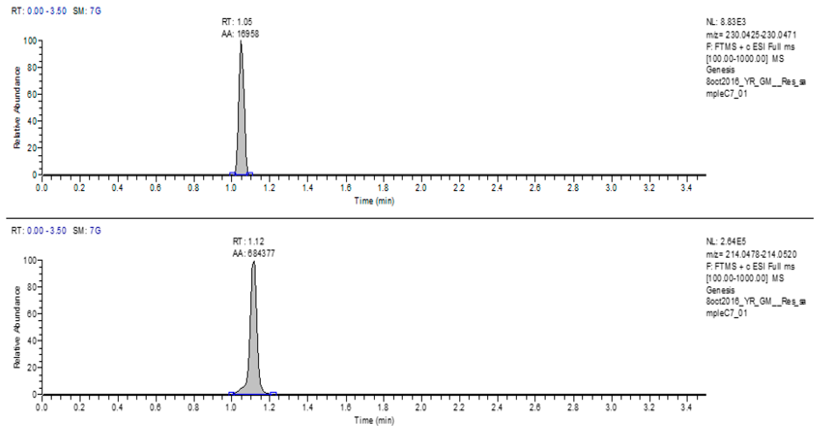
The second set of samples for mass spectrometer were prepared from the wells with assumed bacterial inhibition. A 0.1% incubated resazurin solution prepared in water was used as control. The full scan LC-MS spectrum demonstrated the appearance of both resazurin (m/z 230) and resorufin peaks (m/z 214) (Figure 5).
Interestingly, the retention times of both resazurin and resorufin peaks from the above-mentioned results matched with the RT of the incubated resazurin controls. It was deduced that the bacterial growth was inhibited in the wells from where these samples were taken. Therefore, resazurin was not converted to resorufin, instead both resazurin and resorufin were present.
To summarize, the indicator resazurin gets reduced to resorufin due to bacterial growth. This special feature of resazurin was used as a marker in this study. A 0.1% freshly prepared and incubated resazurin solution was used as control. The different wells showing bacterial growth and inhibition were subjected for LC-MS analysis. LC-MS results revealed the presence of only resorufin peak, which marked the bacterial growth, whereas the presence of both resazurin and resorufin peaks represented the growth inhibition.
In previous studies, LC-MS has only been used for profiling of the chemical compounds responsible for the antibacterial activity of medicinal plants [11,12]. Another study used LC-MS to investigate and identify particularly phenolic compounds in the Erodium glaucophyllum extract from Tunisian Sahara [13]. Interestingly, no study has so far used LC-MS for testing the antibacterial potential of a medicinal plant. This study has set some new research parameters where further elaborative studies could be done toward the direct use of mass spectrometry in determining antibacterial activity.

4. Conclusions

The purpose of this study was to develop the method to determine the antibacterial potential of the dark-colored plant extracts when mixed with indicator in a 96-well plate. This LC-MS method was tried on the plant A. magdalenae, for which the MIC was already determined previously using 96-well plate assay. But in the future, the LC-MS method could be directly used for the antibacterial potential analysis of the dark medicinal plant extracts.
To the best of our knowledge, for the first time, LC-MS assay was specifically aimed to measure masses generated due to the reduction of indicator resazurin to resorufin to determine antibacterial activity of a medicinal plant. The development of the assay first involved the method development, followed by mass spectrometric optimization of the individual masses of resazurin, as well as resorufin in their native form and, then, determination of masses created in the samples.

Author Contributions

G.K.M. contributed to the project developing the methodology, data analysis, validation, and writing the original draft. A.G. helped to develop the methodology, validation, and writing, review and editing the manuscript. D.R. contributed by investigating, project administration, funding acquisition, supervision, writing, review and editing.

Funding

This project was funded by Kean University grants: Students partnering with faculty (SPF), 2018 and Foundation Faculty Research Award (FFRA) 2017-2019>.

Acknowledgments

Authors are thankful to Yuriko Root for all her help.

Conflicts of Interest

The authors declare no conflict of interest

References

  1. Bereket, A.; Samuel, S.; Feleke, M. In vitro antibacterial activity of leaf extracts of Zehneria scabra and Ricinus communis against Escherichia coli and methicillin resistance Staphylococcus aureus. Asian Pac. J. Trop. Biomed. 2014, 4, 816–820. [Google Scholar] [Green Version]
  2. Webster, D.; Taschereau, P.; Belland, R.J.; Sand, C.; Rennie, R.P. Antifungal activity of medicinal plant extracts; Preliminary screening studies. J. Ethnopharmacol. 2008, 115, 140–146. [Google Scholar] [CrossRef] [PubMed]
  3. Ticktin, T. The history of Ixtle in Mexico. Econ. Bot. 2002, 56, 92–94. [Google Scholar] [CrossRef]
  4. Monga, G.K.; Ghosal, A.; Shebitz, D.; Ramanathan, D. Determination of antibacterial activity in rhizome of plant Aechmea magdalenae (andre) andre ex baker. J. Med. Herb Ethnomed. 2017, 3, 13–21. [Google Scholar] [CrossRef]
  5. Balouiri, M.; Sadiki, M.; Ibnsouda, S.K. Methods for in vitro evaluating antimicrobial activity. J. Pharm. Anal. 2016, 6, 71–79. [Google Scholar] [CrossRef] [PubMed]
  6. Cheruiyot, K.R.; Olila, D.; Kateregga, J. N-vitro antibacterial activity of selected medicinal plants from Longisa region of Bomet district, Kenya. Afr. Health Sci. 2009, 9, S42–S46. [Google Scholar] [PubMed]
  7. Bauer, A.W.; Kirby, W.M.; Sherris, J.C.; Turck, M. Antibiotic susceptibility testing by a standardized single disk method. Am. J. Clin. Pathol. 1966, 45, 493–496. [Google Scholar] [CrossRef] [PubMed]
  8. Wali, M.K.; Abed, M.M. Antibacterial activity of acetic acid against different types of bacteria causes food spoilage. J. Food Technol. Preserv. 2019, 3, 1–4. [Google Scholar]
  9. Taye, B.; Giday, M.; Animut, A.; Seid, J. Antibacterial activities of selected medicinal plants in traditional treatment of human wounds in Ethiopia. Asian. Pac. J. Trop. Biomed. 2011, 5, 370–375. [Google Scholar] [CrossRef]
  10. Rampersad, S.N. Multiple applications of alamar blue as an Indicator of metabolic function and cellular health in cell viability bioassays. Sensors (Basel) 2012, 12, 12347–12360. [Google Scholar] [CrossRef] [PubMed]
  11. Belhadj, S.; Jlail, L.; Feki, A.E.; Damak, M.; Sayadi, S.; Allouche, N. LC-MS–MS and GC-MS analyses of biologically active extracts and fractions from Tunisian Juniperus phoenice leaves Henda Keskes. Pharm. Biol. 2017, 55, 88–95. [Google Scholar]
  12. Jang, J.Y.; Shin, H.; Lim, J.; Ahn, J.H.; Jo, Y.H.; Lee, K.Y. Comparison of antibacterial activity and phenolic constituents of bark, lignum, leaves and fruit of Rhus verniciflua. Comparison of antibacterial activity and phenolic constituents of bark, lignum, leaves and fruit of Rhus verniciflua. PLoS ONE 2018, 7, 13. [Google Scholar]
  13. Hajlaoui, S.B.H.; Daoud, A.; Mighri, H.; Ross-Garcia, J.M.; Gharsallah, N.; Kadri, A. Phytochemicals, antioxidant and antimicrobial potentials and LC-MS analysis of hydroalcoholic extracts of leaves and flowers of Erodium glaucophyllum collected from Tunisian Sahara. J. Food Sci. Technol. 2018. [Google Scholar] [CrossRef]
Figure 1. Reduction of resazurin to resorufin.
Separations 06 00037 g001
Figure 2. Fresh resazurin standard showing both resazurin and resorufin with consistent retention time (RT) and area.
Separations 06 00037 g002
Figure 3. Incubated resazurin standard showing both resazurin and resorufin with consistent RT and area.
Separations 06 00037 g003
Figure 4. With bacterial growth, resazurin was reduced to resorufin.
Separations 06 00037 g004
Figure 5. With bacterial inhibition, both resazurin and resorufin peaks were present.
Separations 06 00037 g005
Table 1. The gradient method used with the percentage of aqueous (A%) and organic (B%) solvents drawn into the system at specific intervals of time with the flow rate of 700 μL/min.
cmin/sA %B %μL/min
00.0090.010.0700.0
10.3090.010.0700.0
22.002.098.0700.0
32.902.098.0700.0
43.0090.010.0700.0
53.5090.010.0700.0