Article

Mass Spectrometric Determination of the Effect of Surface Deactivation on Membranes Used for In-Situ Sampling of Cerebrospinal Fluid (CSF)

1
Department of Chemistry-BMC, Analytical Chemistry, Uppsala University, P.O. Box 599, SE-751 24 Uppsala, Sweden
2
Department of Engineering Sciences, Uppsala University, PO Box 534, SE-751 21 Uppsala, Sweden
*
Author to whom correspondence should be addressed.
Separations 2018, 5(2), 27; https://doi.org/10.3390/separations5020027
Submission received: 16 February 2018 / Revised: 23 March 2018 / Accepted: 26 April 2018 / Published: 7 May 2018

Abstract

:
In this paper, a strategy for structured monitoring of surface modifications to control protein adsorption to membrane structures is presented. The already established on-surface enzymatic digestion (oSED) method combined with nano-liquid chromatography and tandem mass spectrometry (LC-MS/MS) analysis was employed for the analysis of proteins in ventricular cerebrospinal fluid (vCSF) from neurointensive care patients. Protein adsorption was studied by in-situ sampling in a temporally resolved manner on both immobilized native and Pluronic-deactivated membranes. Deactivation was significantly reducing the protein adsorption but it also induced novel selective properties of the surface. The proposed versatile strategy will facilitate protein-biomaterial, protein-polymer, protein-protein interaction studies in the future.

Graphical Abstract

1. Introduction

Foreign material in contact with biological matrices—such as tissue, blood, plasma or cerebrospinal fluid (CSF)—instantly attracts dissolved proteins [1,2]. Formation of a protein layer on a foreign surface is the first step of a series of complex dynamic mechanisms that eventually leads to encapsulation of the material. Encapsulation and rejection of foreign material is most often unwanted, and to delay the encapsulation the formation of a protein layer should be fully avoided, decreased, or delayed. A typical example is the membrane used in microdialysis probes for sampling of analytes from body fluids [3], where the transport across the membrane can be severely hindered by biofouling [4]. Novel approaches of surface deactivation and methods for evaluating the effect of surface modifications are therefore required.
Different surface modification methods have been utilized to decrease protein adsorption, and among them coating with Pluronics has proven to be successful [5]. Pluronic is a group of tri-block copolymers that is build up by a hydrophobic core (polypropylene PPO) that covalently binds two hydrophilic strands (Polyethyleneoxide, PEO). When the Pluronic is dissolved in water at a concentration lower than the critical micelle concentration, the hydrophobic units interact with hydrophobic foreign material, resulting in the hydrophilic units of Pluronic protruding into the aqueous solution and forming a cilia-resembling layer. This layer impedes proteins to reach the surface due to steric hindrance and thereby the hydration of the surface is maintained. Recently, our group compared protein adsorption to Poloxamer 407 surface modified microdialysis membranes with adsorption to unmodified membranes [6]. Poloxamer 407 is a triblock copolymer also referred to as Pluronic F-127. The microdialysis sampling was performed in vitro, at 37 °C and for 24 h (one time point was studied) in a cerebrospinal fluid (CSF) sample. In this study, the abundances of 19 proteins were compared and a 30% decrease in protein adsorption was observed on the modified membrane compared to the native membrane. Subunits of hemoglobin, clusterin, and albumin showed the most pronounced decrease in protein adsorption.
In this paper, we provide novel aspects of the characterization of surface deactivation. We investigate competitive protein adsorption in a time-resolved manner. Flat polycarbonate (PC) membrane surfaces were dynamically coated with Pluronic F-127 and the adsorption of proteins from ventricular cerebrospinal fluid (vCSF) was investigated in relation to native membranes. These membranes mimic the probe structures used for microdialysis sampling of CSF. Thus, the evaluation of the membranes is performed in a model system as close to reality as possible. We use the developed, straight forward, and gentle on-surface enzymatic digestion (oSED) protocol [7,8], in which proteins were digested with trypsin while still adsorbed to the membrane. The resulting peptides were collected and analyzed using shotgun proteomics with nano-liquid chromatography and tandem mass spectrometry (LC-MS/MS). The adsorbed proteins identified were compared for coated and native membranes and, compared to previous study, several hundred proteins were monitored.

2. Materials and Methods

2.1. Chemicals and Reagents

The model surface for protein adsorption was electron track etched PC filter membranes with a diameter of 13 mm and a pore size of 0.22 µm from Millipore S.A.S. (Molsheim, Alsace, France). Chemicals, including coating polymer Pluronic F-127 (Pluronic F-127) were purchased from Sigma-Aldrich (St. Louis, MO, USA), if not otherwise stated. LC-MS reagents such as acetonitrile (ACN) and acetic acid (HAc) were obtained from Merck (Darmstadt, Germany). Sequence-grade trypsin was acquired from Roche Diagnostics (Basel, Switzerland).

2.2. Cerebrospinal Fluid Collection

Ventricular CSF was collected and measured for total protein concentration as described by Undin et al. [7]. Briefly, CSF from neurointensive care patients with invasive intracranial pressure monitoring owing to severe spontaneous subarachnoid hemorrhage was collected by ventricular drainage and pooled together. The total protein concentration was measured to be 78 ± 5 mg/L (±SD, n = 3) using Bradford Coomassie® Brilliant Blue G-250 protein assay (595 nm, bovine serum albumin as standard, Bio-Rad, Hercules, CA, USA). Permission to use the vCSF was granted by the Regional Ethics Committee (Dnr UPS 01-367).

2.3. Surface Modification Procedure

The membranes were surface modified at room temperature by soaking them in a 5% w/v solution of Pluronic F-127 for 24 h. After the surface modification, the membranes were washed with water.

2.4. Protein Adsorption in vCSF

Protein adsorption experiments were performed as described by Undin et al. [7]. Briefly, 18 membranes were clamped to 6 holders, resulting in 3 membranes per holder. The distance between each membrane varied between 5–10 mm due to membrane curvature. The holders containing coated membranes and reference membranes were immersed into a beaker containing 200 mL pooled vCSF as presented in Figure 1, and the incubation was performed at 37 °C using gentle agitation. Membranes were sampled from the incubation at 15–480 min by removing them from the beaker, rinsing them with water, and drying them to completeness.

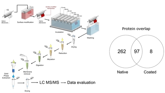
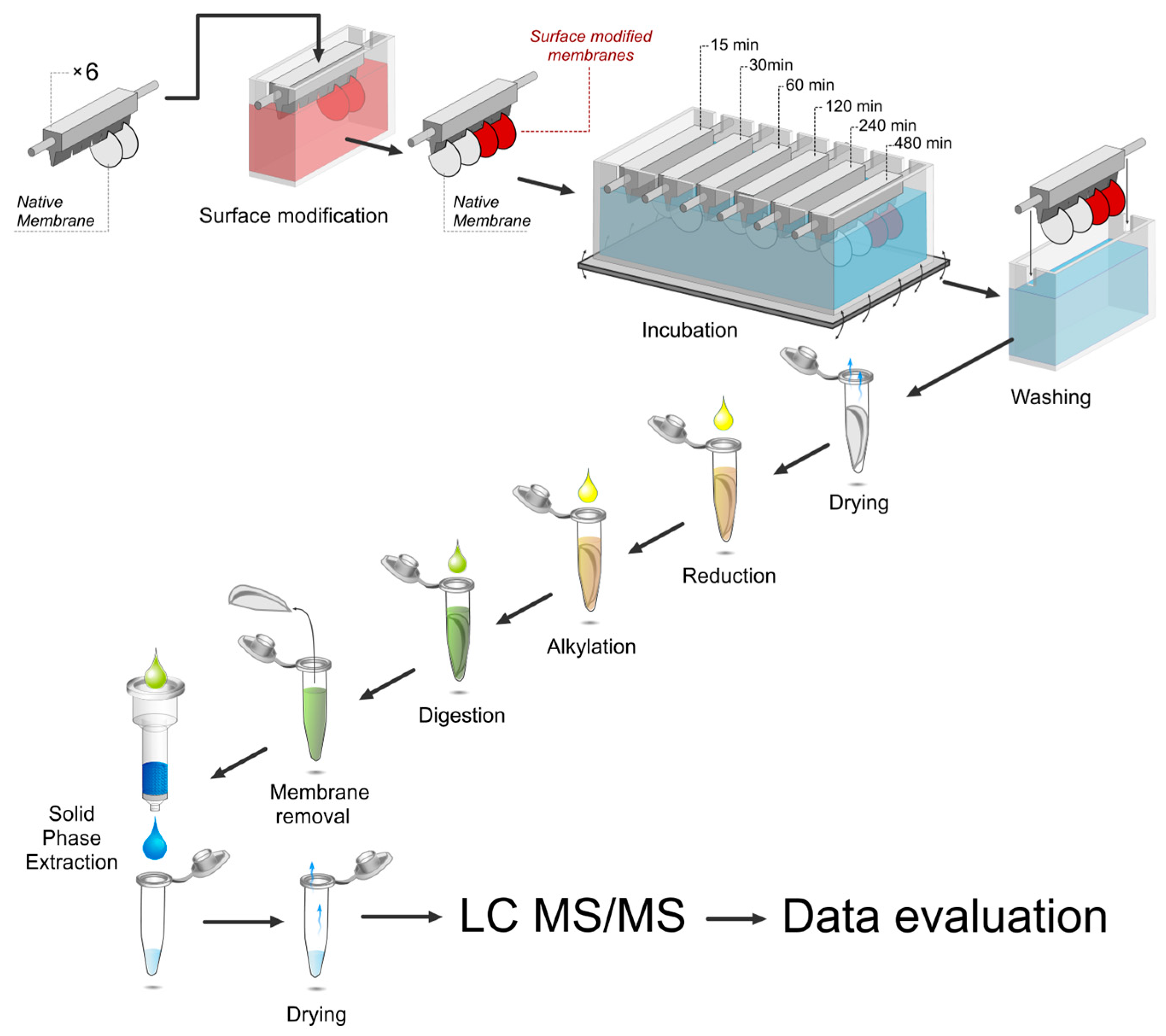
2.5. On-Surface Enzymatic Digestion (oSED)

Dried membranes were divided into four equally sized pieces and covered with 200 µL 50 mM ammonium bicarbonate. Reduction and alkylation of proteins were performed using standard reagents of dithiothreitol (DTT, 4 mM) and iodoacetamide (IAA, 8 mM) after which 1 µg trypsin was added to each membrane-containing tube. Digestion was performed at 37 °C for 24 h. Thereafter, the membranes were removed, and the resulting peptide solution was dried down. Peptides were desalted using C18-based solid phase extraction (SPE) columns according to standard procedures and dried down. The oSED is described in detail by Undin et al. [7,8].

2.6. Nano-LC-MS/MS

The LC-MS/MS analysis was performed as described by Undin et al. [7]. Briefly, in-house packed C18-AQ nano-columns with integrated emitter were used either with an Agilent 1100 converted nano-LC (Agilent Technologies, Santa Clara, CA, USA) system connected to a 7 T hybrid linear in trap Fourier Transform-Ion Cyclotron Resonance (LTQ-FTICR) Ultra MS (Thermo, Thermo Fisher Scientific, Bremen, Germany) or with a nano-LC (EASY-nLC II, Thermo) system connected to a LTQ Orbitrap Velos Pro (Thermo). Peptides were re-suspended in 0.1% FA buffer (mobile phase A) and 4 µL was injected. A linear gradient elution with increased acetonitrile concentration 0–49% was utilized for 88 min. Peptides were identified using a bottom-up/shotgun approach; peptide precursor ions were selected by a high resolution (50–100,000) survey scans while fragment ions were recorded at lower resolution using a data dependent analysis.

2.7. Data Evaluation

Data were searched against the Uniprot/SwissProt database for Homo sapiens (Release: 2013-02-13) using Proteome Discoverer (v.1.4.0.288) (Thermo) with the embedded SEQUEST database search program. The following search criteria were used: Digestion enzyme: Trypsin; Max missed cleavages: 2; Highest charge state: 4; Dynamic modification: Oxidation(M), Deamination (N, Q); Static modification: Carbamidomethylation (C); Precursor mass tolerance: 0.02 Da; Fragment mass tolerance: 0.7 Da. A protein was considered identified after detection of at least two unique (not present in other proteins) and significant (q-value < 0.05) peptides matching the protein. To score the peptides, Percolator version 2.04 was used. The false discovery rate (FDR) level was set to <0.05.

3. Results

This paper describes an unbiased comparison of surface deactivated microdialysis membranes and native membranes with respect to protein adsorption. Membranes were modified with Pluronic F-127, which was previously documented to effect protein adsorption on microdialysis membranes [6]. The novel surface characterization method developed by Undin et al. enabled this in depth, time-resolved study [7,8]. Compared to the total amount of proteins in surrounding CSF solution, the amount of adsorbed proteins was negligible and could not be observed by total protein concentration determinations. A sensitive mass spectrometry detector was therefore required. Using the set-up presented in Figure 1, adsorbed proteins were processed by directly adding trypsin for digestion to a vial containing the membranes to be studied. By analyzing the proteins using high resolved LC-MS/MS, the time dependent adsorption profiles of 114 proteins on coated membranes were obtained. This was compared to 359 proteins adsorbed onto native PC-membranes. The data from coated and native membranes were generated at the same time (in parallel) but the native data were already discussed in a previous publication by Undin et al. [7], however, not in relation to coated surfaces as presented here.

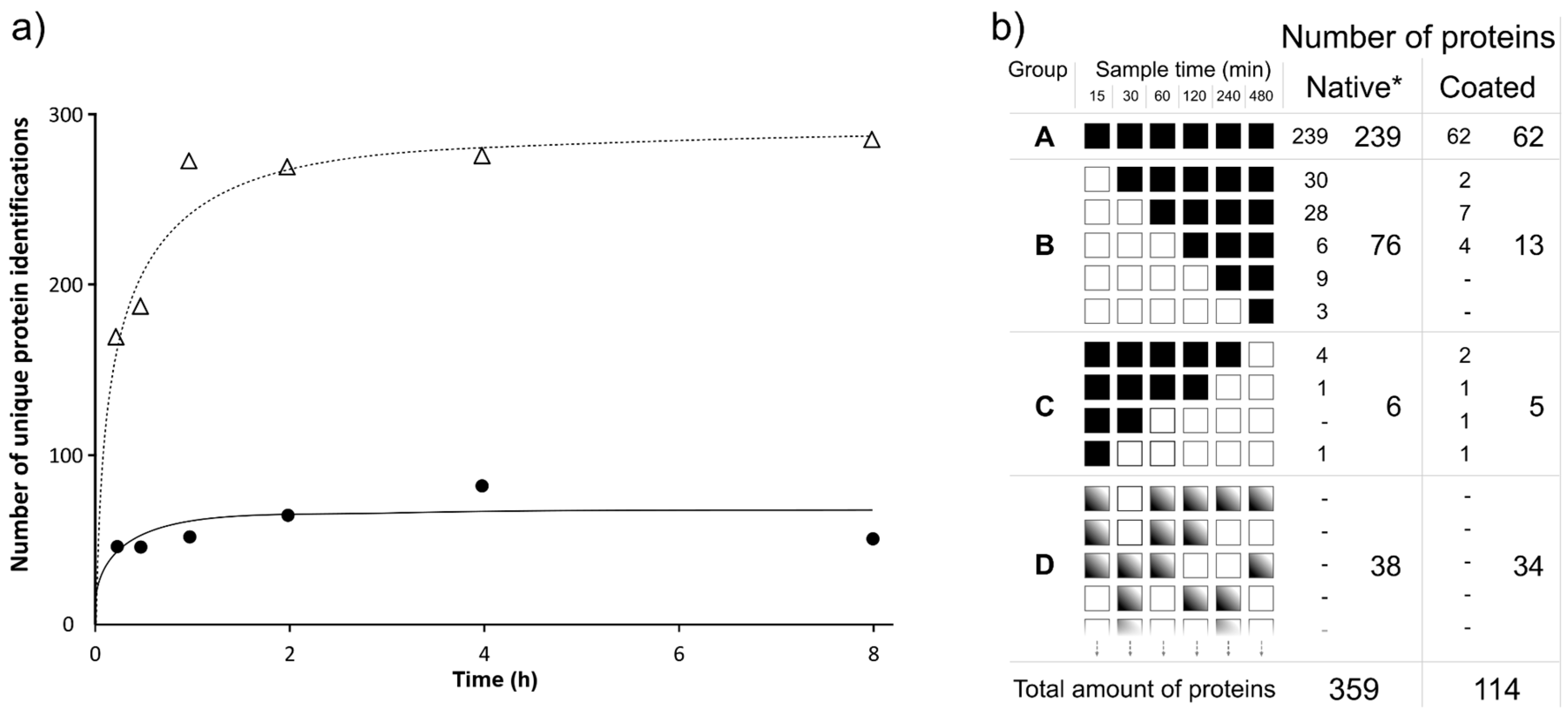
3.1. Protein Adsorption Over Time

As a first step, protein adsorption was studied over time. In Figure 2a the total number of identified adsorbed proteins—for coated and native membranes, respectively—over time is visualized. A clear difference in adsorption profiles for proteins was observed. Most relevant was the observation of reduced adsorption to coated membranes compared to native membranes for all time points of study. The adsorption to native membranes increased with increased time of incubation and reached a steady-state-value after approximately 120 min. For coated membranes, a steady-state-value was observed after 15–30 min of sampling. However, the coating of the membranes did not fully prevent adsorption, and already after 15 min of incubation around 50 proteins were adsorbed. Also, the adsorption to coated membranes increased slightly over time, but the number of identified proteins did not exceed 86 at any time (Figure 2a, 4 h time point). This is lower than the number of identified proteins to native membranes already after only 15 min of incubation. Consequently, there is clearly an advantage in using coated membranes.

3.2. Competitive Protein Adsorption Analysis

The next step in the evaluation of coated/uncoated membranes concerned competitive protein adsorption. As presented in Figure 2b, proteins were separated into different groups dependent on their adsorption behavior. An extended presentation of the protein identifications detected at different time points of adsorption to coated membranes is presented in Supplementary File 1. Group A represents proteins that were identified as adsorbed at the first time point and then remained adsorbed during all time points. Group B represents proteins that adsorbed from 30 min or later and stayed adsorbed. Group C had a reversed adsorption behavior, i.e., the proteins were adsorbed at early time point of incubation but then released or were not detected at later time points. Group D constituted proteins that had a random pattern of adsorption. As much as 67% of the identified proteins adsorbed onto the native membrane are found at all time points, which could be compared to 54% for the coated membrane (Group A). Although, native membranes overall adsorb more than three-times more proteins than coated membranes, the numbers of proteins found adsorbed with reversed adsorption behavior (Group C) and random adsorption behavior (Group D) were almost the same. The biggest relative difference between the coated and native membrane was found in Group D where 30% of all proteins adsorbed onto the coated membranes were found (11% for native membrane). For Group C, a larger proportion of proteins adsorbed to coated membranes belonged to this group, 4%, compared to 1.5% at native membranes. The numbers of proteins in these groups were, however, rather few. The same tendencies, shown as decrease in protein sequence coverage, could be seen in Figure 3a,b,e. Protein characteristics, such as molecular weight and pI-value, were investigated for each of the defined groups, but no common protein features could be related to the clusters generated.

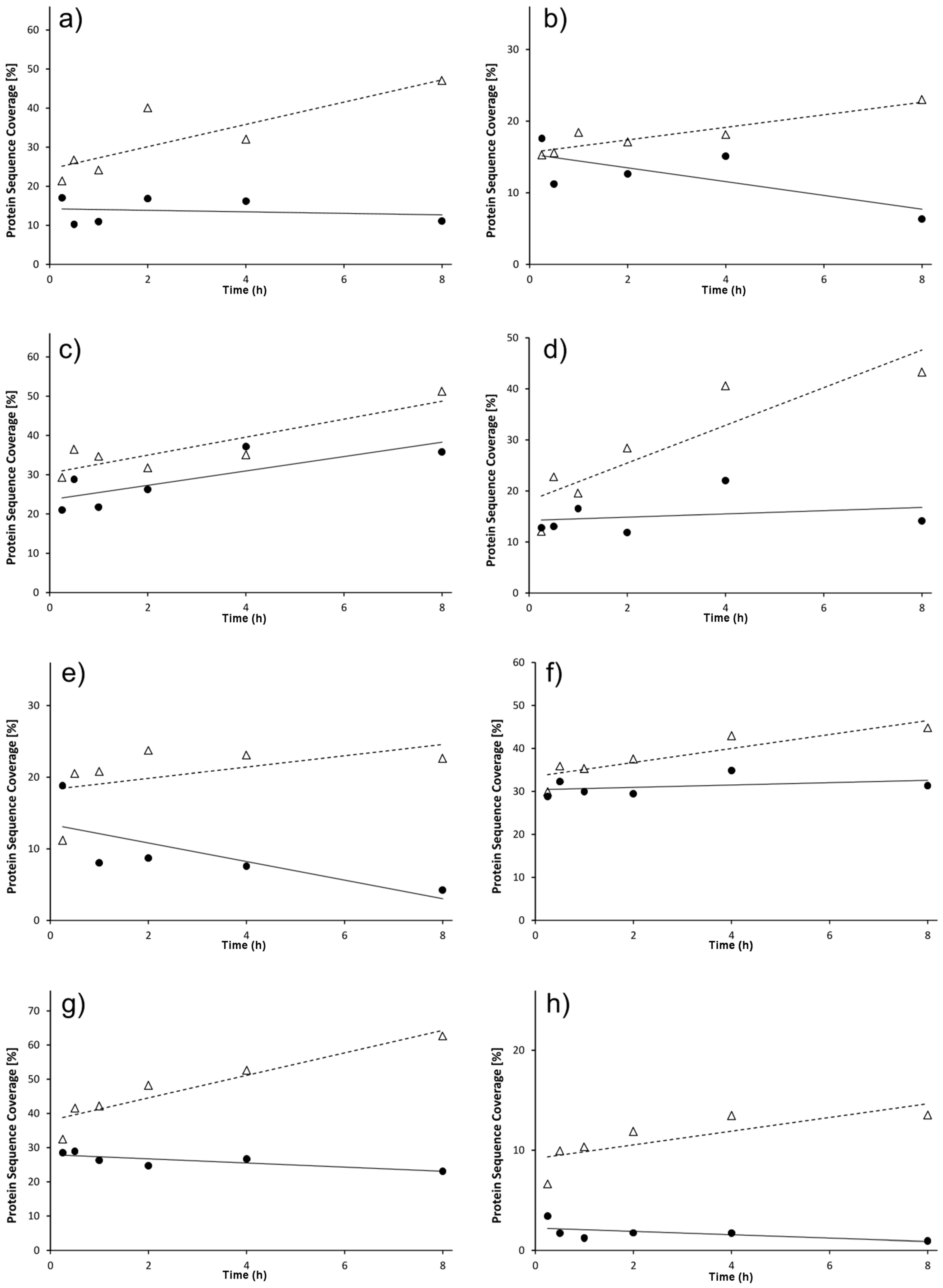
3.3. Individual Protein Adsorption Behavior

To present a strategy for evaluation of how specific proteins change their adsorption to different surfaces with time, the sequence coverages of selected proteins were plotted against incubation times. Figure 3 represents the peptide sequence coverage of the eight most common proteins in CSF, detected at all time points, for both native and coated membranes. In all cases, the sequence coverage was higher for the native membranes, indicating a higher amount of adsorbed proteins onto such surfaces. The slope of the fitted linear curve was also always positive for the native membranes suggesting that the amounts of proteins adsorbed to the surfaces increase with incubation time in vCSF. For the coated membranes, neutral or slightly positive slopes were observed for all selected proteins except for apolipoprotein E (Figure 2c) which had a negative slope. A negative slope indicated a decrease in the adsorbed amount of protein with time.

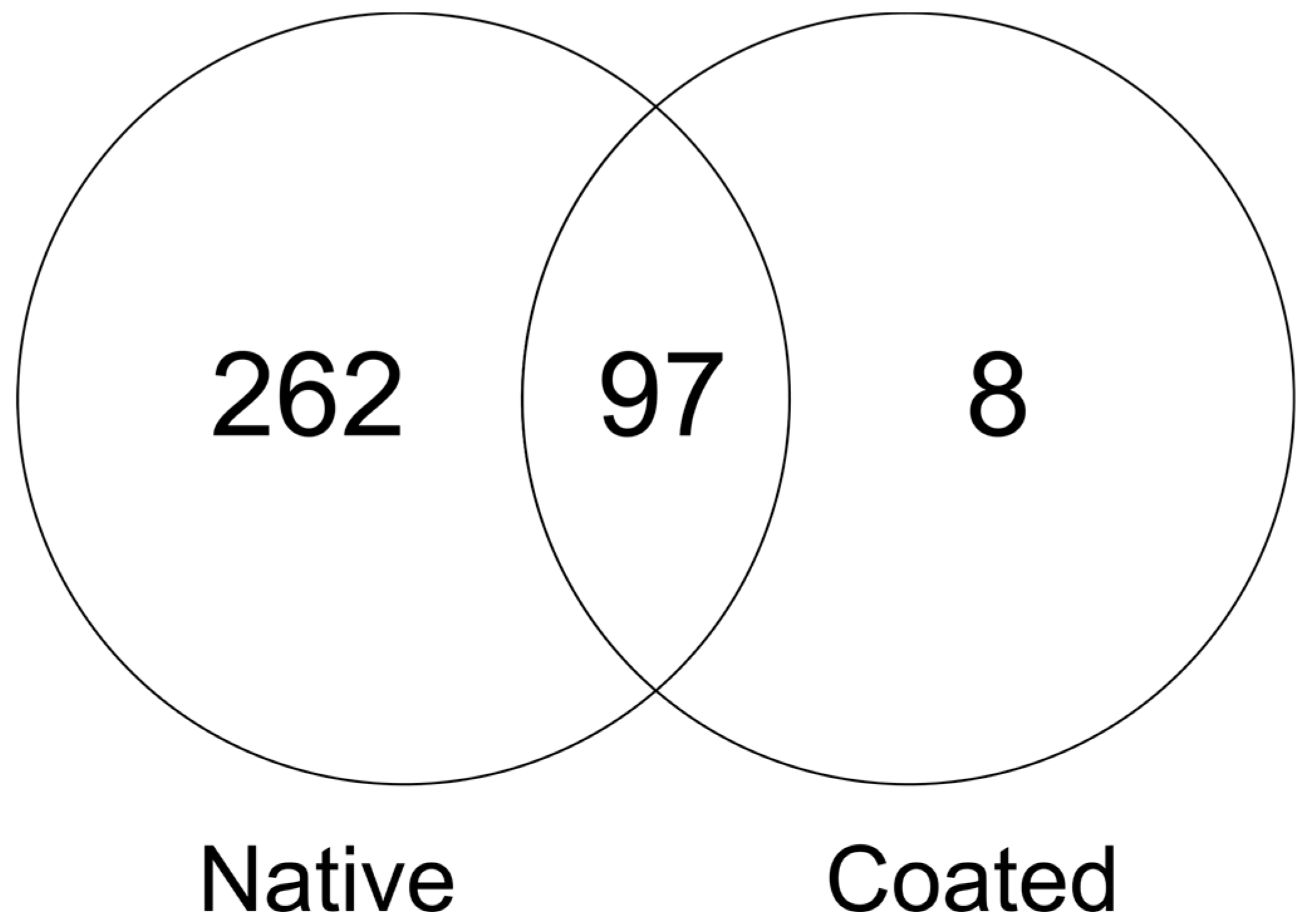
3.4. Proteins Uniquely Adsorbed to Coated Membranes

By comparing the lists of identified proteins (Supplementary File 1), uniquely adsorbed proteins for the different surfaces were identified. As presented in Figure 4, only eight proteins were uniquely adsorbed onto the coated surface. The proteins were desmoglein, protein S100, microtubule-associated tumor suppressor candidate, prolactin-inducible protein, rootletin, RhoGEF and PH domain-containing protein 1, Desmocollin-1, and Small proline-rich protein 2E. These proteins had very different characteristics in terms of length and hydrophobicity and cannot be used to identify typical proteins that preferentially adsorb to coated membranes. The 262 proteins uniquely adsorbed to native membranes were previously discussed in detail [7].

4. Discussion

Microdialysis is one of a few sampling techniques that can be used for on-line sampling to monitor a biological phenomenon. Transport across the membrane, and thus the recovery, can be severely hindered by protein adsorption to the membrane. It is therefore highly relevant to investigate the effect of coatings that can be used to reduce this effect. This is the first comparative study on time-resolved protein adsorption for native versus coated microdialysis membranes. In this study, the Pluronic F-127 coating was used, which significantly decreased protein adsorption. However, several proteins were observed to adsorb rather quickly to the coated surface. Nonetheless, the number of proteins adsorbed to modified membranes was markedly below the number of adsorbed proteins at native surfaces. This agrees with the previous findings by Dahlin et al. [6].
We used sequence coverage as an approximate measure of protein abundance in this study. As discussed by Undin et al. [7], sequence coverage is not an ideal measure of protein abundance, but it is a straightforward way to relate MS data to protein abundance for non-MS specialists and we choose this format to be consistent with a previous study [7]. When the number of peptide spectrum matches (PSMs), a more validated measurement for protein abundance from MS data [9], was used the same trends were observed.
An interesting observation is that the peptide sequence coverage is very similar at the first timepoint (15 min) between the coated and native membranes. This is in agreement with the theory that foreign materials in a biological solution instantly attract proteins and form a protein layer to cover the surface [1,2]. However, in the case of native membranes, our observations indicate that the protein layer continues to grow beyond that observed in coated membranes. The washing steps, applied to separate out unbound proteins, were performed equally for coated and native membranes but could potentially influence proteins adsorbed to the two surfaces differently. This might influence what proteins that are identified as adsorbed.
It is difficult to draw conclusions about time dependency of proteins uniquely adsorbed onto coated membranes. The sequence coverage for these proteins was low and the proteins were observed with two peptides at few measurements, indicating a low over all adsorption on the surface. Further, none of the proteins uniquely identified on the coated surface showed a specific adsorption pattern (i.e., belong to group A–C in Figure 1). The general lower protein amounts on the coated surfaces enhance these identifications of adsorbed proteins at low abundance, due to reduced competition from highly abundant proteins in the MS. This might falsely state them as unique.
It is unknown how enzymatic activity is affected by the presence of Pluronic F-127 on the membrane surface, which is an interesting direction to pursue in the future. The number of proteins was reduced from 359 for the control membrane to 115 for the coated membrane. This is in accordance with previously reported study [10] where ELISA was used to quantify the proteins adsorption. In that study, surface modification of polysulphone membranes with poloxamers gave a considerable decrease in protein adsorption. LC-MS/MS chromatograms were free from polymer contamination, indicating that no polymer was released during digestion, which supports the idea that the consistent results between LC-MS/MS and ELISA are true. It also indicates that the coating did not influence the digestion.

5. Conclusions

The oSED method combined with nanoLC-MS/MS analysis for analysis of vCSF proteins adsorbed in a time dependent manner was successful for the investigation and characterization of native and Pluronic F-127-deactivated membranes. Differences in adsorption patterns, both at the total protein level as well as for specific proteins were observed, where the adsorption was concluded lower—i.e., fewer proteins identified—for the coated compared to native surfaces. The adsorption process was significantly lessened for the coated membranes compared to native membranes. The coating also led to an increased percentage of proteins that showed a random pattern of adsorption. It is expected that this method will be a versatile tool for future investigations of surface modifications to control any wanted or unwanted adsorption of specific proteins and to further understand the intricate nature of adsorption in complex sample mixtures.

Supplementary Materials

The following are available online at https://www.mdpi.com/2297-8739/5/2/27/s1.

Author Contributions

T.U. and A.P.D. conceived and designed the experiments; T.U., A.P.D., and S.B.L. performed the experiments; T.U., A.P.D., and S.B.L. analyzed the data; J.B. contributed reagents/materials/analysis tools; T.U., A.D., J.B., and S.BL. wrote the paper.

Acknowledgments

Financial support from Swedish Research Council (JB, 2015-4870), Åke Wiberg Foundation (SBL, M14-0127), Magnus Bergvall Foundation (SBL, 2015-01200, 2016-01675), Carl Trygger Foundation (SBL, CST 15:57), and Uppsala Berzelii Technology Center for Neurodiagnostics are gratefully acknowledged. Some grants allow for contributions to costs of open access publication.

Conflicts of Interest

The authors declare no conflict of interest. The founding sponsors had no role in the design of the study; in the collection, analyses, or interpretation of data; in the writing of the manuscript, or in the decision to publish the results.

References

  1. Anderson, J.M.; Rodriguez, A.; Chang, D.T. Foreign body reaction to biomaterials. Semin. Immunol. 2008, 20, 86–100. [Google Scholar] [CrossRef] [PubMed]
  2. Rabe, M.; Verdes, D.; Seeger, S. Understanding protein adsorption phenomena at solid surfaces. Adv. Colloid Interface Sci. 2011, 162, 87–106. [Google Scholar] [CrossRef] [PubMed] [Green Version]
  3. Sides, C.R.; Stenken, J.A. Microdialysis sampling techniques applied to studies of the foreign body reaction. Eur. J. Pharm. Sci. 2014, 57, 74–86. [Google Scholar] [CrossRef] [PubMed]
  4. Wisniewski, N.; Klitzman, B.; Miller, B.; Reichert, W.M. Decreased analyte transport through implanted membranes: differentiation of biofouling from tissue effects. J. Biomed. Mater. Res. 2001, 57, 513–521. [Google Scholar] [CrossRef]
  5. Wei, Q.; Becherer, T.; Angioletti-Uberti, S.; Dzubiella, J.; Wischke, C.; Neffe, A.T.; Lendlein, A.; Ballauff, M.; Haag, R. Protein interactions with polymer coatings and biomaterials. Angew. Chem. Int. Ed. Engl. 2014, 53, 8004–8031. [Google Scholar] [CrossRef] [PubMed]
  6. Dahlin, A.P.; Hjort, K.; Hillered, L.; Sjodin, M.O.; Bergquist, J.; Wetterhall, M. Multiplexed quantification of proteins adsorbed to surface-modified and non-modified microdialysis membranes. Anal. Bioanal. Chem. 2012, 402, 2057–2067. [Google Scholar] [CrossRef] [PubMed]
  7. Undin, T.; Bergstrom, L.; Dahlin, A.P. A mass spectrometry based method for investigating time dependent protein adsorption on surfaces in contact with complex biological samples. Future Sci. OA 2015, 1, FSO32. [Google Scholar] [CrossRef] [PubMed]
  8. Undin, T.; Dahlin, A.; Hörnaeus, K.; Bergquist, J.; Bergstrom Lind, S. Mechanistic investigation of the on-surface enzymatic digestion (oSED) protein adsorption detection method using targeted mass spectrometry. Analyst 2016, 141, 1714–1720. [Google Scholar] [CrossRef] [PubMed]
  9. Lundgren, D.H.; Hwang, S.I.; Wu, L.; Han, D.K. Role of spectral counting in quantitative proteomics. Expert Rev. Proteomics 2010, 7, 39–53. [Google Scholar] [CrossRef] [PubMed]
  10. Higuchi, A.; Sugiyama, K.; Yoon, B.O.; Sakurai, M.; Hara, M.; Sumita, M.; Sugawara, S.; Shirai, T. Serum protein adsorption and platelet adhesion on pluronic (TM)-adsorbed polysulfone membranes. Biomaterials 2003, 24, 3235–3245. [Google Scholar] [CrossRef]
Figure 1. Schematics of the experimental set-up for studying protein adsorption to native and coated microdialysis membranes using LC-MS/MS. Native and coated membranes were processed side by side to control the experimental variation.
Separations 05 00027 g001
Figure 2. Time dependent protein adsorption. (a) Total number of adsorbed proteins as function of time. The total number of adsorbed proteins at each time point, detected with at least two unique peptides, plotted against time of adsorption. Triangles represent native membranes and filled circles represent coated surfaces; (b) A schematic heat map and distribution chart of the identified proteins adsorbed onto native and modified membranes. In Group A, proteins are detected at all time points; in Group B, proteins are found after some time of incubation and at all following time points; in Group C, proteins show a pattern of being initially adsorbed and then desorption starts; and finally in Group D, proteins do not have any clear time dependent adsorption pattern. Protein identifications in the respective groups are presented in Supplementary File 1. * Proteins adsorbed to native membranes are in detail discussed by Undin et al. [7].
Separations 05 00027 g002
Figure 3. Protein sequence coverage as a function of time for proteins adsorbed onto native (dashed line and triangles) membranes and coated (solid line and filled circles) membranes. (a) Cystatin C (MW 13 kDa); (b) prostaglandin-H2 D-isomerase (MW 18.5 kDa); (c) apolipoprotein E (MW 34 kDa); (d) apolipoprotein A-IV (MW 43 kDa); (e) Ig gamma-1 chain C region (MW 41 kDa); (f) clusterin (MW 50 kDa); (g) serum albumin (MW 66 kDa); (h) coagulation factor V (MW 250 kDa).
Separations 05 00027 g003
Figure 4. Venn diagram comparing the number of proteins identified uniquely on native surfaces, uniquely on coated surfaces, and on both surfaces.
Separations 05 00027 g004

