Article

Comparison and Validation of QuEChERS Extraction Methods Coupled with UHPLC/Orbitrap HR-MS for the Determination of Antibiotics and Related Compounds in Fish and Fish Feed

by
Kleopatra Miserli
1,
Vasiliki Boti
1,2,
Dimitra Hela
1,2,
Triantafyllos Albanis
1,2 and
Ioannis Konstantinou
1,2,*
1
Department of Chemistry, University of Ioannina, 45110 Ioannina, Greece
2
Unit of Environmental, Organic and Biochemical High-Resolution Analysis–Orbitrap-LC–MS, University of Ioannina, 45110 Ioannina, Greece
*
Author to whom correspondence should be addressed.
Separations 2025, 12(11), 321; https://doi.org/10.3390/separations12110321
Submission received: 20 September 2025 / Revised: 12 November 2025 / Accepted: 14 November 2025 / Published: 18 November 2025

Abstract

The widespread presence of pharmaceutical active compounds (PhACs) in aquatic environments raises significant environmental and public health concerns, particularly through their accumulation in marine biota and potential transfer to humans via seafood. In aquaculture, fish feed is essential for production but may also act as a pathway for contaminants in the marine environment. This study aimed to develop and validate an analytical method for the extraction and quantification of 14 antibiotics and ethoxyquin antioxidant in fish tissue and feed. Two QuEChERS-based extraction protocols were compared: the AOAC 2007.01 method (Method A) using Z-Sep+ as clean-up, and the original QuEChERS method (Method B) employing Enhanced Matrix Removal (EMR)-lipid. Ultra-performance liquid chromatography coupled with Orbitrap mass spectrometry using electrospray ionization in positive and negative mode was applied for identification and quantification. Validation included assessment of recovery, linearity, precision, limits of detection and quantification, uncertainty, matrix effects, and process efficiency. Both methods showed good linearity (R2 > 0.9899) and precision (<19.7%). Method B achieved superior recoveries for most analytes in both fish tissue (70–110%) and feed (69–119%), with lower uncertainties (<18.4%) compared to Method A. Overall, the original QuEChERS method demonstrated better analytical performance, supporting its application as a green, robust tool for monitoring emerging contaminants in aquaculture products.

Graphical Abstract

1. Introduction

At the beginning of the 21st century, the continuous detection of residues of pharmaceutically active compounds (PhACs) in the aquatic environment and biota represented a global problem and has been gaining importance due to the harmful impacts on human health as well as on the environment [1]. Pharmaceutical compounds including a large list of antibiotics, antidepressants, and blood lipid regulators belong to the extent class of emerging contaminants (ECs) that are not removed efficiently through wastewater treatment plants (WWTPs) and released into the environment through the runoff water or outlet water from WWTPs [2]. Among aquatic organisms, fish tissues can be considered as a bioindicator of water pollution due to the direct exposure to wastewater-borne contaminants in rivers and, by extension, in seas [3].
In the last few years, the aquaculture industry has been rapidly growing, resulting in the increased need for fish and seafood products. This means that the productivity of aquaculture may be enforced by the use of several classes of antibiotics such as macrolides, sulfonamides, tetracyclines, quinolones, amphenicols, and streptomycins in order to prevent and treat opportunistic infections in fish and, then, increase the production [4,5]. Many of these pharmaceuticals are not fully metabolized, and once excreted, they enter the aquatic environment as parent compounds or metabolites, demonstrating substantial persistence. The main risks associated with veterinary drug use are the development of antibiotic resistance, potential mutagenicity, and both inhibitory and toxic effects on aquatic organisms [6].
In aquaculture systems, the role of fish feed is crucial, as it serves as a primary nutritional source, as well as a vehicle for medication, introducing contaminants into the food chain either through contaminated raw materials or via environmental exposure during provision [7]. The composition of fish feed depends on the type of farmed fish and its growth stage and dietary requirements. It usually consists of proteins, fats, vitamins, minerals, amino acids, and functional additives like probiotics and enzymes. These are supplied through a mix of ingredients including fishmeal, fish oil, plant materials like soy or wheat, and animal by-products. Fish feed generally contains 14–19% fat and has a dry matter content of approximately 90% [8].
It was estimated that the worldwide fish production in 2020 reached around 178 million tons, with an average global per capita fish intake of 20.2 kg annually [9]. According to FAO 2020 [10], Greece is one of the most important marine and coastal finfish aquaculture producers among the European member countries. Therefore, to prevent the excessive accumulation of antibiotics in the food chain and to ensure food safety, it is essential to consistently monitor multi-class antibiotic residues in aquatic products and to identify risk factors.
Previous studies have reported the presence of pharmaceutical compounds in aquaculture products, raising concerns about their potential impact on food safety and human health [11,12,13,14,15,16,17,18,19,20]. To address such concerns, European Union Commission Regulation 37/2010 [21] has established maximum residue limits (MRLs) for veterinary drugs in foodstuffs of animal origin, including specific MRLs for fish muscle ranging from 0.5 to 600 ng g−1; however, a number of antibiotics still lack specific MRLs, leaving a regulatory gap in residue control.
Sample pretreatment techniques are crucial for the analysis of antibiotic residues since aquatic products are rich in protein, fat, and pigments, and many of these products have complex matrices and large differences in composition ratios [22]. In the case of fish tissues and fish feed, a variety of extraction methods have been widely used, such as solid-liquid extraction [23,24], enzymatic microwave-assisted extraction [25], ultra-sound extraction (USE) [19], pressurized liquid extraction (PLE) [26], and solid-phase microextraction (SPME) [27] utilizing dispersive solid phase extraction (d-SPE) with various types of sorbents as clean-up step [28,29]. Among these, the QuEChERS (quick, easy, cheap, effective, rugged, and safe) method is worth mentioning. It is based on the liquid-liquid separation of the analytes between water and acetonitrile, the use of salts (MgSO4, NaCl, etc.) to produce the salting out effect, and a dispersive solid phase extraction (d-SPE) clean-up step. The QuEChERS approach is advantageous since it offers easy application, small amounts of samples and extraction solvents, low waste volumes, rapid extraction and basic instrumentation, resulting in a time-efficient method [2]. Particularly in fish samples where fat and lipids can be as high as 50%, the availability of chemically diverse sorbents such as primary and secondary amines (PSA), graphitized carbon, octadecylsilica (C18), and zirconia-based C18 sorbent (Z-Sep), as the basis of QuEChERS, plays a crucial role in the clean-up step of such complex matrices [1]. More recently, a next-generation sorbent known as “Enhanced Matrix Removal” (EMR)-lipid has been introduced and is especially designed for removing lipids from QuEChERS extracts of fatty foods without the loss of analytes [30]. To the best of our knowledge, very few studies have applied EMR-Lipid for the analysis of PhACs in fish muscles as well as in fish feed [31,32,33].
Robust, sensitive, and selective multi-residue analysis is essential to identify and quantify PhACs in aquaculture products. Liquid chromatography (LC) combined with High-Resolution Mass Spectrometry (HRMS), such as Orbitrap technology, has been demonstrated to be helpful and appropriate for the analysis of a wide range of compounds including PhACs [2,13,34,35,36,37]. Even when endogenous matrix components of similar mass may co-elute, this instrument provides high resolution and mass accuracy in environmental matrices [38,39,40]. The above analytical technique has been used for quantitative and screening analysis of target compounds, retrospective data analysis, and identifying non-target compounds [39,41,42].
Thus, the present study aims to optimize and validate rapid and sensitive methodologies with UHPLC/LTQ Orbitrap MS for the determination of 15 PhACs in the sea bream (Sparus aurata) and fish feed substrate of aquaculture origin. Different QuEChERS procedures in terms of extraction salts and clean-up sorbents were evaluated and validated according to the Regulation (EU) 2021/808 [43], in terms of recovery, intra- and inter-day precisions, linearity and limits of detection (LODs) and quantification (LOQs) at three spiking levels, measured uncertainty (%U)/Horrat ratio, matrix effects (ME), and process efficiency (PE). This study highlighted also two new sorbents, Z-Sep+ and EMR-Lipid, with limited research on their uses in the clean-up step for fish and fish feed samples. Furthermore, the best extraction method was selected and applied to analyze fish and fish feed samples from Greek aquaculture products collected from local markets.

2. Materials and Methods

2.1. Chemicals and Reagents

The analytical standards of the 14 selected antibiotics compounds and ethoxyquin additive were of high purity (>98%) and in solid state. N-acetylsulfamethoxazole, sulfamethazine, sulfamethizole, sulfamethoxazole, sulfadiazine, sulfamethoxypyridazine, sulfapyridine, sulfaquinoxaline, sulfathiazole, erythromycin, ofloxacin, ciprofloxacin, enrofloxacin, and chloramphenicol were purchased all from Sigma-Aldrich (Darmstadt, Germany). The stock standard solutions were prepared individually (2000 mg L−1 or 1000 mg L−1) in methanol (LC-MS-grade) and stored at −20 °C. A mixed standard working solution with a concentration of 10 mg L−1 was prepared by diluting and mixing the stock solutions. All solvents used in the experiments including methanol, water, and acetonitrile were of high purity (LC-MS grade) and obtained from Fisher Scientific (Waltham, MA, USA) while formic acid (F.A) with a purity of 98% was purchased from Merck (Darmstadt, Germany) and acetic acid >95% from Sigma-Aldrich (Steinheim, Germany). The salts used in QuEChERS method, including anhydrous magnesium sulfate (MgSO4) and Z-Sep+ Bulk, were purchased from Sigma-Aldrich (Steinheim, Germany), whereas sodium acetate (NaOAc) and sodium chloride (NaCl) were supplied from Riedel-de Haën (Hannover, Germany). Enhanced Matrix Removal (EMR)-lipid was supplied from Agilent Technologies (Wilmington, DE, USA). The Evoqua Water Technologies apparatus was used for obtaining ultrapure water. Syringe filters (polytetrafluoroethylene, PTFE, 0.22 μm) were purchased from Millipore (Cork, Ireland), while propylene centrifuge tubes of 50 and 15 mL were also used.

2.2. Sample Collection and Pretreatment

Wild sea bream (Sparus aurata) samples were used to optimize and validate the analytical method. A total of 15 fish samples, 6 samples of sea bream (Sparus aurata) and 9 samples of sea bass (Dicentrarchus labrax), were analyzed using the proposed method. Fish samples were transported to the laboratory in an ice box, filleted, and the muscle tissue was homogenized using a high-speed blender before being stored at −20 °C until extraction. Commercial fish feed, confirmed to be free of the target analytes, was employed for method optimization and validation. The feed samples were collected in sterile polypropylene containers, transported to the laboratory, homogenized with a high-speed blender, and stored at room temperature until extraction. Furthermore, the application of the method was performed on 15 fish feed samples. The procedures followed did not involve the use of live animals, as fish and feed samples were obtained directly from various fish merchants.

2.3. QuEChERS Extraction

Two QuEChERS extraction methods for PhACs were optimized and validated: the American Standard (AOAC 2007.01) [44] (Method A) and the original method (Method B) using Z-Sep+ and Enhanced Matrix Removal (EMR)-lipid, respectively, as clean-up steps. At First, in the optimized Method A, 2 g of wet weight homogenized fish tissue was weighed into a 50 mL propylene centrifuge tube, an appropriate volume of PhACs solution was added, and the sample was left for 10 min to dry. Then, 10 mL of a mixture of acetonitrile (ACN) and methanol at a ratio of 75:25 (v/v) was added, and the tube was shaken for 1 min using a vortex mixer. The AOAC QuEChERS salts (6 g MgSO4 and 1.5 g NaAcetate) were used in this step; the tube was immediately shaken manually for 1 min to prevent coagulation of MgSO4 and then vortexed for 1 min. Next, the sample was centrifuged for 5 min at 4000 rpm and 2 mL of the supernatant organic layer was subjected to d-SPE as a clean-up step. The d-SPE procedure involved the supernatant’s (5 mL) transferring into a 15 mL propylene centrifuged tube, containing 125 mg Z-Sep+. After shaking by hand and vortexing for 1 min, the sample was centrifugated for 5 min at 4000 rpm and the supernatant was evaporated to dryness under a gentle stream of nitrogen at 40 °C. Finally, the sample was reconstituted in 0.5 mL of 0.1% formic acid solution of water:methanol, 90:10 (v/v), then filtered through syringe PTFE membrane filters (0.22 μm) and transferred to the autosampler vial for analysis in positive and negative ionization mode into the UHPLC/Orbitrap MS system. Next, the original QuEChERS extraction method (Method B) with the use of EMR-Lipid as clean-up step was applied for PhACs analysis of fish samples. In brief, 2 g of the homogenized sample was weighed in a 50 mL propylene centrifuge tube; the sample was hydrated with 10 mL of water followed by extraction of the target analytes using 10 mL of acetonitrile containing 1% formic acid. Then, 4 g MgSO4 and 1 g NaCl were added to the tube, which was shaken, vortexed, and centrifuged for 5 min at 4000 rpm. The d-SPE procedure, in this case, involved the activation of the EMR-Lipid (1 g) by adding 5 mL of water and vortexing for 1 min. Then, 8.5 mL of the extract was added to the tube and the suspension was vigorously hand-shaken and vortexed for 1 min. The tube was centrifuged at 4000 rpm for 10 min, and the supernatant was transferred to a polished tube containing 6 g MgSO4 and 1.5 g NaCl to remove the excess water. This was vortexed again for 1 min and centrifuged at 4000 rpm for 10 min; 5 mL of the supernatant was then evaporated under a gentle stream of nitrogen and the residue was dissolved in 0.5 mL of water:methanol, 50:50 (v/v). Prior to the injection into the UHPLC/Orbitrap MS system, the sample was filtered through syringe PTFE membrane filters (0.22 μm). In the case of chloramphenicol, Methods A and B were modified in order to comply with the Minimum Required Performance Limit (MRPL) of 0.3 μg kg−1. Specifically, 6 mL of the supernatant organic layer was subjected to d-SPE with Z-Sep+ and EMR-Lipid as clean-up steps, then evaporated to dryness and reconstituted in 0.2 mL of 0.1% formic acid solution of water:methanol, 90:10 (v/v) and 0.2 mL of water:methanol, 50:50 (v/v), respectively. Finally, fish feed samples underwent the same extraction procedure (Methods A and B) to validate the QuEChERS method and to evaluate its performance.

2.4. UHPLC/LTQ Orbitrap MS Analysis

Chromatographic analysis was carried out on a Thermo Fisher Scientific Accela LC system, which included an Accela AS autosampler (model 2.1.1) and Accela quaternary gradient LC-pump (Thermo Fisher Scientific, Bremen, Germany) coupled with a mass spectrometer (LTQ-FT Orbitrap XL 2.5.5 SP1 (Thermo Fisher Scientific, Bremen, Germany). The separation of the analytes was performed on a Hypersil GOLD C18 column (Thermo Fisher Scientific) analytical column (100 mm × 2.1 mm, 1.9 μm particle size) while the mass spectra were recorded in positive as well as in negative ionization mode (Table 1, Figure S1). The mobile phase consisted of solvent (A): water with 0.1% formic acid and solvent (B): MeOH with 0.1% formic acid. The elution gradient program was initiated at 98% A (initial conditions), maintained for 2 min, progressed to 2% at 13 min, maintained for 2 min, and returned to the initial conditions at 15.1 min, which were maintained until 20 min for the equilibration of the column. The column temperature was 40 °C, the injection volume was set to 5 μL, and the flow rate was 400 μL min−1. Furthermore, the mass spectrometer acquired full-scan MS data at a resolution of 60,000 FWHM (full width at half maximum values) over a mass (m/z) range of 100–1200 Da while dependent MS2 data were collected at a resolution of 15,000 FWHM and a normalized collision energy of (NCE) 35%. The data were processed using the Xcalibur 2.1 software and a mass tolerance window was set at 5 ppm.

2.5. Validation Study

The two analytical methods were validated in terms of recovery, linearity, intra- and inter-day precisions, uncertainty (%U)/Horrat ratio at three spiking levels, matrix effects (ME), process efficiency (PE), decision limit (CCα) and detection capability (CCβ), and limits of quantification (LOQs) and detection (LODs). The current methods were validated on the basis of Regulation (EU) 2021/808 [43]; this regulation requires a validation around the MRL for authorized drugs and at concentrations as low as possible for substances without MRLs. The studied compounds belong to the class of antibiotics and, specifically, in the group of sulfonamides, macrolides, fluoroquinolones, and amphenicols. Sulfonamides and fluoroquinolones have an MRL of 100 μg kg−1 in fish tissue [5] while macrolides have MRLs of 200 μg kg−1 in fish tissue [45]. In contrast, chloramphenicol and ethoxyquin do not have established MRLs, as they are classified as prohibited or unregulated substances in food-producing animals.
Linearity was assessed by a calibration curve of seven points of different concentration levels (10–750 ng g−1) for all target analytes, while recoveries (R %) and precision (repeatability, RSDr, and within-laboratory reproducibility, RSDR) were investigated at three different spiking levels (15, 40, and 90 ng g−1), on six spiked samples over a period of six consecutive days. Regulation 2021/808/EC [43] mentions that RSD of the mean should not exceed 25% for concentrations between 10 and 120 μg kg−1. LOQs and LODs were evaluated as the minimum concentration of analyte in sample extracts providing a signal-to-noise ratio ≥ 10 and ≥3, respectively. Furthermore, matrix effect was evaluated by comparing a calibration curve prepared in fish tissue extract and a calibration curve prepared in the solvent, at the same concentration range, as in Equation (1).
Matrix   Effect   ( M E % ) = { Slope   of   calibration   in   matrix Slope   of   calibration   in   solvent } 1 100
When ME > 0%, there is a positive matrix indicating signal enhancement, while values of ME < 0% correspond to a negative matrix effect and signal suppression. ME% values between −20 and 20% are considered low and acceptable while signal difference between ±20–50% and greater than ±50% is considered moderate and high matrix effect, respectively. Process efficiency of the method was calculated according to Equation (2) where Area (spiked) is the mean chromatographic area of the fortified samples spiked before the extraction, Area (blank) is the peak area of the unspiked sample, and Area (solvent) is the peak area of the analyte in solvent.
P E % = A r e a   ( s p i k e d ) A r e a   ( b l a n k ) A r a e   ( s o l v e n t ) 100
Finally, the expanded measurement uncertainty, expressed by U, was based on the six-replicate analysis on different days, at three spiking levels, while the acceptable expanded MU was determined according to the Horwitz equation as well as the calculation of the Horwitz ratio (HorRat) with acceptable ranges between 0.5 and 2.0 [46,47,48]. The decision limit (CCα) and detection capability (CCβ) were examined by spiking blank fish samples with antibiotics at concentration of the MRL. Calculation of CCα was performed by the MRL concentration (100 µg kg−1) of the majority of the target analytes, as determined by Regulation 37/2010 [21] and 2021/808/EC [43] of the European Commission [49,50,51], plus 1.64 times the SD of duplicate measurements of five samples at MRL, while calculation of the detection capability (CCβ) was based on CCα plus 1.64 times the SD of duplicate measurements of five samples. For unauthorized or prohibited pharmacologically active substances, the CCα shall be calculated by the corresponding concentration at the y-intercept plus 2.33 times the standard deviation of the within-laboratory reproducibility at the intercept, while the corresponding concentration at the decision limit (CCα) plus 1.64 times the standard of the reproducibility equals the detection capability (CCß).

2.6. Green Analytical Chemistry of the Extraction Procedures

The developed analytical methodologies were evaluated in the context of green analytical chemistry (GAC). At first, the Analytical Eco-Scale approach was introduced by Galuszka and his research group [52,53] and it is calculated by penalty points. The sum of penalty points for the entire procedure should be included in the Eco-Scale calculation and based on Equation (3).
A n a l y t i c a l   E c o S c a l e = 100 t o t a l   p e n a l t y   p o i n t s
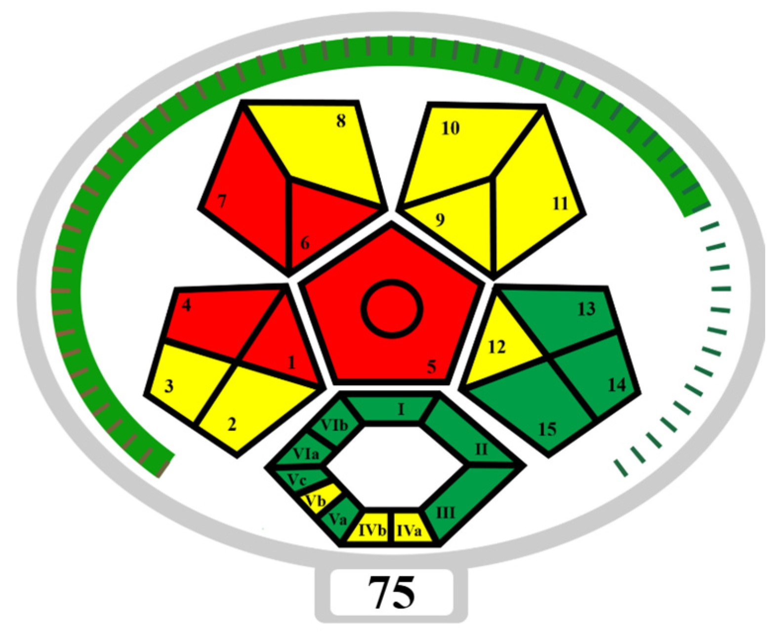
where >75 score represents excellent green analysis, >50 indicates acceptable green analysis, and <50 shows inadequate green analysis. Furthermore, a complex modified green analytical procedure index (ComplexMoGAPI) was evaluated. It included five colored pentagrams to assess the environmental impacts of the analytical process across different steps, and an additional hexagonal segment to represent the activities carried out before sample preparation and the final analysis. This tool allows for accurate and objective method comparisons, providing a comprehensive evaluation of method eco-friendliness using a color code. The green color represents methods with low environmental impact (high sustainability), orange indicates moderate impact, and red signifies procedures with a high environmental impact (low sustainability) [54,55,56,57,58].

3. Results and Discussion

3.1. Optimization of QuEChERS Extraction Method

As a first step, two QuEChERS methods were tested, and in all cases, 2 g of wet weight fish and dry fish feed samples were used. After fortifying the sample with the appropriate level of PhACs (a) the “Original” QuEChERS (4 g MgSO4 and 1 g NaCl) and (b) the “AOAC 2007.01” (6 g MgSO4/1.5 g NaOAc) were used, and a supernatant of 2 mL was submitted to a d-SPE clean-up step using 50 mg Z-Sep+. In the “Original” approach, recoveries ranged from 59% to 89% for fish substrate and between 52% to 89% for fish feed, while in the “AOAC 2007.01”, recoveries were between 70% and 90% and from 63% to 91%, respectively (Table S1). Three replicate samples were performed (n = 3) in all cases. The “AOAC 2007.01” QuEChERS method using Z-Sep+ (Method A) as salt for the clean-up step was selected for the validation study at three spiking levels (15, 40, and 90 ng g−1). Furthermore, the same two QuEChERS methods (a) and (b) were also tested using EMR-Lipid as clean-up step. In one case, the “Original” QuEChERS presented recoveries from 75% to 91% for fish matrix and from 60% to 119% for fish feed, while in the “AOAC 2007.01” approach, recoveries yielded from 58% to 74% and from 52% to 94%, respectively (Table S2). Consequently, the “Original” QuEChERS with EMR-Lipid as clean-up adsorbent (Method B) yielded better extraction recoveries. Both methods were further validated at three spiking levels (15, 40, and 90 ng g−1) for the two substrates.

3.2. Validation Results

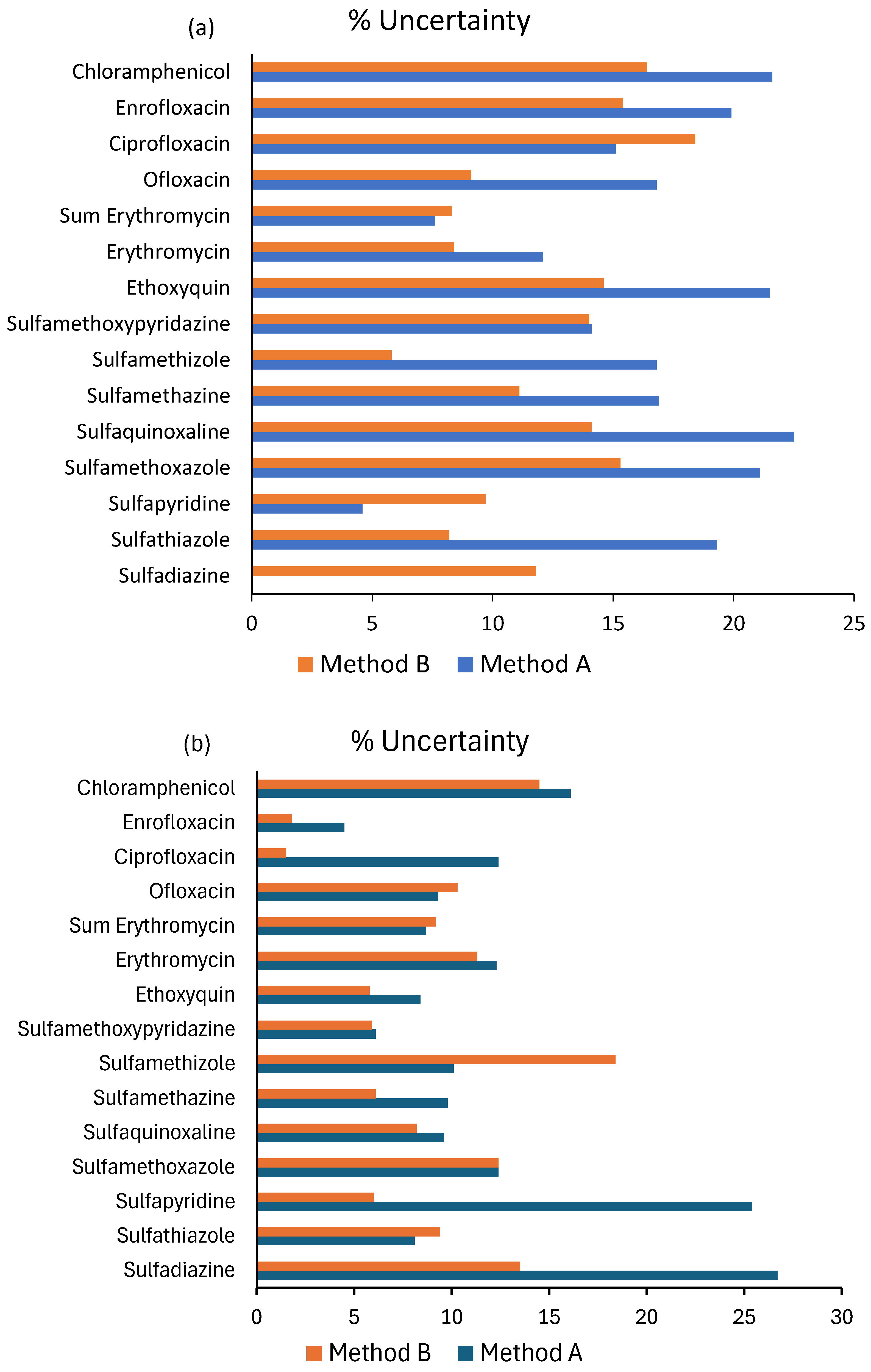
The analytical methodologies were evaluated using the parameters described in Section 2.5, for Sparus aurata and fish feed as matrices. All validation performance parameters of Methods A and B are presented in Table 2 and Table 3, respectively, for fish as well as in Table 4 and Table 5 for fish feed. Method A: Recoveries in all cases ranged at the low spiking level from 62% for sulfamethizole to 97% for sulfapyridine; at the medium spiking level, recoveries were between 73% for sulfamethizole and 93% for sulfadiazine, while at the high concentration level, they were between 74% for sulfamethizole and 102% for ciprofloxacin. High correlation coefficients R2 (>0.9939) were obtained from the calibration curves while LODs and LOQs ranged from 0.2 to 10 ng g−1 and from 0.6 to 25 ng g−1, resulting in high sensitivity of the proposed method. The LOD and LOQ values were below the EU regulation limits for animal-derived food [21,59], which demonstrates the suitability of the developed method for the detection and determination of all the tested antibiotics. Complying with the European Commission Regulation, CCα and CCβ values were calculated after spiking five fish samples with the studied analytes at their MRL level (100 ng g−1). For analytes with no established MRLs such as chloramphenicol and ethoxyquin, the CCα and CCβ values were calculated at the lowest spiking level, at 0.30–0.52 ng g−1 and 0.10–0.20 ng g−1, respectively. Table 6 shows the CCα and CCβ values for fish. Furthermore, the repeatability of the method was satisfactory for all studied compounds below 20% in all cases. Intra-day RSDs (RSDr) ranged from 0.34% to 12.9% and inter-day RSDs (RSDR) from 1.1% to 13.5%. Regarding the matrix effect (ME), as it is shown in Figure 1, the ionization suppression effect was present in most cases between −3.6% for enrofloxacin and −18.7% for sulfadiazine, indicating a low matrix effect, but few compounds showed a moderate enhancement of the signal from up to 26.1% for chloramphenicol. Among all compounds, ethoxyquin exhibited the most pronounced matrix enhancement, with %ME values of +67.6% in Method A, corresponding to a high matrix effect. Coextracted components from biological matrices such as salts (e.g., sulfates and phosphates), carbohydrates (e.g., glucose and nucleotide sugars), amines (e.g., amino acids and urea), and lipids (e.g., cholesterol and triglycerides) increases the matrix effect during the analysis of organic compounds, which is fairly known in the positive ionization mode using ESI and reported in previous studies [11,33]. The results, illustrated in Table S3, indicated PE% values in the range of 47–115%. Finally, the expanded MU was calculated individually for each PhAC, and the results are depicted in Figure 2. MU values for the low spiking ranged between 4.6% for sulfapyridine and 22.5% for sulfaquinoxaline, in medium fortification level from 7.5% for ethoxyquin to 25.2% for sulfathiazole and in the high concentration level from 1.6% for ciprofloxacin to 17.7% for sulfamethizole (Table S3). In all cases, expanded uncertainty values were in accordance with the acceptable expanded MU according to the Horwitz equation (30.1% at 15 ng g−1). In the case of chloramphenicol, the recovery obtained after six replicates at a spiking level of 0.3 ng g−1 was 70%, with a repeatability of 3.6%. The LOD and LOQ values ranged from 0.3 to 1.1 ng g−1, while the associated measurement uncertainty was 36.1% within the acceptable limits defined by the Horwitz equation. For the fish feed matrix, Method A achieved recoveries ranging from 59% to 112% across low, medium, and high spiking levels. Calibration curves showed high linearity (R2 > 0.9894), with LODs and LOQs between 0.98–3.8 and 3.2–12.5 ng g−1, confirming the method’s suitability for antibiotic determination. Repeatability was satisfactory, with intra-day and inter-day RSDs below 19.7%. Most compounds exhibited low ion suppression (−1.5% to −35%) and signal enhancement (1.7% to 35%), although some showed moderate signal enhancement (up to 64%). Process efficiency values ranged between 50% and 121% (Table S4). Expanded measurement uncertainty (MU) values for all fortification levels remained within the acceptable limits defined by the Horwitz equation (≤27.7% at 15 ng g−1).
Moving forward to the validation of (Method B), coefficients of determination (R2) for all the compound calibration curves were >0.9902, confirming an acceptable linearity range. Recoveries at the low spiking level ranged from 69% for ofloxacin to 98% for the sum of erythromycin and its hydrolyzed form. At the medium concentration level, recoveries were between 82% for the erythromycin and ofloxacin and 110% for the sum of erythromycin and its hydrolyzed form, and at the high spiking level, they were between 87% for sulfamethizole and 108% for enrofloxacin. LODs and LOQs ranged from 0.2 to 3.8 ng g−1 and from 0.6 to 12.5 ng g−1, resulting in the highest sensitivity of the proposed method. CCα and CCβ values for fish also indicated the applicability of the method (Table 6). CCα and CCβ values for chloramphenicol and ethoxyquin ranged from 0.14 to 0.24 ng g−1 and 0.11 to 0.19 ng g−1, respectively, comply with the Minimum Required Performance Limit (MRPL) of 0.3 ng g−1 for chloramphenicol. Moreover, RSDr and RSDR were <12.4% and <13%, while PE values in the range of 44–125% resulted in high accuracy and precision of the developed methodology (Table S5). MU values at the low spiking level ranged between 5.8% for sulfamethizole to 18.4% for ciprofloxacin, at the medium concentration level, from 3.1% for sulfamethizole to 15.3% for sulfaquinoxaline, and at the high spiking level, from 1% for sulfathiazole to 11.5% for sulfamethoxazole, in accordance with the acceptable expanded MU according to the Horwitz equation (Figure 2, Table S5). Finally, most of the analytes presented ME ratios within the range of −20% to 20%, suggesting a relatively small matrix effect and fewer interferences caused by co-eluted compounds from fish samples (Figure 1). However, only ethoxyquin presented ME values that exceeded ±50%, suggesting high signal enhancement. In the case of chloramphenicol, the recovery obtained after six replicates at a spiking level of 0.3 ng g−1 was 76%, with a repeatability of 2.3%. The LOD and LOQ values ranged from 0.21 to 0.7 ng g−1, while the associated measurement uncertainty was 31.4% within the acceptable limits defined by the Horwitz equation.
For the fish feed matrix, Method B demonstrated high linearity for all analytes (R2 > 0.9899) and superior sensitivity, with LODs and LOQs between 1.5–3.8 and 5–12.5 ng g−1. Recoveries across low, medium, and high spiking levels ranged from 69% to 119%. Precision was satisfactory, with intra-day and inter-day RSDs below 19.6%, and process efficiency (PE) values between 50% and 104%, indicating high accuracy (Table S6). Expanded measurement uncertainty (MU) values for all fortification levels were within the acceptable Horwitz equation limits (<18.4%, 30.1% at 15 ng g−1). Most analytes exhibited minimal to moderate matrix effects (−1.5% to 29%), although few compounds such as sulfamethazine and sulfapyridine showed higher signal suppression (up to −39% and −35%, respectively), indicating stronger matrix influence in this case.

3.3. Evaluation of the Greenness of the Studied Methods

The two extraction procedures for the determination of PhACs in fish and fish feed samples presented identical Eco-Scale values. As shown in Table 7, both methods reached a high rank of 81, which corresponds to an ideal green analysis. Regarding waste generated during sample preparation, Method A received three penalty points (for 1–10 mL of waste), while Method B was assigned one penalty point (<1 mL). For the LC-MS step, both methods incurred three penalty points for waste [53]. According to Figure 3, both QuEChERS methods presented low environmental impact and high sustainability. The red zones (categories 1, 4, 6, and 7) highlight the most critical weaknesses, namely, off-line sample collection, special storage requirements, macro-scale extraction, and the use of non-green solvents. The yellow zones (categories 2, 3, 8, 9, 10, and 11) reflect intermediate sustainability, mainly linked to sample preservation and transport, moderate solvent consumption, and the use of reagents with moderate toxicity and safety risks [55]. Despite these limitations, the overall assessment still places the method within the green category, confirming its suitability for environmentally conscious analytical applications.

3.4. Comparison of the Validated Methods with Other Studies

A comparison of the main characteristics of the developed methods (Methods A and B) proposed in this study was conducted with previously published studies, which included similar QuEChERS extraction and LC-MS techniques to assess pharmaceuticals in fish and feedstuffs. For instance, Aissaoui et al., 2024 [11] recently developed a method to extract multiclass antibiotics (quinolones, sulfonamides, diaminopyrimidines, and macrolide) from sea bass and sea breams using AOAC 2007.01 QuEChERS and UHPLC-MS/MS analysis. They presented comparable recoveries with this study in the range of 63–97% and a matrix effect above the range of ±50% but much higher LOQs (20.5 to 68.6 ng g−1). Furthermore, a relevant published study, Boti et al., 2024 [41], presented the QuEChERS method with UHPLC-MS/MS for the determination of emerging contaminants in fish feed samples, including some pharmaceuticals and the use of Z-Sep+ adsorbent in the clean-up step, yielded similar outcomes, with recoveries of 53–127, LOQs of 2–50 ng g−1, RSD < 20%, and uncertainty lower than 50%. A simple, selective, and rapid multiresidue original QuEChERS method was developed by Lopes et al., 2012 [60] for the determination of 32 veterinary drug residues belonging to different classes (i.e., macrolides, penicillins, quinolones, sulfonamides, and tetracyclines) in gilthead sea bream (Sparus aurata) samples by UHPLC-MS/MS. Mean recovery ranged from 69% to 125% (at 10, 25, 50, and 100 μg kg−1), intra- and inter-day precision was lower than 20%, LOQs were lower than 25 μg kg−1, and expanded uncertainty was below 25% at 100 μg kg−1. Recently, Dai et al., 2023 [61] developed and validated a QuEChERS preparation method with EMR-Lipid as clean-up using ultrahigh-performance liquid chromatography coupled with quadrupole-Orbitrap mass spectrometry for the determination of chlorpromazine and its metabolites in fish from local markets (Nanjing, China). Satisfactory recoveries ranged from 72% to 117%, RSD from 0.2% to 18%, and LOQs were 2 ng g−1, in agreement with the results of this study. Finally, a simple purification process which tested post-acidic acetonitrile extraction with an EMR cartridge and analyzed by LC-triple quadrupole was established by Xu et al., 2024 [33] and demonstrated satisfactory recoveries (76.2–99.7%), RSD below 13.9%, and LOQs from 0.04 to 5 ng g−1, which are comparable with the results of this study. Furthermore, Konak et al., 2021 [62] used the AOAC 2007.01 QuEChERS method and UHPLC-MS/MS to determine eight mycotoxins and 10 antibiotics in fish feeds. The method showed good linearity R2 (>0.995) and average recoveries ranging from 60 to 102% with relative standard deviations of 2.2 and 15.6%, while LOQ values varied between 1.2 and 5.2 ng g−1.
To conclude, the results from this study demonstrated that the original QuEChERS method with the EMR cartridge (Method B) yielded better recoveries for 12 of 15 analytes in the range of 77 and 98% at the low concentration level than the AOAC QuEChERS (Method A) (62–79%) for fish substrate. Similarly, in fish feed substrate, the application of EMR-Lipid sorbent enabled efficient extraction of all the analytes with higher recoveries (68–78%) compared to the use of Z-Sep+ (59–73%). The majority of the analytes presented measured uncertainty at the low spiking level 15 ng g−1 under < 22.3% in the original QuEChERS (Method B) compared to the AOAC method (Method A) (<27.7%) across both matrices. Both Method A and Method B exhibited low matrix effects for the sulfonamide group in fish samples, with %ME values ranging between −20% and +20%. Notably, the use of EMR-Lipid as clean-up step showed even lower matrix effects within this range (1.9 to 17.1%, signal enhancement), indicating improved control of matrix-induced ionization variability. Although ethoxyquin displayed a relatively high signal enhancement of +67.6% using Method A, this effect was substantially reduced to +51.6% in Method B, demonstrating the effectiveness of the EMR-Lipid clean-up in minimizing matrix interference. Furthermore, the application of the Z-Sep+ sorbent showed that nine compounds presented a medium-to-high matrix effect with values between −43% and 64% in fish feed. On the other hand, the use of the EMR sorbent gave better results in the purification of the extract, presenting a low (−17–6%)-to-medium matrix effect (−35–29%). Consequently, the EMR-Lipid agent demonstrated superior purification capacity, efficiently removing lipids and co-extractants without significant analyte loss, confirming its suitability for accurate analysis of fish and fish feed samples.

3.5. Application to Real Fish and Fish Feed Samples

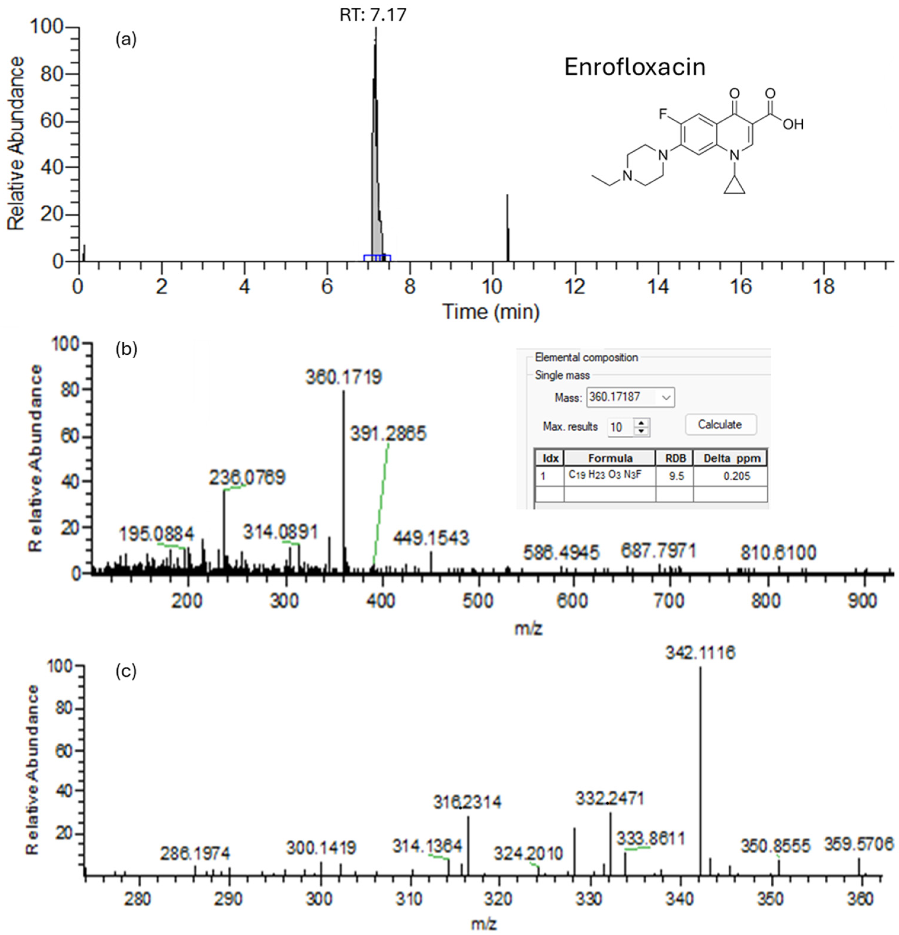
Out of the 15 compounds examined in the developed analytical method, only the group of quinolones was detected in the analyzed fish samples, specifically in three sea bream samples and one sea bass (labrax) sample. Among these, ofloxacin was identified in two sea bream samples, as well as in one labrax sample, all at concentrations below the LOQ. Furthermore, ciprofloxacin was quantified in two sea bream samples at concentrations ranging from 1.4 to 1.6 ng g−1 while enrofloxacin was detected in three sea bream samples at levels varying from below LOQ to 1.9 ng g−1. Notably, one sea bream sample contained all three quinolones antibiotics (ofloxacin, ciprofloxacin, and enrofloxacin) at concentrations between <LOQ and 1.9 ng g−1 (Figure 4). All detected antibiotic residues were found to be well below their established MRLs, thereby indicating that the examined samples are safe for human consumption according to current regulatory standards. Furthermore, the application of Method B in 15 feed samples revealed the presence of sulfamethazine in one feed sample at a concentration of 10.5 ng g−1.

4. Conclusions

Analytical extraction methods based on QuEChERS and UHPLC/LTQ Orbitrap MS technologies were developed in this study in order to monitor 15 multiclass antibiotics and additives in fish and fish feed samples. All analytical procedures presented good performance characteristics, in terms of recovery, linearity, RSD values, and low LOQ values that met the required MRLs established by the European Union. It seems that the original QuEChERS with the dispersive-SPE EMR-Lipid as clean-up step (Method B) yielded better results for most antibiotics across both matrices. Method B achieved superior recoveries for most analytes in both fish tissue at the low concentration level (77–98%) and feed (68–78%), with lower uncertainties (<22.3%) compared to Method A. Matrix effect studies in Method B presented a lower effect with enhancement of the signal in comparison with Method A, in which the signal of the analytes was suppressed. Method B was further applied to real fish and fish feed samples collected from local markets, where only quinolones were detected in the analyzed fish samples, specifically in three sea bream samples and one sea bass sample at concentrations between <LOQ and 1.9 ng g−1, while sulfamethazine was detected in one feed sample at a concentration of 10.5 ng g−1. The developed methodology can potentially be applied to analyze other types of emerging contaminants including pesticides, phthalic acid esters, personal care products, etc., in order to obtain an extensive view of the pollutants that may affect fish in aquaculture farms.

Supplementary Materials

The following supporting information can be downloaded at: https://www.mdpi.com/article/10.3390/separations12110321/s1, Figure S1. A chromatogram for illustration of analytes separation in a spiked fish sample 100 ng g−1 (Method B), Table S1. Recoveries of antibiotics obtained for different clean-up sorbents at spike level 40 ng g−1 in fish samples, Table S2. Recoveries of antibiotics obtained for different clean-up sorbents at spike level 40 ng g−1 in fish feed samples, Table S3. Validation data for 15 antibiotics in sea bream samples; %Process efficiency, expanded uncertainty (%U), and HorRat ratio (Method A), Table S4. Validation data for 15 antibiotics in fish feed samples; %Process efficiency, expanded uncertainty (%U), and HorRat ratio (Method A), Table S5. Validation data for 15 antibiotics in sea bream samples; %Process efficiency, expanded uncertainty (%U), and HorRat ratio (Method B), Table S6. Validation data for 15 antibiotics in fish feed samples; %Process efficiency, expanded uncertainty (%U), and HorRat ratio (Method B).

Author Contributions

K.M.: Writing—review and editing, Writing—original draft, Visualization, Validation, Investigation, Formal analysis, Data curation. V.B.: Writing—review and editing, Conceptualization. D.H.: Writing—review and editing, Writing—original draft. T.A.: Resources, Methodology. I.K.: Writing—review and editing, Writing—original draft, Supervision, Resources, Project administration, Funding acquisition, Conceptualization. All authors have read and agreed to the published version of the manuscript.

Funding

This research is implemented in the framework of H.F.R.I call “Basic research Financing (Horizontal support of all Sciences)” under the National Recovery and Resilience Plan “Greece 2.0” funded by the European Union—Next Generation EU (H.F.R.I. Project Number: 16849).

Institutional Review Board Statement

Ethical approval was not required for this study, since the fish and fish feed samples used were sourced from aquaculture products commercially distributed in local markets, without involving any live animal experimentation.

Data Availability Statement

The original contributions presented in this study are included in the article/Supplementary Material. Further inquiries can be directed to the corresponding author(s).

Conflicts of Interest

The authors declare no conflicts of interest. The funders had no role in the design of the study; in the collection, analyses, or interpretation of data; in the writing of the manuscript; or in the decision to publish the results.

References

  1. Manjarrés-López, D.P.; Vitale, D.; Callejas-Martos, S.; Usuriaga, M.; Picó, Y.; Pérez, S.; Montemurro, N. An Effective Method for the Simultaneous Extraction of 173 Contaminants of Emerging Concern in Freshwater Invasive Species and Its Application. Anal. Bioanal. Chem. 2023, 415, 7085–7101. [Google Scholar] [CrossRef]
  2. Peña-Herrera, J.M.; Montemurro, N.; Barceló, D.; Pérez, S. Development and Validation of an Analytical Method for Determination of Pharmaceuticals in Fish Muscle Based on QuEChERS Extraction and SWATH Acquisition Using LC-QTOF-MS/MS System. Talanta 2019, 199, 370–379. [Google Scholar] [CrossRef] [PubMed]
  3. Baesu, A.; Ballash, G.; Mollenkopf, D.; Wittum, T.; Sulliván, S.M.P.; Bayen, S. Suspect Screening of Pharmaceuticals in Fish Livers Based on QuEChERS Extraction Coupled with High Resolution Mass Spectrometry. Sci. Total Environ. 2021, 783, 146902. [Google Scholar] [CrossRef] [PubMed]
  4. Bondad-Reantaso, M.G.; Arthur, J.R. Improving Biosecurity Through Prudent and Responsible Use of Veterinary Medicines in Aquatic Food Production; FAO: Rome, Italy, 2012; p. 226. ISBN 978-92-5-106975-2. [Google Scholar]
  5. Mukota, A.K.; Gondam, M.F.K.; Tsafack, J.J.T.; Sasanya, J.; Reybroeck, W.; Ntale, M.; Nyanzi, S.A.; Tebandeke, E. Primary Validation of Charm II Tests for the Detection of Antimicrobial Residues in a Range of Aquaculture Fish. BMC Chem. 2020, 14, 32. [Google Scholar] [CrossRef]
  6. Dabestani-Rahmatabad, A.; Nava-Guajardo, L.; Nicol, E.; Stephane, B. A Review on the Main Antibiotic Drugs Used in Fish Farming: Ecotoxicity, Characterization and Remediation. Aquac. Fish. Stud. 2021, 3, 1–11. [Google Scholar] [CrossRef]
  7. Berntssen, M.H.G.; Hoogenveen, R.; Rosenlund, G.; Garlito, B.; Zeilmaker, M.J. Do Background Levels of the Pesticide Pirimiphosmethyl in Plant-Based Aquafeeds Affect Food Safety of Farmed Atlantic Salmon? Food Addit. Contam. Part A 2020, 37, 2109–2122. [Google Scholar] [CrossRef]
  8. Koloka, O.; Boti, V.; Hela, D.; Albanis, T.; Konstantinou, I. PFAS Residue Analysis in Fish and Fish Feed by Applying a Modified QuEChERS Method in Combination with LC-HRMS Detection. Emerg. Contam. 2025, 11, 100467. [Google Scholar] [CrossRef]
  9. FAO. The State of World Fisheries and Aquaculture 2022: Towards Blue Transformation; FAO: Rome, Italy, 2022; Available online: https://www.fao.org/documents/card/en/c/cc0461en (accessed on 15 March 2023).
  10. FAO. The State of World Fisheries and Aquaculture 2020: Sustainability in Action; FAO: Rome, Italy, 2020. [Google Scholar] [CrossRef]
  11. Aissaoui, Y.; Jiménez-Skrzypek, G.; González-Sálamo, J.; Trabelsi-Ayadi, M.; Ghorbel-Abid, I.; Hernández-Borges, J. Determination of Multiclass Antibiotics in Fish Muscle Using a QuEChERS-UHPLC-MS/MS Method. Foods 2024, 13, 1081. [Google Scholar] [CrossRef]
  12. Álvarez-Muñoz, D.; Rambla-Alegre, M.; Carrasco, N.; Lopez De Alda, M.; Barceló, D. Fast Analysis of Relevant Contaminants Mixture in Commercial Shellfish. Talanta 2019, 205, 119884. [Google Scholar] [CrossRef]
  13. Álvarez-Muñoz, D.; Rodríguez-Mozaz, S.; Jacobs, S.; Serra-Compte, A.; Cáceres, N.; Sioen, I.; Verbeke, W.; Barbosa, V.; Ferrari, F.; Fernández-Tejedor, M.; et al. Pharmaceuticals and Endocrine Disruptors in Raw and Cooked Seafood from European Market: Concentrations and Human Exposure Levels. Environ. Int. 2018, 119, 570–581. [Google Scholar] [CrossRef]
  14. Barros, S.C.; Silva, A.S.; Torres, D. Multiresidues Multiclass Analytical Methods for Determination of Antibiotics in Animal Origin Food: A Critical Analysis. Antibiotics 2023, 12, 202. [Google Scholar] [CrossRef]
  15. Dasenaki, M.E.; Thomaidis, N.S. Multi-Residue Determination of 115 Veterinary Drugs and Pharmaceutical Residues in Milk Powder, Butter, Fish Tissue and Eggs Using Liquid Chromatography–Tandem Mass Spectrometry. Anal. Chim. Acta 2015, 880, 103–121. [Google Scholar] [CrossRef] [PubMed]
  16. Desmarchelier, A.; Fan, K.; Minh Tien, M.; Savoy, M.C.; Tarres, A.; Fuger, D.; Goyon, A.; Bessaire, T.; Mottier, P. Determination of 105 Antibiotic, Anti-Inflammatory, Antiparasitic Agents and Tranquilizers by LC-MS/MS Based on an Acidic QuEChERS-like Extraction. Food Addit. Contam. Part A 2018, 35, 646–660. [Google Scholar] [CrossRef] [PubMed]
  17. Kim, K.; Choi, Y.; Mok, S.; Moon, H.B.; Jeon, J. Optimization of the QuEChERS Method for Multi-Residue Analysis of Pharmaceuticals and Pesticides in Aquaculture Products. Food Chem. 2023, 399, 133958. [Google Scholar] [CrossRef] [PubMed]
  18. Mijangos, L.; Ziarrusta, H.; Zabaleta, I.; Usobiaga, A.; Olivares, M.; Zuloaga, O.; Etxebarria, N.; Prieto, A. Multiresidue Analytical Method for the Determination of 41 Multiclass Organic Pollutants in Mussel and Fish Tissues and Biofluids by Liquid Chromatography Coupled to Tandem Mass Spectrometry. ABC 2019, 411, 493–506. [Google Scholar] [CrossRef]
  19. Peña-Herrera, J.M.; Montemurro, N.; Barceló, D.; Pérez, S. Analysis of Pharmaceuticals in Fish Using Ultrasound Extraction and Dispersive Spe Clean-up on Que Z-Sep/C18 Followed by LC-QToF-MS Detection. MethodsX 2020, 7, 101010. [Google Scholar] [CrossRef]
  20. Yang, Y.; Li, X.; Lin, J.; Bao, R. A Modified QuEChERS-Based UPLC-MS/MS Method for Rapid Determination of Multiple Antibiotics and Sedative Residues in Freshwater Fish. Food Chem. X 2024, 22, 101268. [Google Scholar] [CrossRef]
  21. European Commission. Commission Decision 37/2010 of 22 December 2009 on pharmacologically active substances and their classification regarding maximum residue limits in foodstuffs of animal origin. Off. J. Eur. Communities 2010, 15, 1–72. [Google Scholar]
  22. Diallo, T.; Makni, Y.; Lerebours, A.; Thomas, H.; Guérin, T.; Parinet, J. Wide-Scope Screening of Multi-Class Contaminants in Seafood Using a Novel Sample Preparation (QuEChUP) Procedure Coupled with UHPLC-Q-TOF-MS: Application for Semi-Quantitation of Real Seafood Samples. Food Chem. 2023, 426, 136572. [Google Scholar] [CrossRef]
  23. Jung, H.N.; Park, D.H.; Yoo, K.H.; Cho, H.J.; Shim, J.H.; Shin, H.C.; Abd El-Aty, A.M. Simultaneous Quantification of 12 Veterinary Drug Residues in Fishery Products Using Liquid Chromatography-Tandem Mass Spectrometry. Food Chem. 2021, 348, 129105. [Google Scholar] [CrossRef]
  24. Turnipseed, S.B.; Storey, J.M.; Wu, I.L.; Andersen, W.C.; Madson, M.R. Extended Liquid Chromatography High Resolution Mass Spectrometry Screening Method for Veterinary Drug, Pesticide and Human Pharmaceutical Residues in Aquaculture Fish. Food Addit. Contam.—Part Chem. Anal. Control Expo. Risk Assess. 2019, 36, 1501–1514. [Google Scholar] [CrossRef]
  25. Fernandez-Torres, R.; Lopez, B.A.B.; Consentino, M.O.; Mochon, M.C.; Payan, M.R. Enzymatic-Microwave Assisted Extraction and High-Performance Liquid Chromatography-Mass Spectrometry for the Determination of Selected Veterinary Antibiotics in Fish and Mussel Samples. J. Pharm. Biomed. Anal. 2011, 54, 1146–1156. [Google Scholar] [CrossRef]
  26. Subedi, B.; Mottaleb, M.A.; Chambliss, C.K.; Usenko, S. Simultaneous Analysis of Select Pharmaceuticals and Personal Care Products in Fish Tissue Using Pressurized Liquid Extraction Combined with Silica Gel Cleanup. J. Chromatogr. A 2011, 1218, 6278–6284. [Google Scholar] [CrossRef]
  27. Togunde, O.P.; Oakes, K.D.; Servos, M.R.; Pawliszyn, J. Optimization of Solid Phase Microextraction for Non-Lethal in Vivo Determination of Selected Pharmaceuticals in Fish Muscle Using Liquid Chromatography-Mass Spectrometry. J. Chromatogr. A 2012, 1261, 99–106. [Google Scholar] [CrossRef]
  28. Baduel, C.; Mueller, J.F.; Tsai, H.; Gomez Ramos, M.J. Development of Sample Extraction and Clean-up Strategies for Target and Non-Target Analysis of Environmental Contaminants in Biological Matrices. J. Chromatogr. A 2015, 1426, 33–47. [Google Scholar] [CrossRef]
  29. Huerta, B.; Rodríguez-Mozaz, S.; Barcelo, D. Analysis of Pharmaceutical Compounds in Biota. In Comprehensive Analytical Chemistry; Elsevier: Amsterdam, The Netherlands, 2013; Volume 62, pp. 169–193. [Google Scholar] [CrossRef]
  30. DeAtley, A.; Zhao, L.; Lucas, D. Innovative Sample Prep Removes Lipids without Losing Analytes. Am. Lab. 2015, 47, 32–34. [Google Scholar]
  31. Drábová, L.; Dvořáková, D.; Urbancová, K.; Gramblička, T.; Hajšlová, J.; Pulkrabová, J. Critical Assessment of Clean-Up Techniques Employed in Simultaneous Analysis of Persistent Organic Pollutants and Polycyclic Aromatic Hydrocarbons in Fatty Samples. Toxics 2022, 10, 12. [Google Scholar] [CrossRef]
  32. Han, L.; Matarrita, J.; Sapozhnikova, Y.; Lehotay, S.J. Evaluation of a Recent Product to Remove Lipids and Other Matrix Co-Extractives in the Analysis of Pesticide Residues and Environmental Contaminants in Foods. J. Chromatogr. A 2016, 1449, 17–29. [Google Scholar] [CrossRef] [PubMed]
  33. Xu, F.; Zhang, Z.; Shi, Q.; Zhang, R.; Sun, A.; Zhao, J.; Wu, Y.; Shi, X. Rapid Determination and Risk Evaluation of Multi-Class Antibiotics in Aquatic Products by One-Step Purification Process Coupled with Ultra-High Performance Liquid Chromatography–Tandem Mass Spectrometry. Talanta 2024, 277, 126421. [Google Scholar] [CrossRef]
  34. Gómez-Pérez, M.L.; Romero-González, R.; Martínez Vidal, J.L.; Garrido Frenich, A. Analysis of Veterinary Drug and Pesticide Residues in Animal Feed by High-Resolution Mass Spectrometry: Comparison between Time-of-Flight and Orbitrap. Food Addit. Contam. Part A 2015, 32, 1637–1646. [Google Scholar] [CrossRef]
  35. Petrović, M.; Hernando, M.D.; Díaz-Cruz, M.S.; Barceló, D. Liquid Chromatography-Tandem Mass Spectrometry for the Analysis of Pharmaceutical Residues in Environmental Samples: A Review. J. Chromatogr. A 2005, 1067, 1–14. [Google Scholar] [CrossRef]
  36. Villagrasa, M.; López de Alda, M.; Barceló, D. Environmental Analysis of Fluorinated Alkyl Substances by Liquid Chromatography-(Tandem) Mass Spectrometry: A Review. Anal. Bioanal. Chem. 2006, 386, 953–972. [Google Scholar] [CrossRef]
  37. Wu, X.; Lin, Y.; Zhang, X.; Ouyang, N.; Zhou, Y. Optimization of QuEChERS Method for Antibiotic Residue Analysis in Animal Foods via Response Surface Methodology. Separations 2023, 10, 459. [Google Scholar] [CrossRef]
  38. Kalogeropoulou, A.G.; Kosma, C.I.; Albanis, T.A. Simultaneous Determination of Pharmaceuticals and Metabolites in Fish Tissue by QuEChERS Extraction and UHPLC Q/Orbitrap MS Analysis. Anal. Bioanal. Chem. 2021, 413, 7129–7140. [Google Scholar] [CrossRef]
  39. Kosma, C.I.; Koloka, O.L.; Albanis, T.A.; Konstantinou, I.K. Accurate Mass Screening of Pesticide Residues in Wine by Modified QuEChERS and LC-Hybrid LTQ/Orbitrap-MS. Food Chem. 2021, 360, 130008. [Google Scholar] [CrossRef] [PubMed]
  40. Tran-Lam, T.-T.; Pham, P.T.; Bui, M.Q.; Dao, Y.H.; Le, G.T. Organophosphate Esters and Their Metabolites in Marine Fish from Vietnam: Analytical Method Development and Validation. J. Food Compos. Anal. 2024, 131, 106266. [Google Scholar] [CrossRef]
  41. Boti, V.; Martinaiou, P.; Gkountouras, D.; Albanis, T. Target and Suspect Screening Approaches for the Identification of Emerging and Other Contaminants in Fish Feeds Using High Resolution Mass Spectrometry. Environ. Res. 2024, 251, 118739. [Google Scholar] [CrossRef]
  42. Nannou, C.I.; Boti, V.I.; Albanis, T.A. Trace Analysis of Pesticide Residues in Sediments Using Liquid Chromatography-High-Resolution Orbitrap Mass Spectrometry. Anal. Bioanal. Chem. 2018, 410, 1977–1989. [Google Scholar] [CrossRef]
  43. European Commission Decision. Commission Decision 2021/808/EC the Performance of Analytical Methods for Residues of Pharmacologically Active Substances Used in Food-Producing Animals and on the Interpretation of Results as Well as on the Methods to be Used for Sampling and Repealing Decisions 2002/657/EC and 98/179/EC. 2021. Available online: https://eur-lex.europa.eu/eli/reg_impl/2021/808/oj/eng (accessed on 22 March 2021).
  44. AOAC 2007.01; Pesticide Residues in Foods by Acetonitrile Extraction and Partitioning with Magnesium Sulfate: Gas Chromatography/Mass Spectrometry and Liquid Chromatography/Tandem Mass Spectrometry. AOAC International: Rockville, MD, USA, 2007. Available online: https://nucleus.iaea.org/sites/fcris/Shared%20Documents/SOP/AOAC_2007_01.pdf (accessed on 8 September 2025).
  45. Habibi, B.; Ghorbel-Abid, I.; Lahsini, R.; Chehimi Ben Hassen, D.; Trabelsi-Ayadi, M. Development and Validation of a Rapid Hplc Method for Multiresidue Determination of Erythromycin, Clarithromycin, and Azithromycin in Aquaculture Fish Muscles. Acta Chromatogr. 2019, 31, 109–112. [Google Scholar] [CrossRef]
  46. Horwitz, W. Evaluation of Analytical Methods Used for Regulation of Foods and Drugs. Anal. Chem. 1982, 54, 67A–76A. [Google Scholar] [CrossRef]
  47. Horwitz, W.; Albert, R. The Horwitz Ratio (HorRat): A Useful Index of Method Performance with Respect to Precision. J. AOAC Int. 2006, 89, 1095–1109. [Google Scholar] [CrossRef]
  48. Miserli, K.; Kosma, C.; Konstantinou, I. Determination of Pharmaceuticals and Metabolites in Sludge and Hydrochar after Hydrothermal Carbonization Using Sonication-QuEChERS Extraction Method and UHPLC LTQ/Orbitrap MS. Environ. Sci. Pollut. Res. Int. 2023, 30, 1686–1703. [Google Scholar] [CrossRef]
  49. Marinou, E.; Samanidou, V.F.; Papadoyannis, I.N. Development of a High Pressure Liquid Chromatography with Diode Array Detection Method for the Determination of Four Tetracycline Residues in Milk by Using QuEChERS Dispersive Extraction. Separations 2019, 6, 21. [Google Scholar] [CrossRef]
  50. Santos, L.; Rosa, J.; Freitas, A.; Leston, S.; Barbosa, J.; Ramos, F. Detection and Quantification of 47 Antibiotic Residues in Farmed European Sea Bass (Dicentrarchus labrax) Using a Multi-Class and Multi-Residue UHPLC-MS/MS Method. Food Addit. Contam. Part A 2019, 36, 561–570. [Google Scholar] [CrossRef] [PubMed]
  51. Varenina, I.; Bilandžić, N.; Luburić, Đ.B.; Kolanović, B.S.; Varga, I. High Resolution Mass Spectrometry Method for the Determination of 13 Antibiotic Groups in Bovine, Swine, Poultry and Fish Meat: An Effective Screening and Confirmation Analysis Approach for Routine Laboratories. Food Control 2022, 133, 108576. [Google Scholar] [CrossRef]
  52. Essam, H.M.; Saad, M.N.; Elzanfaly, E.S.; Amer, S.M. Optimization and Validation of Eco-Friendly RP-HPLC and Univariate Spectrophotometric Methods for the Simultaneous Determination of Fluorometholone and Tetrahydrozoline Hydrochloride. Acta Chromatogr. 2021, 33, 216–227. [Google Scholar] [CrossRef]
  53. Gałuszka, A.; Migaszewski, Z.M.; Konieczka, P.; Namieśnik, J. Analytical Eco-Scale for Assessing the Greenness of Analytical Procedures. TrAC Trends Anal. Chem. 2012, 37, 61–72. [Google Scholar] [CrossRef]
  54. Kowtharapu, L.P.; Katari, N.K.; Muchakayala, S.K.; Marisetti, V.M. Green Metric Tools for Analytical Methods Assessment Critical Review, Case Studies and Crucify. TrAC Trends Anal. Chem. 2023, 166, 117196. [Google Scholar] [CrossRef]
  55. Mansour, F.R.; Omer, K.M.; Płotka-Wasylka, J. A Total Scoring System and Software for Complex Modified GAPI (ComplexMoGAPI) Application in the Assessment of Method Greenness. Green Anal. Chem. 2024, 10, 100126. [Google Scholar] [CrossRef]
  56. Płotka-Wasylka, J. A New Tool for the Evaluation of the Analytical Procedure: Green Analytical Procedure Index. Talanta 2018, 181, 204–209. [Google Scholar] [CrossRef]
  57. Płotka-Wasylka, J.; Wojnowski, W. Complementary Green Analytical Procedure Index (ComplexGAPI) and Software. Green Chem. 2021, 23, 8657–8665. [Google Scholar] [CrossRef]
  58. Yin, L.; Yu, L.; Guo, Y.; Wang, C.; Ge, Y.; Zheng, X.; Zhang, N.; You, J.; Zhang, Y.; Shi, M. Green Analytical Chemistry Metrics for Evaluating the Greenness of Analytical Procedures. J. Pharm. Anal. 2024, 14, 101013. [Google Scholar] [CrossRef] [PubMed]
  59. Ayasinghe, G.D.T.M.; Szpunar, J.; Lobinski, R.; Edirisinghe, E.M.R.K.B. Determination of Multi-Class Antibiotics Residues in Farmed Fish and Shrimp from Sri Lanka by Ultra Performance Liquid Chromatography-Tandem Mass Spectrometry (UPLC-MS/MS). Fishes 2023, 8, 154. [Google Scholar] [CrossRef]
  60. Lopes, R.P.; Reyes, R.C.; Romero-González, R.; Vidal, J.L.M.; Frenich, A.G. Multiresidue Determination of Veterinary Drugs in Aquaculture Fish Samples by Ultra High Performance Liquid Chromatography Coupled to Tandem Mass Spectrometry. J. Chromatogr. B 2012, 895–896, 39–47. [Google Scholar] [CrossRef]
  61. Dai, J.; Lin, H.; Pan, Y.; Sun, Y.; Wang, Y.; Qiao, J.; Lian, H.; Xu, C. Determination of Chlorpromazine and Its Metabolites in Animal-Derived Foods Using QuEChERS-Based Extraction, EMR-Lipid Cleanup, and UHPLC-Q-Orbitrap MS Analysis. Food Chem. 2023, 403, 134298. [Google Scholar] [CrossRef]
  62. Konak, Ü.İ.; Yatmaz, H.A.; Nilüfer, Ş.; Erkaymaz, T.; Certel, M. Multiresidue Method for the Simultaneous Analysis of Antibiotics and Mycotoxins in Feeds by Ultra-High Performance Liquid Chromatography Coupled to Tandem Mass Spectrometry. Acta Aliment. 2021, 50, 74–82. [Google Scholar] [CrossRef]
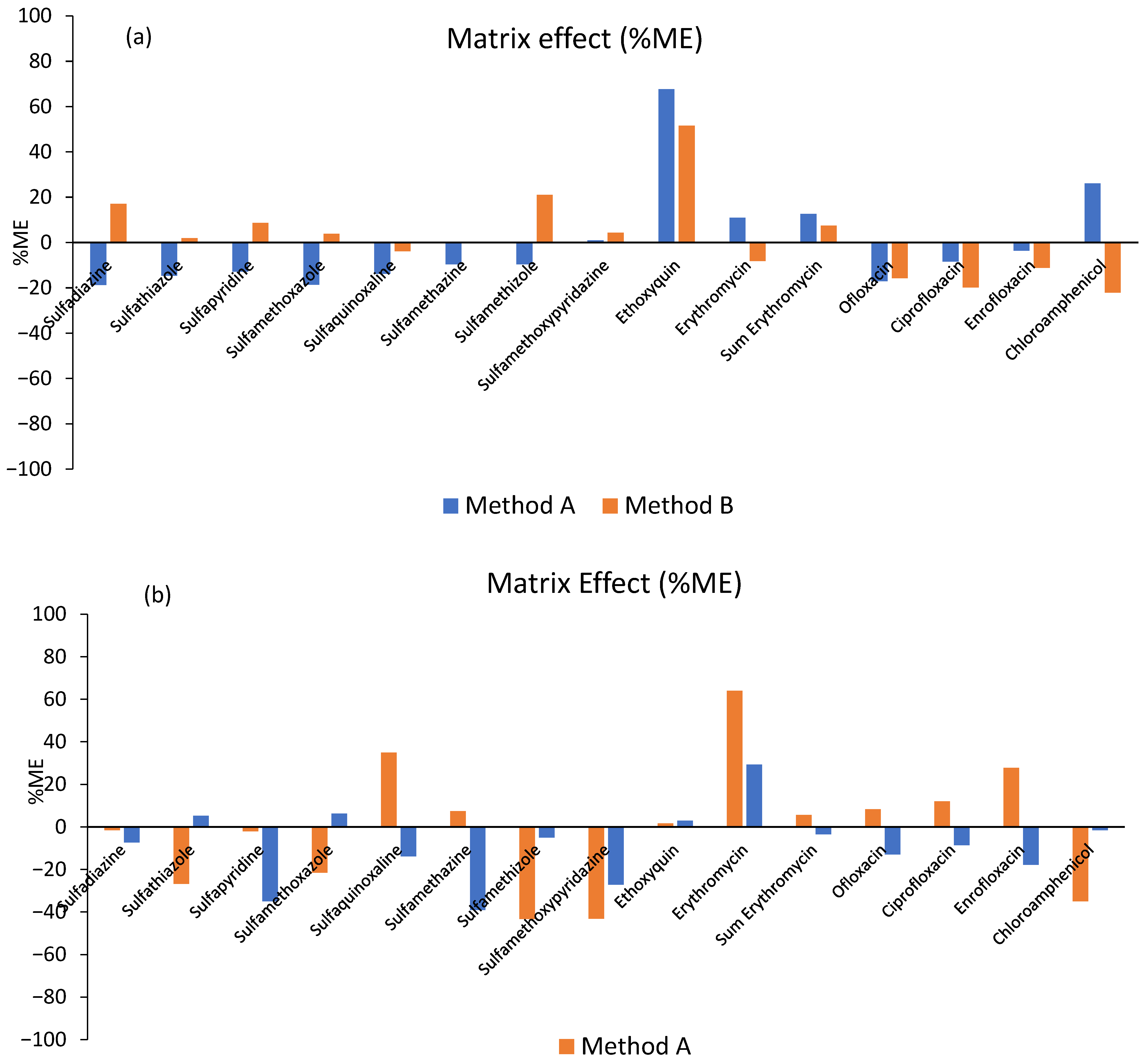
Figure 1. Matrix effects (%ME) calculated from the slopes of the solvent and matrix-matched calibration curves in (a) sea bream samples and (b) fish feed samples.
Separations 12 00321 g001
Figure 2. Expanded measurement uncertainty U% for (a) sea bream samples and (b) fish feed samples in concentration level 15 ng g−1 (n = 5).
Separations 12 00321 g002
Figure 3. Evaluation of greenness of the QuEChERS method and UHPLC-MS/MS using ComplexMo-GAPI.
Separations 12 00321 g003
Figure 4. LC-HR-MS-Orbitrap mass spectra for the detection of enrofloxacin in sea bream sample: (a) extracted ion chromatogram, (b) full scan MS, (c) MS2 fragment ion and sulfamethazine in feed sample, (d) extracted ion chromatogram, (e) full scan MS, and (f) MS2 fragment ion.
Separations 12 00321 g004aSeparations 12 00321 g004b
Table 1. Ultrahigh-performance liquid chromatography MS data for 14 antibiotics from 4 antimicrobial groups as well as ethoxyquin antioxidant.
Pharmaceuticals/
Additive
Retention Time (min)FormulaMolecular Weight (g mol−1)Precursor m/zMass
Accuracy (Δppm)
RDBProduct Ions m/z
Sulfonamides
Sulfadiazine5.81C10H10N4O2S250.276251.0597−0.5396.5156.0168
Sulfathiazole6.29C9H9N3O2S2255.310256.0209−0.5256.5156.0411
Sulfapyridine6.51C11H11N3O2S249.288250.0645−0.4147.5184.0916/156.0060
Sulfamethoxazole7.89C10H11N3O3S253.276254.0594−0.7806.5156.0568/147.0573
Sulfaquinoxaline8.20C14H12N4O2S300.336301.0754−0.82210.5156.0391
Sulfamethazine7.40C12H14N4O2S278.330279.0910−0.7437.5186.0614/156.0796
Sulfamethizole7.29C9H10N4O2S2270.325271.0318−0.3676.5156.0058/108.0688
Sulfamethoxypyridazine7.53C11H12N4O3S280.302281.0703−0.5417.5188.0027/156.0993/
126.0733
Macrolides
Erythromycin11.43C37H67NO13733.937716.4497−1.8325.5558.3030
Erythromycin A11.18C37H69NO14751.900734.46850.3444.5716.4867/558.3203
Quinoline-based additive
Ethoxyquin8.84C14H18NO217.310218.1539−3.2125.5202.1215/176.0662
Quinolones
Ofloxacin7.43C18H20FN3O4361.368362.1511−2.55010.5344.2135/318.2941
Ciprofloxacin7.68C17H18FN3O3331.346332.1405−2.4089.5314.1526/288.2909
Enrofloxacin7.73C19H22FN3O3359.400360.1718−2.7939.5342.2841/316.2802
Amphenicols
Chloramphenicol7.75C11H13Cl2N2O5323.132321.00511.5476.5257.0257/249.1590
Table 2. Analytical performance parameters of the developed methodology for the determination of compounds studied in fish samples (Method A).
Analytes%Recoveries (n = 6)Matrix Match Calibration CurveLOD
(ng g−1)
LOQ
(ng g−1)
Intra-Day RSDr% (n = 6)Inter-Day RSDR% (n = 6)
15
ng g−1
40
ng g−1
90
ng g−1
Linear Range
(ng g−1)
R2 15
ng g−1
40
ng g−1
90
ng g−1
15
ng g−1
40
ng g−1
90
ng g−1
Sulfadiazine-937825–5000.997010.025-9.63.9-11.95.8
Sulfathiazole67747510–5000.99962.27.32.94.43.94.95.25.3
Sulfapyridine97887825–5000.99703.0104.910.62.33.113.510.0
Sulfamethoxazole74859125–5000.99703.0102.612.96.210.39.44.1
Sulfaquinoxaline66788110–5000.99982.78.910.18.53.85.310.82.3
Sulfamethazine71859310–5000.99992.06.611.411.74.411.98.22.8
Sulfamethizole62737410–5000.99953.0101.11.61.34.23.52.1
Sulfamethoxypyridazine72748910–5000.99953.0103.14.94.94.35.25.5
Erythromycin77819910–5000.99992.47.93.12.51.66.12.03.0
Sum Erythromycin86879010–5000.99853.0108.66.82.79.47.32.1
Ethoxyquin70749710–5000.99710.72.35.81.20.86.93.42.5
Ofloxacin71811005–5000.99850.20.71.23.03.15.01.84.3
Ciprofloxacin82881025–5000.99960.41.31.20.340.555.31.11.5
Enrofloxacin7785955–5000.99930.20.71.63.90.66.39.17.9
Chloramphenicol7980875–5000.99390.31.15.33.02.27.88.54.8
Table 3. Analytical performance parameters of the developed methodology for the determination of studied compounds in fish samples (Method B).
Analytes%Recoveries (n = 6)Matrix Match Calibration CurveLOD
(ng g−1)
LOQ
(ng g−1)
Intra-Day RSDr% (n = 6)Inter-Day RSDR% (n = 6)
15
ng g−1
40
ng g−1
90
ng g−1
Linear Range
(ng g−1)
R2 15
ng g−1
40
ng g−1
90
ng g−1
15
ng g−1
40
ng g−1
90
ng g−1
Sulfadiazine959310450–5000.99513.611.93.11.10.86.21.22.3
Sulfathiazole779510150–5000.99263.812.51.30.40.61.11.00.8
Sulfapyridine809910025–5000.99793.611.92.40.80.44.40.90.5
Sulfamethoxazole85899125–5000.99023.912.91.70.96.62.25.38.2
Sulfaquinoxaline88939825–5000.99943.0103.11.72.31.41.54.2
Sulfamethazine80839610–5000.99062.06.60.40.94.74.82.04.6
Sulfamethizole93858750–5000.99873.812.53.40.20.63.60.50.6
Sulfamethoxypyridazine85939725–5000.99743.912.90.71.81.16.51.21.7
Erythromycin7182885–5000.99540.41.33.12.52.14.94.52.8
Sum Erythromycin9811010510–5000.99991.65.32.61.51.15.31.71.9
Ethoxyquin70879610–5000.99340.61.94.7 2.11.19.96.14.1
Ofloxacin6982885–5000.99970.20.75.23.44.65.94.46.7
Ciprofloxacin7085965–5000.99860.41.312.44.96.713.03.97.4
Enrofloxacin84941085–5000.99990.20.77.62.39.311.313.68.9
Chloramphenicol8688935–5000.99420.20.73.11.11.06.21.22.3
Table 4. Analytical performance parameters of the developed methodology for the determination of studied compounds in fish feed samples (Method A).
Analytes%Recoveries (n = 6)Matrix Match Calibration CurveLOD
(ng g−1)
LOQ
(ng g−1)
Intra-Day RSDr% (n = 6)Inter-Day RSDR% (n = 6)
15
ng g−1
40
ng g−1
90
ng g−1
Linear Range
(ng g−1)
R2 15
ng g−1
40
ng g−1
90
ng g−1
15
ng g−1
40
ng g−1
90
ng g−1
Sulfadiazine68858725–5000.98943.411.112.911.97.719.77.65.8
Sulfathiazole60609210–5000.99183.0106.24.21.37.36.45
Sulfapyridine598111210–5000.98962.27.111.210.36.518.612.27
Sulfamethoxazole64829310–5000.98952.89.114.95.40.919.83.14.2
Sulfaquinoxaline68798425–5000.99063.812.55.25.74.18.16.34.8
Sulfamethazine649110710–5000.99561.44.843.60.53.81.40.7
Sulfamethizole63819425–5000.99153.812.59.851.111.87.62.3
Sulfamethoxypyridazine66889310–5000.99552.27.184.93.563.12.7
Erythromycin70829425–5000.99933.411.16.11.70.672.70.1
Sum Erythromycin71829425–5000.99283.812.55.83.51.87.86.54.3
Ethoxyquin728410625–5000.99833.812.54.23.21.16.40.62.2
Ofloxacin63748210–5000.99352.06.712.311.83.313.311.97.8
Ciprofloxacin667911010–5000.99480.983.24.821.22.83.63.7
Enrofloxacin73859910–5000.99531.23.89.62.71.87.62.70.6
Chloramphenicol66819610–5000.99621.44.88.17.93.38.86.56.8
Table 5. Analytical performance parameters of the developed methodology for the determination of studied compounds in fish feed samples (Method B).
Analytes%Recoveries (n = 6)Matrix Match Calibration CurveLOD
(ng g−1)
LOQ
(ng g−1)
Intra-Day RSDr% (n = 6)Inter-Day RSDR% (n = 6)
15
ng g−1
40
ng g−1
90
ng g−1
Linear Range
(ng g−1)
R2 15
ng g−1
40
ng g−1
90
ng g−1
15
ng g−1
40
ng g−1
90
ng g−1
Sulfadiazine7310411525–5000.99013.812.519.65.34.113.49.18.3
Sulfathiazole70788425–5000.99503.411.18.96.31.59.84.21.2
Sulfapyridine72809725–5000.98993.3108.53.10.611.910.92
Sulfamethoxazole768811910–5000.99952.27.15.23.11.69.96.42.1
Sulfaquinoxaline709210625–5000.99283.31011.59.60.915.812.42.7
Sulfamethazine76909310–5000.99961.96.37.56.80.98.67.62.8
Sulfamethizole718610025–5000.99873.3104.72.52.25.43.82.7
Sulfamethoxypyridazine68959910–5000.99861.559.55.47.29.68.47
Erythromycin718110525–5000.99713.31013.63.92.1188.36.3
Sum Erythromycin75749025–5000.99573.812.512.95.14.912.57.15.1
Ethoxyquin70869810–5000.99092.58.39.88.71.513.512.27
Ofloxacin749710710–5000.996026.63.92.20.48.87.35.2
Ciprofloxacin789511010–5000.98251.75.510.80.67.72.80.3
Enrofloxacin69939710–5000.99631.65.39.28.71.515.510.74
Chloramphenicol71889610–5000.99782.27.18.26.73.211.87.33.5
Table 6. Decision limit CCα and detection capability CCβ at the MRL level for fish samples.
AnalytesCCα
(ng g−1)
CCβ
(ng g−1)
CCα
(ng g−1)
CCβ
(ng g−1)
Method AMethod B
Sulfadiazine100.7101.5100.7101.5
Sulfathiazole100.6101.3100.6101.3
Sulfapyridine100.8101.6100.6101.2
Sulfamethoxazole102.2104.4104.8109.6
Sulfaquinoxaline103.1106.2105.5110.9
Sulfamethazine101.5103.1103.5106.9
Sulfamethizole100.1100.2100.7101.5
Sulfamethoxypyridazine100.9101.8100.5101.1
Erythromycin101.5103.1 102.5104.9
Sum Erythromycin103.9107.2102.3106.6
Ethoxyquin0.100.200.110.19
Ofloxacin101.3102.5105.1109.6
Ciprofloxacin100.5101.0104.6109.2
Enrofloxacin101.7103.3100.7102.4
Chloramphenicol0.300.520.090.11
Table 7. The penalty points (PPs) for the determination of studied compounds in aquaculture products by UHPLC/Orbitrap MS analysis.
ReagentsPenalty PointsReagentsPenalty Points
ACN (7.5 mL)4ACN (9 mL)4
MeOH (2.5 mL)6Formic acid (1 mL)8
Extraction Waste3Extraction Waste1
Instruments Instruments
Centrifugation1Centrifugation1
Sonication0Sonication0
LC-MS2LC-MS2
LC-MS Waste3LC-MS Waste3
Total Penalty Points19Total Penalty Points19
Analytical Eco-Scale score (Method A)81Analytical Eco-Scale score
(Method B)
81
Disclaimer/Publisher’s Note: The statements, opinions and data contained in all publications are solely those of the individual author(s) and contributor(s) and not of MDPI and/or the editor(s). MDPI and/or the editor(s) disclaim responsibility for any injury to people or property resulting from any ideas, methods, instructions or products referred to in the content.