Article

Structure and Activity of β-Oligosaccharides Obtained from Lentinus edodes (Shiitake)

1
Institute of Edible Fungi, Shanghai Academy of Agricultural Sciences, Shanghai 201403, China
2
Shanghai Guosen Bio-Tech Co., Ltd., Shanghai 201403, China
3
College of Food Science and Technology, Shanghai Ocean University, Shanghai 201306, China
4
Shanghai Hengrui Pharmaceutical Co., Ltd., Shanghai 200245, China
5
Key Laboratory of Carbohydrate Chemistry and Biotechnology of Ministry of Education, School of Bio-Technology, Jiangnan University, Wuxi 214122, China
6
School of Food Science and Engineering, Yangzhou University, Yangzhou 225127, China
*
Authors to whom correspondence should be addressed.
These authors contributed equally to this work.
Separations 2024, 11(11), 326; https://doi.org/10.3390/separations11110326
Submission received: 11 October 2024 / Revised: 11 November 2024 / Accepted: 11 November 2024 / Published: 14 November 2024
(This article belongs to the Special Issue Research Progress for Isolation of Plant Active Compounds)

Abstract

:
The structure and characteristics of LEOPs, β-oligosaccharides from the fruiting body of Lentinus edodes obtained via acid degradation and gel permeation chromatography, were investigated. We performed high-performance liquid chromatography, infrared spectroscopy, methylation analysis, nuclear magnetic resonance, and correlated activity experiments, including antioxidant, immunomodulatory, and liver injury protection to gain insights. LEOPs comprised an oligosaccharide (Mw 2445 Da) based on six β-1, 3-D-glucose residues as the main chain and six β-1, 6-D-glucose residues as the side chain. Surface plasmon resonance analysis indicated that LEOPs directly bound to dectin-1, which facilitated their immunoenhancing activity via downstream NF-κB activation. The results implied that LEOPs may be the active unit of the shiitake β-glucan. The determination of LEOPs structure was performed to reveal the anti-tumor effect and immune-regulatory function of shiitake β-glucan on a molecular level to provide a basis.

1. Introduction

Lentinus edodes (Berk.) Pegler (shiitake), which belongs to the Tricholoma family, is the second-largest edible mushroom in the world, second only to Agaricus bisporus. It is widely cultivated in China, Japan, and other Asian countries. L. edodes is also a traditional Chinese medicine recorded as “L. edodes has mild medicinal properties, a slightly sweet taste, benefits vital energy, nourishes the stomach, and can treat loss of appetite and chickenpox” in Chinese herbal medicine books, such as “Daily Materia Medica” and “Encountering the Sources of the Classic of Materia Medica”. Modern medical research has found that L. edodes is rich in nutrients, including proteins, polysaccharides, crude fibers, and various vitamins. Notably, shiitake polysaccharides are among the most active compounds.
In 1976, Sasaki et al. [1] showed that shiitake polysaccharides are a type of β-glucan composed of β-D-(1-3)-Glc in the main chain and a 1–6 glucose residue as the side chain using high-performance liquid chromatography (HPLC), infrared spectroscopy, and nuclear magnetic resonance (NMR). L. edodes (shiitake) polysaccharide is a β-glucan with two β-1, 6-D-glucopyranoside branches for every five β-1,3-glucopyranoside linear linkages, and a right-handed triple-helix structure is formed [2]. There is no consensus on the molecular weight of shiitake polysaccharides, and most studies suggest that it is approximately 3 × 10⁵–9.5 × 10⁵ Da [3]. The bioactivity of shiitake polysaccharides is significantly affected by their chain conformation and molecular weight [4].
It was first reported that there is an active center in the β-glucan of the yeast cell wall that inhibits the phagocytosis of enzyme granules by monocytes, with seven glucose residues as repeating units [5]. β-Oligosaccharides have higher immune activity than curdlan treated with yeast endonuclease [6]. In addition, laminarin oligosaccharides with a polymerization degree of 33 and oligogalacturonic acids with an average polymerization degree of 10 extracted from brown algae are the smallest structural units that induce plant defense responses [7]. The antibacterial activity of the degradation product of yeast β-glucan is significantly higher than that of non-degraded high-molecular-weight polysaccharides [8]. Therefore, it can be inferred that β-glucan, like proteins and enzymes, may have one or several active centers of oligosaccharide fragments in it. Studying the mechanism of action between β-glucan oligosaccharides and target proteins at the oligosaccharide level can more directly reveal the anti-tumor and immune-regulatory functions of β-glucan. This provides us with a new perspective that helps us understand the interaction process between polysaccharides and functional protein receptors. The β-glucan structure of L. edodes is very similar to that of laminarin and yeast, and thus, it can be inferred that shiitake β-glucan degraded into oligosaccharide fragments can reduce the molecular weights of polysaccharides and improve their water solubility, retaining or even enhancing their biological activity.
In our preliminary work, the high β-glucan content of L. edodes variety F2 was selected for characterization. The β-glucan compound LF3 was prepared through separation and purification processes [9]. Owing to its high molecular weight and poor water solubility, LF3 was degraded into low-molecular-weight units with good solubility using chemical, physical, and enzymatic methods. Following ultrasound degradation, polysaccharides can only be reduced to a certain molecular weight and cannot be further degraded [10]. Polysaccharide degradation is typically achieved via acidic and enzymatic hydrolysis. Enzymatic degradation has been increasingly valued due to its strong specificity and mild reaction conditions [11]. However, it is difficult for LF3, as a class of large molecular substances with complex molecular structures and poor water solubility, to undergo enzymatic hydrolysis reactions within their molecules. Therefore, enzymatic hydrolysis was deemed ineffective. Hence, using acid hydrolysis to prepare small-molecular-weight active polysaccharides has become a research focus. In recent years, some acid hydrolysis has been used to degrade different kinds of polysaccharides. For example, the molecular weight of fucoidan was decreased from 10,680 kDa to 75.3 kDa, and the yield of low-molecular-weight fucoidan was 62.4% by using 1 mol/L trifluoroacetic acid (TFA) with a concentration of 5 mg/mL, at 121 °C for 1 h [12]. Using different concentrations of TFA to degrade fucoidan from Sargassum pallidum, it was found that increasing the reaction time, TFA concentration, and reaction temperature could further degrade fucoidan to obtain lower-molecular-weight products [13]. Using a new effective process for the production of curdlan oligosaccharides based on alkali-neutralization treatment and the acid hydrolysis of curdlan particles in a water suspension, it can more effectively obtain curdlan β-1,3-oligosaccharide production [14].
In the present study, a β-oligosaccharide was obtained using 0.02 mol/L TFA to hydrolyze LF3 at 110 °C for 2 h. NMR along with methylation was used to analyze the structure of the oligosaccharides and evaluate their antioxidant, liver-injury-repair, and dectin-1 receptor activities in vitro. Our results will provide crucial information for identifying the smallest active unit of shiitake polysaccharides.

2. Materials and Methods

2.1. Materials

The fruiting bodies of L. edodes variety Huxiang F2 were provided by Shanghai Pengshi Mushroom Industry Co., Ltd. (Shanghai, China) LF3 was extracted from F2 using 0.9% NaCl, and the derived precipitate was extracted with hot water at 80 °C and then finally extracted with 5% NaOH [5].

2.2. Isolation and Purification of LEOPs from LF3

A solution containing 10 mg of LF3 and 0.02 mol/L TFA in a ratio of 1 mg to 200 μL was mixed, hydrolyzed at 110 °C for 2 h, and then placed in an ice bath to rapidly cool down and terminate the reaction. After drying, TFA was dissolved in 30 volumes of distilled water, and the first peaks were collected via gel column P-4 and P-2 chromatography. LEOPs were concentrated and dried to a constant weight.

2.3. β-Glucan Content Determination

β-Glucan content was determined using the aniline blue fluorescence detection method [15].

2.4. Polysaccharide Content Determination

The polysaccharide content was determined using the phenol sulfuric acid method [16].

2.5. Determination of Molecular Weight Distribution Using HPLC

The molecular weight was analyzed with HPLC (Waters, Milford, CT, USA) and a Waters 600 Controller System (Waters 717 plus autosampler and Waters 2414 refractive index (RI) detector) (Waters, Milford, CT, USA) fit with a Waters Ultrahydrogel Linear (7.8 mm × 300 mm) gel filtration column (Waters, Milford, CT, USA). A total of 100 μL of LEOPs (2 mg/mL buffer consisting of 0.1 M NaNO3) was applied to the column. Elution was performed using the same buffer at 0.3 mL·min−1. The column was calibrated using a Dextran Standards Kit (Waters, Milford, CT, USA), and column and RI detector temperatures were set at 30 °C.

2.6. Infrared Spectroscopy Analysis

The sample was placed under an infrared lamp for 15 min, the pressure block was lifted, and zinc selenide crystals were carefully wiped with hydrous ethanol and dipped in cotton. After completing the background collection, a small flocculent sample was taken from the testing area and laid flat on a zinc selenide crystal. After pressing the pressure block, the sample collection button was clicked. The above operation was detected and analyzed using a Nicolet Nexus 470 FT-IR spectrometer (Thermo Fisher Scientific Inc., Waltham, MA, USA) in the range of 4000–400 cm−1, with a resolution set to 48 scans/4 cm−1, using Omnic software (version 7.0).

2.7. Determination of Monosaccharide Composition via Anion Chromatography

A total of 2 mg LEOPs was dissolved in 3 mL of 2 mol/L TFA at 110 °C for 5 h in an oil bath, blown dry with a nitrogen blower, mixed with approximately 3 mL of methanol, and blown dry again, repeating the procedure six times to remove TFA. A total of 1.5 mL of distilled water was added, and the mixture was transferred to a 2 mL centrifuge tube. The sample was centrifuged at 12,000× g for 5 min at 25 °C, transferring the supernatant to a sample bottle and diluting it 30 times with distilled water. An ICS-2500 high performance anion chromatograph (using PA-20 column) was used to determine the monosaccharide composition according to the method of Jia et al. [17].

2.8. Methylation Analysis

According to Anumula and Taylor (1992) [18], vacuum-dried LEOPs (2 mg) were dissolved in dimethyl sulfoxide (DMSO) (1 mL) at room temperature and methylated in DMSO (1 mL) and CH3I (0.5 mL) for 30 min with a 2.5% NaOH solution. Methylation analysis was performed using the method reported by Zhang et al. [19].

2.9. NMR Analysis

Twenty milligrams sample was dried in a vacuum dryer containing P2O5 for 24 h, dissolved in deuterated DMSO (0.5 mL of deuterated DMSO), and the nuclear magnetic resonance spectrum was measured. A Varian INOVA 500 NMR spectrometer (Varian Inc., Palo Alto, CA, USA) was used to record the 1H and 13C NMR spectra of the samples. The chemical shift in 1H NMR is based on the hydrogen shift of methyl sulfoxide-d6 (DMSO-d6) and was used as the internal standard. At 25 °C, the spectral lines were locked, referencing DMSO-d6, and corrected to Tetramethylsilane (TMS) (δ (0.00)). 1H spectrum reference standard δ DMSO = 2.50 (5-fold peak) and 13C spectrum reference standard δ DMSO = 39.96 (7 peaks) were used.

2.10. Prophagocytic Effect of LEOPs on Macrophages Determined via Flow Cytometry

The samples were divided into the following four groups: a negative control, a model group, and two LEOPs groups (100 and 200 μg/mL). RAW264.7 macrophages in the logarithmic growth phase were cultured in 24-well plates, and once the cells adhered to the plate, medium containing PBS or 10 μg/mL lipopolysaccharide (LPS) was added into the negative control group or the positive group, respectively. Medium containing 100 or 200 μg/mL LEOPs was added to the two LEOP groups. After incubating at 37 °C and 5% CO2 for 48 h, the cells were treated with 200 μL fluorescent microspheres at 1 × 10⁸ cells/mL for 1 h. Next, each sample was transferred to fresh tubes and analyzed via flow cytometry in triplicate. The phagocytic rate of the samples was calculated using the following formula:
M a c r o p h a g e   p h a g o c y t o s i s   r a t e % = N u m b e r   o f   m a c r o p h a g e s   e n g u l f i n g   f l u o r e s c e n t   m i c r o s p h e r e s T o t a l   n u m b e r   o f   m a c r o p h a g e s × 100 %

2.11. DPPH Radical Scavenging Activity of LEOPs

The DPPH radical scavenging activity of LEOPs was determined according to the method described by Molyneux [20]. The experiments included five LEOP treatment groups (0.2, 0.4, 0.6, 0.8, and 1.0 mg/mL LEOPs) and five positive control groups vitamin C (VC) (0.2, 0.4, 0.6, 0.8, and 1.0 mg/mL). A mixture of 0.2 mL of LEOPs or VC and 2.8 mL of 0.1 mM DPPH solution was prepared. The mixture was kept at room temperature in the absence of light for 30 min. The absorbance of each reaction mixture was measured at 517 nm. Each sample was analyzed five times. The removal rate was calculated as follows:
I % = ( A 2 A 1 A 2 ) × 100

2.12. In Vitro Inducing Effect of LF3 and LEOPs on NF-κB Activation via Dectin-1 Receptor

The NF-κB activation of HEK-BlueTM dectin-1 cells was determined using Liu’s method [21]. The cells were divided into the following 10 groups: a negative control group, three LF3 groups (10, 20, and 50 μg/mL LF3), three LEOPs groups (10, 20, and 50 μg/mL LEOPs), and three positive control groups (10, 20, and 50 μg/mL scleroglucan (SG)). After 24 h culture in an incubator set at 5% CO2 and 37 °C, the absorbance of the reaction solution of each group was measured at 630 nm in triplicate.

2.13. Binding Analysis of LEOPs and Dectin-1 Receptors Using Surface Plasmon Resonance (SPR)

The dectin-1 receptor was immobilized on a Biacore CM5 chip through amine coupling according to the supplier’s instructions. The LEOPs with different concentrations (0–4000 µg/mL) flowed through the surface of the dectin-1 receptor, and SPR monitored the time change curve of the binding and dissociation between the sample and the receptor in real time, and the time-varying curve obtained via real-time monitoring was fitted and analyzed using a 1:1 lammuir model, and the KD (mol) was obtained, which indicates the affinity between the sample and the protein.

2.14. Hepatoprotective Activity of LEOPs on Hepatic Damage

2.14.1. Alcohol-Induced Liver Injury Experiments

According to the method of Madushani [22], this experiment included six groups: a negative control, a positive group, and three LEOP groups (25, 50, 100, and 200 μg/mL). The LO2 cells were cultured in 96-well plates, and after the cells adhered to the plate, medium containing PBS or 1.2M alcohol was added to the negative control group or the model group, respectively, whereas the LEOPs groups were treated with media containing different concentrations of LEOPs. After a 16 h incubation, 1.2 M ethanol was added to the model and sample groups, and after another 8 h of culture, the cell proliferation rate of each group was measured using a 3-(4, 5-dimethylthiazol-2-yl)-2, 5-diphenyltetrazolium bromide assay (MTT).

2.14.2. H2O2-Induced Liver Injury Experiments

According to the method of Wang [23], the samples were divided into the following six groups: a negative control, a positive group, and three LEOP groups (100, 200, and 400 μg/mL). The LO2 cells were cultured in 96-well plates, and after the cells adhered to the wall, medium containing PBS or 0.75 mM H2O2 was added to the negative control group or the model group, respectively, whereas the LEOPs groups were treated with media containing different concentrations of LEOPs. After an 18 h incubation, 0.75 mM H2O2 was added to the model and sample groups. After a final 6 h incubation, the cell proliferation rate of each group was measured using an MTT assay [24].

2.15. Data Statistics

All data are expressed as the mean ± SD. Statistical analysis was conducted using SPSS software (version 22.0) [25]. The data are represented as the mean standard deviation, and ANOVA analysis of variance was used to test the significance of the differences (based on a significance level of p < 0.05 and p < 0.01).

3. Results

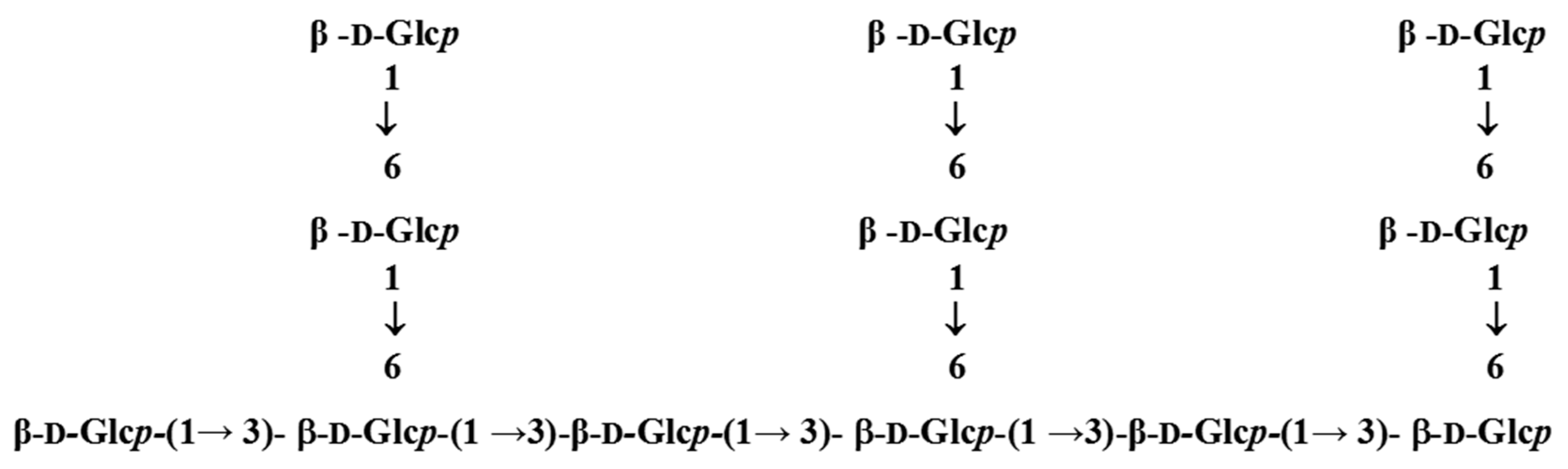
3.1. Elucidation of Properties and Structure of LEOPs Obtained from L. edodes

The total sugar contents of LEOPs and β-glucan were 97.83% and 96.08%, respectively. The lack of absorption at 254 nm and 280 nm indicated the absence of proteins and nucleic acids. The main monosaccharide was glucose, with a molar percentage of 94.85%, and very small amounts of galactose and mannose were also present. HPLC of LEOPs produced a single symmetrical peak, indicating that it was a homogeneous oligosaccharide (Figure 1) with a molecular weight of ~2445 Da based on a calibration curve prepared with dextran standards.
As shown in Figure 2, the infrared spectrum of LEOPs exhibits a typical polysaccharide absorption peak. A broad, smooth, and strong absorption peak with a high absorption intensity appeared at 3300 cm−1, caused by the stretching vibration of hydrogen bonding (-OH) between sugar molecules, indicating the presence of -OH groups in the sample. The absorption peaks of the C-H bond stretching vibration in methylene (-CH2-) and the absorption vibrations of the C-H bond were observed at 2922 and 1420 cm−1, respectively. The absorption peak at 1200 cm−1 indicated the stretching vibration of –C-O. Three absorption peaks appeared between 1000 and 1100 cm−1, indicating the presence of pyranoside. The absorption peak at 890 cm−1 indicated the presence of a β configuration, which indicated that LEOPs were β-polysaccharides or oligosaccharides.
To determine the connection sites between the methylated monosaccharides, the samples were subjected to acid hydrolysis, sodium borohydride reduction, and acetic anhydride acetylation after complete methylation to obtain partially methylated alditol acetate derivatives that evaporated at high temperatures. Gas chromatography-mass spectrometry (GC-MS) analysis was performed to determine the connection mode between the LEOP monosaccharide components.
The GC-MS total ion spectra are shown in Figure 3. The four fragments with retention times of 12.27, 13.43, 13.88, and 15.30 min were terminal glucopyranose (T-Glc), 1,3-, 1,6-, and 1,3,6-linked glucopyranose, respectively. The methylation analysis results of LEOPs (Table 1) showed linkages of terminal glucopyranose (T-Glc) and 1,3-, 1,6-, and 1,3,6-linked glucopyranose at a molar ratio of approximately 1.66:1.81:3.47:1.
LEOP samples were dissolved in DMSO-d6. From the 1H NMR data shown in Figure 4, the chemical shifts ranged from δ3.93 to δ4.04. There were mainly four hydrogen signals in the heteromeric hydrogen region or terminal proton region, with chemical shifts of δ4.02, δ4.00, δ3.99, and δ3.93. The chemical shifts of other hydrogen signals were between δ3.00 and δ3.93. Because the chemical shifts of hydrogen in the residues were all less than δ4.5, all the residues were in a β-configuration.
In the 13C-NMR spectrum (Figure 5), the anomeric carbon signal ranged from δ90 to δ110, and the non-anomeric carbon signal ranged from δ60 to δ85. The anomeric carbon (C1) signal of α bond type connections was approximately δ97–δ101 and a β bond type connection with passes through approximately δ103-δ105 [26]. It can be seen in Figure 6 that there are mainly four carbon signals in the end group carbon region or anomeric carbon region, with chemical shifts of δ103.09, δ103.16, δ103.23, and δ102.94. Usually, the shift of anomeric carbon in (1→3)-β-D-glucan is approximately 100 ppm [27]. Thus, the stronger signals at δ103.09, δ103.23, and δ102.94 ppm correlated to the C-1 of terminal β-D-glucopyranosyl, (1→6)-β-D-glucopyranosyl, (1→3, 1→6)-β-D-glucopyranosyl, and (1→3)-β-D-glucopyranosyl, respectively.
Using published methodology [28,29], the complete structural characterization of LEOPs were achieved following 2D NMR analysis involving 1H-1H correlated spectroscopy (COSY), total correlation spectroscopy (TOCSY), Nuclear Overhauser Effect Spectroscopy (NOESY), heteronuclear multiple quantum coherence (HMQC), and heteronuclear multiple-bond correlation spectroscopy (HMBC), which were used to assign the chemical shift spin systems of sugar subunits (Table 2). The H-1 signals at the high field and C-1 signals at the low field indicated that residues A, B, C, and D were β-linked [30]. Proton chemical shifts from H-2 to H-6 were assigned from 2-dimensional signal NMR, including COSY, NOESY, HMBC, and HMQC spectra. The large JH-2, H-3 value and JH-3, H-4 coupling constants and typical H-1, H-2, and H-4 intercorrelations in the NOESY spectrum indicated that the residue was D-glucopyranose [31]. Except for the downfield shift of C-1 (δ103.09), no carbon signal was evident in the δ78–δ 85 range, indicating that residue A was a 1-linked β-D-glucopyranose. The downfield shifts of the C-6 (δ69.92) carbon signal with respect to the standard values for glucopyranose indicated that residue B was 1, 6-β-D-glucopyranose. The downfield shifts of C-3 (δ85.96) and C-6 (δ69.92) carbon signals with respect to the standard values for glucopyranose indicated that residue C was 1,3,6-β-D-Glop. The downfield shifts of the C-3 (δ86.95) carbon signal with respect to the standard values for glucopyranose indicated that residue D was 1, 3-β-D-glucopyranose.
The sequence of the glycosyl residues was determined from NOESY results (Table 3), followed by confirmation based on HMBC findings (Table 4). Combining the methylation results, monosaccharide composition information, and molecular weight, we determined the structural model of LEOPs as shown in Figure 6.

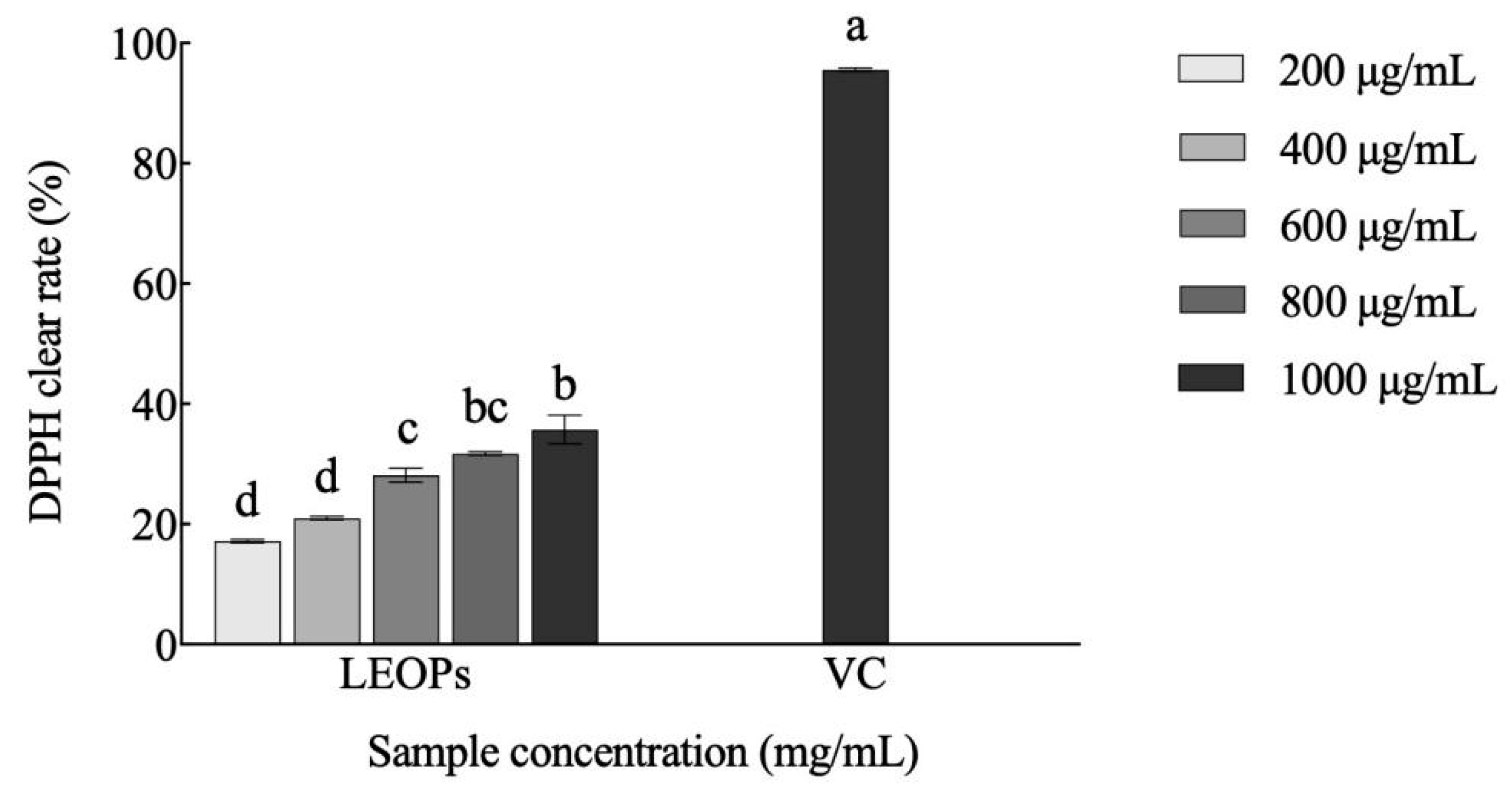
3.2. Antioxidant Activity of LEOPs Obtained from L. edodes

As shown in Figure 7, LEOPs effectively scavenged DPPH· free radicals at all concentrations. The DPPH radical scavenging rates of 200, 400, 600, 800, and 1000 μg/mL LEOPs were 17.18%, 28.13%, 31.72%, and 35.72% respectively. This indicates that LEOPs have good antioxidant activity in a concentration-manner and act as important antioxidant components in the fruiting bodies of L. edodes. There are various macromolecular substances, such as heteropolysaccharides, glucans, and oligosaccharides, in the fruiting body of L. edodes. Previous studies have shown that heteropolysaccharides and glucans have good antioxidant activity. Chen et al. showed the crude polysaccharide from L. edodes exhibited significantly antioxidant activity in a concentration-dependent manner through hydroxyl radicals, superoxide radicals, and Fe2+-chelating ability experiments [32]. Li et al. found that (1→6)-β-d-glucan from L. edodes exhibited good antioxidant activity with a dose-dependence at low concentrations through the assay of hydroxyl radical scavenging activity [33]. Compared with other research, the antioxidant activity of β-oligosaccharides from L. edodes was first reported in our research.

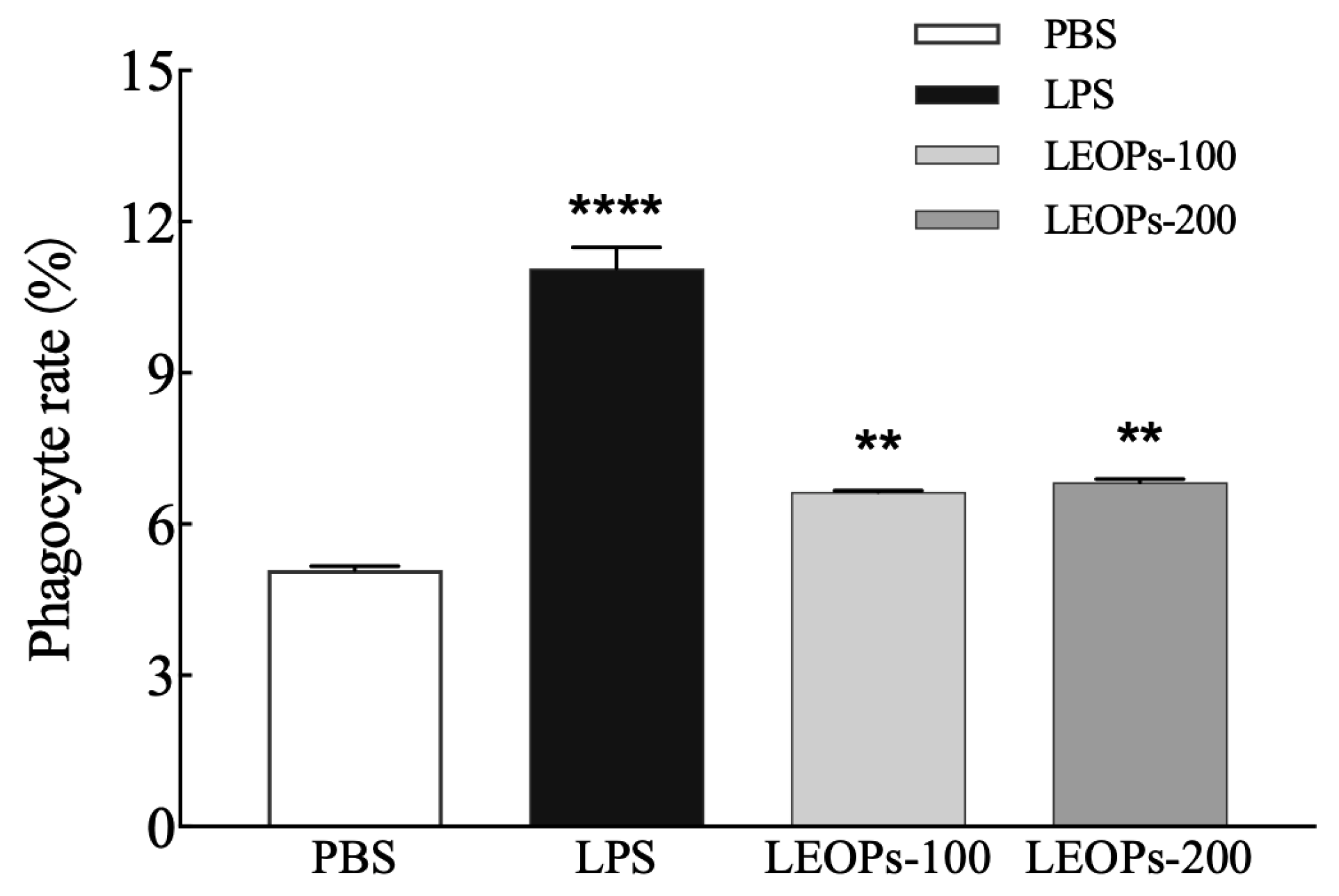
3.3. LEOPs Promote the Phagocytic Function of RAW264.7 Macrophages

Compared with the negative control, 100 and 200 μg/mL LEOPs improved macrophage phagocytic functions by 30.45% and 34.18%, respectively, and the results were highly significant (p < 0.01). For the phagocytic function, 100 and 200 µg/mL LEOPs have approximately the same effect, indicating that 100 μg/mL LEOP achieved optimal promoting effects on macrophage phagocytic functions. This result indicated that LEOPs promoted the phagocytic function of RAW264.7 cells and thus had good immune-promoting activity (Figure 8) and are like other studies on the immunomodulatory activity of polysaccharides from L. edodes. Jeff et al. found the mannogalactoglucan-type polysaccharides from L. edodes could induce an increase in nitrite oxide production in peritoneal macrophages, significantly increase the macrophage phagocytosis of tumor-bearing mice, and augment concanavalin and lipopolysaccharide-induced splenocyte proliferation [34]. Xu et al. reported the polysaccharide from L. edodes could enhance systemic and mucosal immunity through the spatial modulation of intestinal gene expression in mice [35].

3.4. LEOPs Mediate NF-κB Activation via Dectin-1 Receptor

Figure 9 shows that compared with the negative control, LF3 or LEOPs at 10, 20, and 50 μg/mL increased dectin-1 activity by 81.40%, 82.56%, and 72.77% or 126.74%, 147.09%, and 173.26, respectively. This effect was concentration-dependent and provided evidence that LEOPs exhibit immunostimulatory effects by inducing NF-κB activation via dectin-1 receptor. The C-type lectin receptor dectin-1 was originally described as the β-glucan receptor expressed in myeloid cells, with crucial functions in antifungal responses. Son et al. reported ulvan-type polysaccharides from Ulva pertusa demonstrated significant stimulatory effects on various immunocytes, and these effects were closely related to nuclear factor-kappa B pathways through dectin-1 [36].

3.5. LEOPs Bind to Dectin-1 Receptors

A biomolecular interaction analysis enables the real-time observation of inter-molecular interactions. Through sensing maps, response values can be obtained over time, enabling the real-time monitoring of intermolecular binding and dissociation, resulting in richer information. As shown in Figure 10a, the affinity, as determined using the KD values, between LEOPs and dectin-1 was 340.2 nM. The fitting curve showed that the dispersion of scatter points at each concentration was very small; indicating that LEOPs can specifically recognize dectin-1 in a specific and stable manner, further confirming that LEOPs had an affinity for dectin-1. The response value of LEOPs to dectin-1 receptor binding positively correlated with the concentration of LEOPs; its response value was 480.6 RU when the concentration of LEOPs reached 4000 μg/mL (Figure 10b).

3.6. Reparative Effect of LEOPs on Liver Injury

As shown in Figure 11a, 1.2M EtOH could decrease the cell viability, and 200 μg/mL LEOPs showed a good reparative effect on alcohol-induced liver injury (p < 0.01). Like the result of the alcoholic experiment, 100 μg/mL LEOPs showed a good reparative effect on H2O2-induced liver injury (p < 0.05) (Figure 11b).

4. Discussion

The oligosaccharide structure of LEOPs from L. edodes F2 is not consistent with the traditional repeating units of shiitake β-glucan. We believe that the structure of glucan from shiitake mushrooms is not completely consistent. Additional polysaccharides may be present in L. edodes varieties.
The molecular weight of a polysaccharide is a key factor affecting its bioactivity. Acid hydrolysis improves the solubility and antitumor activity of lentinan by reducing its molecular weight [37]. Hua et al. [38] reported that the ultrasonic treatment of polysaccharides from L. edodes improves their anti-immunomodulatory activity. Our study utilized a liver cell oxidative stress injury model to demonstrate that oligosaccharide LEOPs exerted repairing effects on damaged liver cells via their antioxidant effects.
Dectin-1 plays an important role in the immune system and is mainly expressed on the surfaces of antigen-presenting cells, such as dendritic cells, macrophages, and alveolar macrophages. The main function of dectin-1 is to recognize and bind to the fungal cell wall β- glucan. Dectin-1 can activate the NF signaling pathway by reacting with β-glucan, mediating immune cell recognition and the clearance of fungi, activating inflammatory responses, and promoting antifungal immune responses. Dectin-1 is a pattern recognition receptor (PRR) of β-glucans that recognizes the presence of β-(1, 3) and β-(1,6) glycosidic bonds but cannot recognize glucans with α- glycosidic bonds, such as α-(1,3)-D-glucan from Ganoderma lucidum [29]. In a HEK-BlueTM dectin-1 cell model, LEOPs showed strong dectin-1 receptor binding activity in vitro, where the β-glucan content reached 96.08%, higher than that of LF3, and the activity was also higher than that of LF3 (Figure 9). These results are consistent with those of previous studies [9], and the β-glucan content of the shiitake polysaccharide extract was consistent with the dectin-1-receptor-binding activity.
In our study, the binding of LEOPs and dectin-1 receptor was verified using SPR technology, which showed that the binding was specific and stable. These results indicate that LEOPs may be the central active units of shiitake β-glucans. We conclude that degrading shiitake β-glucan into oligosaccharide fragments reduces the molecular weight of polysaccharides, thus enhancing their biological activity.

5. Conclusions

As an important part of polysaccharide research, β-glucan has gradually attracted people’s attention. As the main active compound of L. edodes, shiitake β-glucan has various effects, such as inhibiting tumors and immune activity. In recent years, there have been many reports on the immune activity of shiitake β-glucan, but there is relatively little research on the active units of oligosaccharides with smaller polymerization degrees of β- glucan.
High-molecular-weight β-glucan has the characteristics of easy separation and good activity, but their further development and application are greatly limited due to issues, such as high viscosity and difficulty in dissolving in water caused by their large molecular weight. This study used the selected L. edodes variety F2 with a high β-glucan content as raw material and obtained the β-glucan fraction LF3 using different separation and purification processes. After 0.02 mol/L TFA to hydrolyze LF3 at 110 °C for 2 h, an oligosaccharide, LEOPs, was isolated and purified via gel permeation chromatography.
Compositional analysis showed that LEOPs are composed of glucose and very small amounts of galactose and mannose. Linkage analysis and 1H and 13C and 2D NMR spectroscopy established that LEOPs consisted of six β-1, 3-D-glucose residues as the main chain and six β-1,6-D-glucose residues as the side chain. Surface plasmon resonance analysis indicated that LEOPs directly bound to dectin-1, which facilitated their immunoenhancing activity via downstream NF-κB activation. The results implied that LEOPs may be the active unit of the shiitake β-glucan LF3.

Author Contributions

Conceptualization, H.W., H.Z. (Hongtao Zhang), P.L., H.Z. (Henan Zhang), H.L. and J.Z.; Methodology, Y.Y.; Validation, H.Z. (Hongtao Zhang) and P.L.; Formal analysis, Y.Y., H.W., M.Z. and H.L.; Resources, P.L., M.Z. and Q.L.; Data curation, M.Z., Q.L. and H.Z. (Henan Zhang); Writing—original draft, W.J.; Writing—review & editing, W.W.; Supervision, Q.L., H.Z. (Henan Zhang) and H.L. All authors have read and agreed to the published version of the manuscript.

Funding

This study was supported by the Natural Science Foundation of Shanghai, China (grant No. 20ZR1422900).

Data Availability Statement

Data is contained within the article.

Acknowledgments

The authors thank Wanping Lu of Shanghai Jiao Tong University Instrumental Analysis Center for valuable assistance.

Conflicts of Interest

The authors declare no conflict of interest. Author Huimin Wang was employed by the company Shanghai Hengrui Pharmaceutical Co., Ltd., Shanghai 200245, China. The remaining authors declare that the research was conducted in the absence of any commercial or financial relationships that could be construed as a potential conflict of interest. Author Wei Jia was employed by the company Shanghai Guosen Bio-tech Co., Ltd., Shanghai 201403, China. The remaining authors declare that the research was conducted in the absence of any commercial or financial relationships that could be construed as a potential conflict of interest.

References

  1. Sasaki, T.; Takasuka, N. Further study of the structure of lentinan, an anti-tumor polysaccharide from Lentinus edodes. Carbohydr. Res. 1976, 47, 99–104. [Google Scholar] [CrossRef]
  2. Xu, X.; Chen, P.; Zhang, L.; Ashida, H. Chain structures of glucans from Lentinus edodes and their effects on NO production from RAW 264.7 macrophages. Carbohydr. Polym. 2012, 87, 1855–1862. [Google Scholar] [CrossRef]
  3. Zhang, Y.; Li, S.; Zhang, L. Aggregation Behavior of Triple Helical Polysaccharide with Low Molecular Weight in Diluted Aqueous Solution. J. Phys. Chem. B 2010, 114, 4945–4954. [Google Scholar] [CrossRef] [PubMed]
  4. Yoshida, O.; Nakashima, H.; Yoshida, T.; Kaneko, Y.; Yamamoto, I.; Matsuzaki, K.; Uryu, T.; Yamamoto, N. Sulfation of the immunomodulating polysaccharide lentinan: A novel strategy for antivirals to human immunodeficiency virus (HIV). Biochem. Pharmacol. 1988, 37, 2887–2891. [Google Scholar] [CrossRef] [PubMed]
  5. CZOP, J.K. Isolation of a yeast heptaglucoside that inhibits monocyte phagocytosis of zymosan particles. J. Immunol. 1989, 142, 959–965. [Google Scholar]
  6. Kataoka, K.; Muta, T.; Yamazaki, S.; Takeshige, K. Activation of macrophages by linear (1→3)-β-D-glucans. J. Biol. Chem. 2002, 277, 36825–36831. [Google Scholar] [CrossRef]
  7. Klarzynski, O.; Plesse, B.; Joubert, J.M.; Yvin, J.C.; Kopp, M.; Kloareg, B.; Fritig, B. Linear β-1,3-glucans are elicitors of defense responses in tobacco. Plant Physiol. 2000, 124, 1027–1037. [Google Scholar] [CrossRef]
  8. Khan, A.A.; Gani, A.; Masoodi, F.A.; Amin, F.; Wani, I.A.; Khanday, F.A.; Gani, A. Structural, thermal, functional, antioxidant & antimicrobial properties of β-D-glucan extracted from baker’s yeast (Saccharomyces cerevisiae)-Effect of γ-irradiation. Carbohydr. Polym. 2016, 140, 442–450. [Google Scholar]
  9. Jia, W.; Yu, Y.Z.; Liu, H.L.; Liu, Y.F.; Zhang, M.Y.; Li, Q.Z.; Zhang, J.S.; Wang, W.H. Preparation and In Vitro Activity of β-Glucan-Rich Polysaccharide Extracts from Lentinula edodes Fruiting Bodies. Acta Edulis Fungi 2023, 30, 51–55. [Google Scholar]
  10. Yan, J.K.; Wang, Y.Y.; Ma, H.L.; Wang, Z.-B. Ultrasonic effects on the degradation kinetics, preliminary characterization and antioxidant activities of polysaccharides from Phellinus linteus mycelia. Ultrason. Sonochem. 2016, 29, 251–257. [Google Scholar] [CrossRef]
  11. Wang, D.; Dai, L.; Gao, Y.X. Research progress on enzymaticmodification of polysaccharides. J. Chin. Cereals Oils Assoc. 2017, 32, 134–140. (In Chinese) [Google Scholar]
  12. Sinurat, E.; Saepudin, E.; Hudiyono, S. Immunostimulatory activity of brown seaweed-derived fucoidans at different molecular weights and purity levels towards white spot syndrome virus (WSSV) in shrimp Litopenaeus vanname. J. Appl. Pharm. Sci. 2016, 6, 82–91. [Google Scholar] [CrossRef]
  13. Xue, W.; Li, L.C.; Qi, M.X.; Wu, W.H.; Chi, L.L.; Wang, P.P. Structural characteristic of fucoidans from Sargassum pallidum and their anti-influenza virus activities. J. Shanghai Ocean. Uni. 2023, 32, 227–233. (In Chinese) [Google Scholar]
  14. Li, J.; Zhu, L.; Zheng, Z.Y.; Zhan, X.-B.; Lin, C.-C.; Zong, Y.; Li, W.-J. A new effective process for production of curdlan oligosaccharides based on alkali-neutralization treatment and acid hydrolysis of curdlan particles in water suspension. Appl. Microbiol. Biotechnol. 2013, 97, 8495–8503. [Google Scholar] [CrossRef] [PubMed]
  15. Nitschke, J.; Modick, H.; Busch, E.; Von Rekowski, R.W.; Altenbach, H.J.; Mölleken, H. A new colorimetric method to quantify β-1, 3-1, 6-glucans in comparison with total β-1, 3-glucans in edible mushrooms. Food Chem. 2011, 127, 791–796. [Google Scholar] [CrossRef] [PubMed]
  16. Nowotny, A. Carbohydrate Determination by Phenol-Sulfuric Acid; Springer: Berlin/Heidelberg, Germany, 1979; pp. 171–173. [Google Scholar]
  17. Jia, W.; Zhang, J.S.; Jiang, Y.; Zheng, Z.-Y.; Zhan, X.-B.; Lin, C.-C. Structure of oligosaccharide F21 derived from exopolysaccharide WL-26 produced by Sphingomonas sp. ATCC 31555. Carbohydr. Polym. 2012, 90, 60–66. [Google Scholar] [CrossRef] [PubMed]
  18. Anumula, K.R.; Taylor, P.B. A comprehensive procedure for preparation of partially methylated alditol acetates from glycoprotein carbohydrates. Anal. Biochem. 1992, 203, 101–108. [Google Scholar] [CrossRef]
  19. Zhang, A.Q.; Zhang, J.S.; Tang, Q.J.; Jia, W.; Yang, Y.; Liu, Y.F.; Fan, J.M.; Pan, Y.J. Structural elucidation of a novel fucogalactanthat contains 3-O-methyl rhamnose isolated from the fruiting bodies of the fungus. Hericium Erinaceus. Carbohydr. Res. 2006, 341, 645–649. [Google Scholar] [CrossRef]
  20. Tatarczak, M.M.; Flieger, J. Application of high-performance liquid chromatography with diode array detection to simultaneous analysis of reference antioxidants and 1,1-diphenyl-2-picrylhydrazyl (DPPH) in free radical scavenging test. Int. J. Environ. Res. Public Health 2022, 19, 8288. [Google Scholar] [CrossRef]
  21. Liu, L.P.; Feng, J.; Gao, K.; Zhou, S.; Yan, M.; Tang, C.; Zhou, J.; Liu, Y.; Zhang, J. Influence of carbon and nitrogen sources on structural features and immunomodulatory activity of exopolysaccharides from Ganoderma lucidum. Process Biochem. 2022, 119, 96–105. [Google Scholar] [CrossRef]
  22. Madushani, H.K.; Bing, S.J.; Cho, J.; Kim, A.; Kim, G.; Kim, J.S.; Kim, J.B.; Doh, Y.H.; Jee, Y. Sasa quelpaertensis leaves ameliorate alcohol-induced liver injury by attenuating oxidative stress in HepG2 cells and mice. Acta Histochemica 2018, 120, 477–489. [Google Scholar] [CrossRef]
  23. Wang, M.; Ma, H.L.; Liu, B.; Wang, H.B.; Xie, H.; Li, R.D.; Wang, J.F. Pinus massoniana bark extract protects against oxidative damage in LO2 hepatic cells and mice. Am. J. Chin. Med. 2010, 38, 909–919. [Google Scholar] [CrossRef]
  24. Lee, S.L.; Hsu, W.H.; Tu, C.M.; Wang, W.H.; Yang, C.Y.; Lee, H.K.; Chin, T.Y. The effects of freshwater clam (Corbicula fluminea) extract on activated hepatic stellate cells. Evid.-Based Complement. Altern. Med. 2021, 2021, 6065168. [Google Scholar] [CrossRef]
  25. Lim, H.; Oh, J.S.; Kang, K.R.; Seo, J.Y.; Kim, D.K.; Yu, S.K.; Kim, H.J.; Park, J.C.; Kim, J.S. 25-Hydroxycholesterol induces odontoclastic differentiation through RANK-RANKL upregulation and NF-κB activation in odontoblast-like MDPC-23 cells: An in vitro study. Int. Endod. J. 2023, 56, 432–446. [Google Scholar] [CrossRef]
  26. Bubb, W.A. NMR spectroscopy in the study of carbohydrates: Characterizing the structural complexity. Concept. Magn. Reson. A 2003, 19, 1–19. [Google Scholar] [CrossRef]
  27. Lehtovaara, B.C.; Gu, F.X. Pharmacological, structural, and drug delivery properties and applications of 1, 3-beta-glucans. J. Agric. Food Chem. 2011, 59, 6813–6828. [Google Scholar] [CrossRef] [PubMed]
  28. Duus, J.; Gotfredsen, C.H.; Bock, K. Carbohydrate structural determination by NMR spectroscopy: Modern methods and limitations. Chem. Rev. 2000, 100, 4589–4614. [Google Scholar] [CrossRef] [PubMed]
  29. Hounsell, E.F. 1H NMR in the structural and conformational analysis of oligosaccharides and glycoconjugates. Prog. Nucl. Magn. Reson. Spectrosc. 1995, 27, 445–474. [Google Scholar] [CrossRef]
  30. Fhernanda, R.S.; Elaine, R.C.; Caroline, G.M.; Sassaki, G.L.; Gorin, P.A.; Iacomini, M. Structural characterization of a polysaccharide and a β-glucan isolated from the edible mushroom Flammulina velutipes. Phytochemistry 2006, 67, 2189–2196. [Google Scholar]
  31. Rout, D.; Mondal, S.; Chakraborty, I.; Pramanik, M.; Islam, S.S. Chemical analysis of a new (1→3)-(1→6)-branched glucan from an edible mushroom, Pleurotus florida. Carbohydr. Res. 2005, 340, 2533–2539. [Google Scholar] [CrossRef]
  32. Chen, H.; Ju, Y.; Li, J.; Yu, M. Antioxidant activities of polysaccharides from Lentinus edodes and their significance for disease prevention. Int. J. Biol. Macromol. 2012, 50, 214–218. [Google Scholar] [CrossRef] [PubMed]
  33. Li, J.; Cai, C.; Zheng, M.; Hao, J.; Wang, Y.; Hu, M.; Fan, L.; Yu, G. Alkaline Extraction, Structural Characterization, and Bioactivities of (1→6)-β-d-Glucan from Lentinus edodes. Molecules 2019, 24, 1610. [Google Scholar] [CrossRef] [PubMed]
  34. Jeff, I.B.; Fan, E.; Tian, M.; Song, C.; Yan, J.; Zhou, Y. In vivo anticancer and immunomodulating activities of Manno galactoglucan-type polysaccharides from Lentinus edodes (Berkeley) Singer. Cent. Eur. J. Immunol. 2016, 41, 47–53. [Google Scholar] [CrossRef] [PubMed]
  35. Xu, X.; Yang, J.; Luo, Z.; Zhang, X. Lentinula edodes-derived polysaccharide enhances systemic and mucosal immunity by spatial modulation of intestinal gene expression in mice. Food Funct. 2015, 6, 2068–2080. [Google Scholar] [CrossRef]
  36. Son, S.U.; Kim, T.E.; Park, J.H.; Suh, H.J.; Shin, K.-S. Immunostimulating effects of ulvan type polysaccharide isolated from Korean Ulva pertusa in cyclophosphamide-induced immunosuppressed BALB/c mice. Int. J. Biol. Macromol. 2024, 275 Pt 1, 133518. [Google Scholar] [CrossRef]
  37. Saito, H.; Ohki, T.; Takasuka, N.; Sasaki, T. 13C N.M.R spectral study of a gel-forming (1,3)-β-D-glucan, (lentinan) from Lentinus edodes, and its acid degraded fraction’s structure, and dependence of conformation on the molecular weight. Carbohydr. Res. 1977, 58, 293–305. [Google Scholar] [CrossRef]
  38. Hua, Y.; Zhang, H.; Fu, Q.; Feng, Y.; Duan, Y.; Ma, H. Effects of ultrasound modification with different frequency modes on the structure, chain conformation, and immune activity of polysaccharides from Lentinus edodes. Foods 2022, 11, 2470. [Google Scholar] [CrossRef]
Figure 1. High-performance liquid chromatography (HPLC) of LEOPs.
Separations 11 00326 g001
Figure 2. Infrared spectra of LEOPs.
Separations 11 00326 g002
Figure 3. GC-MS pattern of alditol acetates from the methylation product of LEOPs.
Separations 11 00326 g003
Figure 4. 1H NMR spectra of LEOPs dissolved in DMSO-d6 at 25 °C. f1 represents the frequency offset of the observation channel (first channel). The residues A, B, C, and D are arranged from low to high fields.
Separations 11 00326 g004
Figure 5. 13C NMR spectra of LEOPs dissolved in DMSO-d6 at 25 °C. f1 represents the frequency offset of the observation channel (first channel). The anomeric carbon is labeled based on anomeric hydrogen as A, B, C, and D.
Separations 11 00326 g005
Figure 6. Structure of LEOPs.
Separations 11 00326 g006
Figure 7. Antioxidant activity of LEOPs based on scavenging DPPH· free radicals. Vitamin C is the positive control. Different lowercase letters indicate significant differences (p < 0.05).
Separations 11 00326 g007
Figure 8. Stimulatory effect of LEOPs on RAW264.7 phagocytic function. LPS is the positive control. ** and **** stand for significant differences at p < 0.01 and p < 0.001 levels, respectively, compared with the negative group.
Separations 11 00326 g008
Figure 9. Immunostimulatory effect of LEOPs is mediated via NF-κB activation and the dectin-1 receptor. Scleroglucan is the positive control. Different lowercase letters indicate significant differences, p < 0.05.
Separations 11 00326 g009
Figure 10. SPR analysis showing binding affinities between LEOPs and dectin-1 receptors. (a) 1:1 steady-state affinity fitting curve for dectin-1 receptor; the determined KD value was 340.2 nM; (b) SPR sensograms of LEOPs binding to dectin-1 receptors at different concentrations. The dotted line represents KD value.
Separations 11 00326 g010
Figure 11. Repairing effect of LEOPs on alcoholic liver injury (a) and H2O2 liver injury (b). * and ** stand for significant differences at p < 0.05 and p < 0.01 levels, respectively, compared with the negative control group. All values are presented as means ± SEM (n = 3). Significantly different (### p < 0.001) versus the CK group, significantly different (* p < 0.05, ** p < 0.01) versus the H2O2 or EtOH group.
Separations 11 00326 g011
Table 1. Linkage analysis of LEOPs.
Methylated SugarsLinkagesMolar RatiosRetention Time (s)
2,3,4,6-Me4-GlcpT-Glcp1.6612.27
2,4,6-Me3-Glcp1,3-Glcp1.8113.43
2,3,4-Me3-Glcp1,6-Glcp3.4713.88
2,4-Me2-Glcp1,3,6-Glcp1.0015.30
Table 2. 1H and 13C NMR chemical shift (ppm) of LEOPs at 25 °C.
ResidueProton or Carbon
H-1/C-1H-2/C-2H-3/C-3H-4/C-4H-5/C-5H-6/C-6
A β-D-Glcp- (14.023.133.303.183.493.48 a,3.69 b
103.0976.4071.8671.2377.0361.36
B →6)-β-D-Glcp- (1→4.003.393.623.503.383.18 a, 3.12 b
103.1679.3676.1371.8675.2369.92
C →3,6)-β-D-Glcp- (1→3.993.613.473.243.193.38 a, 3.49 b
103.2374.5885.9670.7075.3169.85
D →3)-β-D-Glcp- (1→3.933.603.483.373.173.70 a,3.49 b
102.9474.8086.9575.3377.0161.11
a Chemical shift for H-6a. b Chemical shift for H-6b. Values shown in bold font indicate linkage positions.
Table 3. Inter-glycosidic correlations from NOESY spectra of LEOPs.
ResidueProtonIntra-Correlation
A β-D-Glcp- (1→4.02 (H-1)3.12 (B:H-6)
B →6)-β-D-Glcp- (1→4.00 (H-1)3.62 (B:H-3),3.38 (C:H-6)
3.12 (H-6)4.02 (A:H-1)
C →3,6-β-D-Glcp- (1→3.99 (H-1)3.61 (C:H-2),3.48 (D H-3),3.38 (C:H-6),3.24 (C:H-4)
3.38,3.49 (H-6)3.93 (D:H-1)
3.47 (H-3)3.93 (D:H-1)
D →3)-β-D-Glcp- (1→3.93 (H-1)3,37 (D:H-4)
3.48 (D:H-3)3.99 (C:H-1)
Inter-residue NOESY is shown in bold font.
Table 4. Inter-glycosidic correlations from of LEOPs.
ResidueProtonProton Correlation
A β-D-Glcp- (1→4.02 (H-1)69.92 (B:C-6)
B →6)-β-D-Glcp- (1→4.00 (H-1)69.85 (C:C-6)
C →3,6)-β-D-Glcp- (1→3.99 (H-1)86.95 (D:C-3)
3.47 (H-3)102.94 (D:C-1),103.23 (C:C-1)
3.38 (H-6)103.16 (B:C-1)
D →3)-β-D-Glcp- (1→3.93 (H-1)86.36 (D:C-3)
3.48 (H-3)103.23 (C:C-1)
Proton correlation HMBC spectra are shown in bold font.
Disclaimer/Publisher’s Note: The statements, opinions and data contained in all publications are solely those of the individual author(s) and contributor(s) and not of MDPI and/or the editor(s). MDPI and/or the editor(s) disclaim responsibility for any injury to people or property resulting from any ideas, methods, instructions or products referred to in the content.

