Article

Antimicrobial Profiling of Piper betle L. and Piper nigrum L. Against Methicillin-Resistant Staphylococcus aureus (MRSA): Integrative Analysis of Bioactive Compounds Based on FT-IR, GC-MS, and Molecular Docking Studies

1
Faculty of Health Sciences, Almarisah Madani University, Makassar 90245, Indonesia
2
Faculty of Pharmacy, Mulawarman University, Samarinda 75242, Indonesia
3
Sekolah Tinggi Ilmu Kesehatan Jayapura, Papua 99352, Indonesia
4
Faculty of Pharmacy, Hasanuddin University, Makassar 90245, Indonesia
5
Drug Discovery and Development Centre, Institute of Research and Community Service, Hasanuddin University, Makassar 90245, Indonesia
6
Faculty of Pharmacy, Gadjah Mada University, Yogyakarta 55281, Indonesia
*
Authors to whom correspondence should be addressed.
Separations 2024, 11(11), 322; https://doi.org/10.3390/separations11110322
Submission received: 11 October 2024 / Revised: 1 November 2024 / Accepted: 4 November 2024 / Published: 8 November 2024

Abstract

:
This study explored the antimicrobial potential of Piper betle L. (PBL) and Piper nigrum L. (PNL) extracts against MRSA. Plant parts including stem, leaf, and fruit were extracted using aquadest, methanol, and hexane, resulting in 18 distinct extracts. FT-IR combined with cluster analysis (CA) categorized the extracts, and anti-MRSA activity was assessed through the paper disk diffusion method. The most potent extracts were further analyzed using GC-MS to identify bioactive compounds. Additionally, molecular docking studies were conducted for MRSA protein targets (4DKI, 6H5O, and 4CJN). The hexane extract of PNL and the aqueous extract of PBL fruit showed the strongest inhibitory effects. GC-MS identified piperine (14.22%) and diisooctyl phthalate (14.67%) as major compounds, with piperolein B, piperanine, β-caryophyllene oxide, and α-caryophylladienol as minor compounds in the hexane extract of PNL, while hydroxychavicol (81.89%) and chavibetol (12.01%) were predominant in the aquadest extract of PBL. Molecular docking revealed that piperolein B and piperine had strong binding affinities to MRSA proteins 4DKI, 6H5O, and 4CJN, comparable to ciprofloxacin. In conclusion, this study confirms the potential of PBL and PNL as sources of novel anti-MRSA agents, supporting further research to develop new therapies.

1. Introduction

Methicillin-resistant Staphylococcus aureus (MRSA) is considered a significant global health threat due to the increasing resistance to multiple antibiotics. Commonly colonizing human skin and nasal passages, S. aureus strains have developed resistance to methicillin, rendering traditional antibiotic treatments less effective [1]. MRSA, alongside other antibiotic-resistant pathogens including Pseudomonas aeruginosa and Enterococcus faecium, has contributed to increased morbidity and mortality rates, as well as escalating healthcare costs [2]. Popovich et al. [3] reported the prevalence of MRSA as a leading cause of nosocomial infections worldwide, underscoring the critical need for effective infection control measures. Moreover, infections are frequently associated with severe complications, including necrotizing pneumonia, and endocarditis that can lead to sepsis. The limited therapeutic options for MRSA infections have led to prolonged hospitalization and increased mortality rates [4].
In response to the increasing threat, natural products have been proposed as promising sources of novel anti-MRSA compounds [5]. Species of the Piper genus, such as Piper betle and Piper nigrum, have garnered significant study interest due to their diverse phytochemical constituents and anti-MRSA activity. Both plants are used traditionally in medicinal systems to treat various diseases.
The rich phytochemical profile, which includes alkaloids, flavonoids, and terpenoids, contributes to the antimicrobial potential. Key bioactive compounds identified in the P. betle include eugenol, chavicol, and chavibetol. Eugenol, for instance, is renowned for its potent antiseptic properties and has been reported to exhibit antimicrobial activity [6]. P. nigrum contains piperine, which imparts the characteristic pungent flavor of black pepper. Piperine has broad-spectrum antimicrobial activity against Gram-positive bacteria such as S. aureus [7].
Therefore, P. betle and P. nigrum have significant potential as natural sources for the development of novel antimicrobial agents, particularly against antibiotic resistance. To maximize the anti-MRSA activity of these plants, optimizing the extraction methodologies is crucial for achieving high yields of bioactive compounds [8]. Factors such as solvent type, solid-to-solvent ratio, extraction time, and temperature can significantly influence the efficiency of extracting target compounds. Previous studies have demonstrated that an appropriate extraction method can substantially enhance the bioactive content of plant extracts [9]. For instance, polar solvents, including methanol or ethanol, are often effective for extracting phenolic compounds such as eugenol and flavonoids from the P. betle and P. nigrum [10]. Furthermore, advanced extraction methods, including ultrasound-assisted and microwave-assisted extraction, can improve efficiency and reduce processing time [11]. This implies that optimization of extraction procedures is a crucial initial step in developing natural products using P. betle and P. nigrum as anti-MRSA agents.
Following the acquisition of plant extracts using optimized extraction methods, metabolomic profiling was performed to identify the bioactive compounds responsible for anti-MRSA activity. Fourier Transform Infrared (FTIR) spectroscopy can be used to obtain the chemical profiles of the extracts and provide preliminary information about the presence of major functional groups [12]. Cluster analysis (CA) was applied to group samples based on FTIR spectral similarity, facilitating the identification of samples with high potential anti-MRSA activity. Gas chromatography-mass spectrometry (GC-MS) is a powerful method for the identification and quantification of volatile and semivolatile compounds in plant extracts [13].
Integrating FTIR, CA, and GC-MS is crucial to obtaining a comprehensive metabolomic profile of plant extracts and identifying key bioactive compounds contributing to anti-MRSA activity. The study demonstrates the effectiveness of a multi-technique approach in characterizing plant metabolites and identifying novel bioactive compounds [14]. FTIR spectroscopy is considered a valuable tool for qualitative and quantitative analysis of major components in plant extracts, while GC-MS is the gold standard for the identification and quantification of volatile compounds. Integrating metabolomic data from in silico and in vitro studies is a crucial synergistic method for drug discovery [15].
Following the identification of potential bioactive compounds from the P. betle and P. nigrum through GC-MS, in silico studies, such as molecular docking, can be used to predict the molecular interactions of compounds with MRSA bacteria protein targets. This in silico information is crucial to filter candidate compounds validated through in vitro antibacterial assays using MRSA clinical isolates [16]. By integrating CA of extract variability profiles, in silico metabolomics, and in vitro experimental validation, this study presents an innovative approach for identifying novel bioactive compounds in the P. betle and P. nigrum. The multi-disciplinary approach not only enables accurate identification of potential anti-MRSA agents but also provides insights into the chemical diversity within plant extracts and their correlation with biological activity.

2. Materials and Methods

2.1. Plant Material

Fresh samples of P. betle (green betel) and P. nigrum (black pepper) stems, fruits, and leaves were procured from Puty Village, Bua District, Luwu Regency, and South Sulawesi, Indonesia (coordinates: −3.109000, 120.217502). Botanical authentication of the collected plant materials was rigorously conducted at the Botanical Laboratory, Department of Biology, Mathematics, and Natural Sciences, Universitas Negeri Makassar, Indonesia. This process ensured the accurate identification and verification of plant species used in the study.

2.2. Extraction Methods

The plant materials were dried in an oven (IL80EN) which was maintained at a temperature of 40–50 °C for three days [17]. The dried plant matter was then pulverized using a blender (IC-10B) set at 25.000 rpm and sieved through a 40/60 mesh sieve to obtain a fine powder. The powdered material was subsequently stored in airtight containers for further analysis. Ultrasonic-assisted extraction (BRANSON 1800) was then performed to extract bioactive compounds from the plant materials. For each sample, 20 g was extracted using 200 mL of aquadest, methanol, or hexane in an ultrasonic bath for 30 min, maintaining a solvent-to-solid ratio of 1:10 throughout the process. Following extraction, the mixtures were filtered, and the solvents were evaporated using either a rotary evaporator (BUCHI) or a freeze-dryer (BUCHI) to obtain concentrated or dry extracts, respectively. The extraction yield was determined using Formula (1), as suggested by Yasir et al. [17].
%   Y i e l d   e x t r a c t   =   ( D r y   e x t r a c t   w e i g h t   W e i g h t   o f   d r i e d   m a t e r i a l s )   ×   100 %

2.3. FTIR Analysis

FTIR Spectroscopy analysis was conducted on 18 Piper extract varieties. To prepare the samples, 5 mg of each extract was mixed with 0.5 mg of potassium bromide (KBr) and 5 mL of analytical-grade methanol. The mixture was homogenized and dried to form a pellet, which was then analyzed using a Perkin Elmer Spectrum GX FTIR spectrometer (8400S, Waltham, MA, USA). Spectra were collected over a wavelength range of 400–4000 cm−1 with 16 scans per sample.
To identify similarities and differences between the extracts, a chemometric analysis using cluster analysis (CA) was performed with Minitab 18 software. The linkage method was set to complete, the distance measure was absolute correlation, and the number of clusters was set to 6. CA was applied to group the 18 extracts based on spectral profiles. Extracts with a similarity above 80% were considered to have similar chemical characteristics [18]. Based on the CA results, six clusters were identified, and each cluster was selected for anti-MRSA activity testing.

2.4. Anti-Methicillin-Resistant Staphylococcus aureus Activity

The MRSA bacteria used in this study were patient isolates from Hasanuddin University Teaching Hospital with a pronumber code (050402003763271). These isolates have shown significant resistance (p > 0.05) against various antimicrobials, including flomoxef, latamoxef, benzylpenicillin, nafcillin, amoxicillin, amoxicillin/clavulanic acid, ampicillin/sulbactam, carbenicillin, ticarcillin, ticarcillin/clavulanic acid, azlocillin, piperacillin, piperacillin/tazobactam, cloxacillin, dicloxacillin, flucloxacillin, and methicillin. Others include oxacillin mic, oxacillin, cefaclor, cefadroxil, cefixime, cefpodoxime, ceftibuten, cefmenoxime, cefoperazone, cefotaxime, cefoxitin, ceftazidime, ceftizoxime, ceftriaxone, cefepime, cefpirome, doripenem, ertapenem, faropenem, imipenem, and meropenem. Based on the antimicrobial testing conducted on 27 September 2023, the isolates showed sensitivity (p < 0.05) to gentamicin, ciprofloxacin, levofloxacin, moxifloxacin, and Ofloxacin, with the card number (AST-GP67) and lot number (1322462503).
For antimicrobial activity testing, Mueller-Hinton agar (MHA) was added to Petri dishes. A 0.5 mL suspension of MRSA bacteria, prepared to match the McFarland standard of 0.5, was evenly spread across the surface of the MHA using a sterile swab [19]. The Petri dishes were then divided into three sections, one each for negative control, positive control, and test samples. Dimethyl Sulfoxide (DMSO) at 0.05 mL was used as the negative control, while ciprofloxacin (15 μg/disk) represented the positive control. The test samples included the following extracts, which are PNL aquadest stem extract, PNL aquadest leaf extract, PNL aquadest fruit extract, PNL hexane fruit extract, PBL methanol stem extract, and PBL aquadest fruit extract. All samples were tested at a concentration of 200 mg/mL using the paper disk method, in which paper disks were soaked in the samples for 5 min before being placed on the surface of MHA inoculated with MRSA. The Petri dishes were then incubated at 37 °C for 24 h. Subsequently, the inhibition zones (clear zones) around the paper disks were observed and measured using a caliper in millimeters (mm) [20]. The data were then analyzed using analysis of variance (ANOVA) and Tukey’s pairwise comparisons to determine significant differences between treatments. The samples with the highest antimicrobial activity were further analyzed by GC-MS for the characterization of active metabolites with anti-MRSA activity.

2.5. GC-MS Analysis

GC-MS analysis was conducted at the Chemical Engineering Laboratory, Politeknik Negeri Ujung Pandang (PNUP), Makassar City, Indonesia. The test samples exhibiting the highest anti-MRSA activity, namely the PNL hexane fruit extract and the PBL aquadest fruit extract (1 g), were dissolved in 5 mL of 96% ethanol (p.a.). The homogenization process was carried out in an ultrasonic bath for 30 min at 55 °C. Subsequently, the mixture was filtered using Whatman filter paper (No. 42), and the obtained filtrate was injected into a GC-MS Ultra QP 2010 (Shimadzu, Japan) instrument [21].
The chromatographic conditions include injector temperature of 250 °C with a splitless mode, pressure of 76.9 kPa, carrier gas flow rate of 14 mL/min, and split ratio of 1:10. The ion source and interface temperatures were set to 200 °C and 280 °C, respectively, with a solvent cut-off time of 3 min and mass ranges of 10–890 m/z. Moreover, the column was an SH-Rxi-5Sil MS with a length of 30 m and an inner diameter of 0.25 mm. The initial column temperature was set at 70 °C with a hold time of 2 min and then increased at a rate of 10 °C/min until 200 °C was reached. Finally, the temperature was increased further to 280 °C at a rate of 5 °C/min with a hold time of 9 min, resulting in a total analysis time of 36 min. The obtained chromatogram data were analyzed using the NIST 17 and Wiley 9 libraries to identify major compounds in the plant extracts with an area percentage of >1% [22].

2.6. Molecular Docking Studies

2.6.1. Sample Preparation (Virtual Screening)

The identified major compounds were then further analyzed using in silico methods to understand the mechanism of action and interaction of the compounds with MRSA target proteins. The compounds identified through GC-MS analysis of the PNL hexane fruit extract and the PBL aquadest fruit extract were subjected to molecular docking. Initially, the structures of these compounds were searched and downloaded from PubChem (accessed on 9 July 2024, link: https://pubchem.ncbi.nlm.nih.gov). The platform provides detailed information on the compound structures, including SMILES (Simplified Molecular Input Line Entry System) data, which can be used to download the 3D structure in PDF format for docking purposes. The target proteins for molecular docking were obtained from the Protein Data Bank (accessed on 9 July 2024, link: https://www.rcsb.org) in PDF [23]. In this study, the MRSA target proteins 4DKI, 6H5O, and 4CJN were prepared for docking.

2.6.2. Molecular Docking

Ligand optimization was performed using Chimera 1.17.3 (UCSF Chimera; accessed on 13 July 2024, link: https://www.cgl.ucsf.edu/chimera/). PyMOL version 2.5 (accessed on 13 July 2024, link: https://pymol.org/2) was used to correct protein structures, remove ligands, and eliminate water molecules. The docking process was conducted using PyRx AutoDock Vina (accessed on 13 July 2024, link: https://pyrx.sourceforge.io/), which was used to calculate binding affinity, RMSD, amino acid residues, and bond types between the optimized ligands and receptors. Furthermore, the interaction results between the ligand and receptor were visualized using the Biovia Discovery Studio Visualizer (accessed on 13 July 2024, link: https://www.3dsbiovia.com). The ligand with the lowest binding energy or docking score, along with hydrogen-bonding interactions, was selected as the best candidate [24].

3. Results and Discussion

3.1. Extraction Optimization

Table 1 shows the extraction yields of various extracts from different parts of P. nigrum (PNL) and P. betle (PBL) using different solvents. Based on the results, the influence of the solvent on the extraction yield followed the order methanol > aquadest > hexane, with the plant parts yielding in the order fruit > leaves > stems. For the PNL stem, methanol produced the highest yield of 5.35%, while hexane produced the lowest at 0.65%. For the leaves, methanol and hexane yields were 2.15% and 3.10%, respectively, with aquadest having a slightly higher yield at 3.15%. In terms of fruit, methanol provided the highest yield at 5.70%, and aquadest produced the lowest at 1.65%. For PBL, methanol had the highest stem yield of 4.10%, with hexane producing the lowest at 0.55%. The methanol extract was found to contain the highest amounts of hydroxychavicol, eugenol, and gallic acid. All three compounds were present at low levels in the P. betle leaf hexane extract [25]. The aquadest extract had the highest yield of 4.45%, while hexane had the lowest at 0.45%. Regarding fruit, methanol achieved the highest yield at 11.75%, followed by aquadest at 6.80%, while hexane provided the lowest at 2.70%. In general, methanol resulted in higher extraction yields across different plant parts for both species, especially for fruits. In a study by Rajopadhye et al. [26], black pepper roots were used for Soxhlet extraction with methanol producing a Peperine concentration of 9.56 ± 0.83 mg/g [26]. Hexane generally produced lower yields, while aquadest treatment was moderately effective, particularly for leaves and fruits. This information is crucial for selecting the most efficient solvent based on the desired yield and plant characteristics.

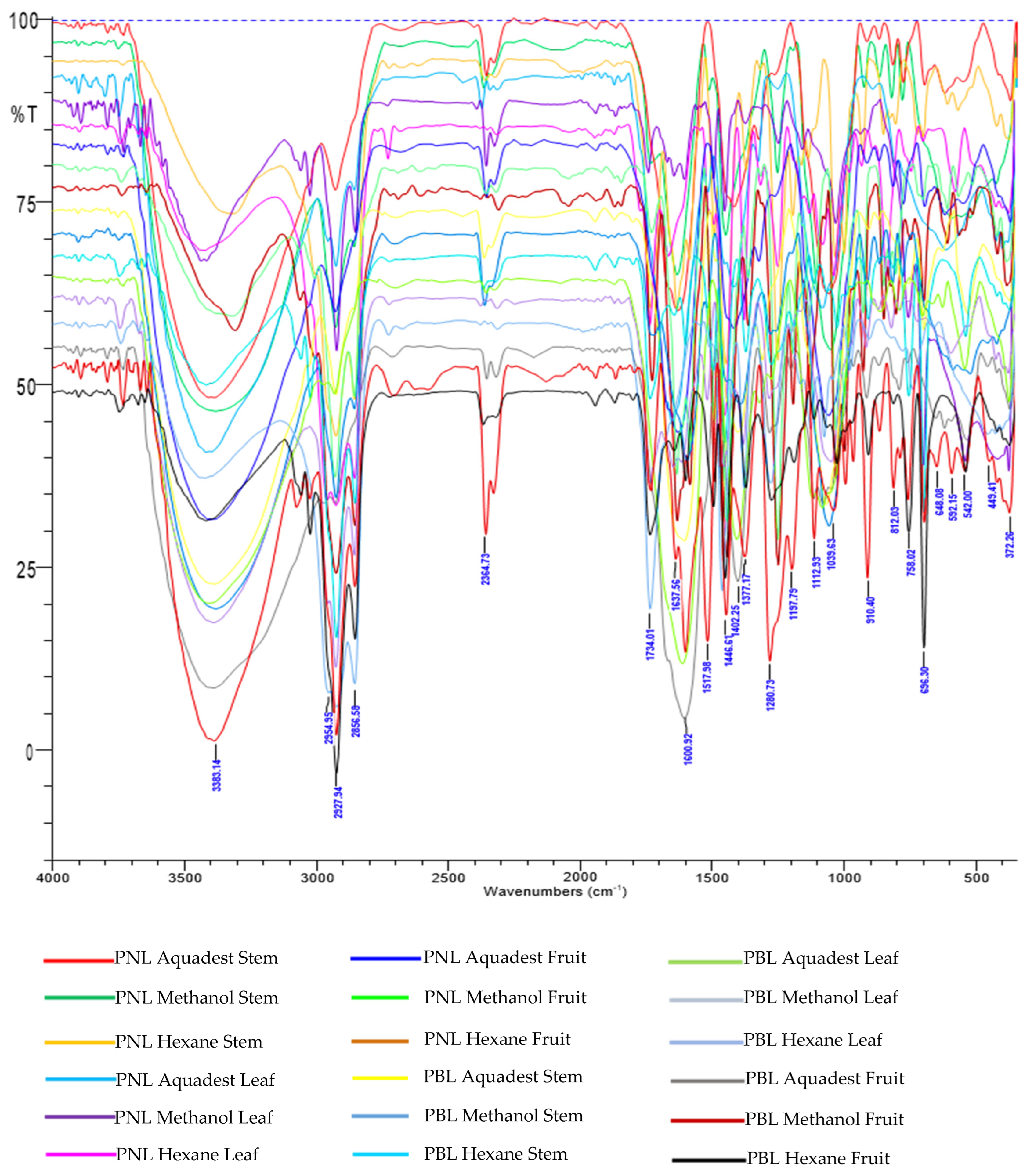
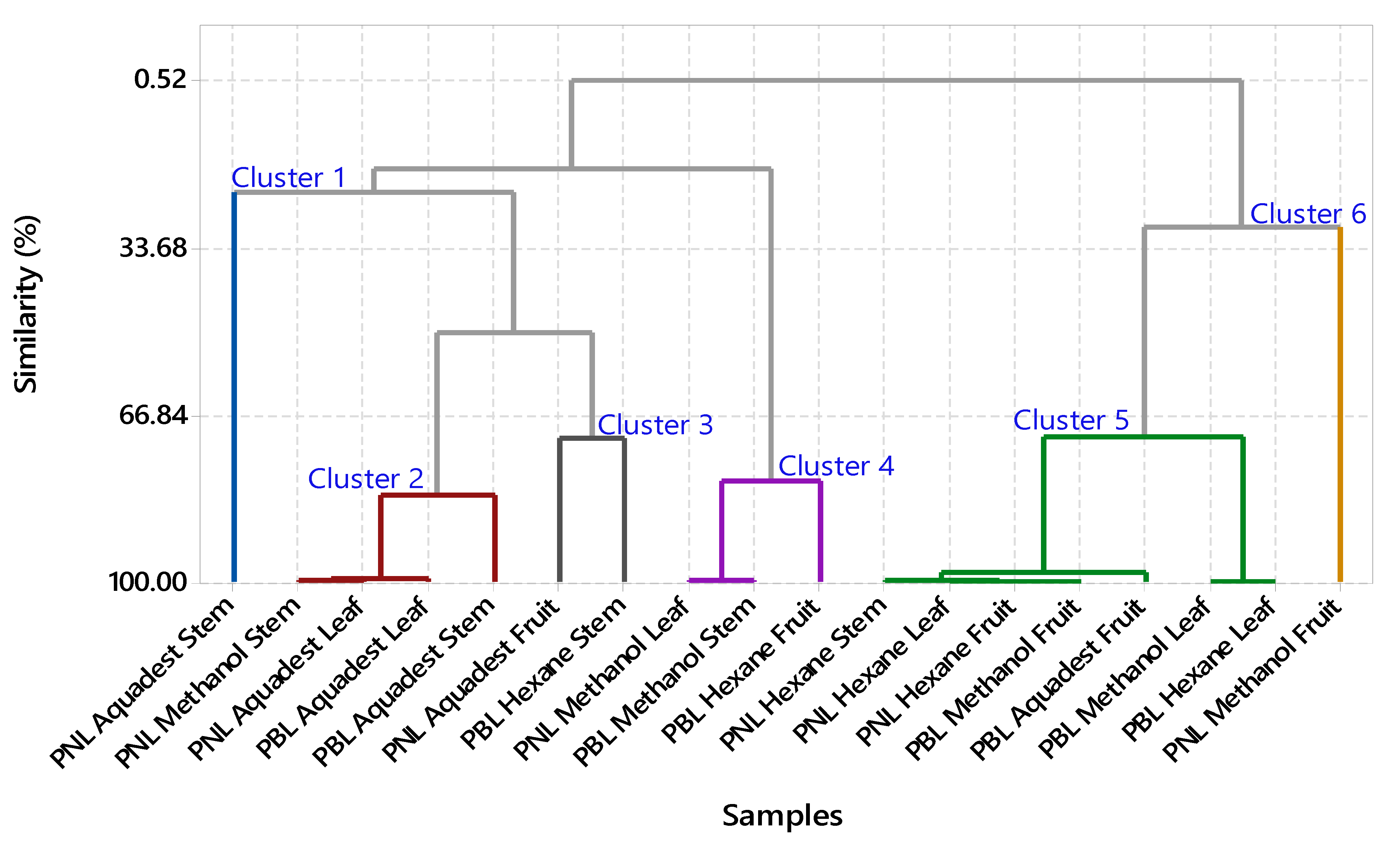
3.2. FTIR Profiling Analysis

Figure 1 shows the FTIR spectral profiles of various active compounds isolated from Piper spp., including P. nigrum (PNL) and P. betle (PBL). The peaks at 3383.14 cm−1 and 3404 cm−1 represent OH stretching bonds, commonly associated with compounds such as piperanine from Piper retrofractum [27]. Furthermore, the peak at 3291.31 cm−1 corresponded to phenolic O–H stretching in hydroxychavicol from PBL [28]. The peak at 3469 cm−1 was related to the stretching bonds of OH groups in piperine from PNL [29]. The range of 2927.94 cm−1 to 2936 cm−1 suggested aliphatic C–H stretching bands associated with compounds such as hydroxychavicol [28] and β-caryophyllene oxide [30]. Moreover, the peak at 2856.58 cm−1 signified aliphatic C–H stretching in piperine and pellitorine [31]. Peaks at 1637.56 cm−1 and 1647.45 cm−1 indicated C=C symmetric aromatic stretching in hydroxychavicol [28] and piperanine [31], while peaks at 1658 cm−1 and 1636 cm−1 represent conjugated diene symmetric stretching in piperanine [27] and piperine [29]. Furthermore, peaks at 1600.92 cm−1 and 1613 cm−1 imply conjugated asymmetric diene stretching associated with piperine [32] and chavibetol [33,34]. The profile also included aromatic C=C stretching at 1582 cm−1 and 1598 cm−1, CH2 bending at 1446.61 cm−1, and C–O stretching at 1197.79 cm−1, representing various active compounds such as chavibetol [33,34], piperine [29], β-caryophyllene oxide [30], and hydroxychavicol [28].
This FTIR profile provides valuable insights into the functional groups and main components in Piper extracts, which are crucial for chemical characterization and phytochemical studies. CA based on functional groups detailed the hierarchical clustering process, in which observations were sequentially merged into larger clusters according to similarity and distance levels [18]. The analysis initially started with 18 distinct clusters, each representing individual observations or small groups. In the initial step, clusters 9 and 17 merged to form a new cluster (cluster 9) containing two observations, at a high similarity level of 99.8523%. This merging process continued, with clusters 14 and 15 combining into cluster 14 in subsequent steps, progressively reducing the total number of clusters. By the fifth step, clusters 3 and 6 merged into a larger cluster (cluster 3), now comprising four observations. This pattern further led to the aggregation of more clusters into increasingly larger groups, and each step reduced the number. For example, clusters 2 and 7 combined in the 14th step to form cluster 2, which included six observation groups.
In the final partition of the CA in Figure 2, six distinct clusters formed with high similarity levels >80%. Cluster 1 includes observations from the initial analysis step. Cluster 2 combines observations from the PNL methanol stem, PNL aquadest leaf, PBL aquadest stem, and PBL aquadest leaf. Cluster 3 aggregates observations from the PNL hexane stem, PNL hexane leaf, PNL fruit hexane, PBL methanol leaf, PBL hexane leaf, PBL aquadest fruit, and PBL methanol fruit, with Piperine as a significant component, and cluster 4 merged observations from PNL methanol leaf, PBL methanol stem, and PBL hexane fruit. Cluster 5 consolidates observations from clusters 7 and 12, while cluster 6 represents observations from cluster 8. The final grouping reflects the hierarchical relationships among observations, capturing similarities and distances throughout the clustering process [35]. Since clusters with similar functional groups based on FTIR data are likely to have the same pharmacological effects [36], representatives from each cluster were selected for further anti-MRSA activity testing.
This novel approach in natural product study simplifies the screening process by focusing on extracts with similar functional group compounds, thereby facilitating the identification of potential candidates without the need to test each extract individually. The extracts selected for anti-MRSA testing were PNL aquadest stem extract from cluster 1, PNL aquadest leaf extract from cluster 2, PNL fruit hexane extract from cluster 3, PBL methanol stem extract from cluster 4, PBL aquadest fruit extract from cluster 5, and PNL methanol fruit extract from cluster 6.

3.3. Anti-MRSA Activity

Based on Table 2, a one-way analysis of variance (ANOVA) was conducted to assess the anti-MRSA activity of seven samples. The ANOVA yielded a highly significant F-value of 122.76 and a p-value of 0.000, indicating notable differences in anti-MRSA activity among the samples at a significance level of 0.05. The samples included six extracts, which are PBL aquadest fruit extract, PNL aquadest leaf extract, PNL aquadest stem extract, PNL hexane fruit extract, PBL methanol stem extract, and ciprofloxacin as a control. The ANOVA provided an adjusted sum of squares of 736.57 and an adjusted mean square of 122.762, resulting in a high R2 value of 98.13%. This suggests that nearly all variability in anti-MRSA activity can be attributed to differences among the extracts. Additionally, the antimicrobial effectiveness of PBL was tested against 20 clinical isolates of S. pseudintermedius (10 MSSP and 10 MRSP) using the Kirby-Bauer disk diffusion method with P. betle extract disks at concentrations of 250, 2500, and 5000 µg. The minimum inhibitory concentration (MIC) for all isolates was determined to be 250 µg/mL [37].
Tukey’s pairwise comparisons provided further clarity on the results. Ciprofloxacin exhibited the highest mean anti-MRSA activity at 26 mm, significantly differing from all other extracts at a 99.58% confidence level. The PBL aquadest fruit extract had a mean activity of 19 mm, significantly outperforming the PNL hexane fruit extract (15 mm) and the PNL aquadest leaf extract (6 mm). The PBL methanol stem extract had a mean of 15 mm, showing significant differences compared to some lower-performing extracts, while the PNL aquadest stem extract (9 mm) also exhibited significant differences from the lowest-performing extracts. This study accumulatively reports the novel potential utility of the Curcuma longa L. and PNL extracts synergistically against MRSA infection by interfering with the mechanism of infectious angiogenesis and bactericidal action [38].

3.4. GC-MS Metabolite Characterization

The GC-MS analysis Table 3 of the Piper plant extracts showed important connections between the identified chemical components and anti-MRSA activity. The aquadest extract of PBL aquadest fruit extract and the PNL hexane fruit extract were subjected to GC profiling to identify the major compounds responsible for anti-MRSA activity. The hexane extract of PNL hexane fruit extract was found to contain piperine, a major component [39] with an area of 14.22% and a similarity index (SI) of 93%. This compound has strong antimicrobial properties and is capable of reducing the secretion of diverse virulence factors from MRSA. Therefore, piperine could be a potential antibiofilm molecule against MRSA-associated biofilm infections [40]. This correlates with the anti-MRSA testing results, where extracts containing piperine showed significant activity against MRSA. Pellitorine, also present in the extract with an area of 5.08% and an SI of 93%, may be able to inhibit bacteria efflux pumps. At 16 µg/mL, pellitorine increased the sensitivity of S. aureus (RN4220) to erythromycin through inhibition of the efflux pumps [41], thereby contributing to antimicrobial activity. Other components such as diisooctyl phthalate and piperanine, though present in significant amounts of 14.67% and 4.03%, respectively, require further investigation to understand specific roles in anti-MRSA activity [42]. For the PBL aquadest fruit extract, hydroxychavicol was identified as the primary compound, making up 81.89% of the extract with an SI of 93%. This suggests that the suggesting that the compound plays a crucial role in the effectiveness of the extract against MRSA.
The PBL leaf extract produced the highest percentage of hydroxychavicol content. Almost up to 90% of the final extract was found to contain hydroxychavicol based on HPLC analysis [43]. This supported the result that extracts from cluster 5, including hydroxychavicol, showed significant anti-MRSA activity. The PBL hydroxychavicol (36.02%) was the major constituent that inhibited the growth and biofilm formation of S. pseudointermedius and MRSP isolated from canine pyoderma in a concentration-dependent manner. Therefore, PBL is a potential candidate for the treatment of MRSP infection and biofilm formation in veterinary medicine [44]. Chavibetol, with an area of 12.01% and SI of 97%, also contributed to the anti-MRSA activity but was less dominant than hydroxychavicol. The PBL leaf extract (200 g) sliced into small pieces and subjected to hydro-distillation for 270 min was found to contain chavibetol (63.78%) [43]. The chemical composition of the ethanolic extract also contained chavibetol (12.03%) [44]. The anti-MRSA activity was correlated with the GC-MS results, demonstrating that extracts containing high concentrations of piperine and hydroxychavicol had stronger antimicrobial effects. This relationship underscores the significance of both compounds in anti-MRSA activity and their potential use in developing effective antimicrobial treatments.

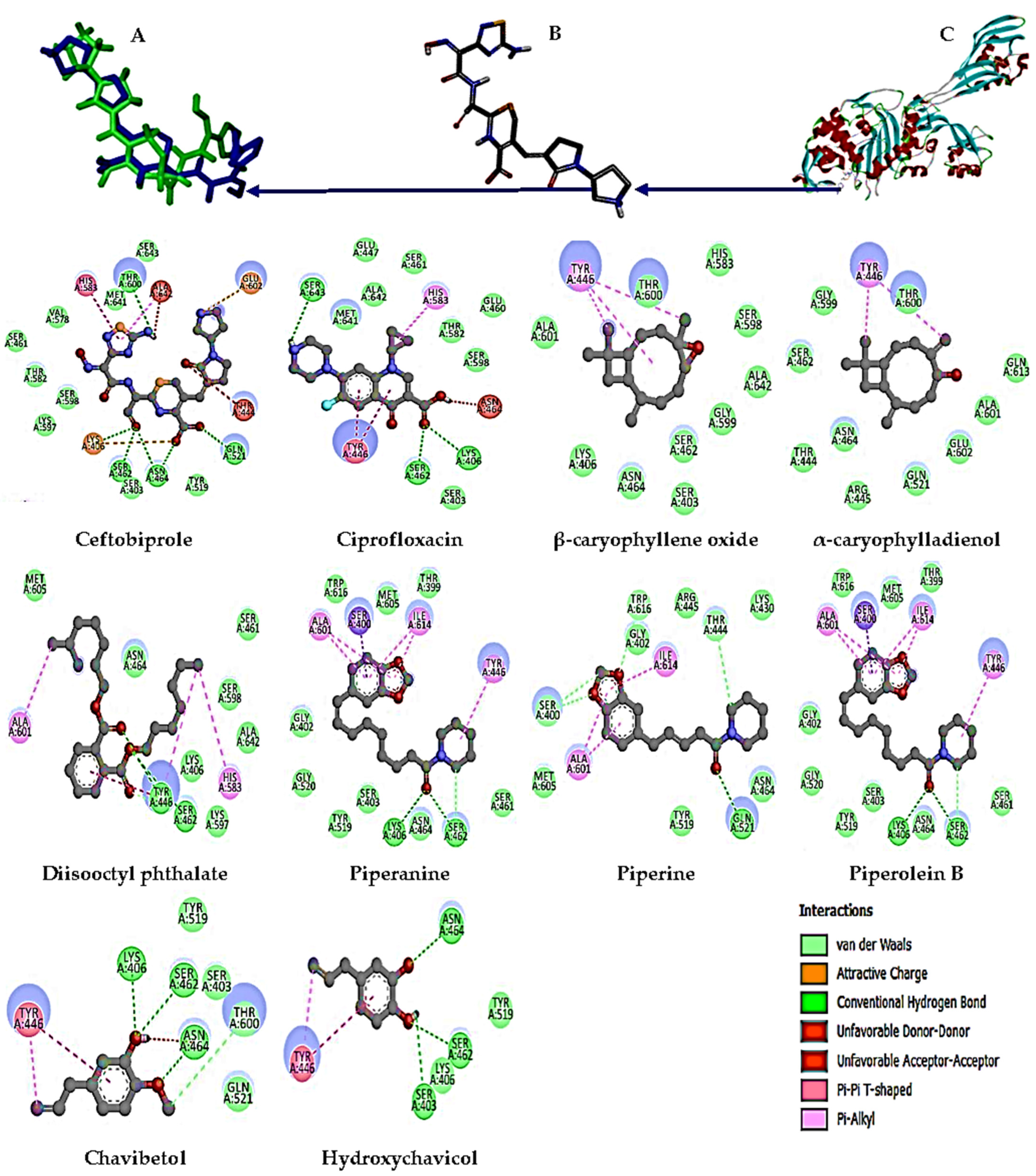
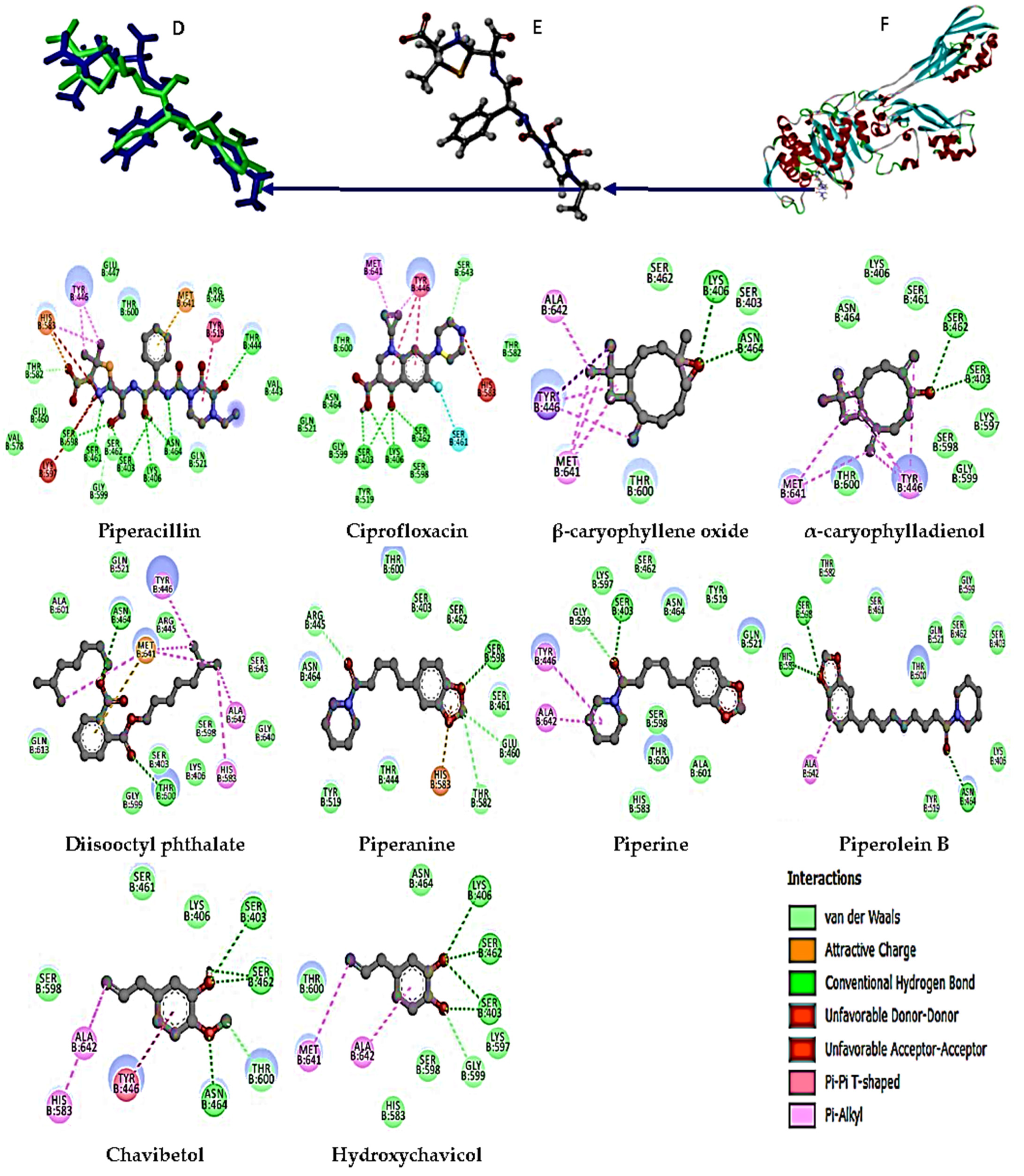
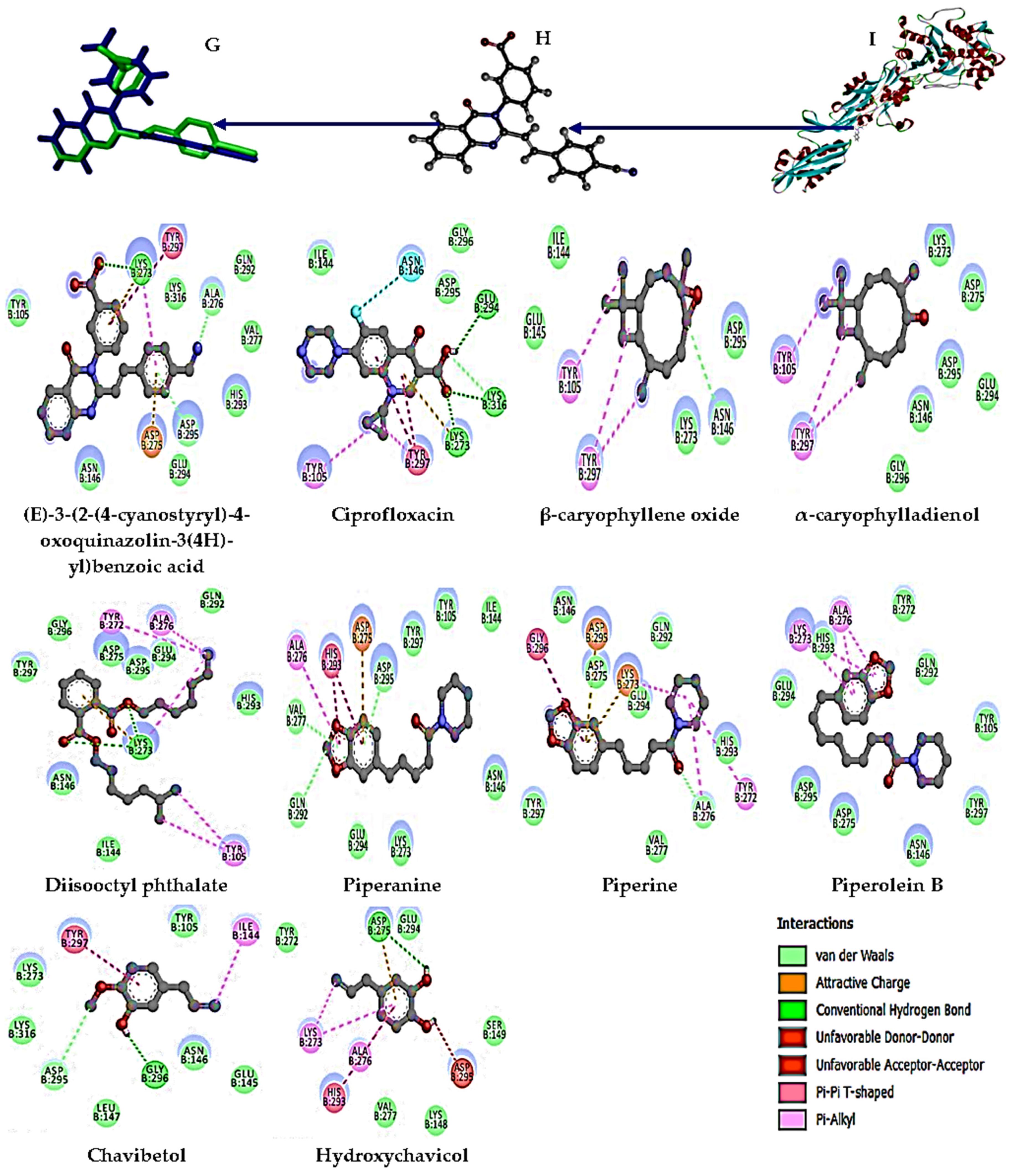
3.5. Molecular Docking Analysis

Table 4 shows the binding free energy values and amino acid residues interacting with the MRSA target proteins 4CJN, 4DKI, and 6H5O [45] for the various tested compounds. These data provide insights into the molecular interactions between these compounds and MRSA target proteins, as well as how the results correlate with anti-MRSA activity assays and chemical profiles of Piper extracts identified through GC-MS.
The native ligand for the 4DKI target protein (Figure 3) has a binding free energy of −9.8 kcal/mol and interacts with the amino acid residues LYS 406, SER 462, ASN 464, GLN 521, and THR 600 through hydrogen bonds [46]. Ciprofloxacin, the positive control, has a binding free energy of −8.3 kcal/mol and interacts with the residues LYS 406, SER 462, and SER 643. The PNL hexane fruit extract, which contains piperine, has a binding free energy of −7.7 kcal/mol and interacts with the residues GLN 521, THR 444, GLY 402, and SER 400, indicating significant potential anti-MRSA activity. Piperine, as the major component with an area of 14.22% in this extract, significantly contributes to the antimicrobial activity. Additionally, Piperolein B (1.5%) showed a binding free energy of −8.3 kcal/mol, equivalent to that of ciprofloxacin, with interactions at residues LYS 406, SER 462, and ASN 464, further enhancing the potential anti-MRSA activity. Piperanine (4.03%) and diisooctyl phthalate (14.67%) also demonstrated significant binding free energies of 7.6 and 7.1 kcal/mol, respectively, with interactions at residues LYS406 and SER462. However, β-caryophyllene oxide and α-caryophylladienol did not show any H-bond interactions with this target protein, suggesting limited anti-MRSA activity.
For the 6H5O target protein (Figure 4), the native ligand has a binding free energy of −9.0 kcal/mol and interacts with the amino acid residues THR 444, SER 598, SER 461, SER 403, LYS 406, and ASN 464 [47]. Ciprofloxacin, as the positive control, has a binding free energy of −8.1 kcal/mol and interacts with LYS 406, SER 403, SER 462, and SER 643. Furthermore, piperine and piperanine from the hexane extract of PNL have a binding free energy of −7.3 kcal/mol and interact with residues SER 403, GLY 599, SER 598, THR 582, GLU 460, and ARG 445, indicating significant anti-MRSA activity supported by the major component diisooctyl phthalate. Minor components such as β-caryophyllene oxide, α-caryophylladienol, and piperolein B also contribute to the anti-MRSA activity of this plant extract, with binding free energies ranging from 6.8 to 7.1 kcal/mol. In the PBL extract, the major compounds hydroxychavicol (81.89%) and chavibetol (12.01%) show binding free energies of 5.4 and 5.6 kcal/mol, respectively, interacting with the residues LYS 406, SER 403, SER 462, GLY 599, ASN 464, and THR 600. This interaction indicates significant anti-MRSA potential, although lower than that of Piperine.
For the 4CJN target protein (Figure 5), the native ligand has a binding free energy of −7.2 kcal/mol and interacts with the residues LYS 273, ALA 276, and ASP 295 [48]. Meanwhile, ciprofloxacin has a binding free energy of −6.2 kcal/mol and interacts with LYS 273, LYS 316, and GLU 294. Piperine in the PNL extract has a binding free energy of −6.5 kcal/mol and a stronger interaction at residue 276 than the positive control. Piperanine shows a favorable binding free energy of −6.0 kcal/mol, with interactions at ASP 295, VAL 277, and GLN 292, supporting the anti-MRSA activity, along with β-caryophyllene oxide and diisooctyl phthalate, although piperolein B does not show an H-bond interaction with this target protein. Meanwhile, in the PBL extract, hydroxyychavicol and chavibetol showed binding free energies of 4.9 and 4.6 kcal/mol, respectively. The extract interacted with residues ASP 275, ASP 295, and GLY 296, indicating a contribution to anti-MRSA activity, although lower compared to PNL. The in vitro assays demonstrated that extracts containing major compounds such as piperine [40], diisooctyl phthalate, and hydroxychavicol [44] from clusters 6 and 5 were significantly effective in inhibiting MRSA growth. These results were consistent with the GC-MS results, which showed high concentrations of piperine (14.22%) and diisooctyl phthalate (14.67%) in the hexane extract of PNL fruit, as well as hydroxychavicol (81.89%) and chavibetol (12.01%) in the aquadest extract of PBL. Both proteins contribute to significant anti-MRSA activity. Docking analysis showed that these compounds strongly bind to key residues in MRSA target proteins, reinforcing their potential as effective antimicrobial agents.

4. Conclusions

This study showed the successful optimization of extraction processes with methanol generally yielding the highest extracts across different parts of both P. betle (PBL) and P. nigrum (PNL), particularly in the fruits. This underscores the suitability of methanol as an extraction solvent. Hexane produced lower yields, while aquadest was moderately effective, especially for leaves and fruits. This optimization is crucial for selecting the most efficient solvent based on the desired yield and plant characteristics. The results demonstrated the significant potential of PBL and PNL as promising anti-MRSA agent sources. By using comprehensive FTIR spectroscopy combined with CA and GC-MS, several bioactive compounds were identified, including piperine, diisooctyl phthalate, and hydroxychavicol. These compounds showed strong binding affinities with key MRSA protein targets (4DKI, 6H5O, and 4CJN), as evidenced by molecular docking studies, with a strong correlation between in silico and in vitro results, confirming the effectiveness in inhibiting MRSA growth. The integration of in silico docking studies with in vitro assays provided a robust framework for identifying and validating potential anti-MRSA agents. Piperine, particularly as a major compound in PNL, demonstrated substantial antimicrobial activity, while hydroxychavicol in PBL showed significant inhibitory effects against MRSA. These results emphasize the importance of using a combined approach in drug discovery for identifying new antimicrobial agents. Further investigations are needed into Piper species as natural sources of novel anti-MRSA compounds with potential for development into effective treatments against resistant bacteria strains.

Author Contributions

Conceptualization, B.Y., A.R. and A.W.J.; methodology, B.Y., A.R., R.M.T., K.P.K. and N.A.; software, B.Y. and A.R.; validation, B.Y., A.R. and G.A.; formal analysis, B.Y. and A.R.; investigation, S.M., S.R., R.R. and N.R.K.N.; resources, B.Y., R.M.T., K.P.K. and N.A.; data curation, B.Y., A.R. and G.A.; writing—original draft preparation, B.Y., A.W.J. and A.R.; writing—review and editing, B.Y., A.W.J. and A.R.; visualization, B.Y. and A.W.J.; supervision, B.Y. and A.R.; project administration, S.M. and S.R.; funding acquisition, B.Y. and A.R. All authors have read and agreed to the published version of the manuscript.

Funding

This research was funded by Kementerian Pendidikan, Kebudayaan, Riset, and Teknologi Direktorat Jenderal Pendidikan Tinggi, Riset, and Teknologi, grant number 2383/E2/DT.01.00/2023. The APC was funded by Abdul Rohman and Budiman Yasir.

Data Availability Statement

The data presented in this study are included within the article. Additional information or datasets generated and analyzed during the research are available from the corresponding author upon reasonable request. No publicly archived datasets were used or generated in this study.

Acknowledgments

The authors are grateful to the Almarisah Madani University for providing the essential laboratory facilities. Furthermore, the authors are grateful to all collaborators and institutions for their invaluable contributions and technical assistance, which have been instrumental in the successful completion of this study.

Conflicts of Interest

The authors declare no conflicts of interest.

References

  1. Bashabsheh, R.H.; Al-Fawares, O.L.; Natsheh, I.; Bdeir, R.; Al-Khreshieh, R.O.; Bashabsheh, H.H. Staphylococcus aureus Epidemiology, Pathophysiology, Clinical Manifestations, and Application of Nano-Therapeutics as a Promising Approach to Combat Methicillin-Resistant Staphylococcus aureus. Pathogens Glob. Health 2024, 118, 209–231. [Google Scholar] [CrossRef] [PubMed]
  2. Nanayakkara, A.K.; Boucher, H.W.; Fowler, V.G., Jr.; Jezek, A.; Outterson, K.; Greenberg, D.E. Antibiotic Resistance in the Patient with Cancer: Escalating Challenges and Paths Forward. CA Cancer J. Clin. 2021, 71, 488–504. [Google Scholar] [CrossRef] [PubMed]
  3. Popovich, K.J.; Aureden, K.; Ham, D.C.; Harris, A.D.; Hessels, A.J.; Huang, S.S.; Maragakis, L.L.; Milstone, A.M.; Moody, J.; Yokoe, D.; et al. SHEA/IDSA/APIC Practice Recommendation: Strategies to Prevent Methicillin-Resistant Staphylococcus aureus Transmission and Infection in Acute-Care Hospitals: 2022 Update. Infect. Control Hosp. Epidemiol. 2023, 44, 1039–1067. [Google Scholar] [CrossRef] [PubMed]
  4. López-Cortés, L.E.; Gálvez-Acebal, J.; Rodríguez-Baño, J. Therapy of Staphylococcus aureus Bacteremia: Evidences and Challenges. Enferm. Infecc. Microbiol. Clin. 2020, 38, 489–497. [Google Scholar] [CrossRef]
  5. Jubair, N.; Rajagopal, M.; Chinnappan, S.; Abdullah, N.B.; Fatima, A. Review on the Antibacterial Mechanism of Plant-Derived Compounds Against Multidrug-Resistant Bacteria (MDR). Evid. Based Complement. Alternat. Med. 2021, 2021, 3663315. [Google Scholar] [CrossRef]
  6. Tran, V.T.; Nguyen, T.B.; Nguyen, H.C.; Do, N.H.; Le, P.K. Recent Applications of Natural Bioactive Compounds from Piper betle (L.) Leaves in Food Preservation. Food Control 2023, 154, 110026. [Google Scholar] [CrossRef]
  7. Ali, H.S.; Mishra, S. Natural Products as Antiparasitic, Antifungal, and Antibacterial Agents. In Drugs from Nature: Targets, Assay Systems and Leads; Springer: Singapore, 2024; pp. 367–409. [Google Scholar]
  8. Ueda, J.M.; Milho, C.; Heleno, S.A.; Soria-Lopez, A.; Carpena, M.; Alves, M.J.; Pires, T.; Prieto, M.A.; Simal-Gandara, J.; Calhelha, R.C.; et al. Emerging Strategies to Combat Methicillin-Resistant Staphylococcus aureus (MRSA): Natural Agents with High Potential. Curr. Pharm. Des. 2023, 29, 837–851. [Google Scholar] [CrossRef]
  9. Ali, R.A. Review on Extraction of Phenolic Compounds from Natural Sources Using Green Deep Eutectic Solvents. J. Agric. Food Chem. 2021, 69, 878–912. [Google Scholar]
  10. Verma, N.; Gautam, B.S. Piper betle: Deep Insights into the Pharmacognostic and Pharmacological Perspectives. Int. J. Med. Phar. Sci. 2024, 13, 4. [Google Scholar] [CrossRef]
  11. Garcia-Vaquero, M.; Ummat, V.; Tiwari, B.; Rajauria, G. Exploring Ultrasound, Microwave and Ultrasound–Microwave Assisted Extraction Technologies to Increase the Extraction of Bioactive Compounds and Antioxidants from Brown Macroalgae. Mar. Drugs 2020, 18, 172. [Google Scholar] [CrossRef]
  12. Chandrakasan, G.; García-Trejo, J.F.; Feregrino-Pérez, A.A.; Aguirre-Becerra, H.; García, E.R.; Nieto-Ramírez, M.I. Preliminary Screening on Antibacterial Crude Secondary Metabolites Extracted from Bacterial Symbionts and Identification of Functional Bioactive Compounds by FTIR, HPLC, and Gas Chromatography–Mass Spectrometry. Molecules 2024, 29, 2914. [Google Scholar] [CrossRef]
  13. Noor, A.; Kamaraj, S.; Bhakti, M.A.; Kumari, C.; Kulshrestha, S.; Pandohee, J.; That, L.F.L.N. Analysis of Volatile Organic Compounds in Honey Using Gas Chromatography and Mass Spectrometry. In Advanced Techniques of Honey Analysis; Academic Press: Cambridge, MA, USA, 2024; pp. 287–307. [Google Scholar]
  14. Cucinotta, L.; Rotondo, A.; Coppolino, S.; Salerno, T.M. Gas Chromatographic Techniques and Spectroscopic Approaches for a Deep Characterization of Piper gaudichaudianum Kunth Essential Oil from Brazil. J. Chromatogr. A 2024, 1732, 465208. [Google Scholar] [CrossRef]
  15. Palermo, A. Metabolomics-and Systems-Biology-Guided Discovery of Metabolite Lead Compounds and Druggable Targets. Drug Discov. Today 2023, 28, 103460. [Google Scholar] [CrossRef]
  16. Taibi, M.; Elbouzidi, A.; Ou-Yahia, D.; Dalli, M.; Bellaouchi, R.; Tikent, A.; Roubi, M.; Gseyra, N.; Asehraou, A.; Hano, C.; et al. Assessment of the Antioxidant and Antimicrobial Potential of Ptychotis verticillata Duby Essential Oil from Eastern Morocco: An In Vitro and In Silico Analysis. Antibiotics 2023, 12, 655. [Google Scholar] [CrossRef] [PubMed]
  17. Yasir, B.; Astuti, A.D.; Raihan, M.; Natzir, R.; Subehan, S.; Rohman, A.; Alam, G. Optimization of Pagoda (Clerodendrum paniculatum L.) Extraction Based on Analytical Factorial Design Approach, Its Phytochemical Compound, and Cytotoxicity Activity. Egypt J. Chem. 2022, 65, 421–430. [Google Scholar] [CrossRef]
  18. Yasir, B.; Alam, G. Chemometrics-Assisted Fingerprinting Profiling of Extract Variation from Pagoda (Clerodendrum paniculatum L.) Using TLC-Densitometric Method. Egypt J. Chem. 2023, 66, 1589–1596. [Google Scholar] [CrossRef]
  19. Roshan, M.; Singh, I.; Vats, A.; Behera, M.; Singh, D.P.; Gautam, D.; Rajput, S.; Tarak, J.; Packirisamy, G.; De, S. Antimicrobial and Antibiofilm Effect of Cannabinoids from Cannabis sativa Against Methicillin-Resistant Staphylococcus aureus (MRSA) Causing Bovine Mastitis. Int. Microbiol. 2024, 1–14. [Google Scholar] [CrossRef]
  20. Martí, M.; Frígols, B.; Serrano-Aroca, A. Antimicrobial Characterization of Advanced Materials for Bioengineering Applications. J. Vis. Exp. 2018, 138, 57710. [Google Scholar]
  21. Alharthi, S.S. Bioactive Extracts of Plant Byproducts as Sustainable Solution of Water Contaminants Reduction. Arab. J. Chem. 2024, 17, 105864. [Google Scholar] [CrossRef]
  22. Sruthi, D.; Zachariah, T.J. Volatile and Non-Volatile Metabolite Profiling of Under-Explored Crop Piper colubrinum Link: With Chromatography Mass Spectrometry Approach and Its Biochemical Diversity from Medicinally Valued Piper Species. J. Chromatogr. B 2024, 1245, 124260. [Google Scholar] [CrossRef] [PubMed]
  23. Gao, K.; Chen, Z.; Zhang, N.; Jiang, P. High Throughput Virtual Screening and Validation of Plant-Based EGFR L858R Kinase Inhibitors Against Non-Small Cell Lung Cancer: An Integrated Approach Utilizing GC–MS, Network Pharmacology, Docking, and Molecular Dynamics. Saudi Pharm. J. 2024, 32, 102139. [Google Scholar] [CrossRef] [PubMed]
  24. Hendrarti, W.; Umar, A.H.; Syahruni, R.; Rafi, M.; Kusuma, W.A. Deciphering the Mechanism of Action Cosmos caudatus Compounds Against Breast Neoplasm: A Combination of Pharmacological Networking and Molecular Docking Approach with Bibliometric Analysis. Indones. J. Sci. Technol. 2024, 9, 527–556. [Google Scholar]
  25. Nguyen, L.T.T.; Nguyen, T.T.; Nguyen, H.N.; Bui, Q.T.P. Simultaneous Determination of Active Compounds in Piper betle Linn. Leaf Extract and Effect of Extracting Solvents on Bioactivity. Eng. Rep. 2020, 2, 10. [Google Scholar]
  26. Rajopadhye, A.A.; Namjoshi, T.P.; Upadhye, A.S. Rapid Validated HPTLC Method for Estimation of Piperine and Piperlongumine in Root of Piper longum Extract and Its Commercial Formulation. Rev. Bras. Farmacogn. 2012, 22, 1355–1361. [Google Scholar] [CrossRef]
  27. Luo, J.; Xiang, J.Y.; Yuan, H.Y.; Wu, J.Q.; Li, H.Z.; Shen, Y.H.; Xu, M. Isolation, Synthesis and Absolute Configuration of 4,5-Dihydroxypiperines Improving Behavioral Disorder in AlCl3-Induced Dementia. Bioorg. Med. Chem. Lett. 2021, 42, 128057. [Google Scholar]
  28. Nik, W.W.; Ravindran, K.; Al-Amiery, A.A.; Izionworu, V.O.; Fayomi, O.S.I.; Berdimurodov, E.; Daoudi, W.; Zulkifli, F.; Dhande, D.Y. Piper betle Extract as an Eco-Friendly Corrosion Inhibitor for Aluminium Alloy in Hydrochloric Acid Media. Int. J. Corros. Scale Inhib. 2023, 12, 948–960. [Google Scholar]
  29. Gorgani, L.; Mohammadi, M.; Najafpour, G.D.; Nikzad, M. Sequential Microwave-Ultrasound-Assisted Extraction for Isolation of Piperine from Black Pepper (Piper nigrum L.). Food Bioprocess Technol. 2017, 10, 2199–2207. [Google Scholar] [CrossRef]
  30. Fan, R.; Qin, X.W.; Hu, R.S.; Hu, L.S.; Wu, B.D.; Hao, C.Y. Studies on the Chemical and Flavour Qualities of White Pepper (Piper nigrum L.) Derived from Grafted and Non-Grafted Plants. Eur. Food Res. Technol. 2020, 246, 2601–2610. [Google Scholar]
  31. Ramesh, B.; Sarma, V.U.M.; Kumar, K.; Babu, K.S.; Devi, P.S. Simultaneous Determination of Six Marker Compounds in Piper nigrum L. and Species Comparison Study Using High-Performance Thin-Layer Chromatography–Mass Spectrometry. J. Planar Chromatogr. 2015, 28, 280–286. [Google Scholar]
  32. Sateesh Reddy, K.; Siva, B.; Divya Reddy, S.; Kumar, K.; Pratap, T.V.; Vidyasagar Reddy, K.; Venkateswara Rao, B.; Suresh Babu, K. Monitoring of Chemical Markers in Extraction of Traditional Medicinal Plants (Piper nigrum, Curcuma longa) Using In Situ ReactIR. J. AOAC Int. 2021, 104, 1181–1187. [Google Scholar] [CrossRef]
  33. Musa, T.A.; Sanagi, M.M.; Wan Ibrahim, W.A.; Ahmad, F.; Aboul-Enein, H.Y. Determination of 4-Allyl Resorcinol and Chavibetol from Piper betle Leaves by Subcritical Water Extraction Combined with High-Performance Liquid Chromatography. Food Anal. Methods 2014, 7, 893–901. [Google Scholar] [CrossRef]
  34. Rahmah, N.L.; Mustapa Kamal, S.M.; Sulaiman, A.; Taip, F.S.; Siajam, S.I. Subcritical Water Extraction of Total Phenolic Compounds from Piper betle L. Leaves: Effect of Process Conditions and Characterization. J. Food Meas. Charact. 2023, 17, 5606–5618. [Google Scholar] [CrossRef]
  35. Bouguettaya, A.; Yu, Q.; Liu, X.; Zhou, X.; Song, A. Efficient Agglomerative Hierarchical Clustering. Expert Syst. Appl. 2015, 42, 2785–2797. [Google Scholar] [CrossRef]
  36. Cao, Z.; Wang, Z.; Shang, Z.; Zhao, J. Classification and Identification of Rhodobryum roseum Limpr. and Its Adulterants Based on Fourier-Transform Infrared Spectroscopy (FTIR) and Chemometrics. PLoS ONE 2017, 12, e0170650. [Google Scholar] [CrossRef]
  37. Jantorn, P.; Tipmanee, V.; Wanna, W.; Prapasarakul, N.; Visutthi, M.; Sotthibandhu, D.S. Potential Natural Antimicrobial and Antibiofilm Properties of Piper betle L. Against Staphylococcus pseudintermedius and Methicillin-Resistant Strains. J. Ethnopharmacol. 2023, 317, 116820. [Google Scholar] [CrossRef]
  38. Khan, G.J.; Humma, Z.E.; Omer, M.O.; Sattar, A.; Altaf, I.; Chen, Z.; He, N. Effects of Curcuma longa L. and Piper nigrum L. Against Methicillin-Resistant Staphylococcus aureus and Infectious Angiogenesis. J. Biobased Mater. Bioenergy 2024, 18, 303–314. [Google Scholar] [CrossRef]
  39. Umapathy, V.R.; Dhanavel, A.; Kesavan, R.; Natarajan, P.M.; Bhuminathan, S.; Vijayalakshmi, P. Anticancer Potential of the Principal Constituent of Piper nigrum, Piperine: A Comprehensive Review. Cureus 2024, 16, e44287. [Google Scholar] [CrossRef]
  40. Das, S.; Malik, M.; Dastidar, D.G.; Roy, R.; Paul, P.; Sarkar, S.; Tribedi, P. Piperine, a Phytochemical, Prevents the Biofilm Formation of Methicillin-Resistant Staphylococcus aureus: A Biochemical Approach to Understand the Underlying Mechanism. Microb. Pathog. 2024, 189, 106601. [Google Scholar] [CrossRef]
  41. Ahirrao, P.; Tambat, R.; Chandal, N.; Mahey, N.; Kamboj, A.; Jain, U.K.; Singh, I.P.; Jachak, S.M.; Nandanwar, H.S. MsrA Efflux Pump Inhibitory Activity of Piper cubeba L.f. and Its Phytoconstituents Against S. aureus RN4220. Chem. Biodivers. 2020, 17, e2000144. [Google Scholar] [CrossRef]
  42. Driche, E.H.; Belghit, S.; Bijani, C.; Zitouni, A.; Sabaou, N.; Mathieu, F.; Badji, B. A New Streptomyces Strain Isolated from Saharan Soil Produces Di-(2-Ethylhexyl) Phthalate, a Metabolite Active Against Methicillin-Resistant Staphylococcus aureus. Ann. Microbiol. 2015, 65, 1341–1350. [Google Scholar] [CrossRef]
  43. Zamakshshari, N.; Ahmed, I.A.; Nasharuddin, M.N.; Hashim, N.M.; Mustafa, M.R.; Othman, R.; Noordin, M.I. Effect of Extraction Procedure on the Yield and Biological Activities of Hydroxychavicol from Piper betle L. Leaves. J. Appl. Res. Med. Aromat. Plants 2021, 24, 100320. [Google Scholar] [CrossRef]
  44. Leesombun, A.; Sungpradit, S.; Bangphoomi, N.; Thongjuy, O.; Wechusdorn, J.; Riengvirodkij, S.; Boonmasawai, S. Effects of Piper betle Extracts Against Biofilm Formation by Methicillin-Resistant Staphylococcus pseudintermedius Isolated from Dogs. Pharmaceuticals 2023, 16, 741. [Google Scholar] [CrossRef] [PubMed]
  45. Benjamin, I.; Louis, H.; Ekpen, O.F.; Gber, T.E.; Gideon, M.E.; Ahmad, I.; Ejiofor, E.U. Modeling the Anti-Methicillin-Resistant Staphylococcus aureus (MRSA) Activity of (E)-6-Chloro-N2-Phenyl-N4-(4-Phenyl-5-(Phenyl Diazinyl)-2λ3,3λ2-Thiazol-2-yl)-1,3,5-Triazine-2,4-Diamine. Polycycl. Aromat. Compd. 2023, 43, 7942–7969. [Google Scholar] [CrossRef]
  46. Lovering, A.L.; Gretes, M.C.; Safadi, S.S.; Danel, F.; De Castro, L.; Page, M.G.; Strynadka, N.C. Structural Insights into the Anti-Methicillin-Resistant Staphylococcus aureus (MRSA) Activity of Ceftobiprole. J. Biol. Chem. 2012, 287, 32096–32102. [Google Scholar] [CrossRef]
  47. Janardhanan, J.; Bouley, R.; Martínez-Caballero, S.; Peng, Z.; Batuecas-Mordillo, M.; Meisel, J.E.; Chang, M. The Quinazolinone Allosteric Inhibitor of PBP 2a Synergizes with Piperacillin and Tazobactam Against Methicillin-Resistant Staphylococcus aureus. Antimicrob. Agents Chemother. 2019, 63, 10–1128. [Google Scholar] [CrossRef]
  48. Bouley, R.; Kumarasiri, M.; Peng, Z.; Otero, L.H.; Song, W.; Suckow, M.A.; Mobashery, S. Discovery of Antibiotic (E)-3-(3-Carboxyphenyl)-2-(4-Cyanostyryl) Quinazolin-4(3H)-One. J. Am. Chem. Soc. 2015, 137, 1738–1741. [Google Scholar] [CrossRef]
Figure 1. FTIR spectra of Piper extracts (PNL, Piper nigrum L.; PBL, Piper betle L.) extracted using aquadest, methanol, and hexane from stem, leaf, and fruit parts.
Separations 11 00322 g001
Figure 2. The dendrogram illustrating the classification of samples through cluster analysis (CA), with samples in clusters 1 to 6 represented by distinct colored lines for each respective cluster.
Separations 11 00322 g002
Figure 3. Structure of the MRSA protein target (4DKI) (C) along with the native ligand (ceftobiprole) (B). The re-docking of the native ligand (co-crystal) into the MRSA protein target pocket validates the method, resulting in a root mean square deviation (RMSD) value of 0.991 Å (A). Additionally, the interactions of the compounds in the extract, the native ligand, and the positive control with the MRSA protein target (4DKI) were illustrated, emphasizing their binding affinities and interactions within the target pocket.
Separations 11 00322 g003
Figure 4. MRSA protein target (6H5O) structure (F), the native ligand (piperacillin) (E), and re-docking native ligand (co-crystal) result in MRSA protein target pocket for validating the method with RMSD value of 0.991 Å (D). Additionally, the interactions of the compounds in the extract, the native ligand, and the positive control with the MRSA protein target (6H5O) were illustrated, emphasizing their binding affinities and interactions within the target pocket.
Separations 11 00322 g004
Figure 5. MRSA protein target (4CJN) Structure (I), the native ligand (E)-3-(2-(4-cyanostyryl)-4-oxoquinazolin-3(4H)-yl)benzoic acid (H) and re-docking native ligand (co-crystal) result in MRSA protein target pocket for validate the method with RMSD value of 0.751 Å (G). Additionally, the interactions of the compounds in the extract, the native ligand, and the positive control with the MRSA protein target (4CJN) were illustrated, emphasizing their binding affinities and interactions within the target pocket.
Separations 11 00322 g005
Table 1. Percentage yield of extracts from different solvents and parts of Piper plants.
CodeSamplePlant PartsSolventExtract (g)Yield (%)
PNLPiper nigrumStemAquadest0.834.15
Methanol1.075.35
Hexane0.130.65
LeafAquadest0.633.15
Methanol0.432.15
Hexane0.623.10
FruitAquadest0.331.65
Methanol1.145.70
Hexane0.42.00
PBLPiper betleStemAquadest0.331.65
Methanol0.824.10
Hexane0.110.55
LeafAquadest0.894.45
Methanol0.673.35
Hexane0.090.45
FruitAquadest1.366.80
Methanol2.3511.75
Hexane0.542.70
Noted: Simplicia was 20 g for all samples.
Table 2. Antibacterial activity of Piper extracts against MRSA based on inhibition zone diameter (mm) for different plant parts and solvents.
Sample Inhibition Zone (mm)Tukey Simultaneous Tests for Differences in MeansStDev
Difference in LevelsDifference in MeansSE of Difference95% CIT-Valuep-Value
PNL Aquadest Stem Extract8PNL Aquadest Leaf Extract—PNL Aquadest Stem Extract−2.6670.797(−5.388, 0.055)−3.350.0561.000
10PNL Aquadest Fruit Extract—PNL Aquadest Stem Extract4.6670.797(1.945, 7.388)5.860.001
9PNL Methanol Fruit Extract—PNL Aquadest Stem Extract5.6670.797(2.945, 8.388)7.110.000
PNL Aquadest Leaf Extract6PBL Methanol Stem Extract—PNL Aquadest Stem Extract5.0000.797(2.279, 7.721)6.270.0000.577
6PBL Hexane Fruit Extract—PNL Aquadest Stem Extract10.3330.797(7.612, 13.055)12.970.000
7Ciprofloxacin—
PNL Aquadest Stem Extract
16.6670.797(13.945, 19.388)20.920.000
PNL Aquadest Fruit Extract14PNL Aquadest Fruit Extract—PNL Aquadest Leaf Extract7.3330.797(4.612, 10.055)9.200.0000.577
14PNL Methanol Fruit Extract—PNL Aquadest Leaf Extract8.3330.797(5.612, 11.055)10.460.000
13PBL Methanol Stem Extract—PNL Aquadest Leaf Extract7.6670.797(4.945, 10.388)9.620.000
PNL Methanol Fruit Extract14PBL Hexane Fruit Extract—PNL Aquadest Leaf Extract13.0000.797(10.279, 15.721)16.310.0000.577
15Ciprofloxacin—
PNL Aquadest Leaf Extract
19.3330.797(16.612, 22.055)24.260.000
15PNL Methanol Fruit Extract—PNL Aquadest Fruit Extract1.0000.797(−1.721, 3.721)1.250.861
PBL Methanol Stem Extract14PBL Methanol Stem Extract—PNL Aquadest Fruit Extract0.3330.797(−2.388, 3.055)0.420.9990.000
14PBL Hexane Fruit Extract—PNL Aquadest Fruit Extract5.6670.797(2.945, 8.388)7.110.000
14Ciprofloxacin—
PNL Aquadest Fruit Extract
12.0000.797(9.279, 14.721)15.060.000
PBL Hexane Fruit Extract20PBL Methanol Stem Extract—PNL Methanol Fruit Extract−0.6670.797(−3.388, 2.055)−0.840.9760.577
19PBL Hexane Fruit Extract—PNL Methanol Fruit Extract4.6670.797(1.945, 7.388)5.860.001
19Ciprofloxacin—
PNL Methanol Fruit Extract
11.0000.797(8.279, 13.721)13.800.000
Ciprofloxacin24PBL Hexane Fruit Extract—PBL Methanol Stem Extract5.3330.797(2.612, 8.055)6.690.0002.080
28Ciprofloxacin—
PBL Methanol Stem Extract
11.6670.797(8.945, 14.388)14.640.000
25Ciprofloxacin—
PBL Hexane Fruit Extract
6.3330.797(3.612, 9.055)7.950.000
Note: 95% CI (95% confidence interval); t-value (test statistic value); p-value (probability value); StDev (standard deviation); pooled StDev = 0.975900; R-sq (R-squared) = 98.22%.
Table 3. GC-MS analysis of compounds in the hexane extract of Piper nigrum fruit and aquadest extract of Piper betle fruit.
Compounds DetectedMolecular FormulaMW
(g/mol)
PubChem (CID)RT
(min)
Area (%)SI (%)
Piper nigrum hexane fruit extract
β-caryophyllene oxideC15H24O220174221013.5631.0197
α-caryophylladienol C15H24O2201452492314.2761.6997
PellitorineC14H25NO223531851618.7725.0893
n-Hexadecanoic acidC16H32O225698519.2792.5196
Octadecanoic acidC18H36O2284528123.8771.4293
Diisooctyl phthalateC24H38O43903393431.08114.6795
(2E,4E)-N-Isobutylhexadeca-2,4-dienamideC20H37NO307644240231.5951.7890
PiperanineC17H21NO3287532061832.8044.0394
PiperineC17H19NO328563802437.76214.2293
Piperolein BC21H29NO33432158021341.6391.5893
Piper betle aquadest fruit extract
ChavibetolC10H12O216459637510.42012.0197
HydroxychavicolC9H10O21507077512.49681.8993
Note: MW (molecular weight); PubChem (PubChem compound identifier); RT (retention time); SI (similarity index).
Table 4. Bond-free energy values and amino acid residues binding to MRSA protein targets (4DKI, 6H5O, and 4CJN).
CompoundsProtein TargetBond-Free Energy (kcal/mol)H-Bond Interaction
Ceftobiprole (Native ligand)4DKI−9.8LYS 406, SER 462, ASN 464, GLN 521, THR 600
Ciprofloxacin −8.3LYS 406, SER 462, SER 643
β-caryophyllene oxid −6.4NI
α-caryophylladienol −6.4NI
Diisooctyl phthalate −7.1SER 462, TYR 446
Piperanine −7.6LYS 406, SER 462
Piperine −7.7GLN 521, THR 444, GLY 402, SER 400
Piperolein B −8.3LYS 406, SER 462, ASN 464
Chavibetol −5.7THR 600, ASN 464, SER 462, LYS 406
Hydroxychavicol −5.7SER 462, ASN 464, SER 403
Piperacillin (Native ligand)6H5O−9.0THR 444, SER 598, SER 461, SER 403, LYS 406, ASN 464
Ciprofloxacin −8.1LYS 406, SER 403, SER 462, SER 643
β-Caryophyllene oxide −6.9ASN 464, LYS 406
α-Caryophylladienol −7.1SER 403, SER 462
Diisooctyl phthalate −6.3ASN 464, THR 600
Piperanine −7.3SER 598, THR 582, GLU 460, ARG 445
Piperine −7.3SER 403, GLY 599
Piperolein B −6.8ASN 464, SER 598, HIS 583
Chavibetol −5.4SER 403, ASN 464, SER 462, THR 600
Hydroxychavicol −5.6LYS 406, SER 403, SER 462, GLY 599
(E)-3-(2-(4-cyanostyryl)-4-oxoquinazolin-3(4H)-yl)benzoic acid (Native ligand)4CJN−7.2LYS 273, ALA 276, ASP 295
Ciprofloxacin −6.2LYS 273, LYS 316, GLU 294
β-caryophyllene oxide −5.2ASN 146
α-caryophylladienol −5.2NI
Diisooctyl phthalate −5.0LYS 273
Piperanine −6.0ASP 295, VAL 277, GLN 292
Piperine −6.5ALA 276
Piperolein B −5.9NI
Chavibetol −4.6ASP 295, GLY 296
Hydroxychavicol −4.9ASP 275
Note: “NI” indicates that the amino acid residues did not interact with the MRSA protein target (4DKI, 6H5O, and 4CJN) via H-bond interactions and were not found in the native ligand interactions.
Disclaimer/Publisher’s Note: The statements, opinions and data contained in all publications are solely those of the individual author(s) and contributor(s) and not of MDPI and/or the editor(s). MDPI and/or the editor(s) disclaim responsibility for any injury to people or property resulting from any ideas, methods, instructions or products referred to in the content.

