Article

Green Bio-Analytical Study of Gabapentin in Human Plasma Coupled with Pharmacokinetic and Bioequivalence Assessment Using UPLC-MS/MS

by
Rehab Moussa Tony
1,
Mohamed A. El Hamd
2,3,*,
Mohammed Gamal
4,*,
Safaa F. Saleh
5,6,
Nujud Maslamani
7,
Wejdan T. Alsaggaf
8 and
Mohamed B. El-Zeiny
1
1
Department of Pharmaceutical Analytical Chemistry, Faculty of Pharmacy, Modern University for Technology and Information, Cairo 11571, Egypt
2
Department of Pharmaceutical Sciences, College of Pharmacy, Shaqra University, Shaqra 11961, Saudi Arabia
3
Department of Pharmaceutical Analytical Chemistry, Faculty of Pharmacy, South Valley University, Qena 83523, Egypt
4
Department of Pharmaceutical Analytical Chemistry, Faculty of Pharmacy, Beni-Suef University, Alshaheed Shehata Ahmed Hegazy St., Beni-Suef 62574, Egypt
5
Department Pharmaceutical Chemistry, College of Pharmacy, Jazan University, Jazan 45142, Saudi Arabia
6
Department of Pharmaceutical Analytical Chemistry, Faculty of Pharmacy, Fayoum University, Fayoum 63514, Egypt
7
Department of Chemistry, College of Science, Imam Abdulrahman Bin Faisal University, Dammam 31441, Saudi Arabia
8
Department of Chemistry, Faculty of Science, King Abdulaziz University, Jeddah 21551, Saudi Arabia
*
Authors to whom correspondence should be addressed.
Separations 2023, 10(4), 234; https://doi.org/10.3390/separations10040234
Submission received: 3 March 2023 / Revised: 21 March 2023 / Accepted: 27 March 2023 / Published: 2 April 2023

Abstract

:
Gabapentin (GAB) is a cyclohexane acetic acid, structurally related to the neurotransmitter gamma-aminobutyric acid (GABA), and considered the principal inhibitory neurotransmitter in the central nervous system (CNS) of mammals. An ultra-performance liquid chromatography–tandem mass spectrophotometry (UPLC-MS/MS) method for assessing pregabalin (PRE) in human plasma, was developed and validated, via PRE usage as an internal standard. The plasma underwent protein precipitation using methanol, prior to analysis. Chromatographic separation was completed using a mobile phase of methanol: 0.1% formic acid solution, (65:35, v/v), at a flow rate of 0.2 mL/min, with an isocratic approach, on an Agilent Eclipse plus column (50 × 2.1 mm and 1.8 μm), in 1.6 min of running time. An Agilent triple quadrupole was used for mass analysis, to detect the ion transitions for GAB and PER, respectively, at m/z of 172.1 → 154.1 and 160.10 → 142.10. The calibration curve, over the linear range of 0.050–10.0 μg/mL, showed a high correlation coefficient, r = 0.9993. The limits of detection and quantitation were 13.37 ng/mL and 40.52 ng/mL, respectively, based on the standard deviation and slope equation. The results for intra- and inter-day measurement accuracy and precision were in acceptable ranges. The method was extended into the assessment of oral administrations of GAB at different doses, of one 600 mg/tablet and two capsules (each one of them has 300 mg of GAB), to volunteers who were used in pharmacokinetics and bioequivalent studies. The AGREE assessment tool was used to visualize the proposed method’s greenness degree, which revealed a high AGREE rating score, supporting the accepted method’s greenness profile.

1. Introduction

Gabapentin (GAB, Figure 1A) is cyclohexane-1-amino acetic acid. It shares structural similarities with the neurotransmitter gamma-aminobutyric acid (GABA), which is regarded as one of the main inhibitory neurotransmitters in the mammalian central nervous system (CNS). It is used to treat seizures, either on its own or in combination with other drugs. Additionally, it is used to lessen shingles-related nerve pain. After 2 to 3 h, GAB is fully absorbed and reaches its peak plasma concentration. Its elimination half-life is between 5 and 7 h, and the kidneys primarily excrete it as an unchanged drug [1]. Therefore, the analysis of GAB in human plasma is very important, as well as for establishing reliable pharmacokinetics and bioequivalent approaches, in case of seizure and pain management.
A literature survey revealed that several analytical techniques have been developed to determine GAB in human plasma, either as a single component or combined with other drugs, including spectrofluorimetry assays [2,3], high-pressure liquid chromatography (HPLC) [4,5,6,7,8,9,10,11,12,13], liquid chromatography–mass spectrophotometry (LC-MS/MS) [14,15,16,17], gas chromatography (GC) [18,19], gas chromatography with a mass spectrophotometer detector (GC-MS) [20], and capillary electrophoresis techniques (CE) [21,22,23,24,25]. Most of the stated LC-MS/MS methods used numerous organic modifiers, buffers, or complicated gradient elution.
Additionally, GAB can be analyzed in urine and plasma samples as well, where comparable recoveries were obtained for GAB analysis in urine and plasma. However, performing a full pharmacokinetic study is preferred in plasma for GAB, in terms of sensitivity, as the quantitation limit was 0.05 μg/mL in the plasma and 0.1 μg/mL in the urine, in the reported bioanalytical method [26]. Furthermore, in this study, we chose to deal with blood, as it is used more in bioavailability and pharmacokinetics studies and it is easier to collect plasma samples. Generally, LC-MS/MS has the advantage of analyzing potent drugs, which are administrated in low concentrations in biological matrices, in addition to its ability to resolve a complex of drug mixtures with good selectivity and sensitivity, compared to traditional detectors coupled with LC. LC-MS/MS, coupled online with a soft ionization technique such as triple quadrupole mass analysis, has been frequently used in pharmacokinetic (PK) studies [27,28,29]. Additionally, MS comes the closest to being the ideal detector to employ with the chromatographic separations, because it combines the highest level of identification potential with the best possible sensitivity in most cases. The MS is related to the mass-to-charge ratio (m/z)-based gas-phase identification of ionized species, and the resulting MS spectrum is a plot of the relative intensity versus m/z. An inlet device, ion source, ion separation system (mass analyzer, which is the “heart” of the mass spectrometer), and detector are the basic parts of an MS [29,30]. Therefore, the principal target of this analytical research was to develop a reliable, simple, green, and cost-effective LC-MS/MS analytical method, for the evaluation of GAB in the real human plasma of volunteers. Additionally, the assessment of GAB has been extended to carrying out a bioequivalent study, for appraisal of a new test product tablet containing 600 mg GAB. Pregabalin (PRE, Figure 1B) is a structural analogue of GABA but functionally unrelated to it. It is presently being used for general anxiety disorders, neuropathic pains, and epilepsy [31,32,33], here, it is used as an internal standard for the assessment of GAB, for a more realistic evaluation.

2. Materials and Methods

2.1. Materials and Chemical Reagents

Gabapentin (GAB) (CAS number: 60142-96-3), of purity 99.49%, was bought from Maps laboratory, Cairo, Egypt, while the purity of pregabalin (PRE) (CAS number: 148553-50-8), which is used as an internal standard, was found to be 99.5%. Test product film-coated tablets, labeled to contain 600 mg GAB, were formulated by EGPI (Egyptian Group for the pharmaceutical industry)—Cairo, Egypt, for Aman Pharma. Additionally, Neurontin hard gelatine capsules are labeled as containing 300 mg GAB, which was manufactured by Pfizer for the pharmaceutical industry (New York, NY, USA).
The purity for all organic solvents and chemicals was aligned with the required standards for the LC approach. Methanol (CAS number: 67-56-1) was purchased from Fischer Scientific UK Ltd. (Loughborough, UK). Formic acid (CAS number: 64-18-6) and acetonitrile (CAS number: 75-05-08), were purchased from Merck (Darmstadt, Germany). Purelab flex, ELGA apparatus was used as a source of deionized water. Human plasma samples were obtained from the VACSERA institution (Giza, Egypt).

2.2. Instruments

An Agilent triple quadrupole mass 6470 LC/TQ, with Agilent Jet Stream (AJS) ion source with optimal sensitivity (Waldbronn, Germany), was the instrument of choice for the UPLC-MS/MS analysis. For MS/MS sensing, multiple reaction monitoring (MRM) and positive electrospray ionization (ESI) modes were used. The ChemStation software B.02.01 was used to perform data acquisition and control hardware. An Eppendorf concentrator plus was used as a solvent concentrator.

2.3. UPLC-MS/MS Conditions

The optimal assay conditions were obtained using the Agilent eclipse plus C18 stationary phase (50 × 2.1 mm and 1.8 μm), and the liquid mobile phase used consisted of a mixture of methanol and 0.1% formic acid (65:35, by volume). It was pumped with an isocratic elution mode at a rate of flow of 0.2 mL per minute. The stationary phase temperature was kept at 30 °C, while the temperature of the autosampler tray was fixed to 15 °C, and the injection volume was 1.0 μL.
To optimize the operating parameters for the detection of GAB and IS, standard solutions of analytes, with concentrations of 100 ng/mL each, in methanol, were infused directly in the MS using ESI in the positive ionization style (ESI+). The MRM mode of the triple quadrupole MS was employed to observe the subsequent transitions (the molecular → the product ions) m/z 172.10 → 154.10 and m/z 160.10 → 142.10 for GAB and PRE, respectively. The gas in the nebulizer was zero-grade air, while N2 gas was used as the collision gas, curtain gas, and auxiliary gas (Table 1).

2.4. Stock Solutions

The parent solutions (100.0 µg/mL) of GAB and PRE were performed disjointedly in methanol. Furthermore, appropriate dilutions were performed using methanol and water (1:1, v/v), in a 10 mL calibrated flask. Both the working and parent flasks were kept at 2–8 °C and set at room temperature before use.

2.5. Establishment of Calibration Curves

The concentrations of eight points (0.05, 0.30, 0.50, 1.00, 2.00, 6.00, 9.00, and 10.00 μg/mL) were obtained, after spiking 50.0 μL of PRE, which is equivalent to a fixed concentration of 20.00 µg/mL, and increasing concentrations of GAB from its working standard solution (50 μL) into aliquots of 450.0 µL blank human plasma. They were then allowed to vortex-mix for 0.5 min. Following this, 1.50 mL of methanol was added, followed by three minutes of vortex-mixing, and centrifugation at 1507× g for 0.5 min, at 5 °C. A protein precipitation (PPT) process was carried out. Thus, 1.0 μL of the clear liquid was injected into the chromatographic system. The previously developed curves were used to estimate the concentrations of GAB in unknown samples. With concentrations of 0.08, 0.80, 3.00, and 0.15 μg/mL, respectively, free plasma was utilized to handle quality control (QC) samples of GAB for each of the three different levels: high (HQC), medium (MQC (A)), (MQC (B)), and low (LQC).

2.6. Method Validation

Following the guidance issued by the FDA for Industry Bioanalytical Method Validation [26], the validation protocol was implemented.

2.6.1. Specificity Study

The specificity of six different blank human plasma samples, which were randomly chosen from various sources, was assessed. To ensure that plasma constituents did not cause any interferences, these plasma models were set up and tested as previously mentioned.

2.6.2. Carryover Study

Carryover was assessed by adding samples of free plasma after adding a high concentration. The acceptable value for carryover in the free plasma for the limit of quantification (LOQ) and PRE, should not be higher than 20% and 5%, respectively.

2.6.3. Estimation of Linearity and Range Study

For the estimation of the linearity, six linear curves were made, in the desired range of 0.050–10.0 µg/mL, for GAB. The regression analysis did not include the zero and blank data. The calibration graph was created by comparing the peak area percentages of GAB to PRE, with the appropriate concentration values (0.050–10.0 µg/mL). The limit of detection (LOD) and quantitation (LOQ) were defined as the concentrations giving peak area at a signal-to-noise ratio (S/N) of 3.3 and 10, respectively. The accepted correlation coefficient (r) for the LOQ concentration, which was obtained at 20%, should be at least 0.99, and the range of recoveries should not exceed 15%.

2.6.4. Precision and Accuracy Study

The accuracy and precision were assessed using spiked plasma trials at the concentrations of QC samples (HQC, MQC, LQC, and LOQ). Six repeats of QC samples were analysed, either on the same day (accuracy) or for the subsequent three days (precision). The precision was calculated as (RSD,%), and the accuracy was calculated as recovery percentage.

2.6.5. Assessment of Recoveries in the Extraction Process

The effectiveness of GAB extraction from the plasma was considered by comparing the peak areas of GAB extracted from the QC samples with those from previously extracted plasma samples at equivalent concentrations.

2.6.6. Investigation of Matrix Effects

To assess the effect of the plasma ingredients on the ionization of GAB, the responses of previously extracted plasma QC trials were compared, at similar concentration levels.

2.6.7. Stability Study

The stability of the analyte was examined using three repetitions of the HQC, MQC, and LQC analyses. Prior to analysis, the QC samples were adjusted to room temperature for six hours to examine the samples’ short-term stability.

Stability in the Autosampler Study

The stability of the GAB extracts was assessed using the QC samples that were kept in the autosampler for a day prior to the analysis.

Bench-Top Stability Study

The stability of the analyzed samples could be assessed by storing the study trials for 24 h at room temperature under standard experimental conditions.

Parent Solutions’ Stability Study

The parent solutions’ stability was assessed both six hours after being kept at room temperature, and one week after being kept at either 5 or 3 degrees.

Freeze-Thaw Stability Study

Examination of the QC specimens’ stability over three freeze and thaw rotations was performed, as the specimen was left to liquefy for longer than 2 h at room temperature, then left to freeze overnight at −86 °C.

Long-Term Stability Study

Long-term stability was evaluated by comparing the mean recovery of stored samples over a period equivalent to, or exceeding, the period between obtaining the first specimen and the last sample analysis time (63 days) at −86 °C. The samples were considered stable when recovery at each level did not exceed ±15% for the zero cycles with % RSD less than 15%.

2.6.8. Investigation of Dilution Integrity

The dilution integrity was assessed by injecting the biological matrix with a substance that was more concentrated than the upper limit of quantitation, dilution of the specimen with blank matrices at six measurements per dilution factor 2 (2-fold ULOQ), and dilution factor 4 (4-fold ULOQ). For acceptance criteria, dilutions of specimens should not influence precision and accuracy.

2.7. Pharmacokinetic and Bioequivalence Studies

The Research Ethics Committee of the Private Research Centre (Cairo, Egypt), approved the consent form and the study protocol to conduct a bioequivalence study for the test product, a 600 mg film-coated tablet. The aims, and possible side effects, of the presented work, were illustrated to the volunteers. The developed UPLC-MS/MS method was used to examine the plasma profile of GAB after an oral intake of a single 600 mg film-coated tablet as a test product (one tablet), and compared with Neurontin 300 mg hard gelatin capsules (two capsules), with one week washout period between the prescriptions, using a randomized open two-period crossover balanced design.
A clinical study was conducted on 34 healthy volunteers. All the involved persons were in the fasting status, to reject any plausible interface between caffeine and food. The participants were knowledgeable about the goals and possible risks concerning this study. Collection of blood samples (of about 3 mL) from all subjects in heparinized tubes was performed in advance of the initial dosing (T0), and then after 0.5, 1.0, 1.25, 1.5, 1.75, 2.0, 2.25, 2.50, 2.75, 3.0, 3.25, 3.50, 3.75, 4.0, 4.50, 5.0, 5.50, 6.0, 8.0, 10.0, 12.0, 24.0, 36.0, and 48.0 h from the administration. After the collection of samples, they were centrifuged for 10 min at 1600× g and then stored at −80 °C till analysis.

3. Results and Discussion

3.1. Treatment of Biological Samples

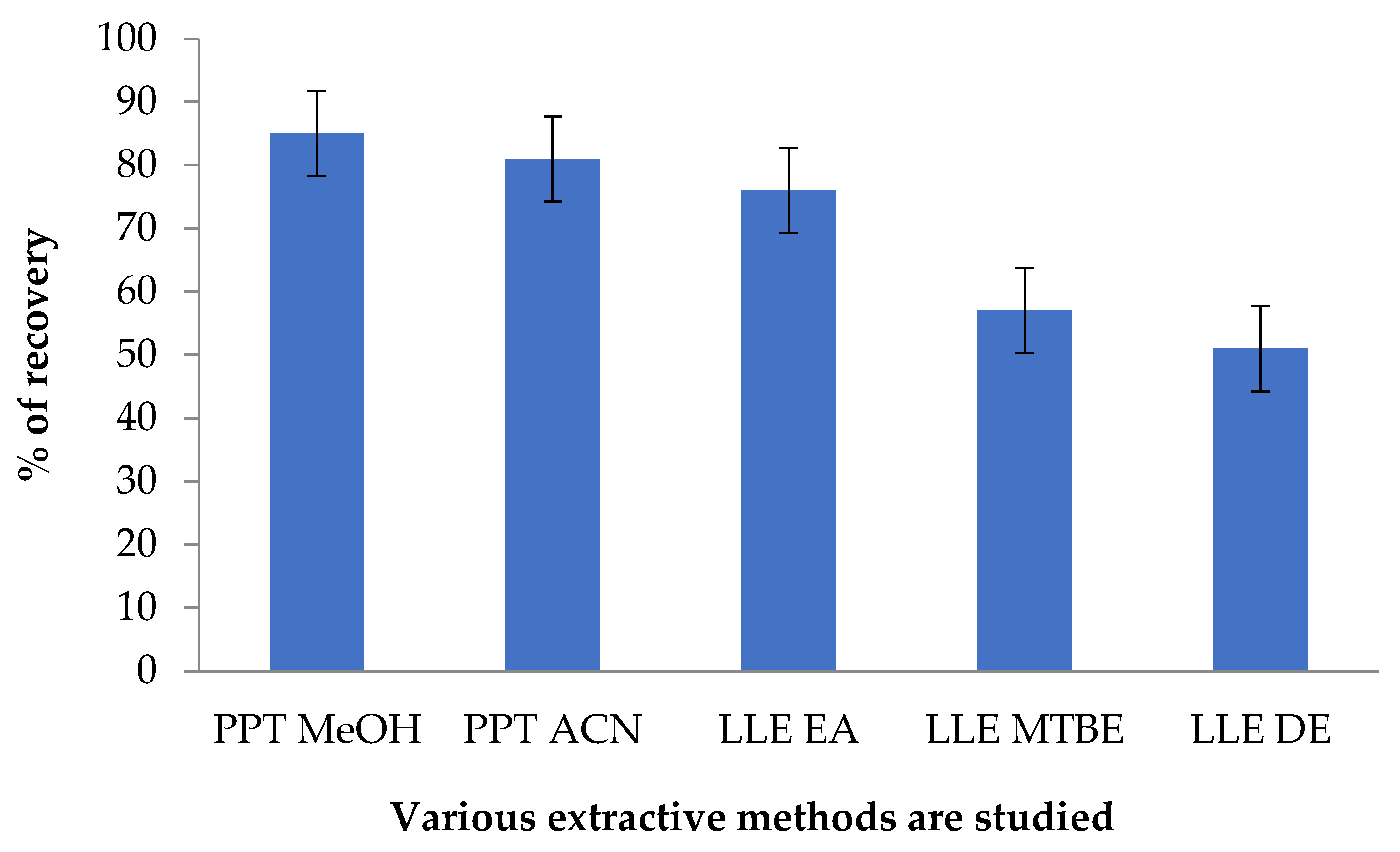
The optimal procedures for determining GAB in human blood were explored using a range of techniques. Liquid/liquid extraction (LLE) using various organic solvents was tested, e.g., diethyl ether, ethyl acetate, and tertiary butyl methyl ether. In contrast, protein precipitation (PPT) using acetonitrile or methanol was tried. A minimal matrix influence for GAB and PRE was observed in the PPT protocol. The maximal recovery for GAB was obtained using 1.5 mL of methanol as the precipitating material (Figure 2).

3.2. Mass Spectrophotometric and Chromatographic Settings

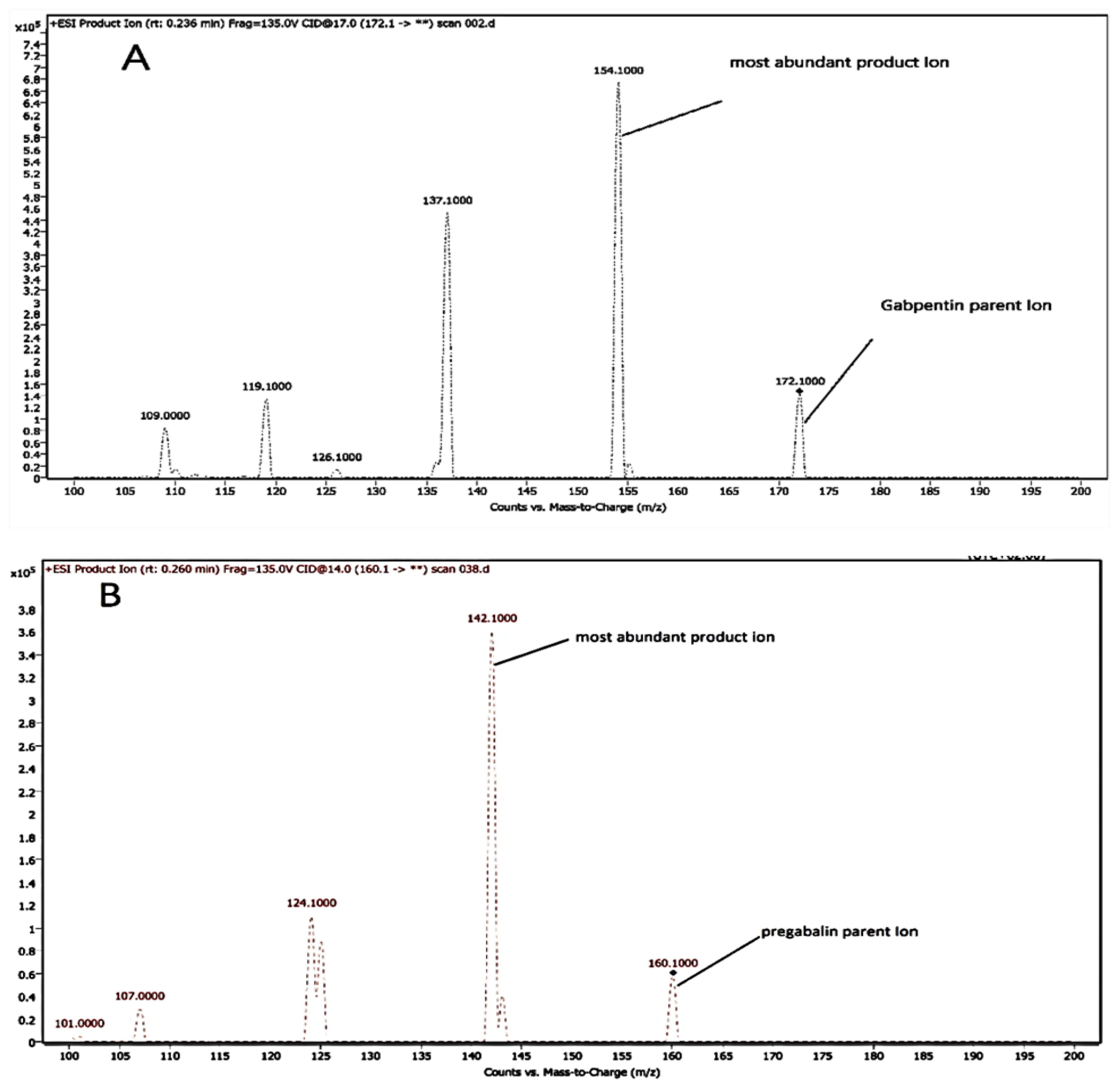
Working organic solutions of GAB and PRE (100 ng/mL) were injected into the MS for adjusting the product and precursor ions using the ESI+ mode. The highest intensities for the product and precursor ions were accomplished in the positive approach for GAB and PRE because their ions could be protonated with ease. The full-scan spectra of precursor ions presented the positive ions [M + H]+ m/z 172.1 and m/z 160.1 for GAB and PRE, respectively. MRM mode was used to explore the resultant ion mass spectrum. Sufficient collision energies were applied, and the subsequent product ions were nominated as m/z 154.1 and m/z 142.1 for GAB and PRE, respectively. Figure 3 shows the most prominent ions.
However, the chromatographic environments were improved by adjusting the type of non-aqueous modifier, its proportion, speed of flow, and the type of stationary phase. The sensitivity and discrimination for the UPLC-MS/MS method were affected markedly by changing the mobile phase composition, so the organic modifiers (acetonitrile and methanol) were used in dissimilar percentages. Using methanol: formic acid (0.10%) (65:35, v/v), with a speed of 0.2 mL/minute, showed the highest sensitivity.
The best peak intensity and shape were obtained using this composition of the mobile phase. The analysis was performed in less than one minute, as shown in (Figure 4). Diverse column materials were tested to estimate GAB and PER, such as C8 XTerra MS (150 × 4.6 mm, 3.5 µm), C18 Zorbax SB (50 × 4.6 mm, 5 µm), and C18 Agilent eclipse plus(50 × 2.1 mm, 1.8 µm). The C18 Agilent eclipse plus showed the best performance and outcomes even for the low QC specimens.
It is clear from the results demonstrated in Table 2, that the new MS method has merged most of the merits that might be provided by the UPLC-MS/MS technique. The use of small column dimensions (50 × 2.1 mm, 1.8 µm) has led to obtaining the shortest run time among all the methods used for the determination of gabapentin (≈1 min) and also has a good resolution. The flow speed, of 0.2 mL/minute, has the merit of being cost-effective, due to low solvent consumption. Additionally, the isocratic mode was used, where a 35% non-aqueous modifier was selected and buffer was avoided. Moreover, we avoided the utilization of liquid–liquid extraction (LLE) or solid-phase extraction (SPE) during specimen preparations, which are sophisticated processes. So, methanol was used for the PPT process, which is a very simple and direct technique.

3.3. Chromatographic Method Validity

Full validation processes were accomplished concerning the FDA protocol for the bioanalytical method [26].

3.3.1. Method Specificity Study

This study exposes the typical UPLC chromatograms for human plasma, plasma that has been PER-enriched, and GAB at lower QC concentrations (Figure 4). The specificity is supported by the absence of any notable interference from the plasma constituents with the GAB peak.

3.3.2. Carryover Study

The results clearly showed that carryover cumulative influence is lessened, as they were 20% and 5% for GAB at LOQ and for PRE, respectively.

3.3.3. Linearity and Range Study

For the assessment of linearity, an average of six determinations was used, and it was discovered that the range for GAB was between 50 and 10,000 ng/mL. The median values for the regression equation were y = 0.7403, x − 0.00127, and r = 0.9993. Where x is the resulting concentration level in ng/mL and y is the percentage of the peak area of GAB to PER. To ensure that there was no interference with the assay, zero and blank specimens were used. At every point along the standard curve, the back-estimated values were less than ± 15% of the nominal concentration levels. For GAB, the S/N ratio was 10 and the RSD was ± 20% for LOQ. The LOD and LOQ were 13.37 ng/mL and 40.52 ng/mL, respectively, based on the standard deviation and slope equation calculation; LOD = 3.3*3/0.7403 = 13.37 ng/m, and LOQ = 10*3/0.7403 = 40.52 ng/mL. Therefore, the selected conditions were directed toward the low plasma concentrations used during the clinical practice of GAB.

3.3.4. Accuracy and Precision Study

According to the results of the four concentrations that were examined (LOQ, HQC, MQC (A, B), and LQC), the intra-day repeatability ranged from 93.24% to 104.40%, with RSD% values from 1.29% to 5.52%. Inter-day accuracy ranged from 90.96% to 103.23% with a precision of 1.14% to 5.16% as shown in Table 3. The data collected demonstrated that the assay is reliable and accurate enough.

3.3.5. Results of Extraction Recovery Study

The effectiveness of GAB and PRE extraction from plasma was evaluated at three concentrations over six iterations. The extraction recoveries for GAB and PRE were reasonable and consistent, ranging from 82.39% to 86.53%, as illustrated in Table 4, demonstrating the effectiveness of the extraction technique used in the presented study.

3.3.6. Matrix Influence Study

Whether matrix elements might enhance or suppress ionization was investigated. The obtained GAB recoveries (Table 4) show that matrix components had no impact on GAB ionization.

3.3.7. Stability Study

The impact of the FDA bioanalytical method validation strategies was confirmed [26]. The stability of GAB in human plasma was evaluated over a range of storage times, and the results were compared to those obtained from fresh samples. GAB was discovered to be stable in plasma for 24 h at 25 °C. Furthermore, there was no discernible degradation after 72 h of GAB storage in the autosampler. The results of the GAB quantification were unaffected by the three freeze–thaw rotations that were performed on the QC specimens. The analyte was stable after 63 days at −86 °C. The information in Table 5 demonstrates that human plasma samples containing GAB can be handled without significantly affecting the quality of the tested analyte if they are kept out of the light. Moreover, in order to assure the stability of the stock solution, the parent solution’s stabilities were also tested for 6 h of storage at room temperature and for one week of brief storage at 5 ± 3 °C, the results are shown in Table 5.

3.3.8. Investigation of Dilution Integrity

After spiking the blank plasma with 16.0 and 32.0 μg/mL of GAB solution, dilution with zero plasma samples (blank) up to 2- and 4-fold were performed, at six determinations per dilution. The accuracy results were 93.83% and 94.13%, while the precision (RSD) was 2.01 and 1.03% for 2- and 4-fold, respectively.

3.4. Pharmacokinetic Study

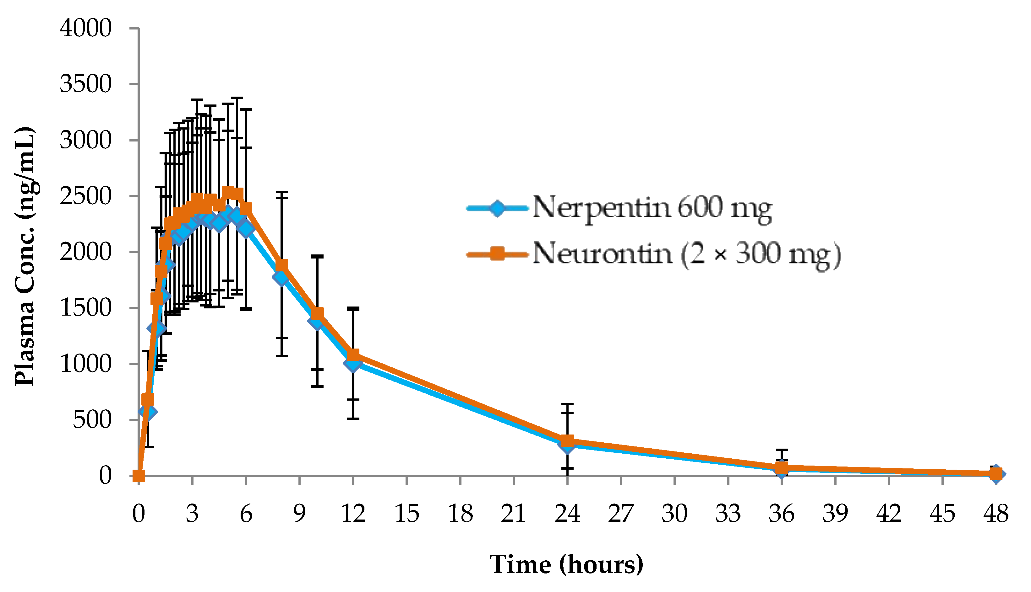
The validated bioanalytical UPLC-MS/MS technique was used, to characterize the pharmacokinetics of GAB in 34 healthy persons, following oral intake of one film-coated tablet test product (containing 600 mg of GAB) while fasting. The pharmacokinetics parameters were also contrasted with the dose of two Neurontin hard gelatin capsules (300 mg/capsule). The statistical analysis of Cmax, AUC0-∞, and AUC0-t in the two GAB formulations, reveals that there is no significant difference between them. The FDA acceptability range for the mean ratio (T/R) of Cmax, AUC0-∞, and AUC0-t was demonstrated in the 90% confidence intervals, as shown in Table 6. According to the results obtained in Figure 5, the test product film-coated tablet (600 mg/tablet GAB) and hard gelatin capsules (300 mg/capsule), were bioequivalent to two Neurontin hard gelatin capsules (300 mg/capsule).
The volunteers’ mean plasma concentration–time outlines after oral intake of one film-coated tablet test product (600 mg/tablet GAB) against two Neurontin hard gelatin capsules (300 mg/each capsule) is shown in Figure 5.

3.5. Greenness Study

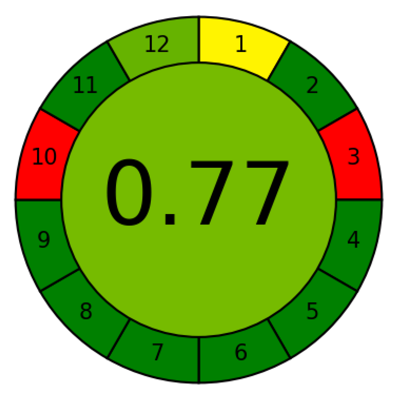
The level of the proposed method’s eco-friendlessness was evaluated using the AGREE tool, due to the growing significance of using green, eco-friendly analytical tools. This instrument has the advantages of automation, simplicity, and dependability [34,35,36]. The overall score was nearly 0.77 and the green colour in the centre of the pictogram presented in Figure 6, confirms the eco-friendlessness features of this technique.
The acetonitrile-free liquid mobile system is an advantage over existing, published techniques, where acetonitrile is recognized as being environmentally hazardous. The red subdivisions 3 and 10, denote off-line sampling and the type of reagents used, respectively. One of the merits of the method is its short analysis time (one min), which allows the analysis of 60 samples per hour and is expressed by the intense green colour for subsector 8.

3.6. Comparing the Suggested Strategy to Other Published Methods That Have a Similar Analytical Purpose

Numerous LC-MS/MS methods have previously been used to assay GAB in human plasma, either alone or in combination with other analytes. Our proposed method was compared to those previously published [14,15,37,38,39,40,41] demonstrating the use of (50 × 2.1 × 1.8 µm) stationary phase, which is relatively small in size, as demonstrated in Table 2. The column’s length resulted in a relatively short run time of 1.6 min, due to the rapid and consistent detection of the target analyte, and the small particle size produced an excellent peak shape, resulting in the high-efficiency analysis of daily samples of human plasma. We compared our proposed method with that of B. Das [37], whose method yielded a comparable run time, but using a stationary system with dimensions of (150 × 4.6 mm, 5 μm), as shown in Table 2. Using 0.2 mL/min flow speed and 1.6 min total run time in the proposed method, resulting in a very low use of solvent, which is environmentally friendly (in addition to being cost-effective). J. Park et al. [38], and Wattananat, T. and Akarawut, W. [15], used similar flow rates to our proposed method, but their solvent consumption for each run was larger, because the run times in their methods were 4 and 2 min, respectively. Moreover, the proposed method used a single organic modifier in a simple isocratic mobile phase, making it equivalent to B. Das [37] and Z. Xiong et al. [36], but it shows a greater simplicity than other methods, which used numerous organic modifiers, buffers [15,38], or gradient elution [14]. As a result, optimising the chromatographic conditions in our approach was simple, requiring minimal effort in terms of selecting an ideal pH or changing a gradient elution programme. Although our proposed method did not have the highest sensitivity for determining GAB (Table 2), it showed the sensitivity needed to quantify GAB in the human plasma matrix, following intake of the test product film-coated tablet (600 mg GAB), making it a standard performance indicator for its intended application. Although many reported methods [7,34,37,38] used methanol with different ratios, the novel LC-MS/MS has the advantage of using less total solvent, as it has the least flow rate and minimal total run time.
The internal standard is also a point of comparison; as shown in (Table 2), the proposed and reported methods [14,15] and [37,38,39,40,41], used a variety of internal standards, whereas N. Ramakrishna et al. [14] and B. Das [37] used isotope-labelled internal standards, having the advantage of being identical in chemical structure to the analytes.

4. Conclusions

A new analytical profile, eco-friendly and bio-validated UPLC-MS/MS chromatographic technique, was used to successfully assess human plasma from real volunteers, for the presence of GAB. In terms of precision, accuracy, and specificity, the outcomes were excellent. Using a quick and simple cleaning protein precipitation method, acceptable recoveries for the extraction of GAB from real human plasma were attained. The method’s advantages over those previously reported, include its portability of affordable IS, speed of the run time, use of the acetonitrile- and buffer-free liquid system, and capacity to conduct the GAB assay under real-world biological circumstances, which has led to the establishment of the method as a standard method for the determination of GAB. Last but not least, the proposed method has some advantages over other published methods, despite using the same component in the mobile phase, which is methanol. These advantages include the low flow rate, of 0.2 mL/min of pumping the mobile phase, the total running time of 1.6 min to complete the separation, and the injection volume of 1.0 μL. Collectively, these features make the proposed method faster, more affordable, and more environmentally friendly.

Author Contributions

R.M.T., M.A.E.H., M.G. and M.B.E.-Z.: conceptualization, data curation, formal analysis, investigation, methodology, validation, and writing—original draft. S.F.S., N.M. and W.T.A.: investigation, methodology, resources, and writing—review and editing. All authors have read and agreed to the published version of the manuscript.

Funding

This research received no external funding.

Data Availability Statement

The corresponding authors will provide the data whenever it is needed.

Acknowledgments

The authors would like to thank the Deanship of Scientific Research at Shaqra University for supporting this work.

Conflicts of Interest

The authors declare no conflict of interest.

References

  1. Physicians’ Desk Reference and Thomson Reuters. Physicians’ Desk Reference, 59th ed.; Thomson PDR: Montvale, NJ, USA, 2005. [Google Scholar]
  2. Al-Zehouri, J.; Al-Madi, S.; Belal, F. Determination of the antiepileptics vigabatrin and gabapentin in dosage forms and biological fluids using Hantzsch reaction. Arzneimittel-Forschung 2001, 51, 97–103. [Google Scholar] [CrossRef] [PubMed]
  3. Belal, F.; Abdine, H.; Al-Majed, A.; Khalil, N.Y. Spectrofluorimetric determination of vigabatrin and gabapentin in urine and dosage forms through derivatization with fluorescamine. J. Pharm. Biomed. Anal. 2002, 27, 253–260. [Google Scholar] [CrossRef] [PubMed]
  4. Forrest, G.; Sills, G.J.; Leach, J.P.; Brodie, M.J. Determination of gabapentin in plasma by high-performance liquid chromatography. J. Chromatogr. B Biomed. Sci. Appl. 1996, 681, 421–425. [Google Scholar] [CrossRef] [PubMed]
  5. Tang, P.H.; Miles, M.V.; Glauser, T.A.; DeGrauw, T. Automated microanalysis of gabapentin in human serum by high-performance liquid chromatography with fluorometric detection. J. Chromatogr. B Biomed. Sci. Appl. 1999, 727, 125–129. [Google Scholar] [CrossRef] [PubMed]
  6. Gauthier, D.; Gupta, R. Determination of Gabapentin in Plasma by Liquid Chromatography with Fluorescence Detection after Solid-Phase Extraction with a C18 Column. Clin. Chem. 2002, 48, 2259–2261. [Google Scholar] [CrossRef] [Green Version]
  7. Zhu, Z.; Neirinck, L. High-performance liquid chromatographic method for the determination of gabapentin in human plasma. J. Chromatogr. B 2002, 779, 307–312. [Google Scholar] [CrossRef] [PubMed]
  8. Chung, T.-C.; Tai, C.-T.; Wu, H.-L. Simple and sensitive liquid chromatographic method with fluorimetric detection for the analysis of gabapentin in human plasma. J. Chromatogr. A 2006, 1119, 294–298. [Google Scholar] [CrossRef]
  9. Sagirli, O.; Çetin, S.M.; Önal, A. Determination of gabapentin in human plasma and urine by high-performance liquid chromatography with UV–vis detection. J. Pharm. Biomed. Anal. 2006, 42, 618–624. [Google Scholar] [CrossRef]
  10. Bahrami, G.; Kiani, A. Sensitive high-performance liquid chromatographic quantitation of gabapentin in human serum using liquid–liquid extraction and pre-column derivatization with 9-fluorenylmethyl chloroformate. J. Chromatogr. B 2006, 835, 123–126. [Google Scholar] [CrossRef]
  11. Jalalizadeh, H.; Souri, E.; Tehrani, M.B.; Jahangiri, A. Validated HPLC method for the determination of gabapentin in human plasma using pre-column derivatization with 1-fluoro-2,4-dinitrobenzene and its application to a pharmacokinetic study. J. Chromatogr. B 2007, 854, 43–47. [Google Scholar] [CrossRef]
  12. Amini, M.; Rouini, M.-R.; Asad-Paskeh, A.; Shafiee, A. A New Pre-column Derivatization Method for Determination of Gabapentin in Human Serum by HPLC Using UV Detection. J. Chromatogr. Sci. 2010, 48, 358–361. [Google Scholar] [CrossRef] [PubMed]
  13. Martinc, B.; Roškar, R.; Grabnar, I.; Vovk, T. Simultaneous determination of gabapentin, pregabalin, vigabatrin, and topiramate in plasma by HPLC with fluorescence detection. J. Chromatography. B Anal. Technol. Biomed. Life Sci. 2014, 962, 82–88. [Google Scholar] [CrossRef] [PubMed]
  14. Ramakrishna, N.V.S.; Vishwottam, K.N.; Koteshwara, M.; Manoj, S.; Santosh, M.; Chidambara, J.; Sumatha, B.; Varma, D.P. Rapid quantification of gabapentin in human plasma by liquid chromatography/tandem mass spectrometry. J. Pharm. Biomed. Anal. 2006, 40, 360–368. [Google Scholar] [CrossRef]
  15. Wattananat, T.; Akarawut, W. Validated LC-MS-MS Method for the Determination of Gabapentin in Human Plasma: Application to a Bioequivalence Study. J. Chromatogr. Sci. 2009, 47, 868–871. [Google Scholar] [CrossRef] [Green Version]
  16. Deeb, S.; McKeown, D.A.; Torrance, H.J.; Wylie, F.M.; Logan, B.K.; Scott, K.S. Simultaneous analysis of 22 antiepileptic drugs in postmortem blood, serum and plasma using LC-MS/MS with a focus on their role in forensic cases. J. Anal. Toxicol. 2014, 38, 485–494. [Google Scholar] [CrossRef] [PubMed] [Green Version]
  17. Nahar, L.; Smith, A.; Patel, R.; Andrews, R.; Paterson, S. Validated Method for the Screening and Quantification of Baclofen, Gabapentin and Pregabalin in Human Post-Mortem Whole Blood Using Protein Precipitation and Liquid Chromatography–Tandem Mass Spectrometry. J. Anal. Toxicol. 2017, 41, 441–450. [Google Scholar] [CrossRef]
  18. Kushnir, M.M.; Crossett, J.; Brown, P.I.; Urry, F.M. Analysis of Gabapentin in Serum and Plasma by Solid-Phase Extraction and Gas Chromatography-Mass Spectrometry for Therapeutic Drug Monitoring. J. Anal. Toxicol. 1999, 23, 1–6. [Google Scholar] [CrossRef] [PubMed] [Green Version]
  19. Wolf, C.E., II; Saady, J.I.; Poklis, A. Determination of Gabapentin in Serum Using Solid-Phase Extraction and Gas-Liquid Chromatography. J. Anal. Toxicol. 1996, 20, 498–501. [Google Scholar] [CrossRef] [PubMed] [Green Version]
  20. Borrey, D.C.R.; Godderis, K.O.; Engelrelst, V.I.L.; Bernard, D.R.; Langlois, M.R. Quantitative determination of vigabatrin and gabapentin in human serum by gas chromatography–mass spectrometry. Clin. Chim. Acta 2005, 354, 147–151. [Google Scholar] [CrossRef]
  21. Liliana Garcia, L.; Shihabi, Z.K.; Oles, K. Determination of gabapentin in serum by capillary electrophoresis. J. Chromatogr. B Biomed. Sci. Appl. 1995, 669, 157–162. [Google Scholar] [CrossRef]
  22. Lin, F.-M.; Kou, H.-S.; Wu, S.-M.; Chen, S.-H.; Wu, H.-L. Capillary electrophoresis analysis of gabapentin and vigabatrin in pharmaceutical preparations as ofloxacin derivatives. Anal. Chim. Acta 2004, 523, 9–14. [Google Scholar] [CrossRef]
  23. Cai, Y.; Yang, J.; Wang, Y.; Bai, K.; Li, H. Determination of gabapentin using capillary electrophoresis with laser-induced fluorescence detection. Se Pu = Chin. J. Chromatogr. 2010, 28, 1179–1184. [Google Scholar]
  24. Cao, L.; Liang, S.; Tan, X.; Meng, J. Determination of gabapentin in human plasma by capillary electrophoresis-laser induced fluorescence detection with and without solid-phase extraction. Microchim. Acta 2012, 178, 285–292. [Google Scholar] [CrossRef]
  25. Lin, X.; Cai, Y.; Yan, J.; Zhang, L.; Wu, D.; Li, H. Determination of Gabapentin in Human Plasma and Urine by Capillary Electrophoresis with Laser-Induced Fluorescence Detection. J. Chromatogr. Sci. 2014, 53, 986–992. [Google Scholar] [CrossRef] [PubMed] [Green Version]
  26. Food and Drug Administration, FDA, Guidance for Industry: Bioanalytical Method Validation, US Department of Health and Human Services, Center for Drug Evaluation and Research (CDER), Center for Veterinary Medicine (CV). 2018. Available online: https://www.fda.gov/files/drugs/published/Bioanalytical-Method-Validation-Guidance-for-Industry.pdf (accessed on 2 March 2023).
  27. Elizabeth, W.; Bruce, J.C.; Thomas, H.; Gary, S. The application of mass spectrometry in pharmacokinetics studies. Spectroscopy 2003, 17, 681–691. [Google Scholar]
  28. El Hamd, M.A.; Wada, M.; Ikeda, R.; Kawakami, S.; Nakashima, K. Validation of an LC-MS/MS Method for the Determination of Propofol, Midazolam, and Carbamazepine in Rat Plasma: Application to Monitor Their Concentrations Following Co-administration. Biol. Pharm. Bull. 2015, 38, 1250–1253. [Google Scholar] [CrossRef] [PubMed] [Green Version]
  29. El Hamd, M.A.; Wada, M.; Ikeda, R.; Kawakami, S.; Kuroda, N.; Nakashima, K. Simultaneous determination of propofol and remifentanil in rat plasma by liquid chromatography–tandem mass spectrometry: Application to preclinical pharmacokinetic drug–drug interaction analysis. Biomed. Chromatogr. 2015, 29, 325–327. [Google Scholar] [CrossRef] [PubMed]
  30. Landersdorfer, C. Modern Pharmacokinetic-Pharmacodynamic Techniques to Study Physiological Mechanisms of Pharmacokinetic Drug-Drug Interactions and Disposition of Antibiotics and to Assess Clinical Relevance; The Bavarian Julius-Maximilians-Universität Würzburg: Würzburg, Germany, 2006. [Google Scholar]
  31. Ryvlin, P.; Perucca, E.; Rheims, S. Pregabalin for the management of partial epilepsy. Neuropsychiatr. Dis. Treat. 2008, 4, 1211–1224. [Google Scholar] [CrossRef] [Green Version]
  32. Ben-Menachem, E. Pregabalin Pharmacology and Its Relevance to Clinical Practice. Epilepsia 2004, 45, 13–18. [Google Scholar] [CrossRef]
  33. Blommel, M.L.; Blommel, A.L. Pregabalin: An antiepileptic agent useful for neuropathic pain. Am. J. Health-Syst. Pharm. 2007, 64, 1475–1482. [Google Scholar] [CrossRef]
  34. Gamal, M.; Naguib, I.A.; Panda, D.S.; Abdallah, F.F. Comparative study of four greenness assessment tools for selection of greenest analytical method for assay of hyoscine N-butyl bromide. Anal. Methods 2021, 13, 369–380. [Google Scholar] [CrossRef] [PubMed]
  35. Płotka-Wasylka, J.; Fabjanowicz, M.; Kalinowska, K.; Namieśnik, J. History and Milestones of Green Analytical Chemistry; Springer: Berlin/Heidelberg, Germany, 2019; pp. 1–17. [Google Scholar]
  36. Xiong, Z.L.; Yu, J.; He, J.F.; Qin, F.; Li, F.M. LC-MS/MS method for quantification and pharmacokinetic study of gabapentin in human plasma. Yao Xue Xue Bao Acta Pharm. Sin. 2011, 46, 1246–1250. [Google Scholar]
  37. Das, B. Estimation of Gabapentin in human plasma using LC-MS/MS method. Asian J. Pharm. Clin. Res. 2013, 6, 213–216. [Google Scholar]
  38. Park, J.H.; Jhee, O.H.; Park, S.H.; Lee, J.S.; Lee, M.H.; Shaw, L.M.; Kim, K.H.; Lee, J.H.; Kim, Y.S.; Kang, J.S. Validated LC-MS/MS method for quantification of gabapentin in human plasma: Application to pharmacokinetic and bioequivalence studies in Korean volunteers. Biomed. Chromatogr. 2007, 21, 829–835. [Google Scholar] [CrossRef] [PubMed]
  39. Ji, H.Y.; Jeong, D.W.; Kim, Y.H.; Kim, H.H.; Yoon, Y.S.; Lee, K.C.; Lee, H.S. Determination of gabapentin in human plasma using hydrophilic interaction liquid chromatography with tandem mass spectrometry. Rapid Commun. Mass Spectrom. RCM 2006, 20, 2127–2132. [Google Scholar] [CrossRef] [PubMed]
  40. Chahbouni, A.; Sinjewel, A.; den Burger, J.C.; Vos, R.M.; Wilhelm, A.J.; Veldkamp, A.I.; Swart, E.L. Rapid quantification of gabapentin, pregabalin, and vigabatrin in human serum by ultraperformance liquid chromatography with mass-spectrometric detection. Ther. Drug Monit. 2013, 35, 48–53. [Google Scholar] [CrossRef]
  41. Nagaraju, P.; Kodali, B.; Datla, P.V.; Kovvasu, S.P. LC-MS/MS quantification of tramadol and gabapentin utilizing solid phase extraction. Int. J. Anal. Chem. 2018, 2018, 1605950. [Google Scholar] [CrossRef]
Figure 1. (A,B): Chemical structures of gabapentin (A), and the internal standard pregabalin (B).
Separations 10 00234 g001
Figure 2. Extraction recovery of GAB from human plasma using different sample preparation techniques and different solvents (MeOH, methanol; CAN, acetonitrile; EA, ethyl acetate; METBE, methyl tert-butyl ether; and DE, diethyl acetate).
Separations 10 00234 g002
Figure 3. Total ion chromatogram (TIC) of an [M + H]+ of GAB (A) and PRE, IS (B).
Separations 10 00234 g003
Figure 4. Mass chromatograms of plain plasma (A), plasma enriched with GAB in LOQ sample (A’), PRE (B) IS blank sample and PRE peak at LOQ sample (B’).
Separations 10 00234 g004
Figure 5. Mean human plasma concentration–time outline after intake of one film-coated tablet (test product, Nerpentin 600 mg of GAB/tablet), and two hard gelatin capsules (300 mg of GAB/capsule).
Separations 10 00234 g005
Figure 6. AGREE approach, indicative of the greenness profile of the proposed method for the rapid assay of GAB.
Separations 10 00234 g006
Table 1. MS/MS factors optimised for the evaluation of GAB and PRE, IS.
AnalyteQ1 * (m/z)Q2 * (m/z)Fragmentor Voltage (V)Collision Energy (V)
GAB172.10154.10100.0012.00
PRE (IS)160.10142.10100.0012.00
* Q1 and Q2 are the precursor ion and product ion, respectively.
Table 2. Comparison of the various parameters and conditions used by the reported and proposed LC-MS/MS methods.
The Injected Volume of Pretreated Human Plasma, μLExtraction Solvent/VolumeInternal StandardLiquid SystemSpeed of Flow, mL/minTotal Analysis Time, minStationary Chromatographic MaterialRange of Linearity for GAB, μg/mLRef.
Not reportedReversed-mode solid-phase extraction (SPE) cartridgeBaclofenSodium acetate buffer–methyl alcohol–acetonitrile (0.04 M) (48:40:12, by volumes)1.18Hypersil HyPurity Elite C (100 × 4.6 mm, 5 mm)0.03–10.0[7]
20Trichloroacetic acid (20 uL), (30%, v/v)GAB-D4Gradient mode: water, acetonitrile (containing 0.1% formic acid and 2 mM ammonium acetate)0.52Kinetex RP-C18 (50 × 2.6 mm, 2.1 μm)0.030–25.0[14]
200Acetonitrile, (PPT), 0.5 mLMetformin hydrochlorideAmmonium formate buffer (10 mM, pH 3.0, adjusted with formic acid) and acetonitrile (40:60, by volumes)0.22Acclaim 120 C8 (100 × 2.1 mm × 3 µm)0.050–5.0[15]
250Methylene chloride, 2 mLGabapentin-D4Methyl alcohol: water (50:50, by volume, pH 3.0)0.82Zorbax Eclipse XDB -C18 (150 × 4.6 mm, 5 μm)0.051–8.0[34]
100Acetonitrile, (PPT) 0.3 mLα-Amino cyclohexane propionic acid hydrateAcetonitrile -ammonium acetate, 10 mM (20:80, by volume, pH 3.2)0.24Gemini C18 (150 × 2.0 mm, 5 µm)0.020–5.0[35]
10Acetonitrile, (PPT), 0.1 mL Metformin hydrochlorideAcetonitrile-ammonium formate (100 mM, pH 3.0) (85:15, by volume)0.53.5Atlantis HILIC silica column (50 × 3 mm × 5 µm)0.050–10.0[36]
100Acetonitrile, (PPT), 0.5 mL1,1-Cyclohexane diacetic acid monoamideAmmonium formate buffer-acetonitrile (20:80, by volume, 10 mM, pH 3.0)1.02Waters symmetry C18 (150 mm × 4.6 mm × 5 µm)0.040–10.0[37]
200SPEPregabalinAmmonium formate buffer (5 mM, pH 3.0 ± 0.3), acetonitrile, and methyl alcohol in the percent of 25:50:25 by volume 0.83.8Phenomenex, Kinetex PFP (50 × 4.6 mm, 5 μm)0.01–6.0[38]
500Methanol, (PPT), the volume is not reportedMetformin hydrochlorideMethyl alcohol, 0.2% formic acid aqueous solution (80:20, by volume)0.32.2Inertsil ODS-3 (50 mm × 2.1 mm ID, 3 μm), Kromasil C18
(50 × 4.6 mm, 5 μm)
0.04–8.0[39]
450Methanol, (PPT), 1.5 mLPregabalinMethyl alcohol: formic acid 0.1%, (65:35, by volume)0.21.6Agilent eclipse (50 × 2.1 mm × 1.8 μm)0.050–10.0The proposed method
Table 3. Precision and accuracy outcomes for the analysis of GAB in human plasma samples.
QC LevelInter-Day (n = 6)Intra-Day (n = 6 × 3)
GAB
Recovery (%)RSD (%)Recovery (%)RSD (%)
LOQ103.231.16104.405.52
LQC90.962.4693.241.29
MQC (A)92.501.1495.053.43
MQC (B)95.793.6298.761.95
HQC94.735.1696.112.94
Table 4. Data for extraction recovery and matrix influence for the assessment of GAB by the new method, in human plasma.
QC LevelsExtraction Recovery *, (%) (n = 3)Matrix Effect * (n = 3)
GABISGABIS
LQC86.27 ± 4.7875.88 ± 1.2484.27 ± 3.7176.08 ± 1.83
MQC (A)85.52 ± 1.8872.84 ± 1.6283.28 ± 1.0874.86 ± 1.76
MQC (B)86.53 ± 0.5674.24 ± 1.3285.34 ± 0.9673.48 ± 1.55
HQC82.39 ± 0.8684.08 ± 0.8983.94 ± 1.0681.04 ± 0.93
* Mean percentage recoveries and RSDs were calculated via six loads of plasma samples.
Table 5. Outcomes of stability study under diverse environments for the assay of GAB QC specimens in human plasma, using the new method.
ConditionsGAB
LQC (n = 3)
Recovery (%) ± RSD
MQC (n = 3)
Recovery (%) ± RSD
HQC (n = 3)
Recovery (%) ± RSD
Autosampler stability101.72 ± 1.68103.29 ± 1.38106.58 ± 1.10
Freeze–thaw stability (6 cycles)103.69 ± 2.53 105.53 ± 2.09107.19 ± 1.36
Short-term stability99.47 ± 2.19102.21 ± 2.87107.36 ± 2.91
Long-term stability (after 63 days)97.17 ± 2.74 99.08 ± 2.48101.15 ± 1.90
Stock solution stability99.28 ± 1.90 101.03 ± 1.8998.29 ± 1.37
Table 6. Pharmacokinetic parameters of GAB in human plasma following a single oral administration of two different dosage forms.
Cmax
(ng/mL)
Tmax (h)AUC0-t
(ng.h/mL)
AUC0-∞
(ng.h/mL)
AUCextra
(%)
AUC0-t/
AUC0-∞ (%)
Kel
(1/h)
t ½ (h)
Neurontin (2 × 300 mg hard gelatin capsules)2966.894.75 32737.5834501.564.1495.860.117.07
The test product (600 mg of GAB film-coated tablet)2745.933.75 29966.7331505.675.7094.290.116.70
Disclaimer/Publisher’s Note: The statements, opinions and data contained in all publications are solely those of the individual author(s) and contributor(s) and not of MDPI and/or the editor(s). MDPI and/or the editor(s) disclaim responsibility for any injury to people or property resulting from any ideas, methods, instructions or products referred to in the content.