Article

Screening the Efficacy of a Microbial Consortium of Bacteria and Fungi Isolated from Different Environmental Samples for the Degradation of LDPE/TPS Films

by
Dajana Kučić Grgić
1,*,
Martina Miloloža
1,
Vesna Ocelić Bulatović
1,
Šime Ukić
1,
Miroslav Slouf
2 and
Veronika Gajdosova
2
1
Faculty of Chemical Engineering and Technology, University of Zagreb, Trg Marka Marulića 19, 10 000 Zagreb, Croatia
2
Institute of Macromolecular Chemistry, Academy of Sciences of the Czech Republic, Heyrovsky Sq. 2, 162 06 Prague, Czech Republic
*
Author to whom correspondence should be addressed.
Separations 2023, 10(2), 79; https://doi.org/10.3390/separations10020079
Submission received: 15 December 2022 / Revised: 16 January 2023 / Accepted: 22 January 2023 / Published: 24 January 2023
(This article belongs to the Section Environmental Separations)

Abstract

:
In this study, a screening of the efficacy of a microbial consortium of bacteria and fungi isolated from activated sludge, river sediment, and compost for the degradation of LDPE/TPS was performed. According to the morphological and biochemical characterization, eight bacteria, Bacillus sonorensis, Bacillus subtilis, Lysinibacillus massiliensis, Bacillus licheniformis, Bacillus indicus, Bacillus megaterium, Bacillus cereus, and Pseudomonas alcaligenes, five molds, Aspergillus sp. 1, Aspergillus sp. 2, Trichoderma sp., Rhizopus sp., Penicillium sp., and Alternaria sp., and a yeast, Candida parapsilosis, were identified. The first experiment E1 was inoculated with microorganisms isolated from activated sludge and river sediment, and E2 with microorganisms isolated from compost. In both experiments, different types of polymeric materials, low density polyethylene (E1-1 and E2-1), thermoplastic starch (E1-2 and E2-2), low density polyethylene + thermoplastic starch (E1-3 and E2-3), low density polyethylene + thermoplastic starch + styrene-ethylene-styrene (E1-4 and E2-4) were added. The obtained results, weight loss, SEM, and FTIR analysis showed that the microorganisms in both experiments were able to degrade polymeric materials. The mixed culture of microorganisms in experiments E1-2 and E2-2 completely degraded TPS (thermoplastic starch). The percent weight losses of LDPE, LDPE+20% TPS, and LDPE+20% TPS+SEBS in experiment E1 were 3.3184%, 14.1152%, and 16.0062% and in experiment E2 were 3.9625%, 20.4520% and 21.9277%, respectively. SEM microscopy shows that the samples with a LDPE matrix exhibited moderate surface degradation and negligible oxidative degradation under the given conditions. FTIR/ATR data demonstrate that degradation was more intense in E2 than in E1.

1. Introduction

Plastics have become a part of humans’ daily lives. They are used for plastic bags, water and milk bottles, food packaging, and toys [1,2]. Synthetic plastics, which account for about 80% of the total global plastics consumption, include polyethylene (PE), polypropylene (PP), polyvinyl chloride (PVC), polystyrene (PS), polyurethane (PU), and polyethylene terephthalate (PET) [3,4]. The widespread use of plastics leads to the generation of large amounts of plastic waste. Unfortunately, the management of plastic waste is generally very poor. For example, in 2015, only 9% of the world’s plastic waste was recycled, 12% was incinerated, and 79% was disposed of in landfills or disposed of improperly [5]. Due to the extensive use of plastics, the entry of plastic particles into the environment cannot be ruled out. Therefore, it is not surprising that more than 4.8 million tons of plastic waste are discharged from land into the sea [6]. Plastic pollution has caused great public concern because it poses an ecological threat in large quantities, e.g., leaching, fragmentation, and additive migration due to rapid colonization by a variety of microorganisms. Furthermore, discarded plastic in the environment can negatively affect various types of organisms, e.g., it can reduce light permeability [7,8]. In addition, plastic can be broken down by mechanical forces or physicochemical processes into smaller particles that can be far more dangerous to organisms.
Nowadays, there is a growing trend towards the development of biodegradable plastics that retain the good properties of plastics, are economically viable, and have no negative impact on the environment. For this purpose, different biodegradable materials (starch, rice husk, coffee grounds) are mixed with synthetic polymers and their properties and biodegradability are examined [9,10,11,12]. Low-density polyethylene (LDPE) is the most widely used plastic, accounting for 34% of the total plastics market. Due to its higher redox potential, it is extremely resistant to biodegradation and requires more energy to break the C-C bonds [5,13]. On the other hand, starch is a natural carbohydrate storage polymer accumulated by plants in the form of intracellular granules. Starch is biodegradable, has a low density, and is non-abrasive, but cost effectiveness is the main reason starch is considered one of the most attractive environmentally friendly biopolymers [12]. Starch can be blended with other synthetic polymers in the form of thermoplastic starch (TPS) to produce plastics with improved mechanical properties as compared to those exhibited by TPS, such as brittleness and susceptibility to water and humid environments (hydrophilic nature) [12]. Plasticizers can be added to starch as a thermoplastic polymer to improve its processability and thus the overall blend. Plasticizers have the task of weakening the strong intermolecular interactions of the hydrogen bonds between the starch molecules and thus improving the flexibility and processability of the starch [14]. TPS is considered as an excellent candidate to partially substitute synthetic polymers, such as LDPE in packaging, agricultural mulch, and other low-cost applications [15]. Abioye and Obuekwe [14] studied the biodegradation of biopolymers made from low-density polyethylene-starch blends and concluded that biodegradability increased with increasing starch content of the blend, with polymer blends containing 80% (w/w) LDPE exhibiting significant but stable biodegradation rates for all starches during the four-week experiment.
Discarded plastic in the environment undergoes many processes, including biodegradation. Biodegradation is a respectable, economical, environmentally sound, and efficient process in which microorganisms degrade pollutants (in this case plastic) to the products CO2, H2O, and monomers while releasing heat. Research on the biodegradation of plastics has shown that many microorganisms are capable of attacking polymer chains. However, the efficiency of degradation was usually low [16,17]. Therefore, the first step is to isolate highly degradable strains of bacteria and fungi from the environment where plastics are most abundant. The isolation of microorganisms from different media is necessary to distinguish different types of microorganisms and to study their metabolic activities for the possible application in the biodegradation of plastics. There are many types of environmental media in which plastics can be found and from which microorganisms can be isolated, e.g., activated sludge, compost, sediments, sewage, rivers, lakes, soils, etc. Each medium represents a specific and unique system, and accordingly, different autochthone microorganisms will be present in each sample. Activated sludge used for biological wastewater treatment consists of bacteria, fungi, protozoa, and metazoans [18,19]. The most important group of microorganisms present in the activated sludge is bacteria, such as the genera Achromobacter, Alcaligenes, Flavobacterium, and Pseudomonas (Pseudomonas alcaligenes, Pseudomonas aeromonas, Pseudomonas putida, Pseudomonas aeruginosa) [18,19,20]. Previous research has shown that the aforementioned bacteria effectively degrade plastics [17]. Another advantage is that wastewater often contains plastic particles (micro and nano plastics), and the bacteria present in the activated sludge are adapted to the plastics. Freshwater lakes as heterogeneous ecosystems are important reservoirs of water and food, which are influenced by metabolic processes in the surface layers of sediments [21]. There are different communities of microorganisms with specific enzymatic compositions and metabolisms [22]. In addition to biological water treatment, solid waste treatment through composting is often used, with the end product being compost [18,23]. Compost is organic material mineralized by mesophilic and thermophilic bacteria and fungi during composting, which means that it contains various microorganisms. Precisely because of the large temperature fluctuations, changes in pH (acidic to alkaline), concentration of dissolved oxygen, and the presence of other polluting organic matter, such as pesticides and microplastics, the microorganisms such as Bacillus licheniformis, Bacillus macerans and Bacillus stearothemophilus, Absidia corymbifera, Aspergillus fumigatus, Penicilium diversum, and Thermomyces lanuginosus have potential for the bioremediation of soils and waters polluted with plastics [23].
Despite continuous research in this field and some notable successes, the biodegradation of polymeric materials from the environment is still a challenge. Therefore, this study contributes to the existing knowledge in the field of microbial degradation of synthetic and biodegradable plastics. Polymer degradation by microorganisms is a promising strategy to convert plastic waste into carbon dioxide, polymer monomers, and potentially value-added compounds. The novelty and importance of this research lies in the finding of a new mixed culture of microorganisms isolated from the environment that possess various enzymes for the biodegradation of polymers that are ubiquitous in the environment. Accordingly, this work focuses on the study of the biodegradation of polymeric materials, namely LDPE, LDPE+20TPS, LDPE+20TPS+SEBS, and TPS, by microorganisms isolated from different environmental media. To gain a more detailed insight into the biodegradation process, scanning electron microscopy (SEM) was used to study the morphological changes on plastic surfaces. The changes in the functional groups of the polymers were analyzed using Fourier transform infrared spectroscopy (FTIR).

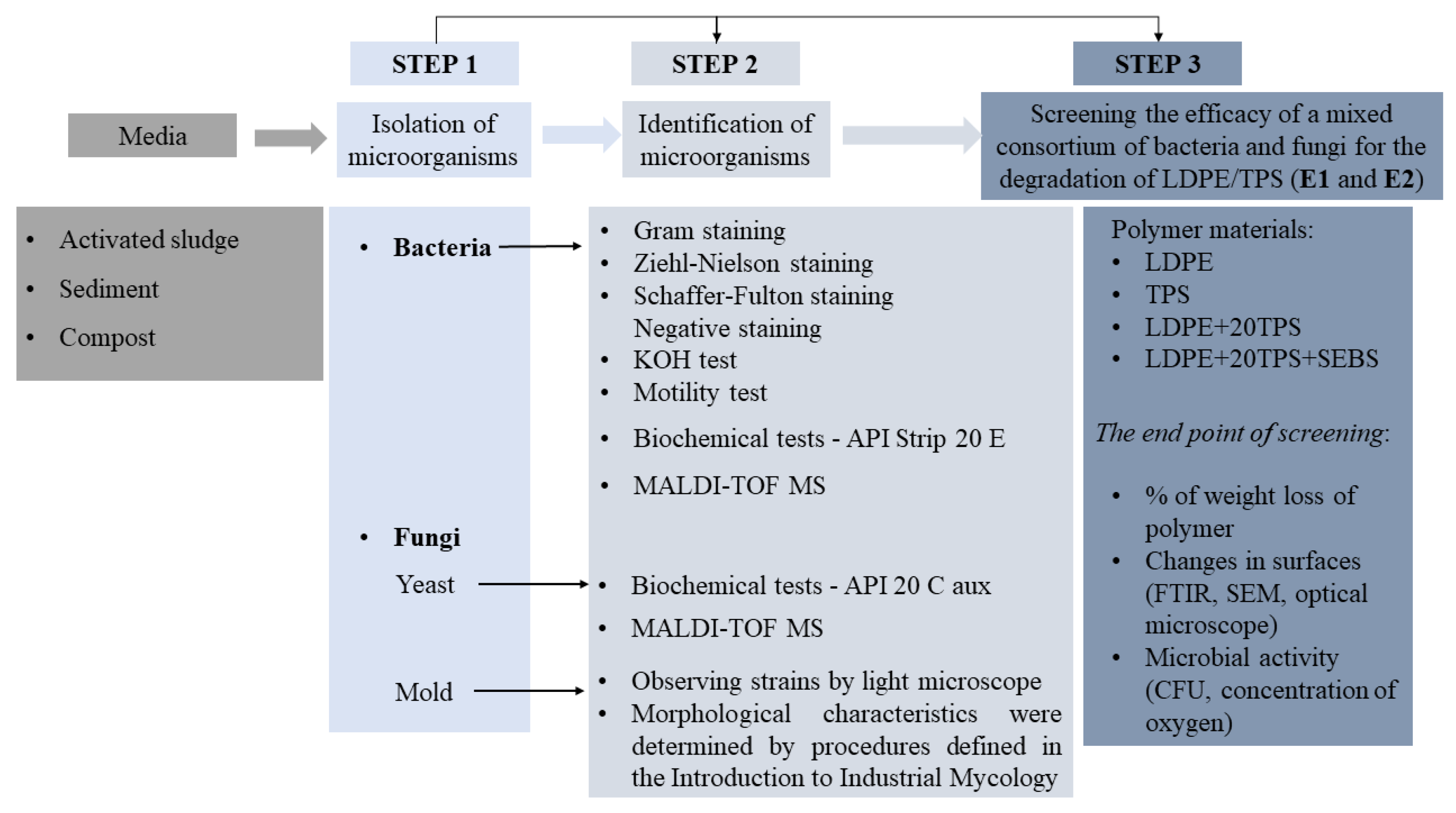
2. Materials and Methods

2.1. Polymer Materials

The polymer materials, low-density polyethylene (LDPE), low-density polyethylene + thermoplastic starch (LDPE+20TPS), low-density polyethylene + thermoplastic starch (20 is the percentage of added TPS in the polymer blend) + styrene-ethylene-styrene (LDPE+20TPS+SEBS), and thermoplastic starch (TPS) were prepared in the form of films (1 × 1 cm) with a thickness of 1 mm. LDPE, Dow 150 E, with a melt flow index 0.25 g/10 min at 190 °C, was purchased from Dow Chemical Company, Midland, MI, USA. The native wheat starch (12.20% moisture) “Srpanjka” (harvest 2008) was obtained from the Agricultural Institute, Osijek, Croatia. The plasticizer, glycerol, was purchased from Gram Mol, Zagreb, Croatia. Maleic anhydride grafted styrene-ethylene-butylene-styrene block copolymer (SEBS-g-MA), Kraton FG 1901X, was manufactured by Shell Chemicals Company, London, UK, and used as a compatibilizer in this study. SEBS-gMA as a triblock polymer was grafted with 2 wt% maleic anhydride. According to the manufacturer, the ratio of styrene/ethylene-butylene is 28/72. The preparation and characterization of the given materials was published in a previous paper [12]. These polymer materials were analyses by light microscope, FTIR, and SEM analysis before and after the experiments.

2.2. Environmental Samples

Microorganisms were isolated from activated sludge, compost, and river sediment. Activated sludge was collected from the municipal wastewater treatment plant Vrgorac—Dalmatia Country, Croatia. Activated sludge contained 98% of water. Characteristics of activated sludge were given in a previously published study by Kučić Grgić et al. [18]. The compost which was used was obtained by the composting process of biodegradable municipal solid waste selected from diverse locations of Zagreb County, Croatia. The characterization of compost was given in a previously published study by Kučić Grgić et al. [24]. River sediment was collected from river Kupa, Karlovac County, Croatia.

2.3. Isolation of Microorganisms from the Environment Samples

The microorganisms were isolated from three different media: activated sludge, river sediment, and compost. Activated sludge and river sediment (V = 100 mL) were placed in sterilized 250 mL Erlenmeyer flasks and shaken at room temperature on the thermostatic rotary shaker at 160 rpm for 24 h. Compost eluate was prepared according to ISO 21268-3:2019, i.e., 10 g of compost (dry matter) was added to 100 mL of sterilized deionized water and shaken for 24 h on the thermostatic shaker. After 24 h the colony-forming units (CFU) of bacteria and fungi were determined on the general-purpose media (nutrient agar (NA)) for bacteria and malt agar (MA) for fungi by the pour plate method according to Briški et al. [25]. For the plate count, a dilution series (0.9% mass of aqueous NaCl solution) was prepared from each sample. The plates were incubated in 80% relative humidity at 28 °C to cultivate the fungi and at 37 °C to cultivate the bacteria. Bacteria were cultured at 37 °C because mesophilic bacteria grow fastest at 37 °C. All isolated bacterial cultures were also cultured at 25 °C to confirm their growth under the conditions in which polymer biodegradation was studied. After incubation, the number of colonies on agar plates was determined. The results were expressed as CFU of bacteria and fungi per mL. Bacterial and fungi colonies which were morphologically different, and which were dominant on NA and MA plates, were collected and transferred onto the NA and MA plates and incubated at 37 °C 24–48 h and 28 °C 3–5 days, respectively [25]. Transfer to the new plates was performed repeatedly until a pure isolate was obtained. After the pure isolates were obtained, they were stored in slant plants for characterizations, Figure 1. Isolates were marked as I1–I15.

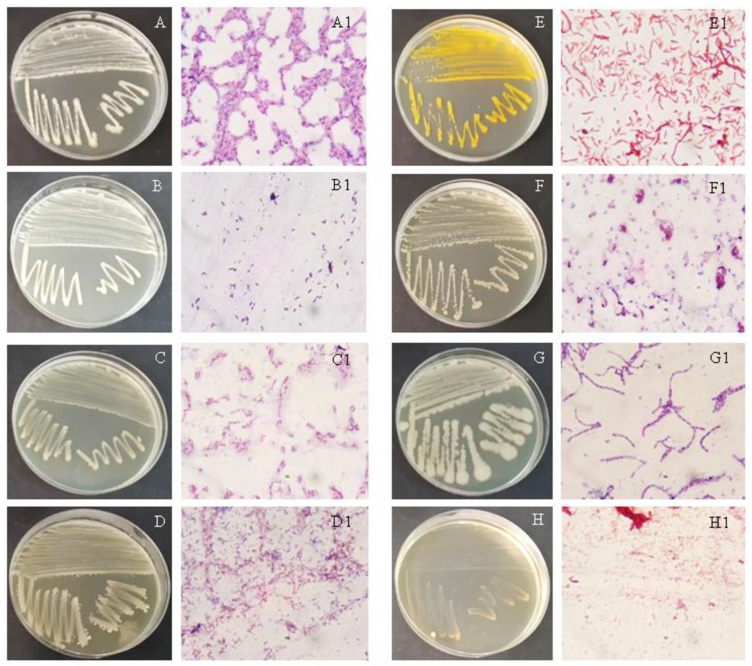
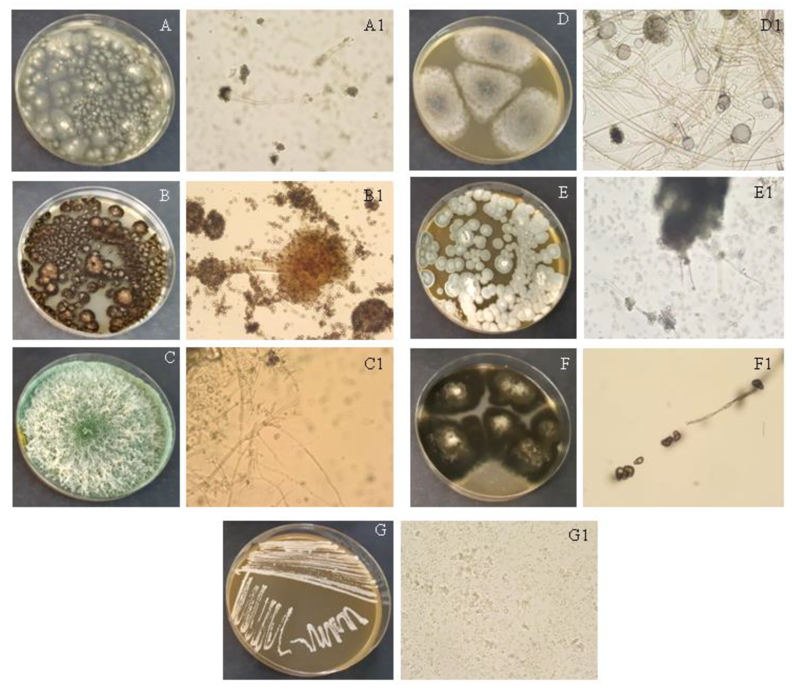
2.4. Identification of Isolated Cultures

Firstly, obtained pure cultures (bacteria, fungi (yeast and mold)) were observed by their growth on agar plates (appearance, color, and shape of colonies) [26] and the cell morphology were observed using a light microscope (Olympus b 201, Tokyo, Japan). Characteristics of isolated bacteria were determinate using the procedures defined in the Manual of Determinative Bacteriology, such as Gram staining, Ziehl-Nielson staining, Schaffer-Fulton staining, and Negative staining, and the KOH test and motility test [26]. After Gram staining, a series of biochemical tests known as API (Analytical Profile Index, BioMérieux®, Lyon, France) were carried out. Gram negative bacterium was identified using API Strip 20 E (Analytical Profile Index, BioMérieux®, Lyon, France). The final step of the identification of bacteria was a matrix-assisted laser desorption/ionization time of flight mass spectrometry (Microflex LT MALDI-TOF MS, Bruker Daltonics, Bremen, Germany) analysis, which is based on the protein identification of pulsed single ionic analytes (pure microbial culture), coupled with a TOF measuring mass analyzer, and the exact protein mass was determined.
Yeast was identified using API 20 C aux (Analytical Profile Index, BioMérieux®, Lyon, France) and MALDI-TOF MS analysis. Molds were identified by studying their growth, change of color during growth, and by observing strains by light microscope. Their morphological characteristics were determined by the procedures defined in the Introduction to Industrial Mycology [27].

2.5. Screening of Polymer-Degrading Bacteria and Fungi

Before carrying out the biodegradation experiment, mixed suspensions of bacteria and fungi were prepared for experiments E1 and E2. First, the bacterial/fungal cultures were inoculated onto nutrient/malt agar 48 h before the start of the biodegradation experiment and incubated at 37 °C (bacteria) or 28 °C (fungi). The grown bacterial/fungal colonies were harvested using a sterile inoculation loop, pooled, and transferred to a sterile Erlenmeyer flask containing 10 mL of physiological saline (0.9 mass % NaCl) to prepare a thick bacterial or fungal suspension [25]. A separate suspension was prepared for each culture with an initial CFU between 107 and 108. In experiments E1 and E2, the mixed culture suspensions contained 10 and 5 different bacterial and fungal cultures, respectively. Accordingly, in experiment E1, 1 mL of each bacterial or fungal suspension was transferred to Erlenmeyer flasks, and in experiment E2, 2 mL was transferred to obtain a total volume of 10 mL of the prepared suspension. All prepared suspensions were mixed well in the homogenizer. The initial CFU of the mixed cultures in the biodegradation experiments, E1 and E2, was approximately 106 cells/mL [24]. The initial optical density of the suspension was 0.2 and was determined spectrophotometrically at λ = 600 nm [13]. The CFU of the fungal spores was determined using the Thoma cell counting chamber (used to prepare the suspension) according to ISO 20391-1:2018.
The screening of polymer-degrading bacteria and fungi was conducted in 250-mL glass bioreactors, which were shaken on the thermostatic rotary shaker at 160 rpm for 56 days at room temperature. Each reactor contained 90 mL of mineral media [28], 10 mL of bacterial and fungi suspension, and one film (1 × 1 cm) of polymer materials (LDPE, TPS, LDPE+20%TPS, LDPE+20%TPS+SEBS). The working volume (Vr) in experiments with microbial consortium isolated from activated sludge and sediment (hereinafter referred to as the solution) (E1), and compost (E2) was 100 mL, respectively. A blank probe was also set up for the E1 and E2 experiments. These blank probes contained mineral medium and a prepared suspension of mixed cultures (E1 or E2) or polymers [8]. Blank probes without the suspension of microbial consortium contained mineral medium and different polymers (LDPE, TPS, LDPE+20% TPS, LDPE+20% TPS +SEBS). These blank probes (with polymers) were intended to exclude the possibility that physicochemical degradation was occurring. All initial conditions in the blank probes were the same as in the biodegradation experiments. The initial pH-value and temperature in E1 and E2 were 6.8 and 25 °C, respectively. The initial working conditions are given in Table 1.
During the experiments, at 0, 14, 28, 42, and 56 day, CFU, the concentration of dissolved oxygen (DO), temperature (T), and pH-value were determined. The concentration of DO, T, and pH-value were measured with a DO electrode and pH electrode connected to the WTW Multi 340i, WTW, Weilheim, Germany. At the end of experiments E1 and E2, the weight loss of each polymer was determinate and the polymer surface was characterized by optical microscope, FTIR, and SEM. Before conducting the above examinations on the polymer’s surface, the polymer materials were washed with 75% ethanol and with deionized water and dried at room temperature for 48 h to remove organic particles [8]. The weight loss (WL) of each polymer material was determinate according to the following equation, Equation (1) [12]:
W L   ( % ) = m 0 m 1 m 0 × 100
where m0 is the mass of polymer film at the beginning of the screening, and m1 is mass of polymer film at the end of the screening.

2.5.1. Light Microscopy

The morphology of each sample before and after composting was characterized by light microscopy (LM). LM was performed with a stereomicroscope SMZ-2T (Nikon, Tokyo, Japan). The samples for the LM were the original thin plates (average thickness 400 μm), which were observed both before and after the degradation experiments using episcopic illumination (reflected light) [12].

2.5.2. Scanning Electron Microscopy

The outer surface and internal morphology of the samples before and after degradation were characterized by scanning electron microscopy (SEM; microscope MAIA3, TESCAN, Brno–Kohoutovice, Czech Republic) [12,16]. The outer surfaces of the samples were observed “as received”. The internal morphologies were observed on fracture surfaces (the samples were submerged in liquid nitrogen, left to equilibrate for at least 5 min, and fractured). Before observation in the SEM microscope, the samples were fixed on the brass stubs with a conductive silver paste (paste Silver DAG 1415, Elmi Consumables, Písek, Czech Republic) and sputter coated with a thin platinum layer (vacuum sputter coater SCD 050, Leica, Wien, Austria) in order to minimize charging and electron beam damage. All micrographs were taken at an accelerating voltage of 3 kV using secondary electron imaging.

2.5.3. FTIR Spectroscopy

ATR FTIR spectra were measured on a Thermo Nicolet Nexus 870 spectrometer, Canada, USA. For all samples, we measured the top surfaces before and after composting using a horizontal micro-ATR Golden Gate unit (SPECAC) with a diamond prism. The ATR FTIR spectra were processed by the advanced ATR correction using the OMNIC software. Each sample was measured at least four times and only the most representative spectra were selected [12,16].

3. Results and Discussion

3.1. Isolation and Identification of Microorganisms

Microorganisms were isolated from activated sludge, compost, and sediment samples and further identified. The isolation was made by serial dilutions (from 10−1 to 10−7) by which the most frequent microorganisms through dilutions were purified by the streaking method. Bacteria and fungi (molds and yeast) were isolated from the investigated samples. In comparison to three investigated media, the highest number of microorganisms was in the sample with compost. The most frequent microorganisms in all three samples were bacteria and molds. Table 2 displays microorganisms which were isolated from activated sludge, sediment, and compost.
After obtaining pure cultures, the next step was to identify each isolated microorganism. Pure bacterial cultures were streaked on NA plates to study their morphological characteristics (Table 3). I1–I8 were bacteria isolated from the mentioned three samples (Table 2). Bacteria grown on NA plates as well as microscopic photographs of bacteria stained by Gram are given in Figure 2A–H. According to the cell morphology observed with the light microscope, all bacteria, I1–I8, were rod-shaped. This was achieved by Gram staining, which was used to classify microorganisms according to their cell wall structure. Isolates I1–I7 (Table 4) were Gram-positive, except for one which was Gram-negative (I8) due to red colored cells after staining (Figure 2H). These results were confirmed by a simple and rapid KOH test, which detected bacterial fibers for the Gram-negative bacterium. KOH penetrates through the cell and cell material (DNA) forming strings by lifting the microbiological eyelet [29]. The additional conformation of cell wall structure can be achieved by Ziehl-Neelson staining. It is a useful staining method for the further characterization of a microorganism’s cell wall structure (referring on lipid content, especially on a high content of mycolic acids). After staining, the acid-fast bacilli stain bright red on the blue background. Isolates I1, I3, I5, and I6 were acid fast, suggesting that lipid content is part of the cell wall [29]. On the other hand, I2 and I8 were not acid-fast; I4 and I7 were variable because some cells were slightly blue, and others stained red/pink. A thicker cell wall allows bacteria to adopt and survive extreme or adverse conditions [30]. The acid-fast bacteria are those Gram-positive bacteria whose cell walls have a high lipid content, i.e., a lipoid capsule with a high molecular weight that is waxy at room temperature [31]. In addition, Schaeffer-Fulton staining allows for the visualization of spores that confer resistance of microorganisms to various pollutants and conditions [32]. Endospores are distinguished from red vegetative cells (when using the counterstain safranin) by their green color. Bacteria isolated from activated sludge and compost form endospores according to the Schaeffer-Fulton staining. All investigated isolates (I1–I7) had the capability to form endospores, except the isolate I8. Bacterial endospores are multilayered structures that allow bacteria to adapt and survive in different environmental conditions, such as heat, starvation, radiation, UV light, the presence of pollutants toxic to the cells, desiccation, etc. [33]. The ability to sporulate is of great importance for bacteria, especially in the environment, as they can survive for a long time under extreme conditions.
However, the last staining used in this study was negative straining. It is the indicator for capsule formation which is the defense barrier [29]. The bacteria forming a capsule take on a blue color on a black background. According to the negative staining, isolates I1–I6 were variable for the determination of the capsule because some blue and colorless cells were noticed after staining. I7 and I8 do not form capsules. It is noteworthy that growth conditions have significant influence on the formation of capsules [34]. According to the motility test with semisolid agar and the stab technique, all isolates I1–I8 are motile (Table 4). Motility is an important property of bacteria enabled by flagella. It allows bacteria to move from the harmful to the favorable environment [33].
Further tests were biochemical tests that are useful for the exploration of the enzymatic content of bacteria. Results of these tests are listed in Table 4. Catalase is an enzyme that allows bacteria to have resistance to H2O2, oxidase is an enzyme that defends cells from oxidative stress, and nitrate-reductase induces the reduction of nitrate to nitrite. Enzymes are key factors for biochemical reactions and allow microorganisms to degrade pollutants.
For the identification of a Gram-negative and rod-shaped bacterium, isolate I8, a simple and quick performing API strip 20 E was used. After 24–48 h of incubation at 37 °C, with the usage of additional reagents and a base of data, the bacterium was identified as bacterium genera of Pseudomonas. The conformation of the genera identified by API and the last step of the identification process was a MALDI-TOF analysis by which bacterium Pseudomonas alcaligenes was identified (Table 2).
The final step of bacterial identification was MALDI-TOF MS analysis (Table 2) which is based on the protein identification of pulsed single ionic analytes (pure microbial culture), coupled with a TOF measuring mass analyzer and the exact protein mass is determined [35]. Most of the bacteria were Bacillus genera that are Gram-positive, rod-shaped, motile, occur in pairs or chains, and spore-forming and, due to this, these bacteria can survive and adjust to different conditions [36]. Bacteria of Bacillus genera are applicable for various purposes, such as in the production of industrial enzymes, bioinsecticides, antibiotics, and others, with usage in industry, agriculture, and in the medical field [37]. According to the previous study, Bacillus sp. and Paenibacillus sp. were isolated from the municipal landfill sediment [38]. Moreover, bacterium genera of Lysinibacillus and Bacillus cereus were isolated from the wastewater and activated sludge that is in agreement with this study [39]. Kalaivani et al. [40] isolated from the municipal solid waste the genera of bacteria Bacillus, Pseudomonas, Azotobacter, Azospirillum, and Lactobacillus.
The results of the identifications of molds are given in Figure 3. The identification of molds, isolates I9–I14 (Table 2), was carried out by studying the cell morphological characteristics by using the procedures defined in the Introduction to Industrial Mycology [27] and by the light microscope (Table 2 and Table 3).
According to Park and Kim [38], five fungal strains were isolated from the soil in which LDPE films were buried; four of them were Aspergillus sp. and one was Fusarium sp. Furthermore, Aspergillus sp., Frankia sp., Streptomyces sp., Trichoderma sp., and Penicillium sp. were isolated from the municipal solid waste [40].
Yeast, isolate I15, was isolated from the sediment (Table 2 and Table 3, Figure 2G). Wet smear was studied by light microscope with the purpose to determinate the cell morphology of the yeast; cells were oval or round with pseudohyphae. Another type of API used in this study was API 20 C aux for the yeast identification. After the suspension was prepared, it was inoculated into the walls and incubated at 28 °C for 72 h. The results were read from the fuzziness of the inoculated suspension. Candida parapsilosis was identified with 99.7% of probability; the identification was confirmed by MALDI-TOF analysis (Table 3). This pathogenic yeast can be isolated from human skin, soil, and marine environments [41]. This species of Candida is rich with various extracellular enzymes which are able to degrade large macromolecules into smaller ones [42] which makes Candida parapsilosis suitable for biodegradation of polymer materials.

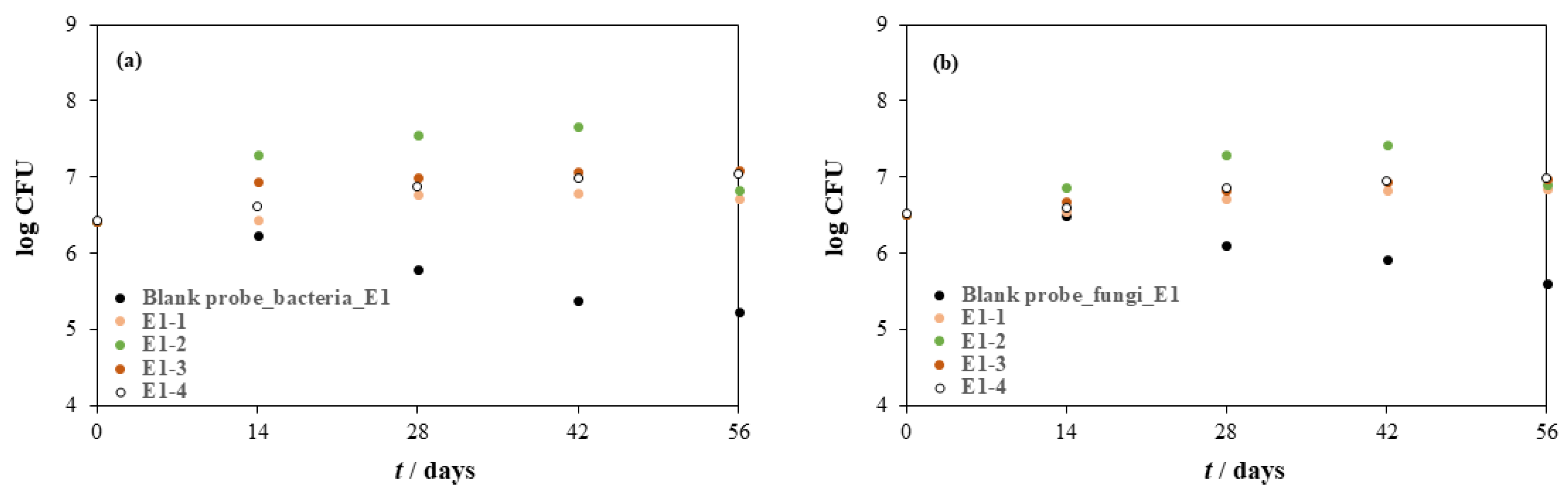
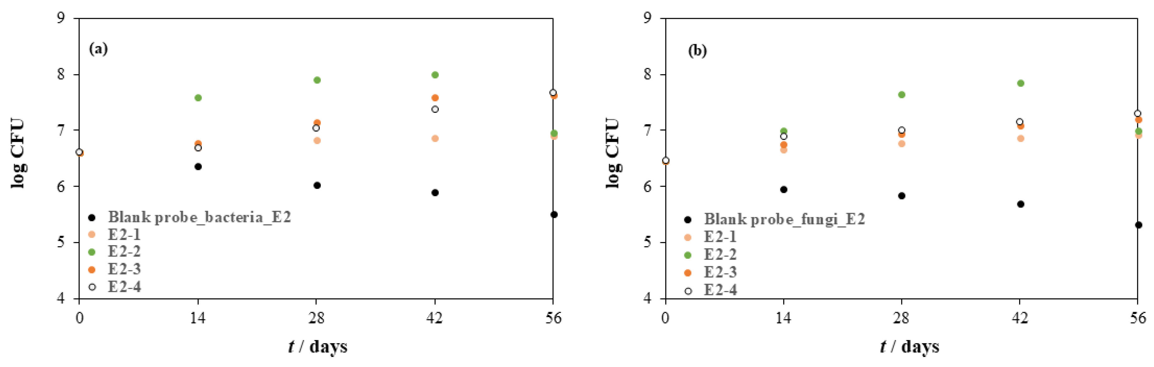
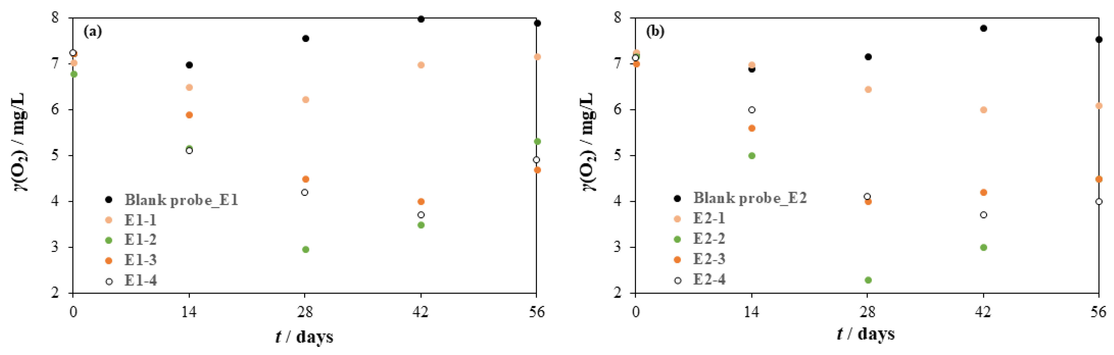
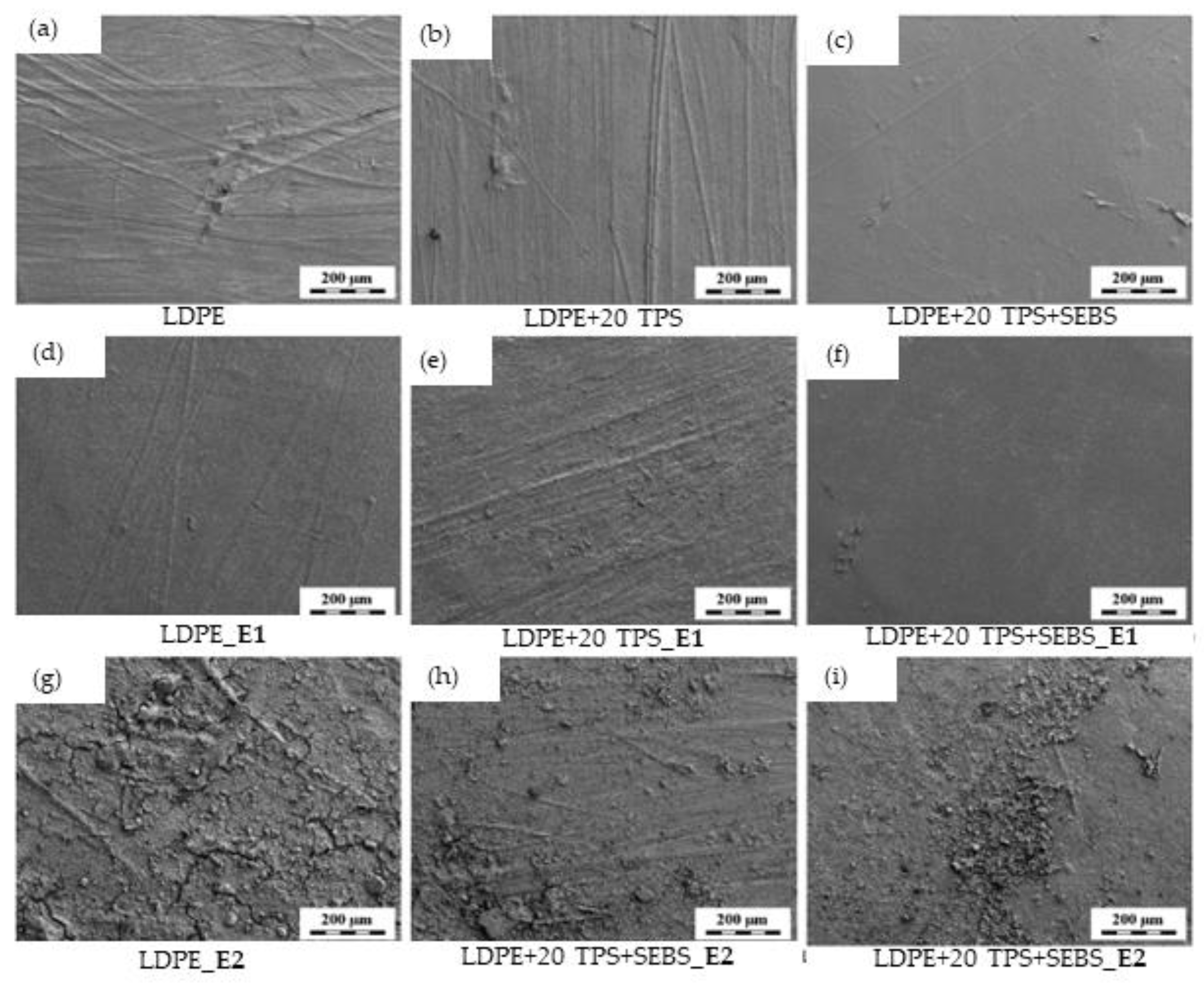
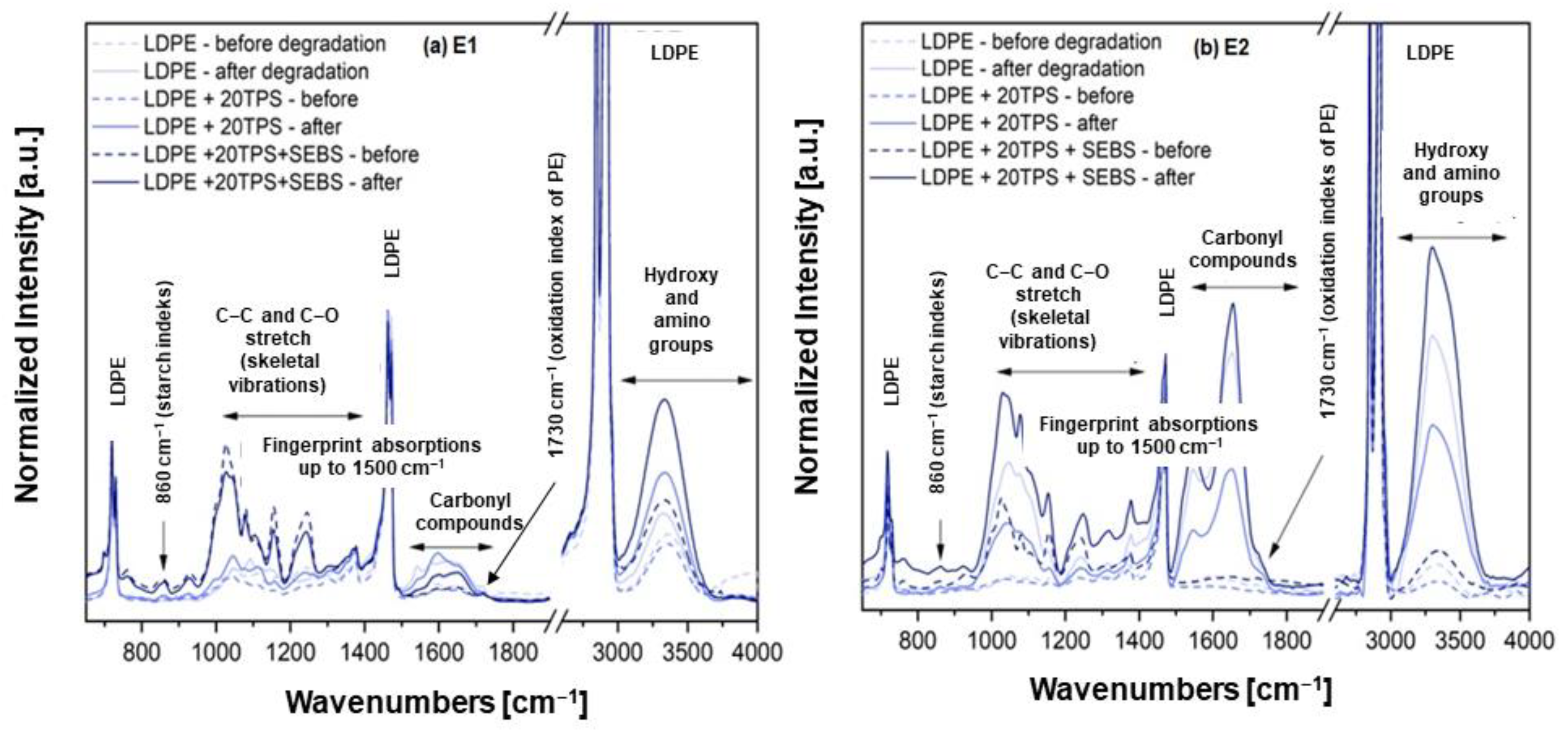
3.2. Biodegradation of Polymer Materials

The biodegradation of LDPE, LDPE+20TPS, LDPE+20TPS+SEBS, and TPS in experiment E1 was carried out by a microbial consortium isolated from activated sludge and river sediment: bacteria, Lysinibacillus massiliensis, Bacillus licheniformis, Bacillus indicus, Bacillus megaterium, Bacillus cereus, and Pseudomonas alcaligenes; molds, Aspergillus sp. 1, Penicillium sp., and Alternaria sp.; and yeast Candida parapsilosis. In contrast to experiment E1, the biodegradation of the polymers in experiment E2 was carried out by only two different bacterial species, Bacillus sonorensis, and Bacillus subtilis, and three different mold species, Aspergillus sp. 2, Trichoderma sp., and Rhizopus sp. Accordingly, it can be seen that in E1, the microbial consortium consisted of Gram-positive and negative bacteria, and molds and yeast, while in E2 the mixed culture consisted of Gram-positive bacteria and molds.
Changes in CFU during the biodegradation of LDPE, LDPE+20TPS, LDPE+20TPS+SEBS, and TPS in experiments E1 and E2 are shown in Figure 4 and Figure 5. In all experiments, E1-1, E1-2, E1-3, E1-4, E2-1, E2-2, E2-3, and E2-4, it was observed that the number of colonies formed by the bacteria and fungi was higher compared to the blank probes, indicating that the tested bacteria and fungi used LDPE, LDPE+20TPS, LDPE+20TPS+SEBS, and TPS as carbon and energy sources [43,44]. In addition, Figure 6 shows that the concentration of dissolved oxygen decreased during the process in all experiments (E1 and E2), except in the blank probes, which is another indication that the microorganisms were consuming oxygen due to biodegradation [45]. In all experiments, the lowest concentration of dissolved oxygen was on day 28, after which the concentration began to increase. As for the blank probes, the results show that the CFU of bacteria and fungi in the blank probes (Figure 5) (a blank containing a mineral medium and a suspension of the tested microorganisms, E1 and E2) of both experiments, E1 and E2, decreased with the longer duration of the experiment due to the lack of a carbon source, which is also confirmed by the slight increase in oxygen concentration (Figure 6). It is known that the more intensive the biodegradation, the more oxygen the microorganisms consume, and, at the same time, the oxygen concentration decreases. If no biodegradation takes place, the oxygen concentration changes slightly. Figure 4 and Figure 5 also show that the number of living cells of bacteria and fungi increased the most in the sample with pure TPS, which is not surprising since TPS is easily accessible to microorganisms [11]. In the LDPE samples, the number of viable bacterial/fungal cells also increased compared to the blank test, but the exponential growth phase was not as significant as in TPS. At the beginning of experiments E1-2, E1-1, E2-1, and E2-2, the number of live bacterial cells was about 3.0 × 10⁶ cell/mL. At the end of the experiments, E1-2, E1-1, E2-1, and E2-2, the numbers of live bacterial cells were 6.2 × 10⁶, 8.1 × 10⁶, 4.5 × 10⁷, and 9.8 × 10⁷ cell/mL, respectively, showing that the number of live bacterial cells in the sample with TPS increased significantly compared to pure LDPE. The same change was observed in the total number of live fungal cells. In experiments E1-3, E1-4, E2-3, and E2-4, where 20% TPS was added to the polymer matrix, the number of live cells of bacteria and fungi was higher than in the sample with pure LDPE, but no significant increase was recorded as in the pure TPS samples. These results are confirmed by the results of weight loss. It can be seen (Table 5) that the greatest weight loss occurred in experiments E1-2 and E2-2 (pure TPS). Starch is an easily degradable substrate for microorganisms, so it is not surprising that TPS was completely degraded. In the experiments without added starch, E1-1 and E2-1, the weight losses were 3.31% and 3.96%, respectively, while in the samples with added starch, E1-3 and E2-3, they were 14.11% and 20.45%, respectively. It can be concluded that TPS improves the biodegradability of the non-degradable polymer [12]. Comparing experiment E1 and E2 (Figure 4 and Figure 5, Table 5), it is noticeable that CFU in all experiments was slightly higher in E2 than in E1. The reason for this could be that there were fewer different types of microorganisms in experiment E2, so competition was not as prominent. In addition, the mixed consortium in experiment E2 were isolated from compost that was in contact with plastics, and it can be assumed that these microorganisms were adapted to the conditions with plastics. Furthermore, the genera of microorganisms present in the medium also play an important role in decomposition. Table 2 shows that different genera of microorganisms were isolated from activated sludge and sediment than from compost. According to the literature [45,46,47,48,49], most of the identified microorganisms (E1 and E2) have the ability to degrade/colonize polymeric materials. For example, the results of Kučić Grgić et al. [45] showed that Bacillus licheniformis and Lysinibacillus massiliensis can biodegrade LDPE and PS microparticles. According to Rani et al. [13], Bacillus licheniformis showed a LDPE degradation rate of 0.069 g per day−1, with a half-life of about 335.32 days to degrade LDPE strips. Research conducted by Suresh et al. [50] has shown that Bacillus cereus, which contains different enzymes such as nitrate reductase and catalase, can partially degrade LDPE over 90 days. In addition, studies by Mukherjee et al. [51] also showed that the bacteria Bacillus licheniformis and Lysinibacillus fusiformis can biodegrade LDPE. The genera Bacillus form endospores that enable them to survive under extreme conditions, such as high temperature, low pH, and under conditions of starvation [29]. Tamnou et al. [48] studied the biodegradation of polyethylene by the bacterium Pseudomonas aeruginosa at different environmental temperatures. The highest percentage weight loss of polyethylene was found to be 6.25% after 30 days at 44 °C. Numerous published papers document the biodegradation potential of fungal organisms towards plastics. Examples include the biodegradation of LDPE by Aspergillus spp. [52,53], Fusarium spp. [52,54], Trichoderma spp., and Penicillium spp. [55]. Munir et al. [47] investigated the biodegradation of LDPE using fungi, Trichoderma viride, and Aspergillus nomius. Results showed that both fungi, Trichoderma viride, and Aspergillus nomius, reduced the weight of LDPE film by 5.13% and 6.63%, respectively, after 45 days of cultivation. In addition, Zahari et al. [49] studied the biodegradation of LDPE and starch-based plastics (SBP) with the yeast Candida tropicalis and the bacterium Bacillus subtilis during an incubation period of 49 days. The percentage of weight losses of LDPE and SBP with C. tropicalis were 3.2% and 22.3%, respectively, while with B. subtilis they were 4.6% and 12.9%, respectively. An important prerequisite for biodegradation is the effective contact of the degrading microorganism and its extracellular enzyme machinery with the polymer, which is ensured by the colonization of the plastic surface with bacterial or fungal biofilms [46]. Microorganisms are able to alter the hydrophobicity of their cell surface to promote hydrophobic interactions with the highly hydrophobic polymer surface [46].
The morphology of selected samples (LDPE, TPS, LDPE/20TPS, and LDPE/20TPS/SEBS) before and after degradation in E1 and E2 were monitored by the LM and SEM. The samples with a LDPE matrix (i.e., LDPE, LDPE/20TPS, and LDPE/20TPS/SEBS) were just slightly degraded (as evidenced below), while the sample with a TPS matrix (i.e., neat TPS) was fully degraded in both the solution and compost (results of TPS after degradation not shown because no sample remained after degradation experiments).
LM micrographs of the samples with a LDPE matrix (figures are not shown) showed that the surfaces of all samples after decomposition in experiment E1 and E2 were compact, clean and without any observable changes. SEM micrographs of the same samples (Figure 7) confirmed that LDPE-matrix samples exhibited just minute degradation and the LDPE matrix remained compact, while the TPS samples were fully decomposed after degradation in both media, as mentioned above. The explanation is that the samples with the majority of LDPE produced co-continual structures with TPS particles within a polyolefin matrix, which prevented degradation, while the TPS consist only of polysaccharide chains, which can be completely degraded and/or decomposed. Fracture surfaces of the LDPE and LDPE/TPS, LDPE/TPS/SEBS blends with a LDPE matrix are shown in Figure 8. It is worth noting that SEBS-g-MA acted as a good compatibilizer that decreased interfacial adhesion, providing smaller TPS particles, which lead to a finer structure of the blends. Nevertheless, the fracture surfaces of the samples before and after degradation looked very similar and did not show any signs of degradation inside the polymer, which corresponded to our main conclusion that the degradation of the samples with a LDPE matrix occurred only on the surface.
Chemical changes on the surface of the LDPE, LDPE/20TPS, and LDPE/20TPS/SEBS systems before and after the degradation in experiments E1 and E2 were characterized by FTIR/ATR spectroscopy. Figure 9 compares representative spectra of systems before degradation (dashed lines) and after degradation (full lines) in experiments E1 and E2. All spectra were baseline-corrected and normalized to the maximum peak around 2920 cm−1, corresponding to aliphatic hydrocarbons [56]. All spectra contained strong peaks of aliphatic hydrocarbons (sharp and intensive bands typical of LDPE matrix around 720, 1470, and 2920 cm−1). Within the fingerprint region (400–1500 cm−1) we could observe a small-to-medium intensity vibration band typical of starch (860 cm−1) and the region of variable intensity (1000–1350 cm−1), which included a C–C stretch, C–O stretch, and skeletal vibrations. The results showed that peak intensities in the fingerprint region varied strongly from place to place for the samples degraded in experiment E2 (FTIR/ATR measurement is local and each sample was measured at least four times). This indicated that the fingerprint region is sensitive to surface contamination, which differed from place to place within a given sample, as confirmed by both LM and SEM results (Figure 7g–i). The intensity of the band at 860 cm−1 is proportional to the starch concentration and it can even be employed in the calculation of the starch index [57]. Nevertheless, the intensity of this band varied from place to place as well, reflecting the local concentration of starch. In the ATR spectra, the intensity indicates only the local starch content in the near-surface region. The intensity of the bands at 1000–1350 cm−1 appears to be related to the presence of SEBS compatibilizer, but also reflects the local starch content. The intensity of the band at 860 cm−1 and the bands at 1000–1350 cm−1 are indeed proportional. The content of SEBS compatibilizer was not enough to increase the band intensity in the range 1000–1350 cm−1.
During the oxidation process, functional groups, such as hydroxyl or carbonyl groups, could be formed via oxidation, which are known to be used in the TCA cycle or in the energy metabolism of bacteria, thereby increasing hydrophilicity [58,59]. The degradation of the samples was visible in the region of carbonyl groups (region 1500–1850 cm−1) and in the region of hydroxy and amino groups (region 3250–3650 cm−1). In both regions, the intensity of vibration bands after biodegradation (Figure 9, full lines) tended to be higher than the intensity of bands before biodegradation (Figure 9, dashed lines). Moreover, the average intensity in these regions was higher for samples degraded in E2 than for the samples degraded in E1, indicating faster degradation in experiment E2 (compare relative height of the bands with respect to LDPE bands at 720 and 1470 cm−1). In the region of carbonyl compounds (1500–1850 cm−1), bands appeared mostly at 1547 cm−1 and 1653 cm−1 for all samples after degradation. According to literature [56], these bands could be assigned to the COO carboxylates (1537 cm−1) and C=C double bonds (1653 cm−1). The bands around 1730 cm−1 (ketones at 1722 cm−1 and C=O ester groups at 1736 cm−1) were very weak; these bands are typical of the oxidation of polyolefins and employed in the calculation of their oxidation index [60,61]. Therefore, the increased number of oxygen atoms on the plastic surface in areas exhibiting microbial growth is direct evidence of LDPE degradation [62]. The broad band around 3330 cm−1 related to hydroxy or amino groups. The intensity of in this region varied strongly not only from sample to sample, but also from place to place. The increase in intensity could be explained by a higher local concentration of TPS (higher local concentration of –OH groups), while the decrease in intensity could have been caused by the possible stronger local degradation (local decrease in TPS concentration resulting in lower local concentration of –OH groups).
FTIR analysis (results are not shown) of the polymers in the blank probes showed that the polymers did not degrade during the 56 days, i.e., the polymers showed the same spectra as on day 0. This confirms that during the 56 days, UV light or some other physicochemical parameters had no influence on the decomposition of plastics.
The main results from FTIR/ATR can be summarized as follows: firstly, the very low intensity of the bands around 1730 cm−1 confirmed the results of LM and SEM microscopy, suggesting that the samples with a LDPE matrix exhibited just moderate surface degradation and negligible oxidative degradation under given conditions [60,61,63]. At the same time, the results suggest that the TPS phase was decomposed rather than oxidized during degradation. Secondly, the variable intensity of the bands around 3300 cm−1 (and the variable intensity of the bands in the fingerprint region) confirmed that the surface degradation of the samples differed from place to place, depending on the local conditions, the local contact with the medium, and perhaps also with the local concentration of starch. Again, this corresponded with the LM and SEM micrographs for samples decomposed in experiments E2, which displayed different damage of sample surfaces at various locations. Finally, the FTIR/ATR data proved that the degradation in E2 was more intensive in comparison with the degradation in E1.

4. Conclusions

Environmental samples represent diverse media with various microorganisms. Microorganisms that are able to adapt to different conditions play an important role in maintaining the ecosystem. In this study, the diversity of microorganisms present in activated sludge, compost, and sediment during exposure to polymeric materials was isolated and identified. The microorganisms isolated were bacteria of the genus Bacillus and a Gram-negative Pseudomonas, while the molds were of the genera Aspergillus, Rhizopus, Alternaria, Penicillium, and Trichoderma, and the yeast was Candida parapsilosis. The results of the screening test show that the microbial consortium of bacteria and fungi isolated from activated sludge and river sediment (E1) and compost (E2) have the ability to degrade prepared films. However, most of material is still intact and the addition of TPS does not significantly prevent or reduce plastic pollution. For this purpose, further research and the development of new biodegradable and compostable materials are needed. In addition, it is necessary to examine the efficiency of each pure culture in degrading LDPE/TPS and compare it with the efficiency of the mixed culture.

Author Contributions

Conceptualization, Š.U., D.K.G. and V.O.B.; writing—original draft preparation, M.M. and V.G.; writing—review and editing, D.K.G., M.S. and Š.U. All authors have read and agreed to the published version of the manuscript.

Funding

The authors would like to acknowledge the financial support of Croatian Science Foundation, project Advanced Water Treatment Technologies for Microplastics Removal (AdWaTMiR) (IP-2019-04-9661).

Institutional Review Board Statement

Not applicable.

Informed Consent Statement

Not applicable.

Data Availability Statement

Data are contained within the article.

Conflicts of Interest

The authors declare no conflict of interest.

References

  1. Sangale, M.K.; Shahnawaz, M.; Ade, A.B. A review on biodegradation of polythene: The microbial approach. J. Bioremed. Biodegrad. 2012, 3, 164. [Google Scholar] [CrossRef]
  2. Wilkes, R.A.; Aristilde, L. Degradation and metabolism of synthetic plastics and associated products by Pseudomonas sp.: Capabilities and challenges. J. Appl. Microbiol. 2017, 123, 582–593. [Google Scholar] [CrossRef] [PubMed] [Green Version]
  3. Auta, H.S.; Emenike, C.U.; Fauziah, S.H. Screening of Bacillus strains isolated from mangrove ecosystems in Peninsular Malaysis for microplastic degradation. Environ. Pollut. 2017, 231, 1552–1559. [Google Scholar] [CrossRef] [PubMed]
  4. Carr, S.A.; Liu, J.; Tesoro, A.G. Transport and fate of microplastic particles in wastewater treatment plants. Water Res. 2016, 91, 174–182. [Google Scholar] [CrossRef] [PubMed]
  5. Geyer, R.; Jambeck, J.R.; Law, K.L. Production, use, and fate of all plastic ever made. Sci. Adv. 2017, 3, e1700782. [Google Scholar] [CrossRef] [PubMed] [Green Version]
  6. Boucher, C.; Morin, M.; Bendell, L.I. The influence of cosmetic microbeads on the sorptive behavior of cadmium and lead within intertidal sediments: A laboratory study. Reg. Stud. Mar. Sci. 2016, 3, 1–7. [Google Scholar] [CrossRef]
  7. Miloloža, M.; Kučić Grgić, D.; Bolanča, T.; Ukić, Š.; Cvetnić, M.; Ocelić Bulatović, V.; Dionysiou, D.D.; Kušić, H. Ecotoxicological assessment of microplastics in freshwater sources—A review. Water 2021, 13, 56. [Google Scholar] [CrossRef]
  8. Miloloža, M.; Bule, K.; Prevarić, V.; Cvetnić, M.; Ukić, Š.; Bolanča, T.; Kučić Grgić, D. Assessment of the influence of size and concentration on the ecotoxicity of microplastics to microalgae Scenedesmus sp., bacterium Pseudomonas putida and yeast Saccharomyces cerevisiae. Polymers 2022, 14, 1246. [Google Scholar] [CrossRef]
  9. Bajsic, E.G.; Persic, A.; Jemric, T.; Buhin, J.; Kucic Grgic, D.; Zdraveva, E.; Zizek, K.; Holjevac Grguric, T. Preparation and characterization of polyethylene biocomposites reinforced by rice husk: Application as potential packaging material. Chemistry 2021, 3, 1344–1362. [Google Scholar] [CrossRef]
  10. Moustafa, H.; Guizani, C.; Dufresne, A. Sustainable biodegradable coffee grounds filler and its effect on the hydrophobicity, mechanical and thermal properties of biodegradable PBAT composites. J. Appl. Polym. Sci. 2016, 134, 44498. [Google Scholar] [CrossRef]
  11. Ocelić Bulatović, V.; Borković, I.; Kučić Grgić, D.; Jozinović, A. Thermal and mechanical properties of thermoplastic starch blends. Chem. Ind. 2018, 67, P21–P31. [Google Scholar] [CrossRef]
  12. Ocelić Bulatović, V.; Kučić Grgić, D.; Mandić, V.; Miloloža, M.; Dybal, J.; Gajdosova, V.; Slouf, M. Biodegradation of LDPE_TPS blends under controlled composting conditions. Polym. Bull. 2022, 177. [Google Scholar] [CrossRef]
  13. Rani, R.; Rathee, J.; Kumari, P.; Singh, N.P.; Santal, A.R. Biodegradation and detoxification of low-density polyethylene by an indigenous strain Bacillus licheniformis. J. Appl. Biol. Biotech. 2022, 10, 9–21. [Google Scholar] [CrossRef]
  14. Abioye, A.A.; Obuekwe, C.C. Investigation of the biodegradation of low-density polyethylene-starch bi-polymer blends. Results Eng. 2020, 5, 100090. [Google Scholar] [CrossRef]
  15. Tena-Salcido, C.S.; Rodríguez-González, F.J.; Méndez-Hernández, M.L.; Contreras-Esquivel, J.C. Effect of morphology on the biodegradation of thermoplastic starch in LDPE/TPS blends. Polym. Bull. 2008, 60, 677–688. [Google Scholar] [CrossRef]
  16. Kale, S.K.; Deshmukh, A.G.; Dudhare, M.S.; Patil, V.B. Microbial degradation of plastic: A review. J. Biochem. Technol. 2015, 6, 952–961. [Google Scholar]
  17. Venkatesh, S.; Mahboob, S.; Govindarajan, M.; Al-Ghanim, K.A.; Ahmed, Z.; Al-Mulhm, N.; Gayathri, R.; Vijayalakshmi, S. Microbial degradation of plastics: Sustainable approach to tackling environmental threats facing big cities of the future. J. King Saud Univ. Sci. 2021, 33, 101362. [Google Scholar] [CrossRef]
  18. Kučić Grgić, D.; Bera, L.; Miloloža, M.; Cvetnić, M.; Ignjatić Zokić, T.; Miletić, B.; Leko, T.; Ocelić Bulatović, V. Treatment of active mud from urban wastewater treatment plants using the composting process. Hrvat. Vode 2020, 28, 1–8. [Google Scholar]
  19. Yan, S.; Subramanian, B.; Surampalli, R.Y.; Narasiah, S.; Tyagi, R.D. Isolation, characterization and identification of bacteria from activated sludge and soluble microbial products in wastewater treatment systems. Pract. Period. Hazard. Toxic Radioact. Waste Manag. 2007, 11, 240–258. [Google Scholar] [CrossRef]
  20. Sharifi-Yazdi, M.K.; Azimi, C.; Khalili, M.B. Isolation and identification of bacteria present in the activated sludge unit, in the treatment of industial waste water. Iranian J. Publ. Health 2001, 30, 91–94. [Google Scholar]
  21. Bhumbla, U.; Majumdar, S.; Jain, S.; Dalal, A.S. A study of isolation and identification of bacteria from lake water in and around Udaipur, Rajasthan. J Family Med Prim Care. 2020, 9, 751–754. [Google Scholar] [CrossRef] [PubMed]
  22. Spring, S.; Schulze, R.; Overmann, J.; Schleifer, K.-H. Identification and characterization of ecologically significant prokaryotes in the sediment of freshwater lakes: Molecular and cultivation studies. FEMS Microbiol. Rev. 2000, 24, 573–590. [Google Scholar] [CrossRef] [PubMed]
  23. Gogoi, G.; Sharma, P.; Buragohain, P.; Phukan, A.; Baruah, R. Isolation and characterization of compost accelerating microbes. Biosci. Trends 2015, 8, 5854–5859. [Google Scholar]
  24. Kučić Grgić, D.; Vuković Domanovac, M.; Domanovac, T.; Šabić, M.; Cvetnić, M.; Ocelić Bulatović, V. Influence of Bacillus subtilis and Pseudomonas aeruginosa BSW and clinoptilolite addition on the biowaste composting process. Arab. J. Sci. Eng. 2019, 44, 5399–5409. [Google Scholar] [CrossRef]
  25. Briški, F.; Kopčić, N.; Ćosić, I.; Kučić, D.; Vuković, M. Biodegradation of tobacco waste by composting: Genetic identification of nicotine-degrading bacteria and kinetic analysis of transformations in leachate. Chem. Pap. 2012, 66, 1103–1110. [Google Scholar] [CrossRef]
  26. Benson, H.J. Microbiological Applications, A Laboratory Manual in General Microbiology, 8th ed.; The McGraw-Hill Companies: New York, USA, 2001; pp. 154–156,160,168,169. [Google Scholar]
  27. Smith, G.; Onions, A.H.S.; Allsopp, D.; Eggins, H.O.W. Smith’s Introduction to Industrial Mycology, 7th ed.; Edward Arnold: London, UK, 1981; pp. 1–398. [Google Scholar]
  28. Kyaw, B.M.; Champakalakshmi, R.; Sakharkar, M.K.; Lim, C.S.; Sakharkar, K.R. Biodegradation of low density polyethylene (LDPE) by Pseudomonas species. Indian J. Microbiol. 2012, 52, 411–419. [Google Scholar] [CrossRef] [PubMed] [Green Version]
  29. Black, J.G. Microbiology: Principles and Explorations, 8th ed.; John Wiley & Sons: Hoboken, NJ, USA, 2012; pp. 68–71. [Google Scholar]
  30. Silhavy, T.J.; Kahne, D.; Walker, S. The bacterial cell envelope. Cold Spring Harb. Perspect. Biol. 2010, 2, a000414. [Google Scholar] [CrossRef] [PubMed]
  31. Yousif, M.Q.; Qasem, S.A. Tissue Processing and staining for histological analyses, Chapter 3. In Skin Tissue Engineering and Regenerative Medicine, 1st ed.; Albanna, M.Z., Holmes, J.H., Eds.; Elsevier: New York, NY, USA, 2016; pp. 49–59. [Google Scholar] [CrossRef]
  32. Oktari, A.; Supriatin, Y.; Kamal, M.; Syafrullah, H. The bacterial endospore stain on Schaeffer Fulton using variation of methylene blue solution. J. Phys. Conf. Ser. 2017, 812, 012066. [Google Scholar] [CrossRef] [Green Version]
  33. Pepper, I.L.; Gerba, C.P.; Gentry, T.J. Environmental Microbiology, Chapter 2. In Microorganisms Found in the Environment, 3rd ed.; Elsevier: New York, NY, USA, 2015; pp. 9–33. [Google Scholar] [CrossRef]
  34. Sue, D.; Hoffmaster, A.R.; Popovic, T.; Wilkins, P.P. Capsule production in Bacillus cereus strains associated with severe pneumonia. J. Clin. Microbiol. 2006, 44, 3426–3428. [Google Scholar] [CrossRef] [Green Version]
  35. Singhal, N.; Kumar, M.; Kanaujia, P.K.; Virdi, J.S. MALDI-TOF mass spectrometry: An emerging technology for microbial identification and diagnosis. Front. Microbiol. 2015, 6, 791. [Google Scholar] [CrossRef] [Green Version]
  36. Anbu, S.; Saranraj, P.; Padma, J. Synergetic effect of microbially fermented soybean and NPK fertilizer on plant growth of grand naine banana. J. Acad. Ind. Res. 2017, 6, 27–31. [Google Scholar]
  37. Du, Y.; Xu, Z.; Yu, G.; Liu, W.; Zhou, Q.; Yang, D.; Li, J.; Chen, L.; Zhang, Y.; Xue, C.; et al. A newly isolated Bacillus subtilis strain named WS-1 inhibited diarrhea and death caused by pathogenic Escherichia coli in newborn piglets. Front. Microbiol. 2019, 10, 1248. [Google Scholar] [CrossRef] [PubMed]
  38. Park, S.Y.; Kim, C.G. Biodegradation of micro-polyethylene particles by bacterial colonization of a mixed microbial consortium isolated from a land site. Chemosphere 2019, 222, 527–533. [Google Scholar] [CrossRef]
  39. Garcha, S.; Verma, N.; Brar, S.K. Isolation, characterization and identification of microorganisms from unorganized dairy sector wastewater and sludge samples and evaluation of their biodegradability. Water Res. Ind. 2016, 16, 19–28. [Google Scholar] [CrossRef] [Green Version]
  40. Kalaivani, S.; Kumar Sahu, A.; Shanthi, K. Screening and isolation of effective microbes from organic wastes for faster and effective degradation of bio-degradable municipal solid waste. In Proceedings of the International Conference on Green Technology and Environmental Conservation, Chennai, India, 15–17 December 2011; pp. 162–166. [Google Scholar] [CrossRef]
  41. Trofa, D.; Gacser, A.; Nosanchuk, J.D. Candida parapsilosis, an emerging fungal pathogen. Clin. Microbiol. Rev. 2008, 21, 606–625. [Google Scholar] [CrossRef] [PubMed] [Green Version]
  42. Neji, S.; Hadrich, I.; Trabelsi, H.; Abbes, S.; Cheikhrouhou, F.; Sellami, H.; Makni, F.; Ayadi, A. Virulence factors, antifungal susceptibility and molecular mechanisms of azole resistance among Candida parapsilosis complex isolates recovered from clinical specimens. J. Biomed. Sci. 2017, 24, 67. [Google Scholar] [CrossRef] [Green Version]
  43. Mor, R.; Sivan, A. Biofilm formation and partial biodegradation of polystyrene by the actinomycete Rhodococcus ruber. Biodegradation 2008, 19, 851. [Google Scholar] [CrossRef] [PubMed]
  44. Yoshida, S.; Hiraga, K.; Takehana, T.; Taniguchi, I.; Yamaji, H.; Maeda, Y.; Toyohara, K.; Miyamoto, K.; Kimura, Y.; Oda, K. A bacterium that degrades and assimilates poly(ethylene terephthalate). Science 2016, 351, 1196. [Google Scholar] [CrossRef]
  45. Kučić Grgić, D.; Miloloža, M.; Lovrinčić, E.; Kovačević, A.; Cvetnić, M.; Ocelić Bulatović, V.; Prevarić, V.; Bule, K.; Ukić, Š.; Markić, M.; et al. Bioremediation of MP-polluted waters using bacteria Bacillus licheniformis, Lysinibacillus massiliensis, and mixed culture of Bacillus sp. and Delftia acidovorans. Chem. Biochem. Eng. Q. 2021, 35, 205–224. [Google Scholar] [CrossRef]
  46. Malachová, K.; Novotný, Č.; Adamus, G.; Lotti, N.; Rybková, Z.; Soccio, M.; Šlosarčíková, P.; Verney, V.; Fava, F. Ability of Trichoderma hamatum isolated from plastics-polluted environments to attack petroleum-based synthetic polymer films. Processes 2020, 8, 467. [Google Scholar] [CrossRef] [Green Version]
  47. Munir, E.; Harefa, R.S.M.; Priyani, N.; Suryanto, D. Plastic degrading fungi Trichoderma viride and Aspergillus nomius isolated from local landfill soil in Medan. IOP Conf. Ser. Earth Environ. Sci. 2018, 126, 012145. [Google Scholar] [CrossRef]
  48. Tamnou, E.B.M.; Arfao, A.T.; Nougang, M.E.; Metsopkeng, C.S.; Ewoti, O.V.N.; Moungang, L.M.; Nana, P.A.; Takang-Etta, L.-R.A.; Perrière, F.; Sime-Ngando, T.; et al. Biodegradation of polyethylene by the bacterium Pseudomonas aeruginosa in acidic aquatic microcosm and effect of the environmental temperature. Environ. Chall. 2021, 3, 100056. [Google Scholar] [CrossRef]
  49. Zahari, N.Z.; Abdullah, S.N.; Tuah, P.M.; Cleophas, F.N. Biodegradation of low-density polyethylene (LDPE) and starch-based plastic (SBP) by thermophiles Bacillus subtilis and Candida tropicalis. IOP Conf. Ser. Mater. Sci. Eng. 2021, 1173, 012035. [Google Scholar] [CrossRef]
  50. Suresh, B.; Maruthamuthu, S.; Palanisamy, N.; Ragunathan, R.; Navaneetha Pandiyaraj, K.; Muralidharan, V.S. Investigation on biodegradability of polyethylene by Bacillus cereus strain Ma-Su isolated from compost soil. Res. J. Microbiol. 2001, 2, 292. [Google Scholar]
  51. Mukherjee, S.; Chaudhuri, U.R.; Kundu, P.P. Bio-degradation of polyethylene waste by simultaneous use of two bacteria: Bacillus licheniformis for production of bio-surfactant and Lysinibacillus fusformis for bio-degradation. RSC Adv. 2016, 6, 2982. [Google Scholar] [CrossRef]
  52. Das, M.P.; Kumar, S. Microbial deterioration of low density polyethylene by Aspergillus and Fusarium sp. Int. J. Chem. Technol. Res. 2014, 6, 299–305. [Google Scholar]
  53. Sindujaa, P.; Padmapriya, M.; Pramila, R.; Vijaya Ramesh, K. Bio-degradation of low density polyethylene (LDPE) by fungi isolated from marine water. Res. J. Biol. Sci. 2011, 6, 141–145. [Google Scholar] [CrossRef]
  54. Singh, J.; Gupta, K.C. Screening and identification of low density polyethylene (LDPE) degrading soil fungi isolated from polythene polluted sites around Gwalior city (M.P.). Int. J. Curr. Microbiol. Appl. Sci. 2014, 3, 443–448. [Google Scholar]
  55. Singh, V.; Dubey, M.; Bhadauria, S. Microbial degradation of polyethylene (low density) by Aspergillus fumigatus and Penicillium sp. Asian J. Exp. Biol. Sci. 2012, 3, 498–503. [Google Scholar]
  56. Coates, J. Interpretation of Infrared Spectra, A Practical Approach. In Encyclopedia of Analytical Chemistry; Meyers, R.A., Ed.; John Wiley & Sons: Chichester, NH, USA, 2000; pp. 10815–10837. [Google Scholar] [CrossRef]
  57. Tai, N.L.; Adhikari, R.; Shanks, R.; Adhikari, B. Aerobic biodegradation of starch–polyurethane flexible films under soil burial condition: Changes in physical structure and chemical composition. Int. Biodeterior. Biodegrad. 2019, 145, 104793. [Google Scholar] [CrossRef]
  58. Bode, H.B.; Zeeck, A.; Jendrossek, D. Physiological and chemical investigations into microbial degradation of synthetic poly(cis-1,4-isoprene). Appl. Environ. Microbiol. 2000, 66, 3680–3685. [Google Scholar] [CrossRef] [PubMed] [Green Version]
  59. Mooney, A.; Ward, P.G.; O’Connor, K.E. Microbial degradation of styrene: Biochemistry, molecular genetics, and perspectives for biotechnological applications. Appl. Microbiol. Biotechnol. 2006, 72, 1–10. [Google Scholar] [CrossRef] [PubMed]
  60. Slouf, M.; Synkova, H.; Baldrian, J.; Marek, A.; Kovarova, J.; Schmidt, P.; Dorschner, H.; Stephan, M.; Gohs, U. Structural changes of UHMWPE after e-beam irradiation and thermal treatment. J. Biomed. Mater. Res. Part B Appl. Biomater. 2008, 85B, 240–251. [Google Scholar] [CrossRef] [PubMed]
  61. Slouf, M.; Michalkova, D.; Gajdosova, V.; Dybal, J.; Pilar, J. Prooxidant activity of phenolic stabilizers in polyolefins during accelerated photooxidation. Polym. Degrad. Stab. 2019, 166, 307–324. [Google Scholar] [CrossRef]
  62. Kim, H.R.; Lee, H.M.; Yu, H.C.; Jeon, E.; Lee, S.; Li, J.; Kim, D.H. Biodegradation of polystyrene by Pseudomonas sp. isolated from the gut of superworms (larvae of Zophobas atratus). Environ. Sci. Technol. 2020, 54, 6987–6996. [Google Scholar] [CrossRef]
  63. Pilar, J.; Michalkova, D.; Slouf, M.; Vackova, T. Long-term accelerated weathering of HAS stabilized PE and PP plaques: Compliance of ESRI, IR and MH data characterizing heterogeneity of photooxidation. Polym. Degrad. Stab. 2015, 120, 114–121. [Google Scholar] [CrossRef]
Figure 1. Procedures for isolation, identification, and screening.
Separations 10 00079 g001
Figure 2. Obtained pure culture by streaking method and microphotographs of Gram staining of bacteria isolates I1 (A,A1), I2 (B,B1), I3 (C,C1), I4 (D,D1), I5 (E,E1), I6 (F,F1), I7 (G,G1), and I8 (H,H1), M = 1000×.
Separations 10 00079 g002
Figure 3. Obtained pure culture by streaking method and microphotographs of fungi isolates I9 (A,A1), I10 (B,B1), I11 (C,C1), I12 (D,D1), I13 (E,E1), I14 (F,F1), and I15 (G,G1), M = 400×.
Separations 10 00079 g003
Figure 4. Change of CFU of bacteria (a) and fungi (b) in experiments E1 during 56 days of biodegradation experiments.
Separations 10 00079 g004
Figure 5. Change of CFU of bacteria (a) and fungi (b) in experiments E2 during 56 days of biodegradation experiments.
Separations 10 00079 g005
Figure 6. Change of dissolved oxygen concentration in experiments (a) E1 and (b) E2 during 56 days of biodegradation experiments.
Separations 10 00079 g006
Figure 7. SEM micrographs showing surface of LDPE, LDPE+20 TPS, and LDPE+20 TPS+SEBS before (ac) and after degradation in experiments E1 (df) and in experiments E2 (gi).
Separations 10 00079 g007
Figure 8. SEM micrographs showing fracture surface of LDPE, LDPE+20 TPS, and LDPE+20 TPS+SEBS before (ac) and after degradation in experiments E1 (df) and in experiments E2 (gi).
Separations 10 00079 g008
Figure 9. FTIR spectra of LDPE, LDPE+20 TPS, and LDPE+20 TPS+SEBS before degradation (dashed lines) and after degradation (full lines) in experiments (a) E1 and (b) E2.
Separations 10 00079 g009
Table 1. The initial working conditions.
ExperimentMedia of Isolated MicroorganismsPolymer Materialm0 (Polymer)/gCFUbacteriaCFUfungi
E1-1Activated sludge and river sedimentLDPE0.11152.7 × 1063.3 × 106
E1-2TPS0.1080
E1-3LDPE+20%TPS0.1424
E1-4LDPE+20%TPS+SEBS0.1287
E2-1CompostLDPE0.09594.1 × 1062.9 × 106
E2-2TPS0.1381
E2-3LDPE+20%TPS0.1770
E2-4LDPE+20%TPS+SEBS0.1245
Table 2. The list of isolated and identified microorganisms from three environmental samples.
IsolatesGroup of MicroorganismsSampleIdentified Microorganisms
I1bacteriacompostBacillus sonorensis
I2Bacillus subtilis
I3activated sludgeLysinibacillus massiliensis
I4sedimentBacillus licheniformis
I5Bacillus indicus
I6Bacillus megaterium
I7activated sludgeBacillus cereus
I8sedimentPseudomonas alcaligenes
I9moldsedimentAspergillus sp. 1 (probably fumigatus)
I10compostAspergillus sp. 2 (probably niger)
I11Trichoderma sp.
I12Rhizopus sp.
I13sedimentPenicillium sp.
I14activated sludgeAlternaria sp.
I15yeastsedimentCandida parapsilosis
Table 3. Morphology of isolated colonies, I1–I15.
IsolatesMorphology of Isolated Colonies
I1Yellowish with flat elevation and irregular edges, rod shaped
I2White to slightly yellow, mucous, and flat with regular round configuration, rod shaped
I3White colonies with yellowish edges, mucous, shiny, and round configuration, rod shaped
I4White yellowish colonies, round and flat with jagged edges, rod shaped
I5Yellowish-orange colonies with regular round configuration, mucous and shiny, rod shaped
I6Yellowish to slightly brown colonies with round configuration, rod shaped
I7Large white to slightly grey, opaque colonies with jagged margins, rod shaped
I8Pale yellowish colonies, mucous with regular round configuration, rod shaped
I9Greyish green, rough with white center, septate hyphae, and black head
I10Formed black colonies with arachnoid structure, non-septate hyphae, black spores, and head from which spores are spreading radiate
I11Greenish with white arachnoid structure and widespread colony, septate hyphae, and head in the shape of flower
I12Greyish colonies with black center, false roots, non-septate hyphae and bow-shaped head
I13Colonies were greyish green, round configuration with white center, septate hyphae, and broom-shaped head
I14Black colonies arachnoid structure with white center, specific conidia, and septate hyphae
I15White, creamy, shiny, and very small with round configuration, cells were oval or round with pseudohyphae
Table 4. Results of staining, KOH test and biochemical tests for each isolate of bacteria.
IsolateGram StainingKOH TestZiehl-Nielson StainingSchaffer-Fulton StainingNegative StainingMotilityOxidaseCatalaseNitrate-Reductase
I1+ve+++/−++++
I2+ve++/−++++
I3+ve+++/-+++
I4+ve+/−++/−++++
I5+ve+++/−++/−
I6+ve+++/−+++
I7+ve+/−++++
I8−ve+++++
Table 5. Weight loss of polymer materials after screening test.
ExperimentPolymer Materialm0 (Polymer)/gm56 (Polymer)/gWL/%
E1-1LDPE0.11150.10783.3184
E1-2TPS0.10800.0000100.00
E1-3LDPE+20%TPS0.14240.122314.1152
E1-4LDPE+20%TPS+SEBS0.12870.108116.0062
E2-1LDPE0.09590.09213.9625
E2-2TPS0.13810.0000100.00
E2-3LDPE+20%TPS0.17700.140820.4520
E2-4LDPE+20%TPS+SEBS0.12450.097221.9277
Disclaimer/Publisher’s Note: The statements, opinions and data contained in all publications are solely those of the individual author(s) and contributor(s) and not of MDPI and/or the editor(s). MDPI and/or the editor(s) disclaim responsibility for any injury to people or property resulting from any ideas, methods, instructions or products referred to in the content.

