Article

Enhancing Trace Metal Extraction from Wastewater: Magnetic Activated Carbon as a High-Performance Sorbent for Inductively Coupled Plasma Optical Emission Spectrometry Analysis

by
Sergio J. Abellán-Martín
1,
David Villalgordo-Hernández
2,
Miguel Ángel Aguirre
1,*,
Enrique V. Ramos-Fernández
2,
Javier Narciso
2,3 and
Antonio Canals
1,*
1
Department of Analytical Chemistry and Food Science, University Institute of Materials, Faculty of Science, University of Alicante, P.O. Box 99, 03080 Alicante, Spain
2
Department of Inorganic Chemistry, University Institute of Materials, Faculty of Science, University of Alicante, P.O. Box 99, 03080 Alicante, Spain
3
Instituto de Investigación Sanitaria y Biomédica de Alicante (ISABIAL), 03010 Alicante, Spain
*
Authors to whom correspondence should be addressed.
Separations 2023, 10(11), 563; https://doi.org/10.3390/separations10110563
Submission received: 3 October 2023 / Revised: 6 November 2023 / Accepted: 8 November 2023 / Published: 10 November 2023

Abstract

:
A new fast, sensitive, and environmentally friendly analytical method has been developed for the simultaneous determination of Ba, Be, Cd, Cr, Cu, Fe, Hg, Mn, Ni, Pb, and Zn in wastewater samples using inductively coupled plasma optical emission spectroscopy (ICP OES). A preconcentration step using a magnetic dispersive solid-phase extraction (MDSPE) technique with a new magnetic sorbent was performed. The new sorbent material was a carbon containing magnetic cobalt and nitrogen groups. This material was synthetized using controlled pyrolysis of a zeolitic imidazolate framework (i.e., ZIF-67). In order to optimize the experimental parameters that affect the MDSPE procedure, a multivariate optimization strategy, using Plackett–Burman and circumscribed central composite designs (CCD), was used. The method has been evaluated employing optimized experimental conditions (i.e., sample weight, 10 g; sample pH, 7.6; amount of sorbent, 10 mg; dispersive agent, vortex; complexing agent concentration, 0.5%; ionic concentration, 0%; eluent, HCl; eluent concentration, 0.5 M; eluent volume, 300 μL; elution time, 3 min and extraction time, 3 min) using external calibration. Limits of detection (LODs) in a range from 0.073 to 1.3 μg L−1 were obtained, and the repeatability was evaluated at two different levels, resulting in relative standard deviations below 8% for both levels (n = 5). An increase in the sensitivity was observed due to the high enrichment factors (i.e., 3.2 to 13) obtained compared with direct ICP OES analysis. The method was also validated through carrying out recovery studies that employed a real wastewater sample and through the analysis of a certified reference material (ERM®-CA713). The recovery values obtained with the real wastewater were between 94 and 108% and between 90 and 109% for the analysis of ERM®-CA713, showing negligible matrix effects.

1. Introduction

An important effect of industrial processes and human activities is the pollution of several ecosystems [1,2,3]. In this regard, heavy metals pollution has risen in recent years and has become an ongoing issue for environmental organizations, being one of the most serious ecological concerns at present [4,5]. These pollutants may enter the human body through air, water, food, or absorption through the skin [6]. It is well known that exposure to certain heavy metals has been linked to a number of disorders that are harmful to human health since, some of them are not metabolized and can accumulate in soft tissues [7,8]. According to the World Health Organization (WHO), long-term exposure to environmental pollution, such as air, soil, and water pollution, causes around 25% of the illnesses that humans face today [9]. Therefore, the determination of these toxic metal ions in water and aquatic ecosystems is of great importance, as these contaminants are detrimental to the environment and have serious impacts on human health [10].
Various analytical techniques such as inductively coupled plasma optical emission spectrometry (ICP OES) [11,12,13], inductively coupled plasma mass spectrometry (ICP-MS) [14,15,16], electrothermal atomic absorption spectrometry (ETAAS) [17,18,19], flame atomic absorption spectrometry (FAAS) [20,21,22], and atomic fluorescence spectrometry (AFS) [23,24] have been applied for the determination of trace amounts of heavy metal ions in water and biological samples. Because of its superior sensitivity, broad dynamic linear range, reduced detection limits, excellent accuracy, and simultaneous multi-elemental analysis capacity, ICP OES is one of the most popular analytical techniques for elemental analysis [11,25]. Despite these benefits, trace metals concentration in real samples is extremely low and the complexity of matrices in real samples may substantially interfere with the determination of analytes [26,27]. Therefore, to solve such problems, it is necessary to develop simple, selective, reliable, efficient, and eco-friendly sample pretreatment methods for the extraction of trace amounts of heavy metals in environmental and biological samples before ICP OES determination [28]. Under these conditions, a separation and enrichment step such as liquid–liquid extraction (LLE) [29,30,31], coprecipitation [32,33,34], filtration [35], ion exchange [36,37,38], cloud point extraction (CPE) [39,40], or solid-phase extraction (SPE) [41,42,43,44] have been used prior to its determination.
Among the above-mentioned separation and preconcentration techniques, solid-phase extraction (SPE) is one of the most widely used sample pretreatment techniques [45]. SPE has some advantages over other sample pretreatment techniques due to its simplicity, reduced extraction time, greater enrichment factors, cheaper cost, and the reduction in the consumption of organic solvents [46,47]. The fundamental concept of SPE is the transfer of analytes from the liquid phase to the solid phase’s adsorption sites where they remain during the extraction process [48]. Then, both phases are separated by centrifugation or simply recovered by flotation [49,50]. After adsorption, the analytes retained in the solid sorbent can be eluted [51], thermally desorbed [52], or directly determined using a suitable analytical technique [53]. SPE has been extensively employed for the preconcentration of metals in polluted waters [54].
Despite all these advantages, there is growing interest in developing ecological analytical methods based on the Green Analytical Chemistry principles [55]. The Green Analytical Chemistry principles and the processes which are adhered to them aim to replace harmful chemicals, reduce waste in laboratories, and, as a result, miniaturize traditional analytical procedures. Furthermore, in recent years, interest in the sample preparation step has increased in relation to the principles of Green Analytical Chemistry. For that reason, the continuous search for innovative sample preparation methods has led to the creation of new procedures such as solid-phase microextraction (SPME) [56] and dispersive solid-phase extraction (DSPE) [57]. In DSPE, the solid sorbent is dispersed in the solution. This strategy encourages quick interaction of the analytes with the sorbent, minimizing the consumption of sorbent and leading to a faster extraction process in comparison with traditional SPE [58]. New advances in sample preparation that employ DSPE try to reduce the consumption of sorbents by using smaller amounts (i.e., microgram); this tendency has led to the development of dispersive micro-solid-phase extraction (DμSPE) [59].
Magnetic materials have recently been reported to be used as sorbents in several papers [6,60,61]. Magnetic dispersive solid-phase extraction (MDSPE) is gaining popularity because it decreases sample preparation time and allows for easier sorbent manipulation [62,63]. In MDSPE, the magnetic sorbent material is dispersed into the liquid phase; after the extraction, it is simply removed from the sample solution using a magnet [64], avoiding time-consuming filtration or centrifugation steps to carry out the phase separation [61,65,66]. The sorbents decorated with magnetic solids simultaneously combine the exceptional sorbent capability and the easy sorbent handling, reducing analysis time, sorbent amounts, and the consumption of solvents [67,68]. Finally, analytes can be desorbed employing an eluent solvent or heating for further analysis [69].
One of the most important MDSPE-related research areas at present is the quest for novel magnetic sorbent materials with high selectivity and strong adsorption capacity that are chemically and thermally stable, inexpensive, and environmentally benign [14,70].
Different magnetic sorbents have been used in magnetic dispersive solid-phase extraction, such as inorganic oxides, porous polymers, molecularly imprinted polymers (MIP), biosorbents, or carbon nanomaterials [71,72]. In addition, magnetic composites based on zeolitic materials have been described for the extraction and preconcentration of organic compounds and metals from different samples, including water, environment, food, and biological samples [6,73,74,75].
The aim of this work is to develop a new analytical method for the determination of trace metals (i.e., Ba, Be, Cd, Cr, Cu, Fe, Hg, Mn, Ni, Pb, and Zn) in wastewater samples through MDSPE using a new magnetic material developed specifically for this application. This material is carbon, containing magnetic cobalt and nitrogen groups. These groups can serve as anchoring centers for the metals and have been prepared using controlled pyrolysis of a zeolitic imidazolate framework (ZIF). These materials are composed of tetrahedrally coordinated transition metal ions (e.g., Fe, Co, Cu, Zn) that are connected by imidazolate linkers; this is further carbonized in order to obtain Co-doped magnetic carbon. More information about the employed material can be found in the bibliography [76]. To the best of our knowledge, the new magnetic material has not been employed for the determination of trace metals before.

2. Materials and Methods

2.1. Instrumentation

All the measurements were carried out using an Agilent 720 inductively coupled plasma optical emission spectrometer (Agilent Technologies, Melbourne, Australia). The operating conditions employed using the ICP OES are shown in Table S1. The instrumental parameters were optimized, accomplishing the maximum signal intensity for the set of emission lines evaluated. In addition, the sample uptake rate (i.e., 100 μL min−1) was the minimum liquid flow required in order to measure all emission lines analyzed. A pH meter Basic 20 (Crison Instrument, Barcelona, Spain) provided with a combined glass electrode was used for the pH measurements. In order to disperse the extractant into the sample, a Vortex Reax Top (Heidolph Instruments, Schwabach, Germany) was used. A neodymium magnet (1.32–1.37 T) was used to bring about the phase separation (Supermagnete, Gottmadingen, Germany). NemrodW statistical software (NemrodW® v.2007/2010, LPRAI, Marseille, France) and Statgraphics® Centurion (Statpoint Technologies, Warreton, VA, USA) were employed to construct the experimental designs and evaluate the optimization results and the desirability function. NemrodW was used to evaluate the screening and the central circumscribed design of the developed method and Statgraphics® Centurion was employed to construct the desirability function to obtain a common experimental condition for all the analytes evaluated.
Characterization of the porosity of the samples was performed using N2 adsorption–desorption isotherms as well as CO2 adsorption isotherms [77]. Before measurement, samples were outgassed at 150 °C during 4 h (Autosorb degasser, Quantachrome, Boynton Beach, FL, USA). The nitrogen adsorption–desorption isotherms were measured at −196 °C (Autosorb-6, Quantachrome). The Brunauer–Emmett–Teller (BET) equation was utilized for the estimation of the surface area, which was determined from the N2 adsorption branch. The number of points used to apply the BET equation was higher than five, and the BET constant was always positive. The Dubinin–Radushkevich method was utilized to obtain the micropore volume (Vmicro) and determine whether any diffusional restrictions in the adsorption took place. Pore volume has been obtained at P/P0 = 0.95. All data treatment related to sorbent characterization was carried out using the software Quadrawin (Quantachrome). The same device was used for the CO2 adsorption isotherms at 0 °C. No diffusional restrictions were observed. Powder X-Ray diffraction was utilized for the identification of crystallographic phases using a D8-Advanced diffractometer with a Goëbel mirror and a Kristalloflex K 760-80F X-Ray generation system, fitted with a Cu cathode and a Ni filter (Bruker, Billerica, MA, USA). Spectra were registered between 5° and 80° with an angular step of 0.05° and a three second step time. X-ray photoelectron spectroscopy (XPS) analysis was carried out in a VG-Microtech Multilab device with a MgKalpha (Hv:1253.6 eV) radiation source, pass energy of 50 eV, and background pressure of 5·10−7 Pa (VG-Microtech, Uckfield, UK). The spectra were carefully deconvoluted and the peak areas estimated through integration of each peak, prior subtraction of a Shirley background, and fitting the experimental peaks to a 30/70 proportion of a combination of Lorentzian/Gaussian lines. Field emission scanning electron microscopy (FESEM model Merlin VP Compact, Zeiss, Jena, Germany) with energy-dispersive X-ray spectroscopy (EDS, model Quantax 400, Bruker) in both backscattered electron (BSE) and secondary electron (SE) modes were employed to study the morphology of the samples. Raman spectroscopy characterization was carried out in an NRS-5100 device from Jasco (Tokyo, Japan) using a 633 nm He laser. Magnetic susceptibility measurements were carried out at 300 K in a magnetic field ranging from −30 to 30 kOe using a MPMS XL (SQUID) magnetometer from Quantum Design (San Diego, CA, USA). Finally, the zeta potential was measured using a Colloid Metrix Stabino II instrument (Meerbusch, Germany).

2.2. Samples and Reagents

Standard reference solutions of Hg and Be used for ICP OES calibration and for addition and recovery experiments were purchased from High Purity Standard (Charleston, SC, USA) with a concentration of 1000 mg L−1. Standard reference solutions of Ba, Cd, Cr, Cu, Fe, Mn, Ni, Pb, and Zn were obtained from Merck IV solution of 1000 mg L−1 (Merck, Darmstadt, Germany). Working solutions of analytes were prepared using appropriate dilution of the commercial standard reference solutions. To prepare the buffer solutions during the optimization and measurement process, H3PO4 85% purity (Scharlau, Barcelona, Spain) and KH2PO4 and K2HPO4 pro-analysis (Merck) were employed. Sodium diethyldithiocarbamate (DDTC, 99%, Merck) was used as a complexing agent. HCl 30% purity (Merck) and HNO3 65% purity (Scharlau) were employed as eluent phases during the measurement and optimization process. During the optimization process, NaCl pro-analysis (Scharlau) was used to adjust the ionic strength of the samples. All the experiments were prepared using ultrapure water with resistivity higher than 18.0 MΩ cm obtained using a PURELAB flex 3 purification system (Elga LabWater, High Wycombe, UK). Reagents employed during the synthesis of ZIF-67 were provided by Merck in analytical grade and were used without further purification.
The standard reference material employed in this work was ERM®-CA713 wastewater (Joint Research Center, European Commission, Geel, Belgium). A real water sample used was obtained from a wastewater treatment plant (Aguas Municipalizadas de Alicante E.M., Spain). Sample was collected in amber glass containers and then stored in the dark at 4 °C. To perform the analysis, the sample supernatant was collected and measured (i.e., dissolved metals) after decantation during 24 h without any sample digestion or filtration step.

2.3. Synthesis of the Magnetic Sorbent

The synthetic procedure followed the classical ZIF-67 synthesis [78,79], in which 144 mmol of 2-Methylimidazole were dissolved in 50 mL of deionized water. This solution was blended with a 25 mL deionized water solution in which 12 mmol of cobalt acetate were previously dissolved. The mixture was stirred vigorously for 5 min and kept at room temperature for 72 h. ZIF-67 was recovered by centrifugation at 12,000 rpm and washed three times with methanol. Then, the powder was dried at 60 °C for 24 h in a conventional oven. Lastly, the ZIF was put in a tubular furnace under a 100 mL min−1 N2 dynamic atmosphere, heated up at 5 °C min−1, and carbonized at 900 °C for 2 h. The carbon material was then washed with HCl following the procedure of Villalgordo-Hernández et al. [76] (i.e., ZIF67C_900_l meaning activated carbon obtained by carbonization at 900 °C and washed once with HCl). Previously, a screening study of different magnetic carbons was conducted, finding that this material was the most suitable for this specific application.

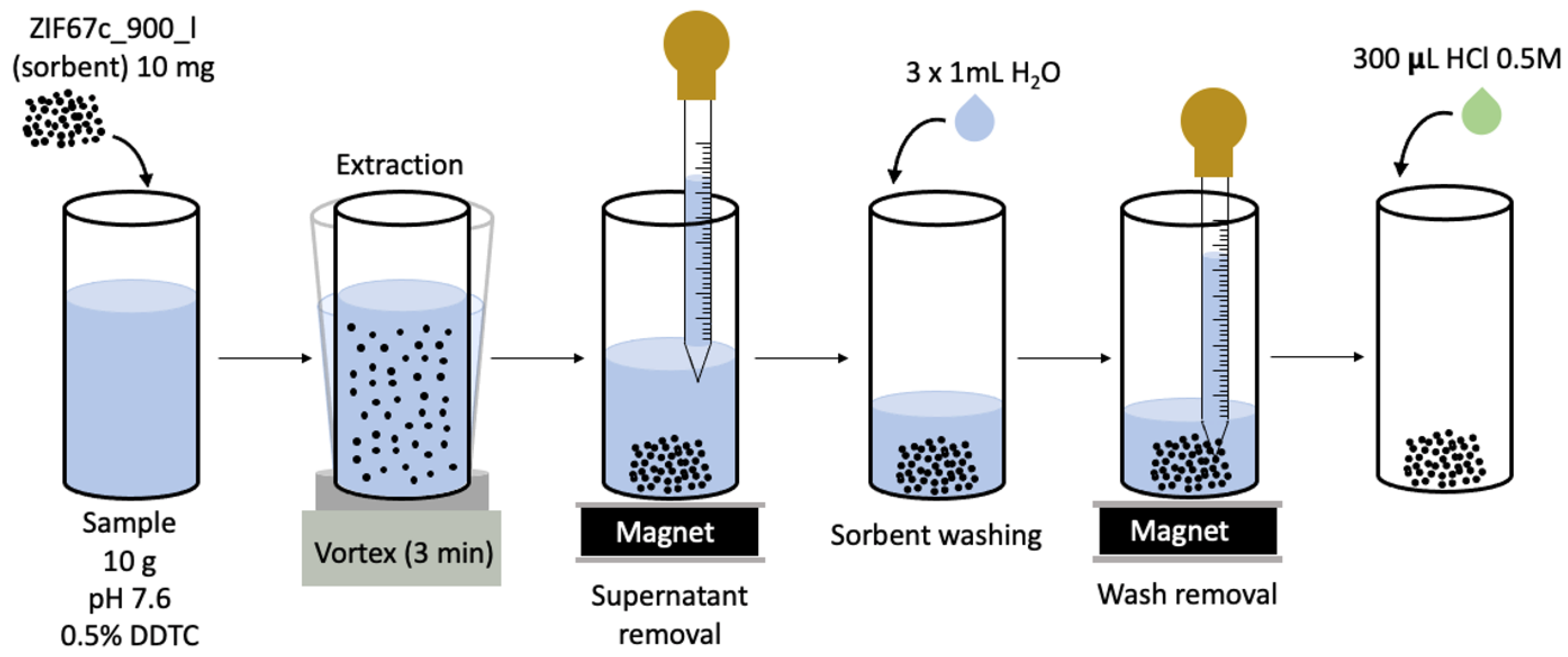
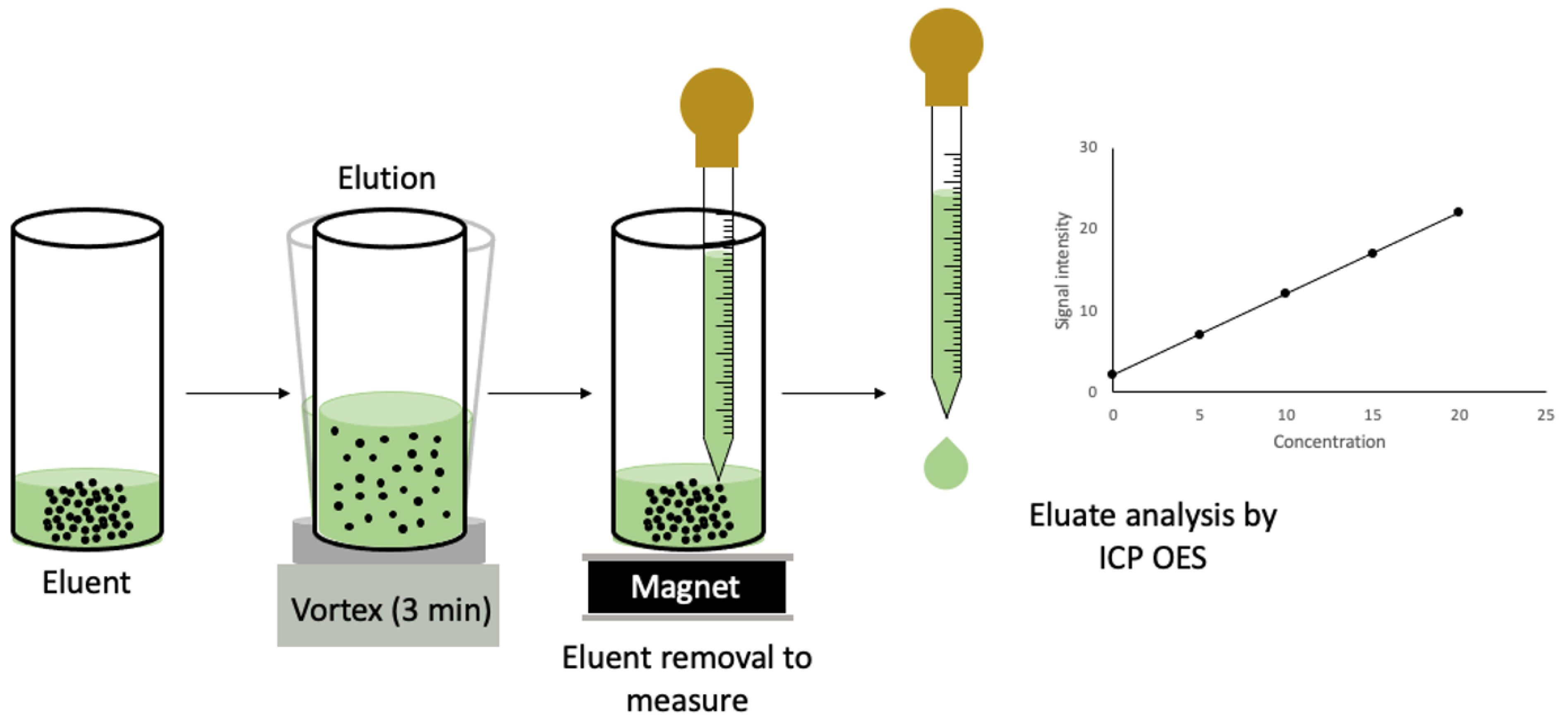
2.4. MDSPE Procedure

For the magnetic microextraction process, the first step was to adjust the pH of the sample or standards at 7.6 using the necessary amount of KH2PO4 and K2HPO4. Subsequently, DDTC was added as a complexing agent until its concentration was 0.5%. Once the sample (or standard) was prepared, it was transferred (i.e., 10 g) to a glass centrifugation tube and 10 mg of the magnetic solid extractant (ZIF67C_900_l) was introduced in the tube that contains the sample or standard. Then, the mixture was shaken using the vortex for 3 min to extract the analytes. After shaking, the supernatant (i.e., sample) was removed and the sorbent was washed 3 times with 1 mL of ultrapure water and the wash water was removed. Afterwards, 300 μL of HCl 0.5 M was added to the sorbent in order to elute the analytes. The elution process was carried out using the vortex for 3 min. Finally, the eluate was removed from the solution containing the sorbent and measured directly with the ICP OES. During the extraction, washing, and elution process, the neodymium magnet was used to ease the separation step between the solid and liquid phases. The entire MDSPE process is shown in Figure 1.

3. Result and Discussion

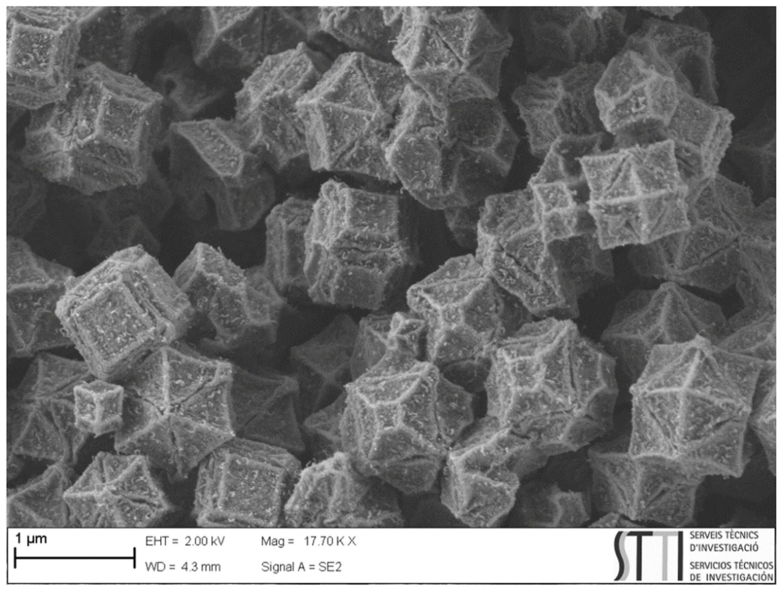
3.1. Characterization of ZIF67C_900_l

As presented in Figure S1, three diffraction peaks appear at 44.2°, 51.5°, and 75.9° in the XRD pattern, which could be indexed to Co(0) (JCPDS card No. 15-0806). Moreover, the peak found at 25.85° could be assigned to the (002) plane of graphite (JCPDS card No. 89-8487). In all cases, well-defined peaks can be observed, which indicate that both carbon and cobalt are crystalline, but there are crystallite sizes of less than 100 nm, since the peaks are very broad.
The N2 adsorption isotherm (Figure S2) of the original carbon and the washed one are very similar; in both cases, they show a very wide distribution of porosity, which is easily deduced from the wide shoulder that shows the adsorption. Although the specific surface area is not very high in either case (400 and 480 m2 g−1), the washed sample has an area that is 20% higher. It should also be noted that it presents a certain hysteresis (desorption branch above the adsorption branch); this indicates that we have ink-bottle-type pores that have been generated by acid washing the carbon due to the extraction of the unbound cobalt particles [76].
The secondary electron micrograph (Figure 2) shows the cobalt-doped carbon crystallites. The microstructure reminds us of the original ZIF, where there has simply been a contraction of the particles without losing their original cubo-octahedral shape [80]. In addition, it is possible to observe from the secondary electron micrograph that the particle size of the ZIF is 1 μm.
The XPS spectra shown in Figure S3 are very revealing. Firstly, we will analyze Co. The XPS spectrum shows the 2p1/2 transition where a peak associated with Co(0) and a peak at higher energy associated with Co(II) can be clearly seen. The presence of the satellites is unequivocal that we have Co(II) and not Co(III). A fact to highlight is that the presence of Co(0), which is what we have seen by other techniques, indicates that we only have a small nanometric layer of cobalt oxide. Regarding the nitrogen spectrum, we observe the 1s transition, where three peaks can be seen that we can associate with three N positions in the graphene sheet, as indicated in the figure.
The Raman spectrum shows four easily identifiable bands [81] as shown in Figure S4. First of all, highlight the two bands called G and 2D, which indicate that the carbon is not turbostratic but rather graphitic, as the intensity of the G band is much higher than that of the carbon. The 2D band indicates that there are stacks of more than 10 layers. The presence of the D band, also called the defects, is indicative that the graphene sheets have a considerable number of defects, and that there are areas where the carbon is not perfectly ordered.
Figure S5 shows the magnetic susceptibility of the studied ZIF67C_900_l material at 300 K in a magnetic field that ranges from -30 to 30 kOe. At the magnetization curve it is possible to observe a sigmoid shape that passes approximately through the origin having an extremely narrow hysteresis loop. In addition, the coercivity field (HC) and residual magnetization (MR) values are significantly small while saturation magnetization (Ms) is relatively high, the values are 200 KOe, 3.91 emu g−1, and 15.34 emu g−1, respectively, showing a ferromagnetic behavior [82].
To investigate the surface charges of the ZIF67C_900_l in the same matrix sample performed the MDSPE procedure, their zeta potentials were measured at different sample pH. The zeta potential of ZIF67C_900_l as function of the sample pH of the suspension is given in Figure S6. In the suspension with an initial pH of 4.0 the zeta potential was 20.3 mV. As can be seen it continuously decreases when the pH is increased up to a pH value of 7.0, where the zeta potential is stabilized; the isoelectric point of the magnetic material is 5.0. Therefore, the surface of the magnetic material is positively charged at pH values below the isoelectric point and negatively charged above this point. The negative charge surface of the magnetic sorbent is of great importance, since it affects the extraction process of positively charged metals. For that reason, the optimum pH value was 7.6 (Section 3.2) where the charge was negative (i.e., the material was deprotonated), increasing the interactions with metals and, thus, increasing the extraction efficiency.

3.2. MDSPE Multivariate Optimization

Since MDSPE is affected by many experimental variables, it is necessary to optimize the entire extraction process. Many experiments would have been necessary to optimize all experimental variables if they had been done through univariate optimization, increasing the consumption of sample, reagents, energy and time. To overcome this fact, multivariate optimization has recently emerged as an alternative to univariate optimization, since the number of experiments was significantly reduced. For that reason, a multivariate optimization design has been employed in order to obtain the optimal experimental conditions, considering the simultaneous determination of all analytes. The optimization process was carried out using a Plackett–Burman design as a screening approach to distinguish between significant and non-significant factors, followed by a central composite design (CCD) in order to obtain the optimal values of the significant factors. To perform the MDSPE optimization experiments a standard of 500 μg L−1 containing the elements was used. Then, the analytical signals obtained measuring with ICP OES were individually used as response function for optimization. In this way, individual optimum conditions were obtained for each analyte. In order to obtain a global optimum condition for all the analytes, a desirability function was employed to extract all the analytes with the best conditions.

3.2.1. Screening Study

The eleven factors that affect the MDSPE were selected based on previous experience and experiments carried out by our research group. The factors evaluated in the Plackett–Burman design were the following: (i) sample weight, (ii) sample pH, (iii) amount of sorbent, (iv) dispersion mode, (v) complexing agent concentration, (vi) ionic concentration, (vii) eluent, (viii) eluent concentration, (ix) eluent volume, (x) extraction time, and (xi) elution time. The positive and negative levels of each factor are shown in Table S2. A matrix of twelve experiments was constructed and randomly performed in order to obtain the analytical signals.
Once the obtained signals were used in the Plackett–Burman design, Pareto charts were constructed, showing the significant and non-significant factors (Figure S7). The length of each bar in these Pareto charts is related to the relative importance of that factor in the MDSPE approach. Furthermore, the bars to the right show a positive effect in the response, when the value of the factor is increased from the lower to the upper level. On the other hand, the bars to the left indicate a negative effect. Additionally, the bars that surpass reference vertical lines (dashed lines) are 95% likely to be significant [83].
Regarding the results in Figure S7, it can be seen that only the eluent (i.e., HCl), the dispersion mode (i.e., vortex), the sample pH, and the eluent volume significantly affect the metal extraction procedure for the majority of the analytes that were evaluated. In addition, MDSPE is favored with the positive effects of the sample weight (10 g), the amount of sorbent (10 mg), and the extraction and elution time (3 min). On the other hand, it is favored with the negative effect of the complexing agent concentration (0.5%), the ionic concentration (0%), and the eluent concentration (0.5 M). Among the four factors that significantly affect the MDSPE, two of them are binary (i.e., dispersion mode and eluent) and the others are continuous (i.e., sample pH and eluent volume). Therefore, the binary and the non-significant factors have been directly fixed at the most favorable levels for the central composite design.

3.2.2. Central Composite Design (CCD)

Once the significant parameters affecting the MDSPE were determined, a central composite design (CCD) was formulated to obtain the optimum sample pH and eluent volume for each element. In this design, there were five levels (Table S3). The response surfaces obtained are shown in Figure S8 and the optimum conditions for each element are shown in Table S4.

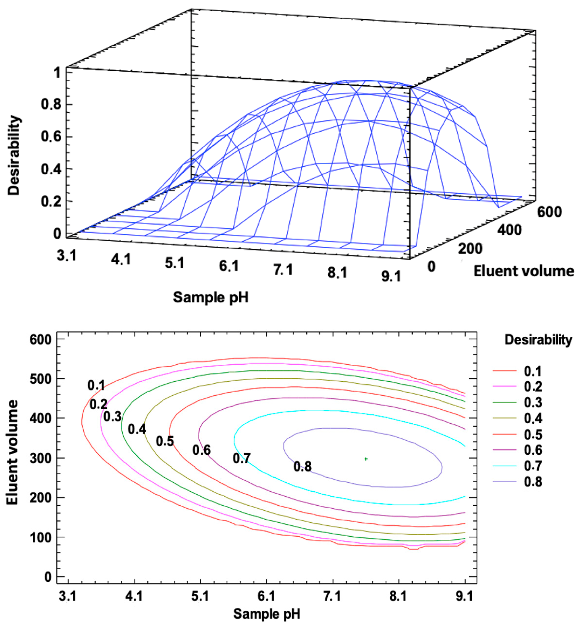
3.2.3. Desirability Function

Due to the different results obtained in the sample for the optimum pH and eluent volume for each element, it is necessary to find the same optimum conditions for all the analytes evaluated. For that reason, the desirability function was employed to obtain a common set of optimum extraction conditions to facilitate the experimental procedure. Figure 3 shows the optimum values of sample pH and eluent volume for all the elements by applying the desirability function. In the graphics obtained using this function, the closest result to a value of 1 is the best fit for all the elements. Therefore, once the desirability function was applied, the obtained optimized conditions for simultaneous extraction of all evaluated metals were the ones shown in Table S5. It was possible to observe that the optimum pH value obtained (i.e., 7.6) was above the isoelectric point (i.e., pH = 5), as predicted in Section 3.1. This was due to the fact that, above the isoelectric point, the material was negatively charged; this affects the extraction process by increasing the affinity for metals (i.e., positively charged).

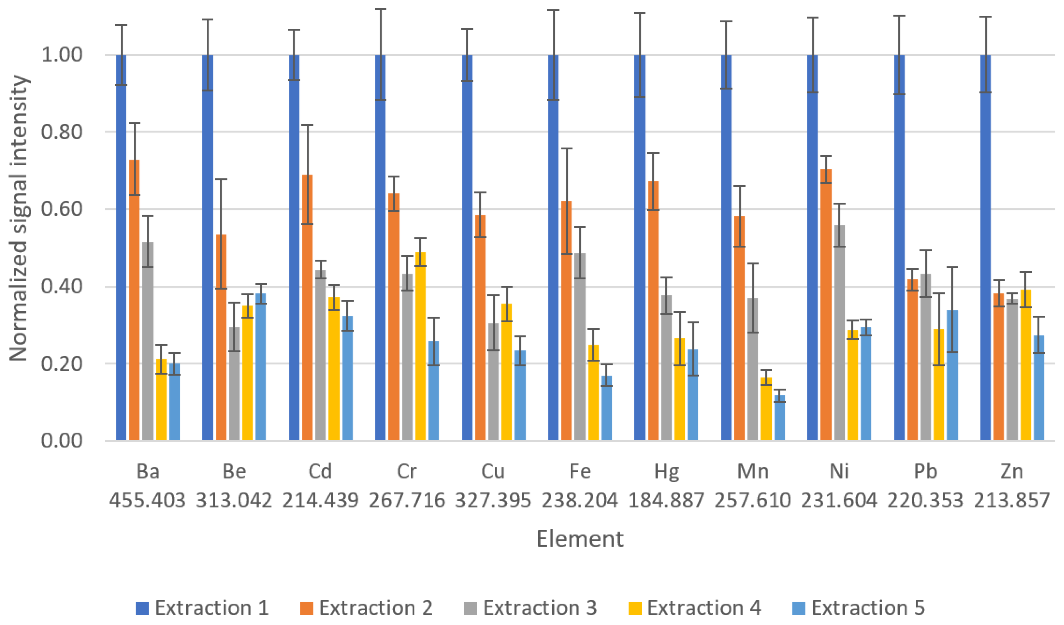
3.3. Sorbent Reutilization Study

The possibility of reusing the sorbent was also studied. To that end, five consecutive extractions were carried out in triplicate, employing the same MDSPE procedure; then, the magnetic sorbent was washed three times with 1 mL of ultrapure water. Finally, the magnetic composite was dried at 130 °C for 1 h after each extraction process in order to eliminate the excess water. The experiments were performed using a standard of 5 µg L−1. As shown in Figure 4, the signal intensity decreases after each extraction, which may be due to the corrosive nature of the acid used in the elution process and the washing out of the functional groups. In addition, the phase separation using the magnet took longer time after each extraction. For these reasons, the material cannot be used more than once. Nonetheless, the synthesis of ZIF67C_900_l is very simple and inexpensive. Thus, the disadvantage of not being able to reuse the material is not a limiting factor in carrying out the developed method. In addition, new synthesis routes are currently being investigated in our laboratory to provide a reusable material with the same extraction efficiency.

3.4. Method Validation

All the analytical figures of merit of the proposed method (Table 1) were assessed under optimum conditions, as shown in Table S5. For the purposes of comparison, the proposed method (i.e., MDSPE-ICP OES) was contrasted with the direct ICP OES analysis (i.e., without the MDSPE procedure). In the case of the proposed method, the calibration curves showed coefficients of determination (R2) that ranged from 0.993 to 0.9996 (i.e., N = 7), demonstrating good linearity. In the direct ICP OES analysis, good linearities were also obtained (i.e., 0.98–0.9994). Five independent measurements of samples spiked at 25 and 300 µg L−1 of each target element were used to assess the repeatability of the proposed method. The obtained relative standard deviations were between 4 and 8%. The sensitivity was also evaluated for both methods and compared through the enrichment factor. Sensitivities ranged between 0.39 and 877 cps µg−1 L in the direct ICP OES analysis. A significant increase in the sensitivity was observed in the proposed method, obtaining values that ranged from 3.03 to 7103 cps µg−1 L. Finally, in order to compare the sensitivity values, the enrichment factor was calculated using the ratio between the obtained sensitivities with and without the MDSPE procedure; the results of enrichment factors were between 3.2 and 13. These values of the enrichment factor enhanced the significant increase in the sensitivity of the applied extraction method.
Limits of detection (LODs) and limits of quantification (LOQs) were calculated according to the guidelines established by Eurachem [84], from the value of the standard deviation of 10 measurements of the blank signal. This value is divided by the sensitivity (i.e., slope of the calibration line) and multiplied by factor k. The k value established by the IUPAC is 3 for the limit of detection and 10 for the limit of quantification. As can be seen, the LOD values were found to be between 0.073 and 0.22 µg L−1 for all the analytes except for Fe, Ni, and Pb, with a LODs of 1.3, 1.1 and 1.1 µg L−1, respectively. The limits of detection obtained in direct ICP OES analysis ranged from 0.39 to 21 µg L−1. In other words, the LOD values obtained performing the MDSPE procedure were 14 times lower on average in comparison with those obtained through direct ICP OES analysis. In addition, the LOD values obtained with the developed method are on average 17 times lower than those established in the official method of the Environmental Protection Agency (EPA) (i.e., method 200.7). Finally, a certified reference material (ERM-CA713) was analyzed using the optimal conditions of the developed method in order to evaluate the method validation. The certified reference material analysis obtained recovery values between 90 and 109%; these results demonstrate the reliability of the method which was developed for the analysis of trace metals in wastewater samples. Table 2 shows the results of the certified reference material analysis.

3.5. Method Applicability

Real wastewater sample was analyzed and recovery studies were carried out to assess the applicability of the proposed method. Real sample was spiked at two different levels (i.e., 5.05 and 25.4 µg L−1) and analyzed in triplicate. All the measurements of the real sample were performed under optimal conditions. As can be seen in Table 3, the results show recovery values ranging between 94 and 108%, with standard deviations from 2 to 8% demonstrating that the developed method is valid for applications in performing analyses of real wastewater samples.

3.6. Comparison with Other Methods

A comparison between this work and previously described procedures that combined different SPE techniques and spectrochemical detection methods for the determination of trace metals in water and environmental samples is shown in Table S6. As can be seen, the developed method has the shortest extraction time (i.e., 3 min) and the lowest sorbent and sample amounts (i.e., 10 mg and 10 g, respectively) compared with previous studies. This is a great advantage according to the second (i.e., minimal sample size and minimal number of samples are goals), fifth (i.e., automated and miniaturized methods should be selected), and ninth (i.e., the use of energy should be minimized) principles of Green Analytical Chemistry (GAC) [85]. In addition, the developed method in this work has been evaluated for use with a higher number of analytes compared with the rest of the experimental studies; this is in accordance with the eighth principle of GAC (i.e., multi-analyte or multi-parameter methods are preferred versus methods using one analyte at a time). In some cases, the differences are outstanding: for example, in the method developed by Dogru et al. [1], a duration of at least 85 min was needed in order to prepare the column and perform the solid-phase extraction. In addition, the sorbent amount required in the proposed method was found to be between 2 and 25 times lower than other methods presented in the literature (Table S6), reducing the consumption of reagents and the cost of the MDSPE procedure. On the other hand, this work provides LOD values (i.e., 0.073–0.22 µg L−1) that are lower or comparable with previous ICP OES-based studies; except for Fe, Ni, and Pb, where the limits of detection were 1.3, 1.1, and 1.1 µg L−1, respectively.

4. Conclusions

The developed MDSPE-ICP OES method has been successfully applied for the preconcentration, separation, and determination of trace metals (i.e., Ba, Be, Cd, Cr, Cu, Fe, Hg, Mn, Ni, Pb, and Zn) in wastewater samples. The proposed sorbent is a new carbon material that contains magnetic cobalt; it was prepared using controlled pyrolysis of a ZIF-67. This magnetic material combines the excellent extraction properties of zeolites with the simple handling of the magnetic sorbent in the presence of an external magnetic field. Using this method, high enrichment factors (i.e., between 3.2 and 13) and lower limits of detection and quantification have been obtained compared with those obtained by direct ICP OES analysis. The limits of detection obtained in the developed method range from 0.073 to 1.1 µg L−1; these values are, on average, 17 times lower than the values established in the official method of the Environmental Protection Agency (i.e., method 200.7). In addition, the MDSPE-ICP OES has been validated through the analysis of a certified reference material; this analysis obtained recovery values ranging from 90 to 109% of the certified value. Furthermore, the applicability of the method was evaluated using recovery experiments at two different spiking levels (i.e., recovery values from 94 to 108%) in a real wastewater sample. The magnetic nature of the sorbent material enables the separation through the application of an external magnetic field; this permits the centrifugation step to be avoided and reduces the consumption of energy and time used in the analysis. In addition, the proposed method has been developed for a high number of elements, and the sample and sorbent amounts required have been reduced compared with previous studies; these difference meet the requirements outlined by the GAC principles and ensure that this method is an ecofriendly option. Moreover, the synthesis of the magnetic sorbent is inexpensive, and can be disposed of after a single use. For all these reasons, the developed method is a fast, reliable, multi-elemental, and ecofriendly alternative to conventional SPE methods for use in the analysis of trace metals in wastewater samples. Furthermore, taking into account all the extraction properties and the low synthesis cost, the developed magnetic material and the new analytical method could comprise a potential option to perform the extraction of analytes in industrial wastewater treatment in comparison with current materials.

Supplementary Materials

The following supporting information can be downloaded at: https://www.mdpi.com/article/10.3390/separations10110563/s1, Figure S1: XRD pattern of ZIF67c_900_l; Figure S2: N2 adsorption isotherm of the original carbon (i.e., ZIF67c_900) and the washed material ZIF67c_900_l; Figure S3: XPS spectra of cobalt and nitrogen in ZIF67c_900_l; Figure S4: Raman spectrum of ZIF67c_900_l; Figure S5: Magnetization curve of ZIF67c_900_l: (a) representation of the magnetization curve (i.e., X axis from −25,000 to 25,000 Oe); (b) amplified representation of the magnetization curve (i.e., X axis from −2000 to 2000 Oe); Figure S6: Zeta potential of ZIF67c_900_l/DDTC complex as function of the pH value; Figure S7: Pareto charts obtained in the screening study of the factors affecting the MDSPE. Orange means significant effect while blue means non-significant effect on MDSPE; Figure S8: Response surface from CCD for each analyte evaluated; Table S1: Operational parameters employed in Agilent 720 ICP OES; Table S2: Considered experimental factors and levels in the Plackett–Burman design; Table S3: Considered experimental levels in CCD design; Table S4: Optimum sample pH and eluent volume for each determined element; Table S5: Optimum experimental conditions for MDSPE; Table S6: Comparison of analytical figures of merit of the proposed method and other published methods.

Author Contributions

Conceptualization, A.C. and J.N.; Methodology, D.V.-H., E.V.R.-F., S.J.A.-M. and M.Á.A.; Software, S.J.A.-M., D.V.-H. and M.Á.A.; Validation, S.J.A.-M., D.V.-H., E.V.R.-F. and M.Á.A.; Formal Analysis, S.J.A.-M. and D.V.-H.; Investigation, D.V.-H., E.V.R.-F., S.J.A.-M. and M.Á.A.; Resources, A.C. and J.N.; Data Curation, D.V.-H., E.V.R.-F., S.J.A.-M. and M.Á.A.; Writing—Original Draft Preparation, D.V.-H. and S.J.A.-M.; Writing—Review and Editing, D.V.-H., E.V.R.-F., J.N., S.J.A.-M., M.Á.A. and A.C.; Visualization, D.V.-H., E.V.R.-F., J.N., S.J.A.-M., M.Á.A. and A.C.; Supervision, A.C. and J.N.; Project Administration, A.C. and J.N.; Funding Acquisition, A.C. and J.N. All authors have read and agreed to the published version of the manuscript.

Funding

This research was funded by the Ministry of Spain grant number (PID2021-126155OB-I00, and PID2020-116998RB-I00) and by the Regional Government of Valencia (Spain) grant number (CIPROM/2021/062, CIPROM/2021/022, INVEST/2022/70, and PRTR-C17.I1).

Data Availability Statement

The data presented in this study are available on request from the corresponding authors.

Acknowledgments

The authors are grateful to Ingeniatrics for the OneNeb® Series 2 provided. This article is based upon work from the Sample Preparation Study Group and Network, supported by the Division of Analytical Chemistry of the European Chemical Society.

Conflicts of Interest

The authors declare no conflict of interest.

References

  1. Dogru, M.; Gul-Guven, R.; Erdogan, S. The Use of Bacillus Subtilis Immobilized on Amberlite XAD-4 as a New Biosorbent in Trace Metal Determination. J. Hazard. Mater. 2007, 149, 166–173. [Google Scholar] [CrossRef] [PubMed]
  2. Zouboulis, A.I.; Loukidou, M.X.; Matis, K.A. Biosorption of Toxic Metals from Aqueous Solutions by Bacteria Strains Isolated from Metal-Polluted Soils. Process Biochem. 2004, 39, 909–916. [Google Scholar] [CrossRef]
  3. Bermejo-Barrera, P.; Martínez Alfonso, N.; Díaz López, C.; Bermejo Barrera, A. Use of Amberlite XAD-2 Loaded with 1-(2-Pyridylazo)-2-Naphthol as a Preconcentration System for River Water Prior to Determination of Cu2+, Cd2+ and Pb2+ by Flame Atomic Absorption Spectroscopy. Microchim. Acta 2003, 142, 101–108. [Google Scholar] [CrossRef]
  4. Sardans, J.; Montes, F.; Peñuelas, J. Determination of As, Cd, Cu, Hg and Pb in Biological Samples by Modern Electrothermal Atomic Absorption Spectrometry. Spectrochim. Acta B 2010, 65, 97–112. [Google Scholar] [CrossRef]
  5. Wang, J.; Chen, C. Biosorbents for Heavy Metals Removal and Their Future. Biotechnol. Adv. 2009, 27, 195–226. [Google Scholar] [CrossRef]
  6. Giakisikli, G.; Anthemidis, A.N. Magnetic Materials as Sorbents for Metal/Metalloid Preconcentration and/or Separation. A Review. Anal. Chim. Acta 2013, 789, 1–16. [Google Scholar] [CrossRef]
  7. Harvey, P.J.; Handley, H.K.; Taylor, M.P. Identification of the Sources of Metal (Lead) Contamination in Drinking Waters in North-Eastern Tasmania Using Lead Isotopic Compositions. Environ. Sci. Pollut. Res. 2015, 22, 12276–12288. [Google Scholar] [CrossRef]
  8. Ma, Y.; Egodawatta, P.; McGree, J.; Liu, A.; Goonetilleke, A. Human Health Risk Assessment of Heavy Metals in Urban Stormwater. Sci. Total Environ. 2016, 557–558, 764–772. [Google Scholar] [CrossRef] [PubMed]
  9. Zhou, L.; Gao, C.; Xu, W. Magnetic Dendritic Materials for Highly Efficient Adsorption of Dyes and Drugs. ACS Appl. Mater. Interfaces 2010, 2, 1483–1491. [Google Scholar] [CrossRef]
  10. Bagheri, H.; Afkhami, A.; Saber-Tehrani, M.; Khoshsafar, H. Preparation and Characterization of Magnetic Nanocomposite of Schiff Base/Silica/Magnetite as a Preconcentration Phase for the Trace Determination of Heavy Metal Ions in Water, Food and Biological Samples Using Atomic Absorption Spectrometry. Talanta 2012, 97, 87–95. [Google Scholar] [CrossRef]
  11. Suleiman, J.S.; Hu, B.; Huang, C.; Zhang, N. Determination of Cd, Co, Ni and Pb in Biological Samples by Microcolumn Packed with Black Stone (Pierre Noire) Online Coupled with ICP-OES. J. Hazard. Mater. 2008, 157, 410–417. [Google Scholar] [CrossRef] [PubMed]
  12. Yamini, Y.; Faraji, M.; Shariati, S.; Hassani, R.; Ghambarian, M. On-Line Metals Preconcentration and Simultaneous Determination Using Cloud Point Extraction and Inductively Coupled Plasma Optical Emission Spectrometry in Water Samples. Anal. Chim. Acta 2008, 612, 144–151. [Google Scholar] [CrossRef] [PubMed]
  13. Davis, A.C.; Calloway, C.P.; Jones, B.T. Direct Determination of Cadmium in Urine by Tungsten-Coil Inductively Coupled Plasma Atomic Emission Spectrometry Using Palladium as a Permanent Modifier. Talanta 2007, 71, 1144–1149. [Google Scholar] [CrossRef]
  14. Huang, C.; Hu, B. Silica-Coated Magnetic Nanoparticles Modified with γ-Mercaptopropyltrimethoxysilane for Fast and Selective Solid Phase Extraction of Trace Amounts of Cd, Cu, Hg, and Pb in Environmental and Biological Samples Prior to Their Determination by Inductively Co. Spectrochim. Acta B 2008, 63, 437–444. [Google Scholar] [CrossRef]
  15. Zheng, F.; Hu, B. Thermo-Responsive Polymer Coated Fiber-in-Tube Capillary Microextraction and Its Application to on-Line Determination of Co, Ni and Cd by Inductively Coupled Plasma Mass Spectrometry (ICP-MS). Talanta 2011, 85, 1166–1173. [Google Scholar] [CrossRef]
  16. Zhang, L.; Chen, B.; Peng, H.; He, M.; Hu, B. Aminopropyltriethoxysilane-Silica Hybrid Monolithic Capillary Microextraction Combined with Inductively Coupled Plasma Mass Spectrometry for the Determination of Trace Elements in Biological Samples. J. Sep. Sci. 2011, 34, 2247–2254. [Google Scholar] [CrossRef]
  17. Naghizadeh, M.; Taher, M.A.; Behzadi, M.; Moghaddam, F.H. Preparation a Novel Magnetic Natural Nano Zeolite for Preconcentration of Cadmium and Its Determination by ETAAS. Environ. Nanotechnol. Monit. Manag. 2017, 8, 261–267. [Google Scholar] [CrossRef]
  18. Jorhem, L.; Åstrand, C.; Sundström, B.; Baxter, M.; Stokes, P.; Lewis, J.; Grawe, K.P. Elements in Rice from the Swedish Market: 1. Cadmium, Lead and Arsenic (Total and Inorganic). Food Addit. Contam.-Part A 2008, 25, 284–292. [Google Scholar] [CrossRef]
  19. Montoro-Leal, P.; García-Mesa, J.C.; Siles Cordero, M.T.; López Guerrero, M.M.; Vereda Alonso, E. Magnetic Dispersive Solid Phase Extraction for Simultaneous Enrichment of Cadmium and Lead in Environmental Water Samples. Microchem. J. 2020, 155, 104796. [Google Scholar] [CrossRef]
  20. Xie, F.; Lin, X.; Wu, X.; Xie, Z. Solid Phase Extraction of Lead (II), Copper (II), Cadmium (II) and Nickel (II) Using Gallic Acid-Modified Silica Gel Prior to Determination by Flame Atomic Absorption Spectrometry. Talanta 2008, 74, 836–843. [Google Scholar] [CrossRef]
  21. Tuzen, M.; Soylak, M. Column Solid-Phase Extraction of Nickel and Silver in Environmental Samples Prior to Their Flame Atomic Absorption Spectrometric Determinations. J. Hazard. Mater. 2009, 164, 1428–1432. [Google Scholar] [CrossRef] [PubMed]
  22. Ghaedi, M.; Tavallali, H.; Shokrollahi, A.; Zahedi, M.; Montazerozohori, M.; Soylak, M. Flame Atomic Absorption Spectrometric Determination of Zinc, Nickel, Iron and Lead in Different Matrixes after Solid Phase Extraction on Sodium Dodecyl Sulfate (SDS)-Coated Alumina as Their Bis (2-Hydroxyacetophenone)-1, 3-Propanediimine Chelates. J. Hazard. Mater. 2009, 166, 1441–1448. [Google Scholar] [CrossRef] [PubMed]
  23. Bryce, D.W.; Izquierdo, A.; Luque De Castro, M.D. Continuous Microwave Assisted Pervaporation/Atomic Fluorescence Detection: An Approach for Speciation in Solid Samples. Anal. Chim. Acta 1996, 324, 69–75. [Google Scholar] [CrossRef]
  24. Zhang, W.; Su, Z.; Chu, X.; Yang, X. Evaluation of a New Electrolytic Cold Vapor Generation System for Mercury Determination by AFS. Talanta 2010, 80, 2106–2112. [Google Scholar] [CrossRef] [PubMed]
  25. Cheng, G.; He, M.; Peng, H.; Hu, B. Dithizone Modified Magnetic Nanoparticles for Fast and Selective Solid Phase Extraction of Trace Elements in Environmental and Biological Samples Prior to Their Determination by ICP-OES. Talanta 2012, 88, 507–515. [Google Scholar] [CrossRef] [PubMed]
  26. Zeini Jahromi, E.; Bidari, A.; Assadi, Y.; Milani Hosseini, M.R.; Jamali, M.R. Dispersive Liquid-Liquid Microextraction Combined with Graphite Furnace Atomic Absorption Spectrometry. Ultra Trace Determination of Cadmium in Water Samples. Anal. Chim. Acta 2007, 585, 305–311. [Google Scholar] [CrossRef]
  27. Shabani, A.M.H.; Dadfarnia, S.; Nasirizadeh, N.; Shishehbore, M.R. Preconcentration of Copper with Dithizone-Naphthalene for Subsequent Determination by Atomic Absorption Spectrometry. J. Anal. Chem. 2007, 62, 46–50. [Google Scholar] [CrossRef]
  28. Miró, M.; Hansen, E.H. Recent Advances and Future Prospects of Mesofluidic Lab-on-a-Valve Platforms in Analytical Sciences—A Critical Review. Anal. Chim. Acta 2012, 750, 3–15. [Google Scholar] [CrossRef]
  29. Baghdadi, M.; Shemirani, F. Cold-Induced Aggregation Microextraction: A Novel Sample Preparation Technique Based on Ionic Liquids. Anal. Chim. Acta 2008, 613, 56–63. [Google Scholar] [CrossRef]
  30. Soylak, M.; Yilmaz, E. Ionic Liquid Dispersive Liquid-Liquid Microextraction of Lead as Pyrrolidinedithiocarbamate Chelate Prior to Its Flame Atomic Absorption Spectrometric Determination. Desalination 2011, 275, 297–301. [Google Scholar] [CrossRef]
  31. Pena-Pereira, F.; Lavilla, I.; Bendicho, C. Miniaturized Preconcentration Methods Based on Liquid-Liquid Extraction and Their Application in Inorganic Ultratrace Analysis and Speciation: A Review. Spectrochim. Acta B 2009, 64, 1–15. [Google Scholar] [CrossRef]
  32. Soylak, M.; Aydin, A. Determination of Some Heavy Metals in Food and Environmental Samples by Flame Atomic Absorption Spectrometry after Coprecipitation. Food Chem. Toxicol. 2011, 49, 1242–1248. [Google Scholar] [CrossRef] [PubMed]
  33. Soylak, M.; Kars, A.; Narin, I. Coprecipitation of Ni2+, Cd2+ and Pb2+ for Preconcentration in Environmental Samples Prior to Flame Atomic Absorption Spectrometric Determinations. J. Hazard. Mater. 2008, 159, 435–439. [Google Scholar] [CrossRef] [PubMed]
  34. Saberyan, K.; Zolfonoun, E.; Shamsipur, M.; Salavati-Niasari, M. Separation and Preconcentration of Trace Gallium and Indium by Amberlite XAD-7 Resin Impregnated with a New Hexadentates Naphthol-Derivative Schiff Base. Sep. Sci. Technol. 2009, 44, 1851–1868. [Google Scholar] [CrossRef]
  35. Uludag, Y.; Özbelge, H.Ö.; Yilmaz, L. Removal of Mercury from Aqueous Solutions via Polymer-Enhanced Ultrafiltration. J. Membr. Sci. 1997, 129, 93–99. [Google Scholar] [CrossRef]
  36. Bruno, P.; Caselli, M.; De Gennaro, G.; Ielpo, P.; Ladisa, T.; Placentino, C.M. Ion Chromatography Determination of Heavy Metals in Airborne Particulate with Preconcentration and Large Volume Direct Injection. Chromatographia 2006, 64, 537–542. [Google Scholar] [CrossRef]
  37. Da̧browski, A.; Hubicki, Z.; Podkościelny, P.; Robens, E. Selective Removal of the Heavy Metal Ions from Waters and Industrial Wastewaters by Ion-Exchange Method. Chemosphere 2004, 56, 91–106. [Google Scholar] [CrossRef]
  38. Sánchez Rojas, F.; Ojeda, C.B.; Pavón, J.M.C. On-Line Preconcentration of Rhodium on an Anion-Exchange Resin Loaded with 1,5-Bis(2-Pyridyl)-3-Sulphophenyl Methylene Thiocarbonohydrazide and Its Determination in Environmental Samples. Talanta 2004, 64, 230–236. [Google Scholar] [CrossRef]
  39. Shariati, S.; Yamini, Y.; Zanjani, M.K. Simultaneous Preconcentration and Determination of U(VI), Th(IV), Zr(IV) and Hf(IV) Ions in Aqueous Samples Using Micelle-Mediated Extraction Coupled to Inductively Coupled Plasma-Optical Emission Spectrometry. J. Hazard. Mater. 2008, 156, 583–590. [Google Scholar] [CrossRef]
  40. Meeravali, N.N.; Jiang, S.J. Microwave Assisted Mixed-Micelle Cloud Point Extraction of Au and Tl from Environmental Samples without Using a Chelating Agent Prior to ICP-MS Determination. J. Anal. At. Spectrom. 2008, 23, 1365–1371. [Google Scholar] [CrossRef]
  41. Tuzen, M.; Karaman, I.; Citak, D.; Soylak, M. Mercury(II) and Methyl Mercury Determinations in Water and Fish Samples by Using Solid Phase Extraction and Cold Vapour Atomic Absorption Spectrometry Combination. Food Chem. Toxicol. 2009, 47, 1648–1652. [Google Scholar] [CrossRef] [PubMed]
  42. Tuzen, M.; Uluozlu, O.D.; Karaman, I.; Soylak, M. Mercury(II) and Methyl Mercury Speciation on Streptococcus Pyogenes Loaded Dowex Optipore SD-2. J. Hazard. Mater. 2009, 169, 345–350. [Google Scholar] [CrossRef] [PubMed]
  43. Fan, J.; Qin, Y.; Ye, C.; Peng, P.; Wu, C. Preparation of the Diphenylcarbazone-Functionalized Silica Gel and Its Application to on-Line Selective Solid-Phase Extraction and Determination of Mercury by Flow-Injection Spectrophotometry. J. Hazard. Mater. 2008, 150, 343–350. [Google Scholar] [CrossRef] [PubMed]
  44. Faraji, M.; Yamini, Y.; Rezaee, M. Extraction of Trace Amounts of Mercury with Sodium Dodecyle Sulphate-Coated Magnetite Nanoparticles and Its Determination by Flow Injection Inductively Coupled Plasma-Optical Emission Spectrometry. Talanta 2010, 81, 831–836. [Google Scholar] [CrossRef]
  45. Poole, C.F. New Trends in Solid-Phase Extraction. TrAC-Trends Anal. Chem. 2003, 22, 362–373. [Google Scholar] [CrossRef]
  46. Zhang, C.; Xing, H.; Yang, L.; Fei, P.; Liu, H. Development Trend and Prospect of Solid Phase Extraction Technology. Chin. J. Chem. Eng. 2022, 42, 245–255. [Google Scholar] [CrossRef]
  47. Giakisikli, G.; Anthemidis, A.N. Automated Magnetic Sorbent Extraction Based on Octadecylsilane Functionalized Maghemite Magnetic Particles in a Sequential Injection System Coupled with Electrothermal Atomic Absorption Spectrometry for Metal Determination. Talanta 2013, 110, 229–235. [Google Scholar] [CrossRef]
  48. Rao, T.P.; Metilda, P.; Gladis, J.M. Preconcentration Techniques for Uranium(VI) and Thorium(IV) Prior to Analytical Determination-an Overview. Talanta 2006, 68, 1047–1064. [Google Scholar] [CrossRef]
  49. Zhu, F.; Ma, Z.; Qiu, K.; Peng, W. Separation of Ilmenite from Vanadium Titanomagnetite by Combining Magnetic Separation and Flotation Processes. Separations 2023, 10, 95. [Google Scholar] [CrossRef]
  50. Córdova Udaeta, M.; Dodbiba, G.; Ponou, J.; Sone, K.; Fujita, T. Recovery of Phosphorus from Sewage Sludge Ash (SSA) by Heat Treatment Followed by High Gradient Magnetic Separation and Flotation. Adv. Powder Technol. 2017, 28, 755–762. [Google Scholar] [CrossRef]
  51. Jiménez-Soto, J.M.; Cárdenas, S.; Valcárcel, M. Evaluation of Single-Walled Carbon Nanohorns as Sorbent in Dispersive Micro Solid-Phase Extraction. Anal. Chim. Acta 2012, 714, 76–81. [Google Scholar] [CrossRef] [PubMed]
  52. Galán-Cano, F.; Lucena, R.; Cárdenas, S.; Valcárcel, M. Direct Coupling of Dispersive Micro-Solid Phase Extraction and Thermal Desorption for Sensitive Gas Chromatographic Analysis. Anal. Methods 2011, 3, 991–995. [Google Scholar] [CrossRef]
  53. Skorek, R.; Zawisza, B.; Marguí, E.; Queralt, I.; Sitko, R. Dispersive Micro Solid-Phase Extraction Using Multiwalled Carbon Nanotubes for Simultaneous Determination of Trace Metal Ions by Energy-Dispersive x-Ray Fluorescence Spectrometry. Appl. Spectrosc. 2013, 67, 204–209. [Google Scholar] [CrossRef] [PubMed]
  54. Ramutshatsha-Makhwedzha, D.; Mbaya, R.; Mavhungu, M.L.; Nomngongo, P.N. Ultrasonic Assisted Dispersive-Solid Phase Extraction for Preconcentration of Trace Metals in Wastewater Samples. J. Environ. Chem. Eng. 2022, 10, 108187. [Google Scholar] [CrossRef]
  55. Armenta, S.; Garrigues, S.; de la Guardia, M. Green Analytical Chemistry. TrAC-Trends Anal. Chem. 2008, 27, 497–511. [Google Scholar] [CrossRef]
  56. Duan, C.; Shen, Z.; Wu, D.; Guan, Y. Recent Developments in Solid-Phase Microextraction for on-Site Sampling and Sample Preparation. TrAC-Trends Anal. Chem. 2011, 30, 1568–1574. [Google Scholar] [CrossRef]
  57. Socas-Rodríguez, B.; Herrera-Herrera, A.V.; Asensio-Ramos, M.; Hernández-Borges, J. Dispersive Solid-Phase Extraction. In Analytical Separation Science; Wiley-VCH Verlag GmbH & Co. KGaA: Weinheim, Germany, 2015; pp. 1525–1570. [Google Scholar] [CrossRef]
  58. Ruiz, F.J.; Ripoll, L.; Hidalgo, M.; Canals, A. Dispersive Micro Solid-Phase Extraction (DµSPE) with Graphene Oxide as Adsorbent for Sensitive Elemental Analysis of Aqueous Samples by Laser Induced Breakdown Spectroscopy (LIBS). Talanta 2019, 191, 162–170. [Google Scholar] [CrossRef]
  59. Lord, H.; Pawliszyn, J. Evolution of Solid-Phase Microextraction Technology. J. Chromatogr. A 2000, 885, 153–193. [Google Scholar] [CrossRef]
  60. Kharissova, O.V.; Dias, H.V.R.; Kharisov, B.I. Magnetic Adsorbents Based on Micro- and Nano-Structured Materials. RSC Adv. 2015, 5, 6695–6719. [Google Scholar] [CrossRef]
  61. Tian, J.; Xu, J.; Zhu, F.; Lu, T.; Su, C.; Ouyang, G. Application of Nanomaterials in Sample Preparation. J. Chromatogr. A 2013, 1300, 2–16. [Google Scholar] [CrossRef]
  62. Płotka-Wasylka, J.; Szczepańska, N.; de la Guardia, M.; Namieśnik, J. Miniaturized Solid-Phase Extraction Techniques. TrAC-Trends Anal. Chem. 2015, 73, 19–38. [Google Scholar] [CrossRef]
  63. Wierucka, M.; Biziuk, M. Application of Magnetic Nanoparticles for Magnetic Solid-Phase Extraction in Preparing Biological, Environmental and Food Samples. TrAC-Trends Anal. Chem. 2014, 59, 50–58. [Google Scholar] [CrossRef]
  64. Safarikova, M.; Kibrikova, I.; Ptackova, L.; Hubka, T.; Komarek, K.; Safarik, I. Magnetic Solid Phase Extraction of Non-Ionic Surfactants from Water. J. Magn. Magn. Mater. 2005, 293, 377–381. [Google Scholar] [CrossRef]
  65. Aliyari, E.; Alvand, M.; Shemirani, F. Modified Surface-Active Ionic Liquid-Coated Magnetic Graphene Oxide as a New Magnetic Solid Phase Extraction Sorbent for Preconcentration of Trace Nickel. RSC Adv. 2016, 6, 64193–64202. [Google Scholar] [CrossRef]
  66. Asfaram, A.; Ghaedi, M.; Goudarzi, A.; Soylak, M.; Mehdizadeh Langroodi, S. Magnetic Nanoparticle Based Dispersive Micro-Solid-Phase Extraction for the Determination of Malachite Green in Water Samples: Optimized Experimental Design. New J. Chem. 2015, 39, 9813–9823. [Google Scholar] [CrossRef]
  67. Costa dos Reis, L.; Vidal, L.; Canals, A. Determination of Siloxanes in Water Samples Employing Graphene Oxide/Fe3O4 Nanocomposite as Sorbent for Magnetic Solid-Phase Extraction Prior to GC–MS. J. Sep. Sci. 2018, 41, 4177–4184. [Google Scholar] [CrossRef]
  68. Aguilar-Arteaga, K.; Rodriguez, J.A.; Barrado, E. Magnetic Solids in Analytical Chemistry: A Review. Anal. Chim. Acta 2010, 674, 157–165. [Google Scholar] [CrossRef]
  69. Chen, J.; Wang, Y.; Wei, X.; Xu, P.; Xu, W.; Ni, R.; Meng, J. Magnetic Solid-Phase Extraction for the Removal of Mercury from Water with Ternary Hydrosulphonyl-Based Deep Eutectic Solvent Modified Magnetic Graphene Oxide. Talanta 2018, 188, 454–462. [Google Scholar] [CrossRef]
  70. Mollahosseini, A.; Toghroli, M.; Kamankesh, M. Zeolite/Fe3O4 as a New Sorbent in Magnetic Solid-Phase Extraction Followed by Gas Chromatography for Determining Phthalates in Aqueous Samples. J. Sep. Sci. 2015, 38, 3750–3757. [Google Scholar] [CrossRef]
  71. Tolmacheva, V.V.; Apyari, V.V.; Kochuk, E.V.; Dmitrienko, S.G. Magnetic Adsorbents Based on Iron Oxide Nanoparticles for the Extraction and Preconcentration of Organic Compounds. J. Anal. Chem. 2016, 71, 321–338. [Google Scholar] [CrossRef]
  72. Hemmati, M.; Rajabi, M.; Asghari, A. Magnetic Nanoparticle Based Solid-Phase Extraction of Heavy Metal Ions: A Review on Recent Advances. Microchim. Acta 2018, 185, 160. [Google Scholar] [CrossRef] [PubMed]
  73. Baile, P.; Vidal, L.; Canals, A. A Modified Zeolite/Iron Oxide Composite as a Sorbent for Magnetic Dispersive Solid-Phase Extraction for the Preconcentration of Nonsteroidal Anti-Inflammatory Drugs in Water and Urine Samples. J. Chromatogr. A 2019, 1603, 33–43. [Google Scholar] [CrossRef]
  74. Baile, P.; Fernández, E.; Vidal, L.; Canals, A. Zeolites and Zeolite-Based Materials in Extraction and Microextraction Techniques. Analyst 2019, 144, 366–387. [Google Scholar] [CrossRef] [PubMed]
  75. Baile, P.; Vidal, L.; Canals, A. Magnetic Dispersive Solid-Phase Extraction Using ZSM-5 Zeolite/Fe2O3 Composite Coupled with Screen-Printed Electrodes Based Electrochemical Detector for Determination of Cadmium in Urine Samples. Talanta 2020, 220, 121394. [Google Scholar] [CrossRef]
  76. Villalgordo-Hernández, D.; Grau-Atienza, A.; García-Marín, A.A.; Ramos-Fernández, E.V.; Narciso, J. Manufacture of Carbon Materials with High Nitrogen Content. Materials 2022, 15, 2415. [Google Scholar] [CrossRef]
  77. Zabiegaj, D.; Caccia, M.; Casco, M.E.; Ravera, F.; Narciso, J. Synthesis of Carbon Monoliths with a Tailored Hierarchical Pore Structure for Selective CO2 Capture. J. CO2 Util. 2018, 26, 36–44. [Google Scholar] [CrossRef]
  78. Narciso, J.; Ramos-Fernandez, E.V.; Delgado-Marín, J.J.; Affolter, C.W.; Olsbye, U.; Redekop, E.A. New Route for the Synthesis of Co-MOF from Metal Substrates. Microporous Mesoporous Mater. 2021, 324, 111310. [Google Scholar] [CrossRef]
  79. Ramos-Fernandez, E.V.; Redondo-Murcia, A.; Grau-Atienza, A.; Sepúlveda-Escribano, A.; Narciso, J. Clean Production of Zeolitic Imidazolate Framework 8 Using Zamak Residues as Metal Precursor and Substrate. J. Clean. Prod. 2020, 260, 121081. [Google Scholar] [CrossRef]
  80. Delgado-Marín, J.J.; Martín-García, I.; Villalgordo-Hernández, D.; Alonso, F.; Ramos-Fernández, E.V.; Narciso, J. Valorization of CO2 through the Synthesis of Cyclic Carbonates Catalyzed by ZIFs. Molecules 2022, 27, 7791. [Google Scholar] [CrossRef]
  81. Caccia, M.; Giuranno, D.; Molina-Jorda, J.M.; Moral, M.; Nowak, R.; Ricci, E.; Sobczak, N.; Narciso, J.; Fernández Sanz, J. Graphene Translucency and Interfacial Interactions in the Gold/Graphene/SiC System. J. Phys. Chem. Lett. 2018, 9, 3850–3855. [Google Scholar] [CrossRef]
  82. Costa, D.A.S.; Mambrini, R.V.; Fernandez-Outon, L.E.; Macedo, W.A.A.; Moura, F.C.C. Magnetic Adsorbent Based on Cobalt Core Nanoparticles Coated with Carbon Filaments and Nanotubes Produced by Chemical Vapor Deposition with Ethanol. Chem. Eng. J. 2013, 229, 35–41. [Google Scholar] [CrossRef]
  83. Baile, P.; Vidal, L.; Aguirre, M.Á.; Canals, A. A Modified ZSM-5 Zeolite/Fe2O3 Composite as a Sorbent for Magnetic Dispersive Solid-Phase Microextraction of Cadmium, Mercury and Lead from Urine Samples Prior to Inductively Coupled Plasma Optical Emission Spectrometry. J. Anal. At. Spectrom. 2018, 33, 856–866. [Google Scholar] [CrossRef]
  84. Magnusson, B.; Örnemark, U. Eurachem Guide: The Fitness for Purpose of Analytical Methods—A Laboratory Guide to Method Validation and Related Topics, 2nd ed.; LGC: Teddington Ltd.: Teddington, UK, 2014; ISBN 9789187461590. [Google Scholar]
  85. Gałuszka, A.; Migaszewski, Z.; Namieśnik, J. The 12 Principles of Green Analytical Chemistry and the SIGNIFICANCE Mnemonic of Green Analytical Practices. TrAC-Trends Anal. Chem. 2013, 50, 78–84. [Google Scholar] [CrossRef]
Figure 1. Schematic representation of MDSPE for preconcentration of trace metals using the optimized experimental conditions (i.e., (i) sample weight, 10 g; (ii) sample pH, 7.6; (iii) amount of sorbent, 10 mg; (iv) dispersion mode, vortex; (v) complexing agent concentration, 0.5%; (vi) ionic concentration, 0%; (vii) eluent, HCl; (viii) eluent concentration, 0.5 M; (ix) eluent volume, 300 μL; (x) extraction time, 3 min; (xi) elution time, 3 min).
Separations 10 00563 g001aSeparations 10 00563 g001b
Figure 2. Secondary electron micrograph of ZIF67C_900_l.
Separations 10 00563 g002
Figure 3. Graphics with obtained optimum results of sample pH and eluent volume applying the desirability function. Experimental fixed conditions: (i) sample weight, 10 g; (ii) amount of sorbent, 10 mg; (iii) dispersion mode, vortex; (iv) complexing agent concentration, 0.5%; (v) ionic concentration, 0%; (vi) eluent, HCl; (vii) eluent concentration, 0.5 M; (viii) extraction time, 3 min; (ix) elution time, 3 min.
Separations 10 00563 g003
Figure 4. Study of sorbent reusability using the same sorbent in five consecutive extractions of 5 µg L−1 standard solution. The sorbent was washed three times with 1 mL of ultrapure water and heated at 130° for 1 h; it was dried between each extraction. The error bars were evaluated through the standard deviation of three replicates. Experimental conditions used: (i) sample weight, 10 g; (ii) sample pH, 7.6; (iii) amount of sorbent, 10 mg; (iv) dispersion mode, vortex; (v) complexing agent concentration, 0.5%; (vi) ionic concentration, 0%; (vii) eluent, HCl; (viii) eluent concentration, 0.5 M; (ix) eluent volume, 300 μL; (x) extraction time, 3 min; (xi) elution time, 3 min.
Separations 10 00563 g004
Table 1. Analytical figures of merit for direct ICP OES and the MDSPE-ICP OES method, developed for the analysis of water samples.
Direct ICP OES
Emission Line (nm)Working Range (µg L−1)R2Sensitivity a (cps µg−1 L)Repeatability (RSD%)LOD (µg L−1)LOQ (µg L−1)EF
Ba 455.403250–10000.998534 ± 17-2.68.8-
Be 313.042250–10000.998877 ± 28-0.391.3-
Cd 214.439250–10000.9988.3 ± 0.2-1.55.0-
Cr 267.716250–10000.999411.37 ± 0.19-1.34.4-
Cu 327.395250–10000.99811.0 ± 0.4-1.23.9-
Fe 238.204250–10000.9989.1 ± 0.2-9.833-
Hg 184.887250–10000.99912.86 ± 0.06-5.117-
Mn 257.610250–10000.99634.3 ± 1.6-0.822.7-
Ni 231.604250–10000.981.24 ± 0.12-9.933-
Pb 220.353250–10000.980.39 ± 0.04-2170-
Zn 213.857250–10000.99225.0 ± 1.6-0.812.7-
MDSPE-ICP OES d
Emission line (nm)Working range (µg L−1)R2Sensitivity a (cps µg−1 L)Repeatability (RSD%) bLOD (µg L−1)LOQ (µg L−1)EF c
25 (µg L−1)300 (µg L−1)
Ba 455.4030.5–5000.99917103 ± 93550.0850.2913
Be 313.0420.5–5000.9975638 ± 131760.0730.246.4
Cd 214.4390.5–5000.99384 ± 3640.120.4110
Cr 267.7160.5–5000.99850.2 ± 1.0760.140.454.4
Cu 327.3950.5–5000.99885.9 ± 1.5660.100.347.8
Fe 238.2045–5000.99764.6 ± 1.8851.34.37.1
Hg 184.8870.5–5000.99733.8 ± 0.8760.130.4312
Mn 257.6100.5–5000.997109 ± 3660.110.373.2
Ni 231.6045–5000.9988.23 ± 0.16771.13.86.6
Pb 220.3535–5000.99963.03 ± 0.03871.13.87.9
Zn 213.8571–5000.99892.2 ± 1.6860.220.753.7
a Slope of the calibration curve ± standard deviation (N = 7). b Repeatability of the method evaluated at two levels through the RSD (n = 5). c Enrichment factor (EF): ratio between the slope with and without the MDSPE procedure. d Experimental conditions used: (i) sample weight, 10 g; (ii) sample pH, 7.6; (iii) amount of sorbent, 10 mg; (iv) dispersion mode, vortex; (v) complexing agent concentration, 0.5%; (vi) ionic concentration, 0%; (vii) eluent, HCl; (viii) eluent concentration, 0.5 M; (ix) eluent volume, 300 μL; (x) extraction time, 3 min; (xi) elution time, 3 min.
Table 2. Results of certified reference material analysis a.
AnalyteCdCrCuFeHgMnNiPb
Certified value
(µg L−1) b
5.09 ± 0.2020.9 ± 1.3101 ± 7445 ± 271.84 ± 0.1195 ± 450.3 ± 1.449.7 ± 1.7
Found value
(µg L−1) c
4.8 ± 0.421.6 ± 1.394 ± 8424 ± 331.66 ± 0.1490 ± 255 ± 350 ± 4
Recovery (%)95 ± 8103 ± 693 ± 895 ± 790 ± 895 ± 2109 ± 5100 ± 7
a Experimental conditions used: (i) sample weight, 10 g; (ii) sample pH, 7.6; (iii) amount of sorbent, 10 mg; (iv) dispersion mode, vortex; (v) complexing agent concentration, 0.5%; (vi) ionic concentration, 0%; (vii) eluent, HCl; (viii) eluent concentration, 0.5 M; (ix) eluent volume, 300 μL; (x) extraction time, 3 min; (xi) elution time, 3 min. b Certified value ± expanded uncertainty. c Found value ± expanded uncertainty (coverage factor of 2).
Table 3. Results of recovery study on spiked real sample a.
Analyte
Added Value (µg L−1)BaBeCdCrCuFeHgMnNiPbZn
Found value
(µg L−1) b
-<LOQ<LOQ<LOQ308.4 ± 0.553.6 ± 1.062.6 ± 1.31.27 ± 0.052.41 ± 0.02<LOQ<LOQ108.4 ± 1.4
5.055.4 ± 0.2
(106 ± 4)
5.2 ± 0.2
(101 ± 6)
5.02 ± 0.16
(99 ± 3)
313.8 ± 0.4
(108 ± 8)
58.4 ± 0.3
(95 ± 6)
67.6 ± 0.2
(98 ± 4)
6.1 ± 0.2
(94 ± 3)
7.2 ± 0.2
(94 ± 5)
5.2 ± 0.2
(103 ± 4)
5.2 ± 0.2
(103 ± 4)
113.4 ± 0.3
(98 ± 6)
25.426.1 ± 1.1
(102 ± 5)
24.0 ± 0.9
(94 ± 3)
24.0 ± 0.8
(94 ± 4)
334.2 ± 1.4
(101 ± 5)
79.6 ± 1.9
(102 ± 6)
88.8 ± 0.7
(103 ± 3)
27.0 ± 1.4
(100 ± 7)
26.5 ± 0.8
(95 ± 3)
27.2 ± 0.7
(107 ± 2)
27.2 ± 1.3
(107 ± 4)
133.4 ± 0.8
(98 ± 4)
a Experimental conditions used: (i) sample weight, 10 g; (ii) sample pH, 7.6; (iii) amount of sorbent, 10 mg; (iv) dispersion mode, vortex; (v) complexing agent concentration, 0.5%; (vi) ionic concentration, 0%; (vii) eluent, HCl; (viii) eluent concentration, 0.5 M; (ix) eluent volume, 300 μL; (x) extraction time, 3 min; (xi) elution time, 3 min. b Found value ± standard deviation. Recovery (%) ± standard deviation is shown in parentheses.
Disclaimer/Publisher’s Note: The statements, opinions and data contained in all publications are solely those of the individual author(s) and contributor(s) and not of MDPI and/or the editor(s). MDPI and/or the editor(s) disclaim responsibility for any injury to people or property resulting from any ideas, methods, instructions or products referred to in the content.

