La démence frontotemporale: aspects cliniques et morphologiques
Summary
Introduction
Présentation clinique de la démence frontotemporale
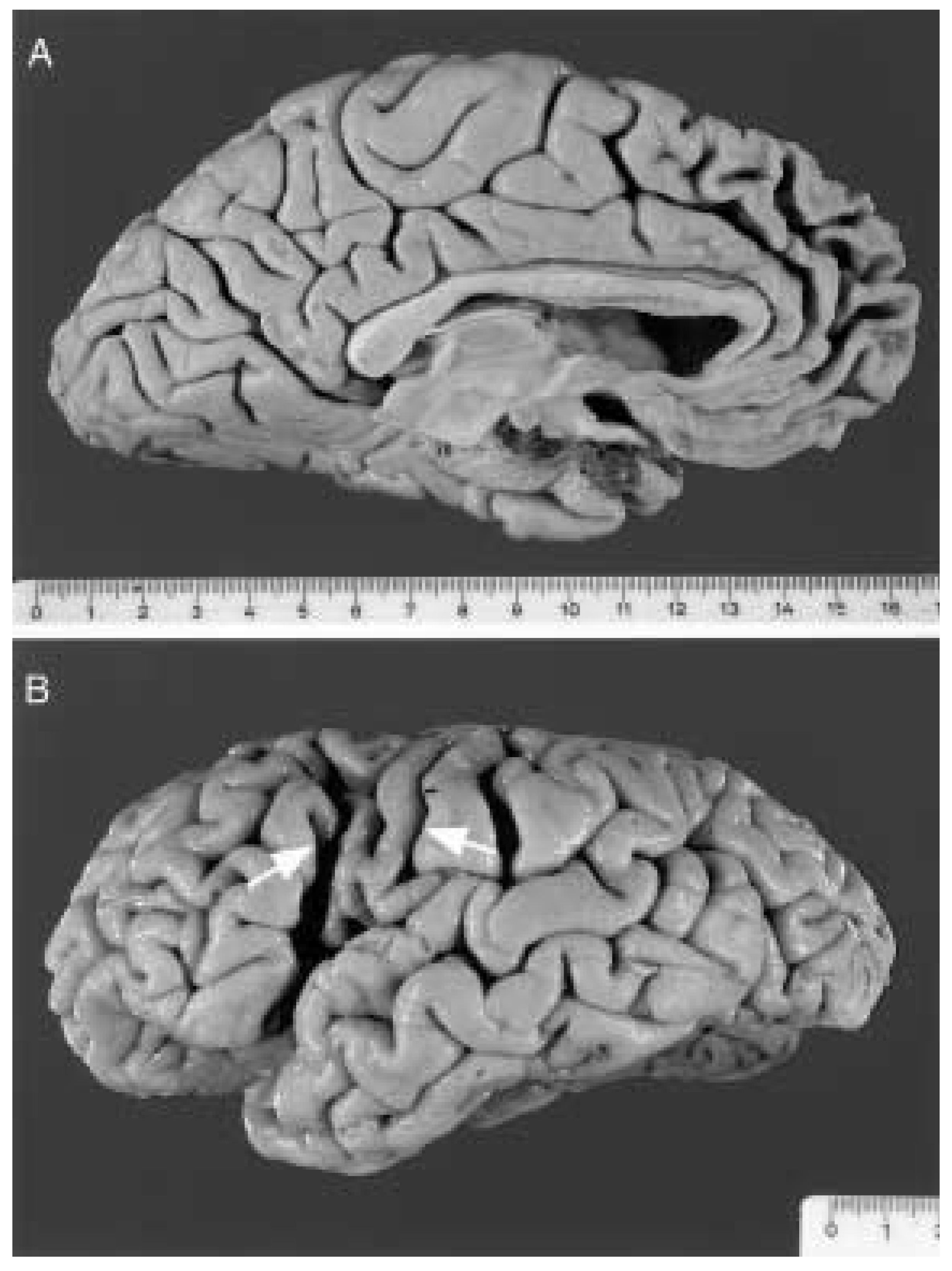
- Le premier mode de présentation clinique, le plus fréquent, correspond à la variante frontale de la démence frontotemporale, avec atrophie à prédominance frontale et surtout de la convexité frontale. Le tableau clinique consiste en une altération précoce de la personne et des conduites sociales (social conduct). Le malade est souvent inerte ou apathique, présente une difficulté grandissante à adapter son comportement au contexte social. Son comportement est désinhibé, inapproprié, compulsif, souvent répétitif et stéréotypé. Des troubles cognitifs peuvent apparaître dans le domaine de l’abstraction, de l’attention, de la planification et de la solution de problèmes complexes. La mémoire est par contre bien conservée du moins aux premiers stades de la maladie.
- Chez une partie des patients avec démence frontotemporale, la symptomatologie clinique se présente sous la forme d’une dissolution isolée (non accompagnée d’autres troubles cognitifs) du langage dont il convient de distinguer deux formes, l’aphasie primaire progressive [15] et la démence sémantique [16]. Le tableau clinique de l’aphasie primaire progressive se caractérise par une réduction progressive de la production du langage – d’où le terme d’aphasie non fluente utilisé pour ce type de trouble du langage – accompagnée d’erreurs phonologiques et grammaticales. Au début, les sujets ont un manque du mot, en particulier des noms propres. S’ajoutent par la suite des difficultés lors de la lecture et de l’écriture. Cependant, la connaissance du sens des mots est bien préservée chez ces sujets. La progression des troubles du langage peut aboutir, avec le temps, à un mutisme complet. La dyspraxie (apraxie) gestuelle ajoutée au mutisme rend alors toute communication avec ces malades virtuellement impossible. L’aphasie primaire progressive doit être distinguée de la dysarthrie pure progressive ou de la désintégration phonologique dans lesquelles la formation plutôt que l’usage des mots est progressivement compromise [17].Dans la démence sémantique, au contraire de l’aphasie primaire progressive, la production du langage n’est pas réduite, d’ou le terme d’aphasie fluente souvent utilisé dans ce cas. Les sujets ont de sérieuses difficultés à nommer les objets – manque du mot – mais surtout à comprendre la signification tant des mots parlés que des mots écrits. Le langage perd progressivement de sa substance, le répertoire lexical se rétrécit, le langage devient stéréotypé. La mémoire, les fonctions visuo-spatiales, la capacité de jugement peuvent par contre rester longtemps intactes (jusqu’à plusieurs années).
Démence frontotemporale et maladie d’Alzheimer
Démence sémantique et démence d’Alzheimer
Imagerie cérébrale
Neuropathologie de la démence frontotemporale
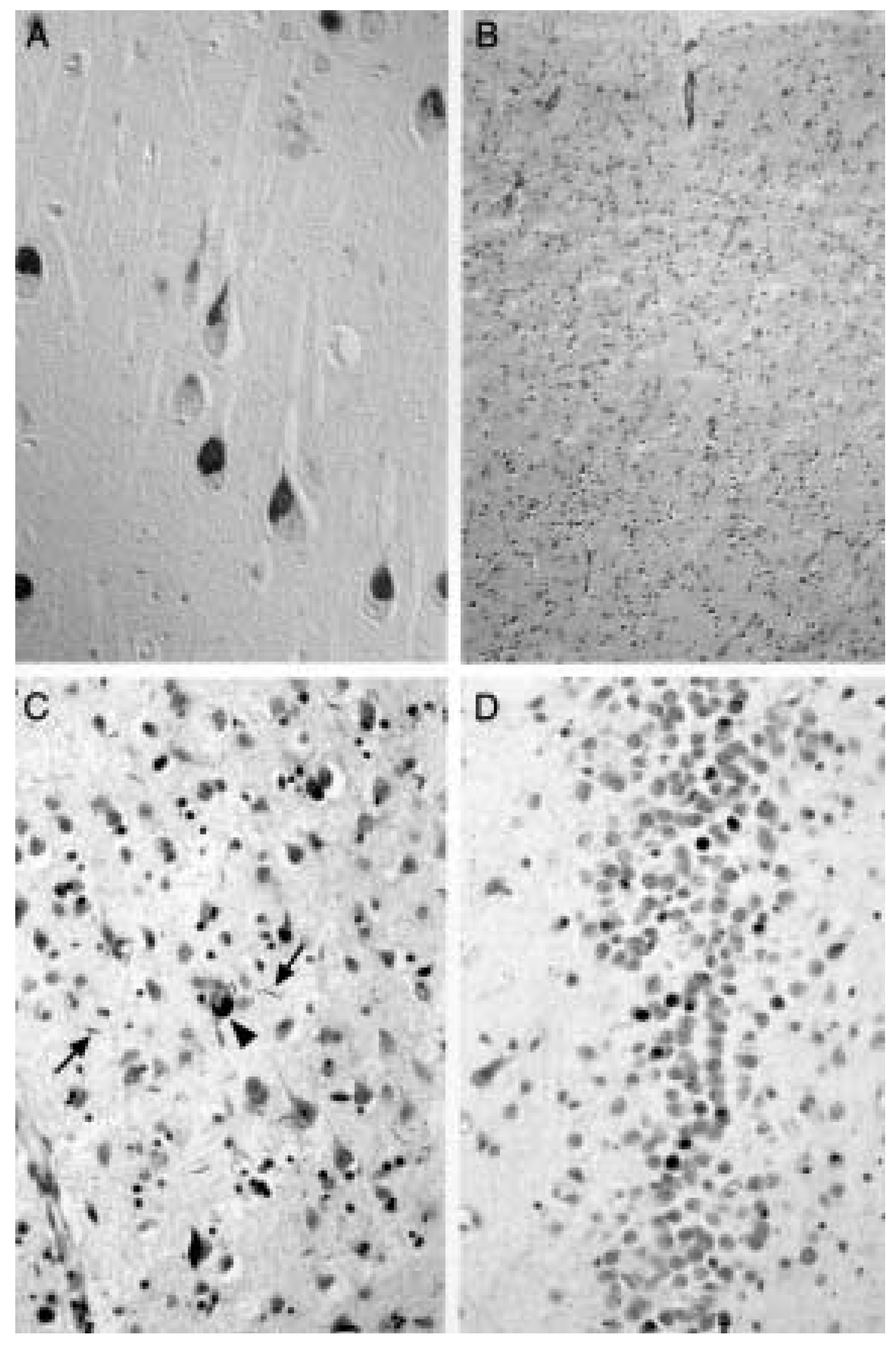
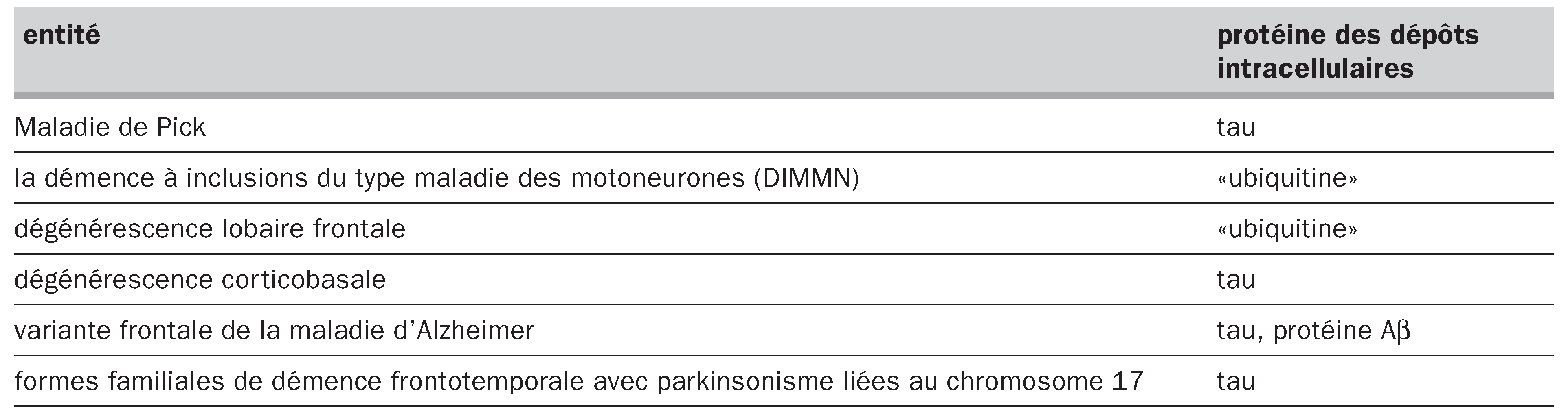
La maladie de Pick


La dégénérescence lobaire frontale ou la démence de type frontal
La démence à inclusions du type maladie des motoneurones (DIMMN)
Démence frontotemporale familiale sans mutations du gène de la protéine tau
La dégénérescence corticobasale
La variante frontale de la maladie d’Alzheimer
Remarques concernant la morphologie

 |
Le problème de la protéine tau manquante dans la démence frontotemporale
Références
- Foster, NL; Wilhelmsen, K; Sima, AAF; Jones, MZ; D’Amato, C; Gilman, S; et al. Frontotemporal dementia and Parkinsonism linked to chromosome 17: a consensus statement. Ann Neurol 1997, 41, 706–15. [Google Scholar]
- Spillantini, MG; VanSwieten, JC; Goedert, M. Tau mutations in frontotemporal dementia and parkinsonism linked to chromosome 17 (FTDP-17). Neurogenetics 2000, 2, 193–205. [Google Scholar]
- Spillantini, MG; Goedert, M. Tau gene mutations and tau pathology in frontotemporal dementia and parkinsonism linked to chromosome 17. Adv Exp Med Biol 2001, 487, 21–37. [Google Scholar]
- Stevens, M; van Duijn, CM; Kamphorst, W; de Knijff, P; Heutink, P; van Gool, WA; et al. Familial aggregation in frontotemporal dementia. Neurology 1998, 50, 1541–5. [Google Scholar] [CrossRef]
- Rizzu, P; Van Swieten, JC; Joosse, M; Hasegawa, M; Stevens, M; Tibben, A; et al. High prevalence of mutations in the microtubule-associated protein tau in a population study of frontotemporal dementia in the Netherlands. Am J Hum Genet 1999, 64, 414–21. [Google Scholar] [CrossRef]
- Morris, HR; Khan, MN; Janssen, JC; Brown, JM; Perez-Tur, J; Baker, M; et al. The genetic and pathological classification of familial frontotemporal dementia. Arch Neurol 2001, 58, 1813–6. [Google Scholar][Green Version]
- Kertesz, A; Kawarai, T; Rogaeva, E; St George-Hyslop, P; Poorkaj, P; Bird, TD; et al. Familial frontotemporal dementia with ubiquitin-positive, tau-negative inclusions. Neurology 2000, 54, 818–27. [Google Scholar]
- Rosso, SM; Kamphorst, W; de Graaf, B; Willemsen, R; Ravid, R; Niermeijer, MF; et al. Familial frontotemporal dementia with ubiquitin-positive inclusions is linked to chromosome 17q21–22. Brain 2001, 124, 1948–57. [Google Scholar] [CrossRef][Green Version]
- Savioz, A; Kovari, E; Anastasiu, R; Rossier, C; Saini, K; Bouras, C; et al. Search for a mutation in the tau gene in a Swiss family with frontotemporal dementia. Exp Neurol 2000, 161, 330–5. [Google Scholar]
- Kövari, E; Leuba, G; Savioz, A; Saini, K; Anastasiu, R; Miklossy, J; et al. Familial frontotemporal dementia with ubiquitin inclusion bodies and without motor neuron disease. Acta Neuropathol 2000, 100, 421–6. [Google Scholar] [CrossRef]
- Ashworth, A; Lloyd, S; Brown, J; Gydesen, S; Sorensen, SA; Brun, A; et al. Molecular genetic characterisation of frontotemporal dementia on chromosome 3. Dement Geriatr Cogn Disord 1999, 10 (Suppl 1), 93–101. [Google Scholar]
- Hosler, BA; Siddique, T; Sapp, PC; Sailor, W; Huang, MC; Hossain, A; et al. Linkage of amyotrophic lateral sclerosis with frontotemporal dementia to chromosome 9q21–22. JAMA 2000, 284, 1664–9. [Google Scholar] [CrossRef]
- Neary, D; Snowden, JS; Gustafson, L; Passant, U; Stuss, D; Black, S; et al. Frontotemporal lobar degeneration: a consensus on clinical diagnostic criteria. Neurology 1998, 51, 1546–54. [Google Scholar] [CrossRef]
- McKhann, GM; Albert, MS; Grossman, M; Miller, B; Dickson, D; Trojanowski, JQ. Clinical and pathological diagnosis of frontotemporal dementia: report of the Work Group on Frontotemporal Dementia and Pick’s disease. Arch Neurol 2001, 58, 1803–9. [Google Scholar] [CrossRef]
- Mesulam, MM. Primary progressive aphasia: differentiation from Alzheimer’s disease. Ann Neurol 1987, 22, 533–4. [Google Scholar]
- Hodges, JR; Patterson, K; Ward, R; Garrard, P; Bak, T; Perry, R; et al. The differentiation of semantic dementia and frontal lobe dementia (temporal and frontal variants of frontotemporal dementia) from early Alzheimer’s disease: a comparative neuropsychological study. Neuropsychology 1999, 13, 31–40. [Google Scholar]
- Mesulam, MM. Primary progressive aphasia. Ann Neurol 2001, 49, 425–32. [Google Scholar]
- Binetti, G; Locascio, JJ; Corkin, S; Vonsattel, JP; Growdon, JH. Differences between Pick’s disease and Alzheimer’s disease in clinical appearance and rate of cognitive decline. Arch Neurol 2000, 57, 225–32. [Google Scholar]
- Lowe, J; Lennox, G; Leigh, PN. Disorders of movements and system degenerations. In Greenfield’s Neuropathology, 6th ed. Vol. 2; Graham, DI, Lantos, P, Eds.; Arnold Publishers: London, 1997; pp. 281–366. [Google Scholar]
- Grimes, DA; Lang, AE; Bergeron, CB. Dementia as the most common presentation of cortico-basal ganglionic degeneration. Neurology 1999, 53, 1969–74. [Google Scholar] [CrossRef]
- Galton, CJ; Patterson, K; Graham, K; Lambon-Ralph, MA; Williams, G; Antoun, N; et al. Differing patterns of temporal atrophy in Alzheimer’s disease and semantic dementia. Neurology 2001, 57, 216–25. [Google Scholar] [CrossRef]
- Miller, BL; Cummings, JL; Villanueva-Meyer, J; Boone, K; Mehringer, CM; Lesser, IM; et al. Frontal lobe degeneration: clinical, neuropsychological, and SPECT characteristics. Neurology 1991, 41, 1374–82. [Google Scholar]
- Neary, D; Snowden, JS; Shields, RA; Burjan, AW; Northen, B; MacDermott, N; et al. Single photon emission tomography using 99mTc-HM-PAO in the investigation of dementia. J Neurol Neurosurg Psychiatry 1987, 50, 1101–9. [Google Scholar]
- Kamo, H; McGeer, PL; Harrop, R; McGeer, EG; Calne, DB; Martin, WR; et al. Positron emission tomography and histopathology in Pick’s disease. Neurology 1987, 37, 439–45. [Google Scholar]
- Frisoni, GB; Laakso, MP; Beltramello, A; Geroldi, C; Bianchetti, A; Soininen, H; et al. Hippocampal and entorhinal cortex atrophy in frontotemporal dementia and Alzheimer’s disease. Neurology 1999, 52, 91–100. [Google Scholar] [CrossRef]
- Hartikainen, P; Laakso, MP; Lehtovirta, M; Riekinnen, P; Partanen, K; Soininen, H. Volumes of the hippocampus in the MRI-based diagnosis of frontotemporal dementia with a reference to Alzheimer’s disease and Parkinson’s disease. Neurology 1998, 50 (Suppl 4), A161. [Google Scholar]
- Laakso, MP; Frisoni, GB; Kononen, M; Mikkonen, M; Beltramello, A; Geroldi, C; et al. Hippocampus and entorhinal cortex in frontotemporal dementia and Alzheimer’s disease: a morphometric MRI study. Biol Psychiatry 2000, 15, 1056–63. [Google Scholar]
- Knopman, DS; Mastri, AR; Frey, WH, 2nd; Sung, JH; Rustan, T. Dementia lacking distinctive histologic features: a common non-Alzheimer degenerative dementia. Neurology 1990, 40, 251–66. [Google Scholar]
- Mann, DM; South, PW. The topographic distribution of brain atrophy in frontal lobe dementia. Acta Neuropathol 1993, 85, 334–40. [Google Scholar]
- Sinnatamby, R; Antoun, NA; Freer, CE; Miles, KA; Hodges, JR. Neuroradiological findings in primary progressive aphasia. Neuroradiology 1996, 38, 232–8. [Google Scholar] [CrossRef]
- Chawluk, JB; Mesulam, MM; Hurtig, H; Kushner, M; Weintraub, S; Saykin, A; et al. Slowly progressive aphasia without generalized dementia: studies with positron emission tomography. Ann Neurol 1986, 19, 68–74. [Google Scholar] [CrossRef]
- Molina, JA; Probst, A; Villanueva, C; Jimenez-Jimenez, FJ; Madero, S; Torres, N; et al. Primary progressive aphasia with glial cytoplasmic inclusions. Eur Neurol 1998, 40, 71–7. [Google Scholar] [CrossRef]
- Tyrell, PJ; Warrington, EK; Frackoviak, RS; Rossor, MN. Heterogeneity in progressive aphasia due to focal cortical atrophy. A clinical and PET study. Brain 1990, 113, 1321–36. [Google Scholar]
- Kempler, D; Metter, EJ; Riege, WH; Jackson, CA; Benson, DF; Hanson, WR. Slowly progressive aphasia: three cases with language, memory, CT, and PET data. J Neurol Neurosurg Psychiatry 1990, 53, 987–93. [Google Scholar] [CrossRef]
- Mummery, CJ; Patterson, K; Price, CJ; Ashburner, J; Frackowiak, RS; Hodges, JR. A voxel-based morphometry study of semantic dementia: relationship between temporal lobe atrophy and semantic memory. Ann Neurol 2000, 47, 36–45. [Google Scholar] [CrossRef]
- Chan, D; Fox, NC; Jenkins, R; Scahill, RI; Crum, WR; Rossor, MN. Rates of global and regional cerebral atrophy in AD and frontotemporal dementia. Neurology 2001, 57, 1756–63. [Google Scholar] [CrossRef]
- Rosen, HJ; Gorno-Tempini, ML; Goldman, WP; Perry, RJ; Schuff, N; Weiner, M; et al. Patterns of brain atrophy in frontotemporal dementia and semantic dementia. Neurology 2002, 58, 198–208. [Google Scholar] [CrossRef]
- Pick, A. Ueber die Beziehungen der senilen Hirnatrophie zur Aphasie. Prager Med Wochenschr 1892, 17, 165–7. [Google Scholar]
- Alzheimer, A. Ueber eigenartige Krankheitsfälle des späteren Alters. Z ges Neurol Psychiat 1911, 4, 356–85. [Google Scholar]
- Dickson, DW. Pick’s disease: a modern approach. Brain Pathol 1998, 8, 339–54. [Google Scholar]
- Probst, A; Tolnay, M; Langui, D; Goedert, M; Spillantini, M. Pick’s disease: hyperphosphorylated tau protein segregates to the somatoaxonal compartment. Acta Neuropathol 1996, 92, 588–96. [Google Scholar] [CrossRef]
- Delacourte, A; Sergeant, N; Wattez, A; Gauvreau, D; Robitaille, Y. Vulnerable neuronal subsets in Alzheimer’s and Pick’s disease are distinguished by their tau isoform distribution and phosphorylation. Ann Neurol 1998, 43, 193–204. [Google Scholar]
- Chin, SS; Goldman, JE. Glial inclusions in CNS degenerative diseases. J Neuropathol Exp Neurol 1996, 55, 499–508. [Google Scholar]
- Tolnay, M; Probst, A. Review: tau protein pathology in Alzheimer’s disease and related disorders. Neuropathol Appl Neurobiol 1999, 25, 171–87. [Google Scholar]
- Lee, VM-L; Goedert, M; Trojanowski, JQ. Neurodegenerative tauopathies. Annu Rev Neurosci 2001, 24, 1121–59. [Google Scholar]
- Murrell, JR; Spillantini, MG; Zolo, P; Guazzelli, M; Smith, MJ; Hasegawa, M; et al. Tau gene mutation G389R causes a tauopathy with abundant Pick-like inclusions and axonal deposits. J Neuropathol Exp Neurol 1999, 58, 1207–26. [Google Scholar] [CrossRef]
- Rizzini, C; Goedert, M; Hodges, JR; Smith, MJ; Jakes, R; Hills, R; et al. Tau gene mutation K257T causes a tauopathy similar to Pick’s disease. J Neuropathol Exp Neurol 2000, 59, 990–1001. [Google Scholar]
- Pickering-Brown, S; Baker, M; Yen, SH; Liu, WK; Hasegawa, M; Cairns, N; et al. Pick’s disease is associated with mutations in the tau gene. Ann Neurol 2000, 48, 859–67. [Google Scholar]
- Neumann, M; Schulz-Schaeffer, W; Crowther, RA; Smith, MJ; Spillantini, MG; Goedert, M; et al. Pick’s disease associated with the novel tau gene mutation K369I. Ann Neurol 2001, 50, 503–13. [Google Scholar]
- Hutton, M; Lendon, CL; Rizzu, P; Baker, M; Froelich, S; Houlden, H; et al. Association of missense and 5’-splicesite mutations in tau with the inherited dementia FTDP-17. Nature 1998, 393, 702–5. [Google Scholar]
- Sergeant, N; David, JP; Lefranc, D; Vermersch, P; Wattez, A; Delacourte, A. Different distribution of phosphorylated tau protein isoforms in Alzheimer’s and Pick’s diseases. FEBS Lett 1997, 412, 578–82. [Google Scholar] [CrossRef]
- Mailliot, C; Sergeant, N; Bussiere, T; Caillet-Boudin, ML; Delacourte, A; Buee, L. Phosphorylation of specific sets of tau isoforms reflects different neurofibrillary degeneration processes. FEBS Lett 1998, 433, 201–4. [Google Scholar] [CrossRef]
- Tolnay, M; Probst, A. Frontal lobe degeneration: novel ubiquitin-immunoreactive neurites within frontotemporal cortex. Neuropathol Appl Neurobiol 1995, 21, 492–7. [Google Scholar]
- Giannakopoulos, P; Hof, PR; Bouras, C. Dementia lacking distinctive histopathology: clinicopathological evaluation of 32 cases. Acta Neuropathol 1995, 89, 346–55. [Google Scholar]
- Jackson, M; Lowe, J. The new neuropathology of degenerative frontotemporal dementias. Acta Neuropathol 1996, 91, 127–34. [Google Scholar] [CrossRef]
- Neary, D; Snowden, JS; Mann, DM; Northern, B; Goulding, PJ; MacDermott, N. Frontal lobe dementia and motor neuron disease. J Neurol Neurosurg Psychiatry 1990, 53, 23–32. [Google Scholar]
- Okamoto, K; Hirai, S; Yamazaki, T; Sun, XY; Nakazato, Y. New ubiquitin-positive intraneuronal inclusions in the extra-motor cortices in patients with amyotrophic lateral sclerosis. Neurosci Lett 1991, 129, 233–6. [Google Scholar] [CrossRef]
- Wightman, G; Anderson, VE; Martin, J; Swash, M; Anderton, BH; Neary, D; et al. Hippocampal and neocortical ubiquitin-immunoreactive inclusions in amyotrophic lateral sclerosis with dementia. Neurosci Lett 1992, 139, 269–74. [Google Scholar] [CrossRef]
- Ulrich, J; Probst, A; Langui, D; Greenberg, S; Dukas, L; Stähelin, HB. Rapidly progressing dementia with filipodia-like cytoskeletal neuritic anomalies, but without Alzheimer changes. Clin Neuropathol 1993, 143, 39–45. [Google Scholar]
- Komori, T; Arai, N; Oda, M; Nakayama, H; Mori, H; Yagishita, S; et al. Astrocytic plaques and tufts of abnormal fibers do not coexist in corticobasal degeneration and progressive supranuclear palsy. Acta Neuropathol 1998, 96, 401–8. [Google Scholar]
- Spillantini, MG; Yoshida, H; Rizzini, C; Lantos, PL; Khan, N; Rossor, MN; et al. A novel tau mutation (N296N) in familial dementia with swollen achromatic neurons and corticobasal inclusion bodies. Ann Neurol 2000, 48, 939–43. [Google Scholar]
- D’Souza, I; Poorkaj, P; Hong, M; Nochlin, D; Lee, VM; Bird, TD; et al. Missense and silent tau gene mutations cause frontotemporal dementia with parkinsonism-chromosome 17 type, by affecting multiple alternative RNA splicing regulatory elements. Proc Natl Acad Sci U S A 1999, 96, 5598–603. [Google Scholar]
- Hasegawa, M; Smith, MJ; Iijima, M; Tabira, T; Goedert, M. FTDP-17 mutations N279K and S305N in tau produce increased splicing of exon 10. FEBS Lett 1999, 443, 93–6. [Google Scholar] [CrossRef]
- Iijima, M; Tabira, T; Poorkaj, P; Schellenberg, GD; Trojanowski, JQ; Lee, VM; et al. A distinct familial presenile dementia with a novel missense mutation in the tau gene. Neuroreport 1999, 10, 497–501. [Google Scholar]
- Sergeant, N; Wattez, A; Delacourte, A. Neurofibrillary degeneration in progressive supranuclear palsy and corticobasal degeneration: tau pathologies with exclusively “exon 10” isoforms. J Neurochem 1999, 72, 1243–9. [Google Scholar]
- Johnson, JK; Head, E; Kim, R; Starr, A; Cotman, CW. Clinical and pathological evidence for a frontal variant of Alzheimer’s disease. Arch Neurol 1999, 56, 1233–9. [Google Scholar]
- Zhukareva, V; Vogelsberg-Ragaglia, V; Van Deerlin, VM; Bruce, J; Shuck, T; Grossman, M; et al. Loss of brain tau defines novel sporadic and familial tauopathies with frontotemporal dementia. Ann Neurol 2001, 49, 165–75. [Google Scholar]
- Lendon, CL; Lynch, T; Norton, J; McKeel, DW, Jr.; Busfield, F; Craddock, N; et al. Hereditary dysphasic disinhibition dementia. A frontotemporal dementia linked to 17q21–22. Neurology 1998, 50, 1546–55. [Google Scholar] [CrossRef]
- Adamec, E; Chang, HT; Stopa, EG; Hedreen, JC; Vonsattel, JP. Tau protein expression in frontotemporal dementias. Neurosci Lett 2001, 315, 21–34. [Google Scholar]
This is an open-access article distributed under the terms of the Creative Commons Attribution Non-Commercial No Derivatives (CC BY-NC-ND) 4.0 License.
Share and Cite
Probst, A.; Tolnay, M. La démence frontotemporale: aspects cliniques et morphologiques. Swiss Arch. Neurol. Psychiatry Psychother. 2002, 153, 165-174. https://doi.org/10.4414/sanp.2002.01276
Probst A, Tolnay M. La démence frontotemporale: aspects cliniques et morphologiques. Swiss Archives of Neurology, Psychiatry and Psychotherapy. 2002; 153(4):165-174. https://doi.org/10.4414/sanp.2002.01276
Chicago/Turabian StyleProbst, Alphonse, and M. Tolnay. 2002. "La démence frontotemporale: aspects cliniques et morphologiques" Swiss Archives of Neurology, Psychiatry and Psychotherapy 153, no. 4: 165-174. https://doi.org/10.4414/sanp.2002.01276
APA StyleProbst, A., & Tolnay, M. (2002). La démence frontotemporale: aspects cliniques et morphologiques. Swiss Archives of Neurology, Psychiatry and Psychotherapy, 153(4), 165-174. https://doi.org/10.4414/sanp.2002.01276