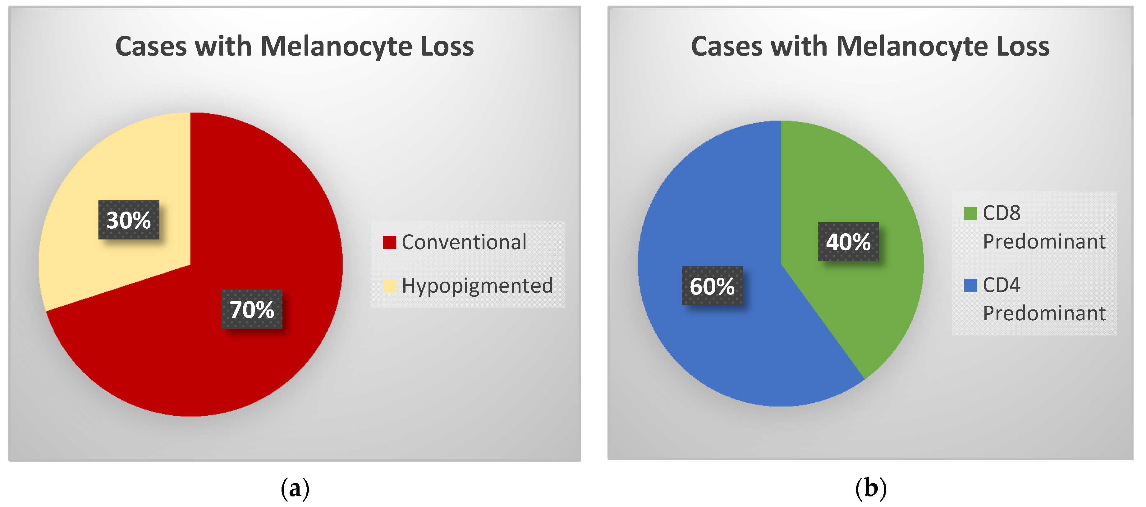

Evaluation of Melanocyte Loss in Mycosis Fungoides Using SOX10 Immunohistochemistry


Abstract
1. Introduction
2. Materials and Methods
3. Results
4. Discussion
Author Contributions
Funding
Institutional Review Board Statement
Informed Consent Statement
Acknowledgments
Conflicts of Interest
References
- Bagherani, N.; Smoller, B.R. An overview of cutaneous T cell lymphomas. F1000Res 2016, 5. [Google Scholar] [CrossRef] [Scilit]
- Hristov, A.C.; Tejasvi, T.; Wilcox, R.A. Mycosis fungoides and Sezary syndrome: 2019 update on diagnosis, risk-stratification, and management. Am. J. Hematol. 2019, 94, 1027–1041. [Google Scholar] [CrossRef] [Scilit]
- Kaufman, A.E.; Patel, K.; Goyal, K.; O’Leary, D.; Rubin, N.; Pearson, D.; Bohjanen, K.; Goyal, A. Mycosis fungoides: Developments in incidence, treatment and survival. J. Eur. Acad. Dermatol. Venereol. 2020, 34, 2288–2294. [Google Scholar] [CrossRef] [Scilit] [PubMed]
- Smoller, B.R. Mycosis fungoides: What do/do not we know? J. Cutan. Pathol. 2008, 35 (Suppl. 2), 35–39. [Google Scholar] [CrossRef] [Scilit] [PubMed]
- Agar, N.S.; Wedgeworth, E.; Crichton, S.; Mitchell, T.J.; Cox, M.; Ferreira, S.; Robson, A.; Calonje, E.; Stefanato, C.M.; Wain, E.M.; et al. Survival outcomes and prognostic factors in mycosis fungoides/Sezary syndrome: Validation of the revised International Society for Cutaneous Lymphomas/European Organisation for Research and Treatment of Cancer staging proposal. J. Clin. Oncol. 2010, 28, 4730–4739. [Google Scholar] [CrossRef] [Scilit] [PubMed]
- Broggi, G.; Lacarrubba, F.; Verzi, A.E.; Micali, G.; Caltabiano, R. Confocal microscopy features of patch-stage mycosis fungoides and their correlation with horizontal histopathological sections. A case series. J. Cutan. Pathol. 2019, 46, 163–165. [Google Scholar] [CrossRef] [Scilit] [PubMed]
- Bilgic, S.A.; Cicek, D.; Demir, B. Dermoscopy in differential diagnosis of inflammatory dermatoses and mycosis fungoides. Int. J. Dermatol. 2020, 59, 843–850. [Google Scholar] [CrossRef] [Scilit] [PubMed]
- Hodak, E.; Amitay-Laish, I. Mycosis fungoides: A great imitator. Clin. Dermatol. 2019, 37, 255–267. [Google Scholar] [CrossRef] [Scilit] [PubMed]
- Castano, E.; Glick, S.; Wolgast, L.; Naeem, R.; Sunkara, J.; Elston, D.; Jacobson, M. Hypopigmented mycosis fungoides in childhood and adolescence: A long-term retrospective study. J. Cutan. Pathol. 2013, 40, 924–934. [Google Scholar] [CrossRef] [Scilit] [PubMed]
- Rodney, I.J.; Kindred, C.; Angra, K.; Qutub, O.N.; Villanueva, A.R.; Halder, R.M. Hypopigmented mycosis fungoides: A retrospective clinicohistopathologic study. J. Eur. Acad. Dermatol. Venereol. 2017, 31, 808–814. [Google Scholar] [CrossRef] [Scilit] [PubMed]
- Luo, Y.; Liu, Z.; Liu, J.; Liu, Y.; Zhang, W.; Zhang, Y. Mycosis Fungoides and Variants of Mycosis Fungoides: A Retrospective Study of 93 Patients in a Chinese Population at a Single Center. Ann. Dermatol. 2020, 32, 14–20. [Google Scholar] [CrossRef] [Scilit] [PubMed]
- Mollaaghababa, R.; Pavan, W.J. The importance of having your SOX on: Role of SOX10 in the development of neural crest-derived melanocytes and glia. Oncogene 2003, 22, 3024–3034. [Google Scholar] [CrossRef] [Scilit] [PubMed]
- Shakhova, O.; Zingg, D.; Schaefer, S.M.; Hari, L.; Civenni, G.; Blunschi, J.; Claudinot, S.; Okoniewski, M.; Beermann, F.; Mihic-Probst, D.; et al. Sox10 promotes the formation and maintenance of giant congenital naevi and melanoma. Nat. Cell Biol. 2012, 14, 882–890. [Google Scholar] [CrossRef] [Scilit] [PubMed]
- Furlan, F.C.; de Paula Pereira, B.A.; da Silva, L.F.; Sanches, J.A. Loss of melanocytes in hypopigmented mycosis fungoides: A study of 18 patients. J. Cutan. Pathol. 2014, 41, 101–107. [Google Scholar] [CrossRef] [Scilit] [PubMed]
- Pimpinelli, N.; Olsen, E.A.; Santucci, M.; Vonderheid, E.; Haeffner, A.C.; Stevens, S.; Burg, G.; Cerroni, L.; Dreno, B.; Glusac, E.; et al. Defining early mycosis fungoides. J. Am. Acad. Dermatol. 2005, 53, 1053–1063. [Google Scholar] [CrossRef] [Scilit] [PubMed]
- Amorim, G.M.; Niemeyer-Corbellini, J.P.; Quintella, D.C.; Cuzzi, T.; Ramos, E.S.M. Hypopigmented mycosis fungoides: A 20-case retrospective series. Int. J. Dermatol. 2018, 57, 306–312. [Google Scholar] [CrossRef] [Scilit] [PubMed]
- Martinez Villarreal, A.; Gantchev, J.; Lagace, F.; Barolet, A.; Sasseville, D.; Odum, N.; Charli-Joseph, Y.V.; Hernandez Salazar, A.; Litvinov, I.V. Hypopigmented Mycosis Fungoides: Loss of Pigmentation Reflects Antitumor Immune Response in Young Patients. Cancers 2020, 12, 2007. [Google Scholar] [CrossRef] [Scilit] [PubMed]
- Chuang, G.S.; Wasserman, D.I.; Byers, H.R.; Demierre, M.F. Hypopigmented T-cell dyscrasia evolving to hypopigmented mycosis fungoides during etanercept therapy. J. Am. Acad. Dermatol. 2008, 59, S121–S122. [Google Scholar] [CrossRef] [Scilit] [PubMed]
- Martinez-Escala, M.E.; Posligua, A.L.; Wickless, H.; Rutherford, A.; Sable, K.A.; Rubio-Gonzalez, B.; Zhou, X.A.; Kaplan, J.B.; Pro, B.; Choi, J.; et al. Progression of undiagnosed cutaneous lymphoma after anti-tumor necrosis factor-alpha therapy. J. Am. Acad. Dermatol. 2018, 78, 1068–1076. [Google Scholar] [CrossRef] [Scilit] [PubMed]
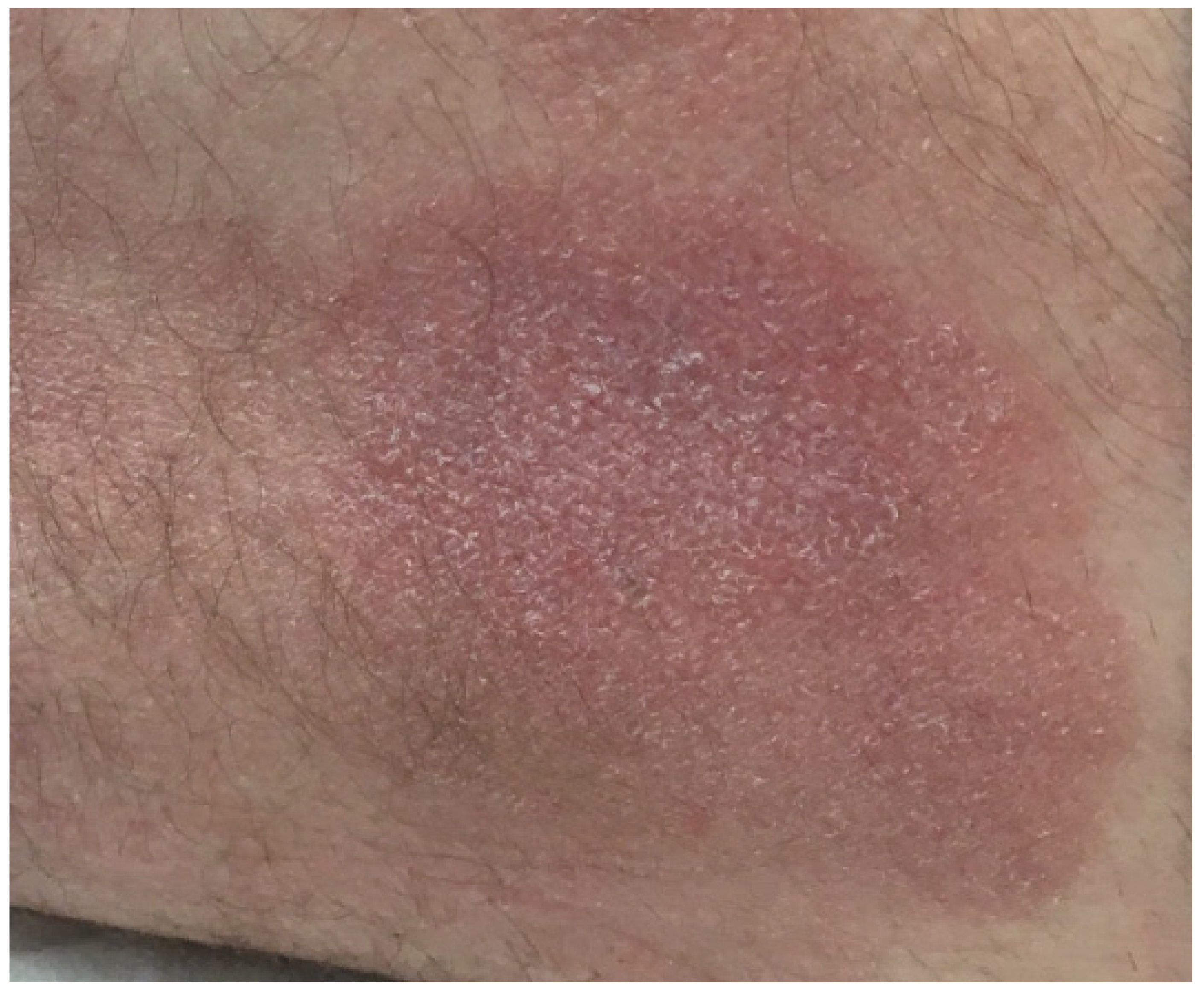
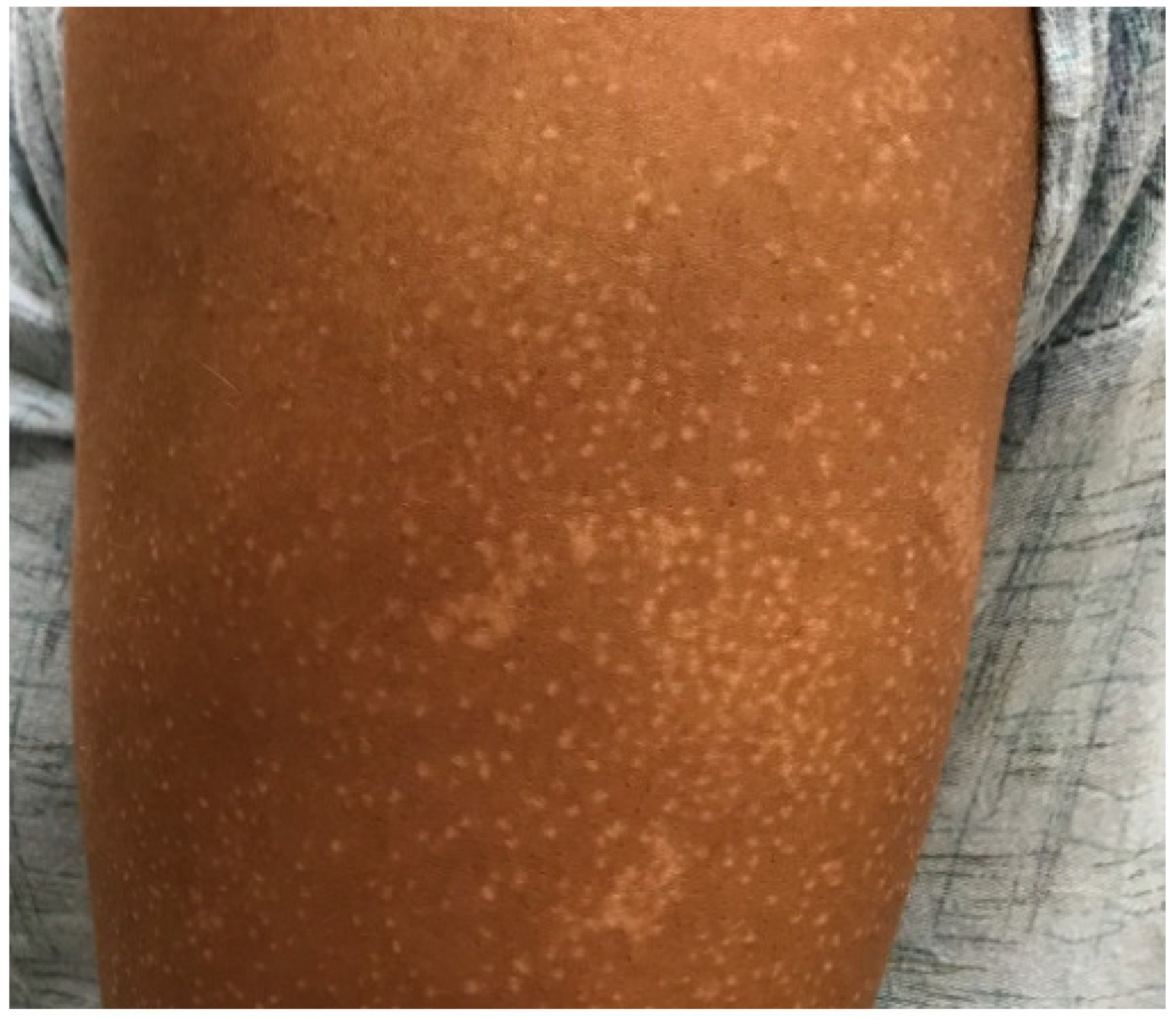

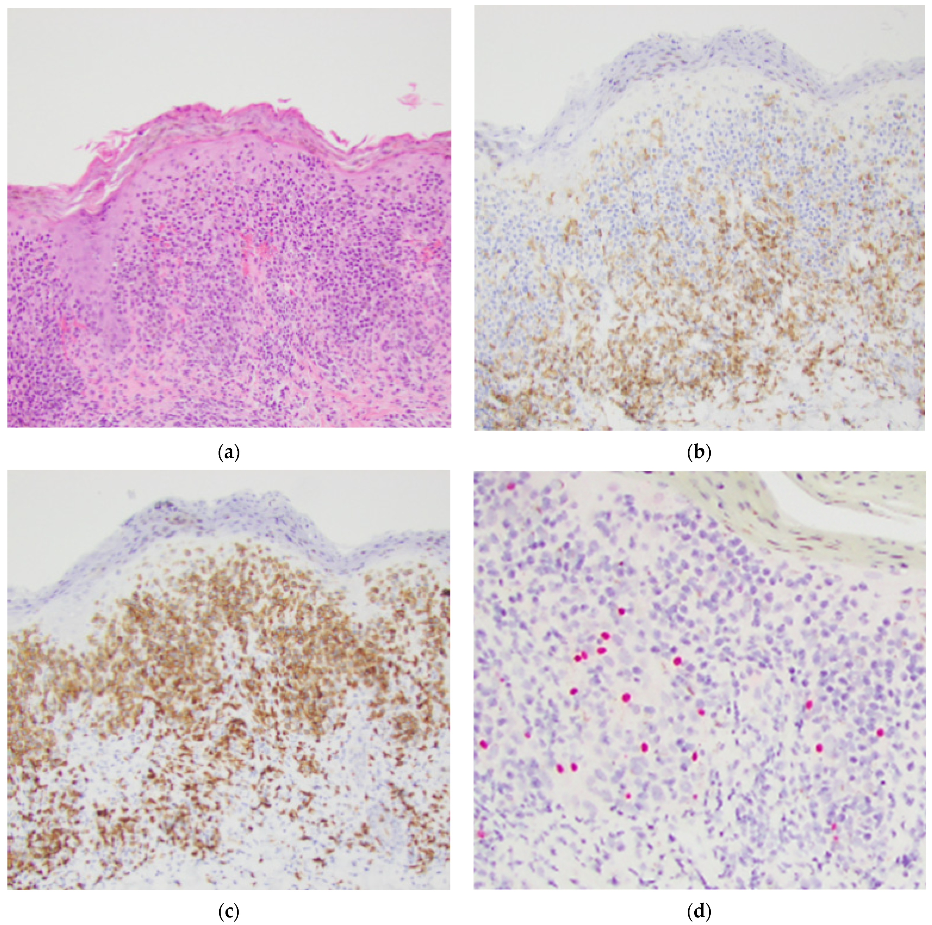

| Results | Hypopigmented | Conventional | All |
|---|---|---|---|
| Melanocyte loss | 3 (43%) | 7 (54%) | 10 (50%) |
| CD4 predominant | 4 (57%) | 9 (69%) | 13 (65%) |
| Ethnicity non-Caucasian | 6 (86%) | 2 (15%) | 8 (40%) |
| Gender (female) | 5 (71%) | 4 (31%) | 9 (45%) |
| Age (Average) | 43 | 54 | 50 |
| Range | 30–62 | 15–86 | 15–86 |
Publisher’s Note: MDPI stays neutral with regard to jurisdictional claims in published maps and institutional affiliations. |
© 2021 by the authors. Licensee MDPI, Basel, Switzerland. This article is an open access article distributed under the terms and conditions of the Creative Commons Attribution (CC BY) license (https://creativecommons.org/licenses/by/4.0/).
Share and Cite
Barron, C.R.; Smoller, B.R. Evaluation of Melanocyte Loss in Mycosis Fungoides Using SOX10 Immunohistochemistry. Dermatopathology 2021, 8, 277-284. https://doi.org/10.3390/dermatopathology8030034
Barron CR, Smoller BR. Evaluation of Melanocyte Loss in Mycosis Fungoides Using SOX10 Immunohistochemistry. Dermatopathology. 2021; 8(3):277-284. https://doi.org/10.3390/dermatopathology8030034
Chicago/Turabian StyleBarron, Cynthia Reyes, and Bruce R. Smoller. 2021. "Evaluation of Melanocyte Loss in Mycosis Fungoides Using SOX10 Immunohistochemistry" Dermatopathology 8, no. 3: 277-284. https://doi.org/10.3390/dermatopathology8030034
APA StyleBarron, C. R., & Smoller, B. R. (2021). Evaluation of Melanocyte Loss in Mycosis Fungoides Using SOX10 Immunohistochemistry. Dermatopathology, 8(3), 277-284. https://doi.org/10.3390/dermatopathology8030034