Case Report

Histological Changes Related to Symptomatic Improvement of Spontaneous Keloids Treated with a Low-Dosage Regimen of UVA-1 Phototherapy

by
Carlos Cuenca-Barrales
1,*,
José Aneiros-Fernández
2,
Israel Pérez-López
1,
Julia Rodríguez-Pérez
3,
Ricardo Ruiz-Villaverde
1 and
Jorge Luis Espelt-Otero
1
1
Dermatology Department, Hospital Universitario San Cecilio, 18016 Granada, Spain
2
Pathology Department, Hospital Universitario San Cecilio, 18016 Granada, Spain
3
Nursing Department, Hospital Universitario San Cecilio, 18016 Granada, Spain
*
Author to whom correspondence should be addressed.
Dermatopathology 2020, 7(3), 53-56; https://doi.org/10.3390/dermatopathology7030009
Submission received: 18 October 2020 / Revised: 27 November 2020 / Accepted: 1 December 2020 / Published: 3 December 2020
(This article belongs to the Section Clinico-Pathological Correlation in Dermatopathology)

Abstract

:
Keloids are a difficult-to-treat disease characterized by an imbalance in mechanisms of tissue reparation. We present the case of a middle-aged woman with spontaneous keloids which histologically and clinically improved after UVA-1 phototherapy treatment. There are few reported cases of keloids treated with high doses of UVA-1 phototherapy. We used a low-dosage regimen with a good response in only one cycle, which could diminish the risk of skin cancer development.

1. Introduction

Keloids represent a form of abnormal wound healing, characterized by local fibroblast proliferation and excessive collagen production due to a loss of control in regulatory mechanisms of tissue reparation. Multiple treatments have been reported for this pathology, such as local corticosteroids, fluorouracil, imiquimod, silicone gel, cryotherapy, radiation and laser therapy; however, many of them are not effective enough and recurrences are frequent [1].

2. Case Report

A healthy middle-aged woman with Fitzpatrick skin type III presented at our dermatologic outpatient clinic complaining of keloids on her back, chest and breasts. She had no family history of keloids and reported that these lesions had been spontaneously appearing since she was 15 years old; the latest one appeared two years ago on her breasts and were still growing (Figure 1A).
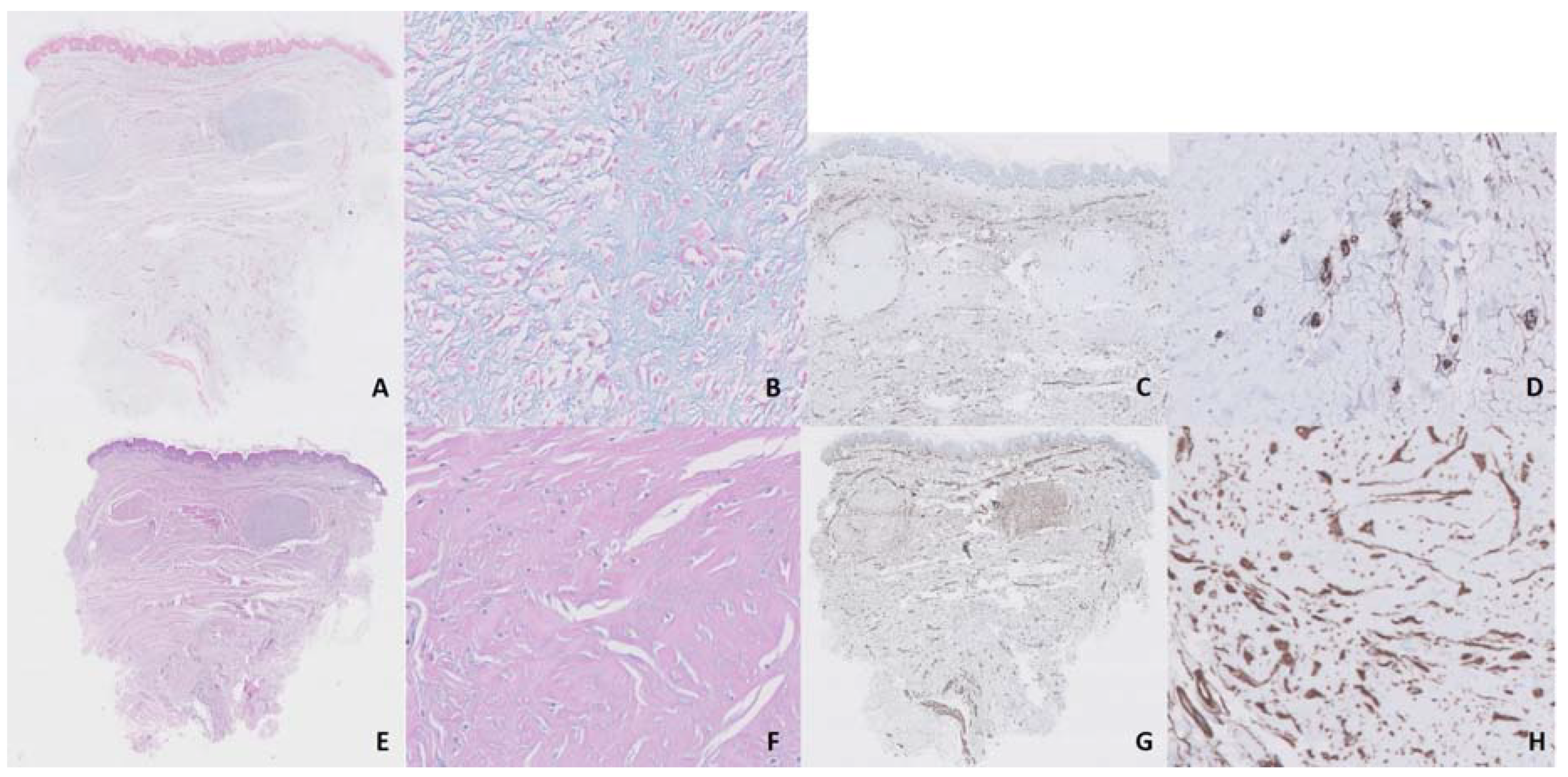
Several treatments with topical and intralesional corticosteroids and intralesional bleomycin had not been effective. We started treatment with UVA-1 phototherapy at 370 nm in a low-dosage regimen (starting dosage of 5 J/cm2, 3 sessions/week, with an increase of 5% in each session and a maximum dose of 15.95 J/cm2). After 25 sessions, with a total accumulated dose of 237.5 J, we observed an important reduction in pruritus and discomfort reported by the patient and a flattening and softening of keloids, although erythema response was limited and there was no significant aesthetic improvement (Figure 1B). Moreover, this response was maintained 3 months after treatment discontinuation. Histologic comparison between samples taken from the same keloid on the breast before and after treatment showed significant improvement, with denser and less disorganized collagen, a reduction in mucin and cellularity (after counting 10 mm2, we observed a reduction from 350 to 265 cells/mm2 after treatment with UVA-1 phototherapy) and neoangiogenesis (after counting 10 mm2, we observed an increase in vascular structures from 7 to 12/mm2 after treatment with UVA-1 phototherapy) (Figure 2 and Figure 3).
The case presented follows the Declaration of Helsinki. As this is a single, completely anonymous case, the approval of the Institutional Review Board was not required. The patient gave her informed consent.

3. Discussion

UVA-1 is a modality of phototherapy with a long wavelength, which allows it to achieve greater penetration and makes it especially effective for the treatment of sclerotic skin diseases [2]. This longer wavelength is also associated with a lower risk of burning during treatment than other phototherapy modalities. The main mechanism of action is the activation of fibroblasts, which produce metalloproteinases, with the consequent reduction in collagen production [3]. Other mechanisms include immediate and retarded apoptosis (due to the generation of reactive oxygen species), neovascularization, activation of the FAS/FAS ligand system (which activates programmed cell death) and reduction in TGFβ, TNFα, IL-5, -6, -8, -13 and -31 and IFNγ [2]. Therefore, according to what we observed in our samples, it seems that on one hand, there is a reduction in the excess cellularity due to apoptosis, and on the other hand, there is an activation of the remaining fibroblasts which produces a rupture of the disorganized collagen due to the action of metalloproteinases and a new, denser and better organized collagen. Modulation of endothelial dysfunction induced by UVA-1, with an increase in CD34 expression [4], may also play a role, as endothelial dysfunction and abnormal blood vessel regulation have been proposed as pathogenic factors in keloid development [5]. In our samples, we observed an increase in vascular structures after the treatment. Finally, mucin, which is found at increased levels in keloids [6], was reduced after the UVA-1 sessions. All these mechanisms are responsible for the striking changes observed in the histological images of our case, which correlated with patient-reported outcomes. The aesthetic impairment barely changed, maybe because these changes occur mainly in the dermis instead of the epidermis; however, when the patient was asked, she said she was satisfied with the treatment.
There are various UVA-1 dosage regimen recommendations. Generally, each cycle consists of 15–20 sessions with increasing doses, and no more than two cycles per year are recommended [2]. In our case, the use of only one cycle of 25 sessions with a low-dosage regimen showed striking histological changes and an improvement in the patient’s symptoms. There have been reports of several skin diseases being successfully treated with UVA-1 phototherapy—for example, sclerotic diseases, such as systemic sclerosis and morphea, inflammatory conditions, such as eczema and prurigo, granulomatous diseases, such as sarcoidosis and annular granuloma, and T cell lymphomas and related disorders, such as pityriasis lichenoides and lymphomatoid papulosis [7]. To the best of our knowledge, there have only been two cases of keloids treated with UVA-1 laser [8] and a few cases treated with UVA-1 phototherapy, all with high doses [9,10,11], but no cases treated with a low-dosage regimen have been reported to date. UVA-1 phototherapy has less risk of burns and skin cancer development than other types of phototherapy [9], and the use of low doses further decreases this risk.

4. Conclusions

UVA-1 phototherapy can improve keloids both histologically and clinically and could be a first-line treatment for this challenging condition. Future research will determine the role of UVA-1 phototherapy in the management of keloids and the most appropriate dosages.

Author Contributions

J.L.E.-O. and J.R.-P. were responsible for the treatment and follow-up of the patient; J.A.-F. carried out the histological analyses; C.C.-B., I.P.-L., R.R.-V. and J.L.E.-O. collected the data and wrote the manuscript. All the authors critically revised the manuscript and approved the final version. All authors have read and agreed to the published version of the manuscript.

Funding

This research received no external funding.

Acknowledgments

We would like to thank Charlotte Bower for improving the English of the manuscript.

Conflicts of Interest

The authors declare no conflict of interest.

References

  1. Viera, M.H.; Vivas, A.C.; Berman, B. Update on Keloid Management: Clinical and Basic Science Advances. Adv. Wound Care 2012, 1, 200–206. [Google Scholar] [CrossRef] [PubMed] [Green Version]
  2. Dawe, R.S. Ultraviolet A1 phototherapy. Br. J. Dermatol. 2003, 148, 626–637. [Google Scholar] [CrossRef] [PubMed]
  3. Petersen, M.J.; Hansen, C.; Craig, S. Ultraviolet A irradiation stimulates collagenase production in cultured human fibroblasts. J. Investig. Dermatol. 1992, 99, 440–444. [Google Scholar] [CrossRef] [PubMed] [Green Version]
  4. Breuckmann, F.; Stuecker, M.; Altmeyer, P.; Kreuter, A. Modulation of endothelial dysfunction and apoptosis: UVA1-mediated skin improvement in systemic sclerosis. Arch. Dermatol. Res. 2004, 296, 235–239. [Google Scholar] [CrossRef] [PubMed]
  5. Ogawa, R.; Akaishi, S. Endothelial dysfunction may play a key role in keloid and hypertrophic scar pathogenesis-Keloids and hypertrophic scars may be vascular disorders. Med. Hypotheses 2016, 96, 51–60. [Google Scholar] [CrossRef] [PubMed]
  6. Kelly, A.P. Keloids. Dermatol. Clin. 1988, 6, 413–424. [Google Scholar] [CrossRef]
  7. Kerr, A.C.; Ferguson, J.; Attili, S.K.; Beattie, P.E.; Coleman, A.J.; Dawe, R.S.; Eberlein, B.; Goulden, V.; Ibbotson, S.H.; Menage Hdu, P.; et al. Ultraviolet A1 phototherapy: A British Photodermatology Group workshop report. Clin. Exp. Dermatol. 2012, 37, 219–226. [Google Scholar] [CrossRef] [PubMed]
  8. Polat, M.; Kaya, H.; Sahin, A. A New Approach in the Treatment of Keloids: UVA-1 Laser. Photomed. Laser Surg. 2016, 34, 130–133. [Google Scholar] [CrossRef]
  9. Asawanonda, P.; Khoo, L.S.; Fitzpatrick, T.B.; Taylor, C.R. UV-A1 for keloid. Arch. Dermatol. 1999, 135, 348–349. [Google Scholar] [CrossRef]
  10. Hannuksela-Svahn, A.; Grandal, O.J.; Thorstensen, T.; Christensen, O.B. UVA1 for treatment of keloids. Acta Derm. Venereol. 1999, 79, 490. [Google Scholar] [CrossRef] [PubMed]
  11. Tuchinda, C.; Kerr, H.A.; Taylor, C.R.; Jacobe, H.; Bergamo, B.M.; Elmets, C.; Rivard, J.; Lim, H.W. UVA1 phototherapy for cutaneous diseases: An experience of 92 cases in the United States. Photodermatol. Photoimmunol. Photomed. 2006, 22, 247–253. [Google Scholar] [CrossRef] [PubMed]
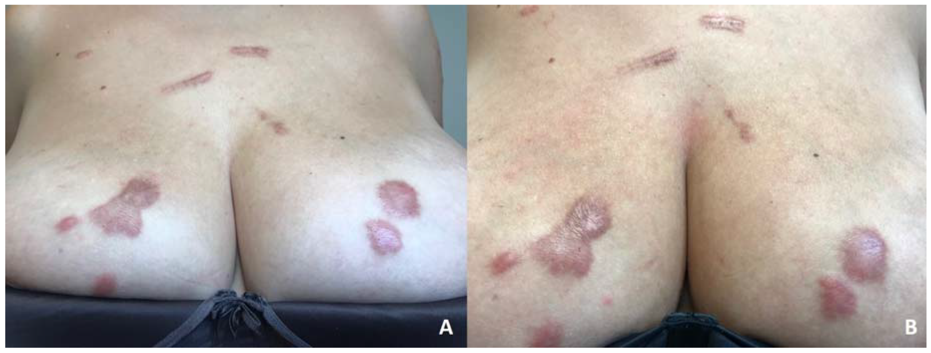
Figure 1. Spontaneous keloids on the chest and breasts of the patient before (A) and after (B) the treatment with UVA-1 phototherapy.
Dermatopathology 07 00009 g001
Figure 2. Before treatment, we observed basal deposits of mucin (A,B, 10× and 40×, respectively, alcian blue staining), few vascular structures (C,D, 10× and 40×, respectively, CD34 staining), disorganized collagen (E,F, 10× and 40×, respectively, hematoxylin–eosin staining) and a rich cellularity (G,H, 10× and 40×, respectively, vimentin staining).
Dermatopathology 07 00009 g002
Figure 3. After treatment with a low-dosage regimen of UVA-1 phototherapy, we observed a reduction in mucin (A,B, 10× and 40×, respectively, alcian blue staining), neoangiogenesis (C,D, 10× and 40×, respectively, CD34 staining), denser and less disorganized collagen (E,F, 10× and 40×, respectively, hematoxylin–eosin staining) and a reduction in cellularity (G,H, 10× and 40×, respectively, vimentin staining) in comparison with Figure 2.
Dermatopathology 07 00009 g003
Publisher’s Note: MDPI stays neutral with regard to jurisdictional claims in published maps and institutional affiliations.