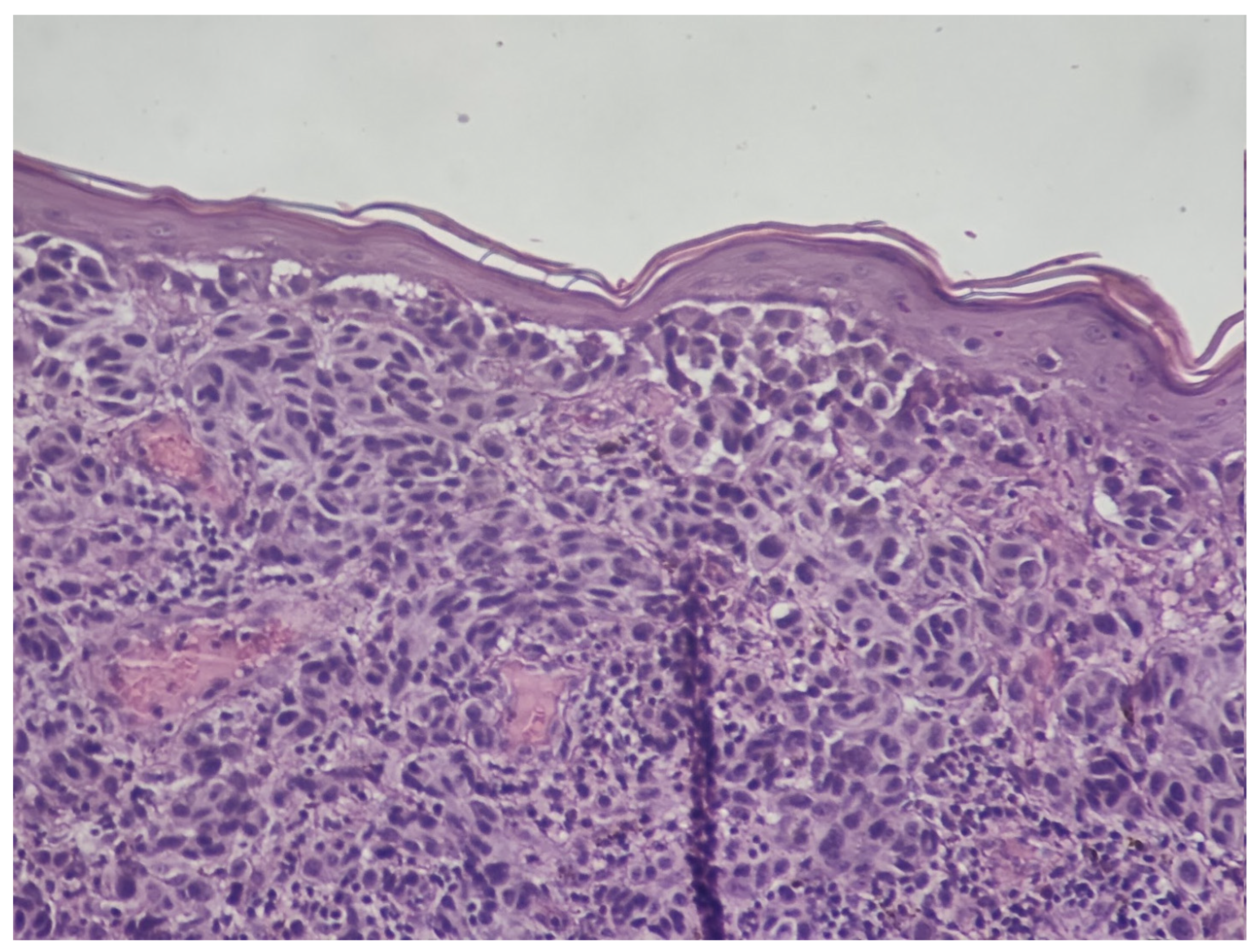

Non-Melanocytic Histopathological Clues for Melanoma Diagnosis: A Practical Review of Solar Elastosis, Stromal Regression, and Epidermal Reaction Patterns. Do Old-School Clues Still Matter?


Simple Summary
Abstract
1. Introduction
2. Solar Elastosis Patterns
2.1. The Umbrella Sign
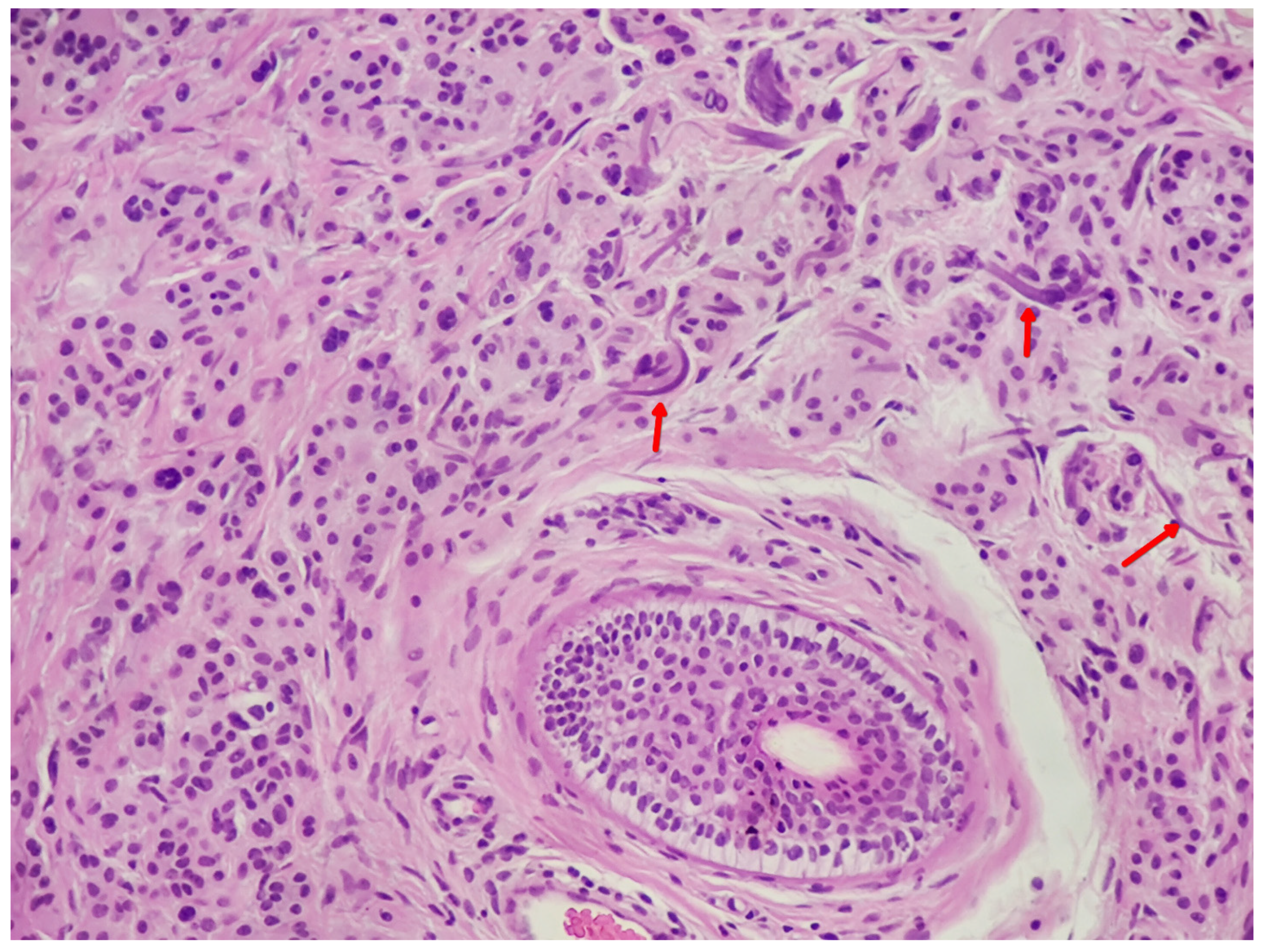
2.2. The Purple Fiber Sign (High Specificity Pro-Nevus)
2.3. Displacement/Compression of Solar Elastosis (Pro-Melanoma)
3. Stromal Reaction and Regression
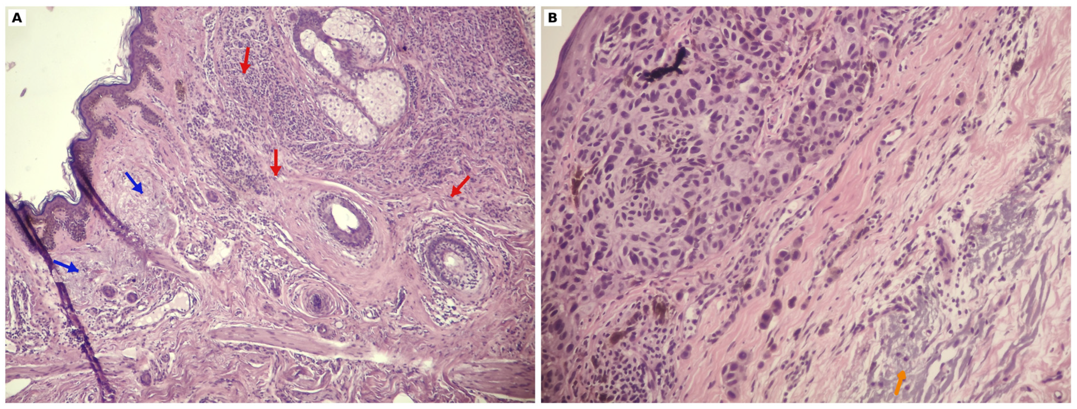
3.1. Regression-Type Fibrosis, Melanophages, and Vascular Change (Pro-Melanoma)
3.2. Immunophenotype of Regression
3.2.1. Functional Heterogeneity of the Regression Infiltrate
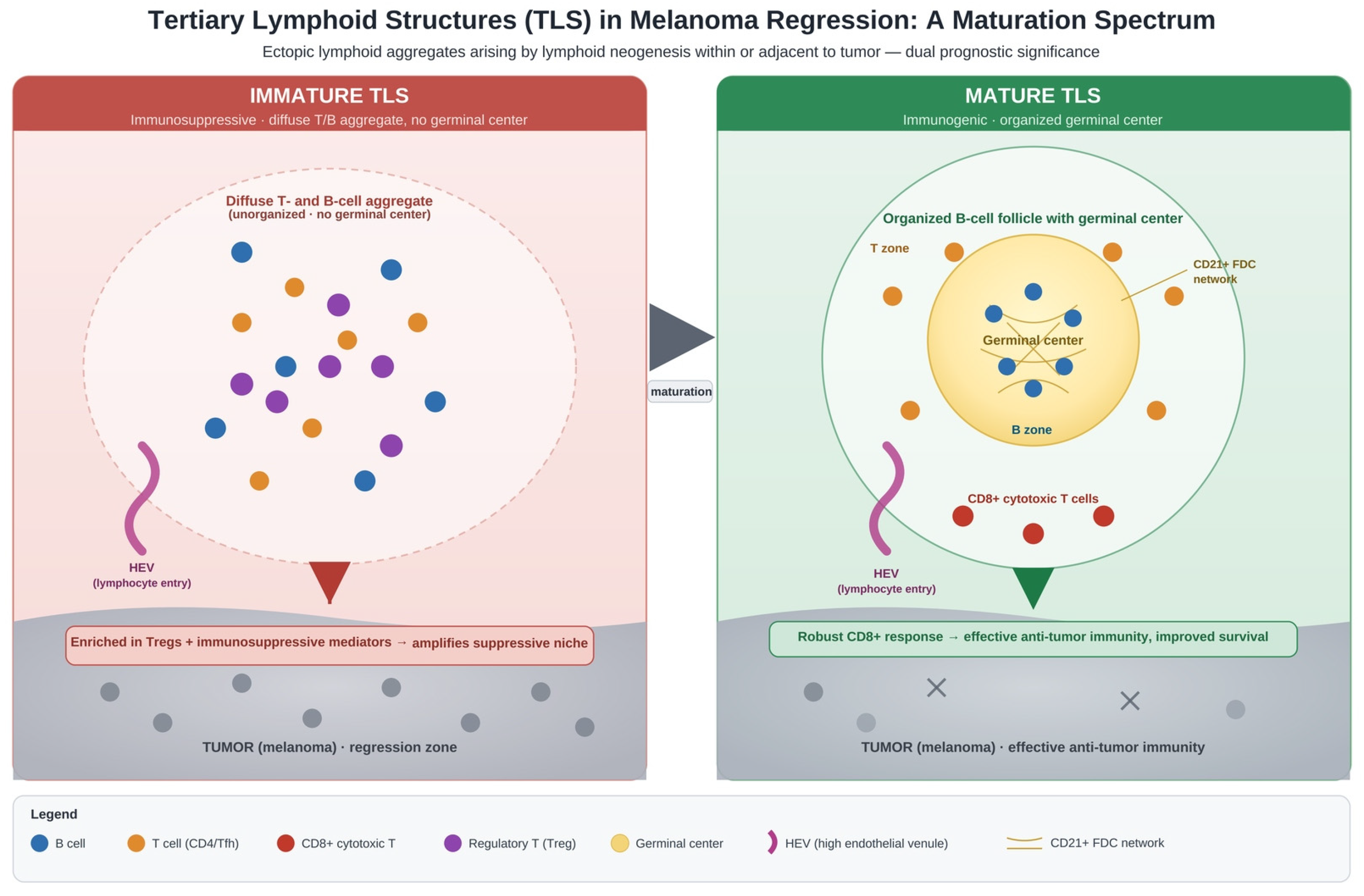
3.2.2. Tertiary Lymphoid Structures and Maturation-Dependent Immune Polarity
3.3. Fibroplasia out of Proportion to Cytology
3.4. Bland Stroma
3.5. Prognostic Implications of Regression
4. Epidermis, Adnexa, and Inflammation
4.1. Epidermal Context
4.2. Adnexal Preservation vs. Destruction
4.3. Host Inflammatory Response
5. Epidermal Reaction Patterns: Hyperplasia, Pseudoepitheliomatous Hyperplasia, and Effacement
5.1. Terminology and Definitions
5.2. Epidermal Hyperplasia and Melanoma-Driven Angiogenesis
5.3. Pseudoepitheliomatous Hyperplasia (PEH) Associated with Melanoma
5.4. Epidermal Effacement and Prognostic Associations
6. A Practical Stepwise Diagnostic Framework

| Histological Clue | Favors | Level of Evidence | Suggested Ancillary Test |
|---|---|---|---|
| Umbrella sign (central reduction in elastosis) | Nevus | Single-cohort, qualitatively corroborated [3,6,7,8] | Elastin histostain to confirm pattern [5] |
| Purple fiber sign (purple-tinged entrapped elastotic fibers) | Nevus | Single-cohort, preliminary [3] | None specific; H&E-dependent [3] |
| Stromal blending/adnexal cuffing | Nevus | Established morphologic criteria [1] | SOX10/Melan-A to map extent [25] |
| Displacement/compression of solar elastosis | Melanoma | Established [3,4,5] | Elastin histostain [5] |
| Regression (compressed elastic layer, melanophages, fibrosis) | Melanoma (context-dependent) | Established morphology; prognostic value variable [5,9] | Elastin histostain or immunostain to separate regression from scar [5]; SOX10 for residual melanocytes [25] |
| Disproportionate fibrosis/adnexal destruction | Melanoma | Established morphologic criteria [1,29] | p16, Ki-67, FISH/CGH if atypia present [22] |
| Epidermal effacement (“consumption”) | Melanoma (not specific) | Established but non-specific [29,31] | SOX10/Melan-A [25] |
6.1. Low Power Assessment (Step 1)
6.2. Characterize Epidermal Change
6.3. Evaluate Melanocytic Component (Step 2)
6.4. Evaluate Stromal Features and Elastosis (Step 2)
6.5. Distinguish Key Entities
- Melanoma with overlying simple hyperplasia: Expect dermal invasion, cytologic atypia, lack of maturation, possible mitoses; hyperplastic epidermis usually symmetric and regular (acanthotic but not PEH-like); this may correlate with angiogenesis in thicker melanomas [23].
- Melanoma with PEH: Irregular squamoid cords extend into dermis with SCC-like or SK-like patterns; be vigilant for interspersed atypical melanocytes; use melanocytic immunostains (SOX10, Melan-A, MITF) liberally if the melanocytic component is suspected but obscured [25].
- Watch for benign or dysplastic nevus on sun-damaged skin with umbrella sign and/or purple fiber sign, limited cytologic atypia, and maturation [3].
- Atypical junctional melanocytic hyperplasia/AIMP (Atypical Intraepidermal Melanocytic Nevus) on actinic skin: This includes worrisome features for melanoma in situ but incomplete criteria; discuss as such in the report and manage with excision and margins similar to melanoma in situ (5–10 mm) often [22,32].
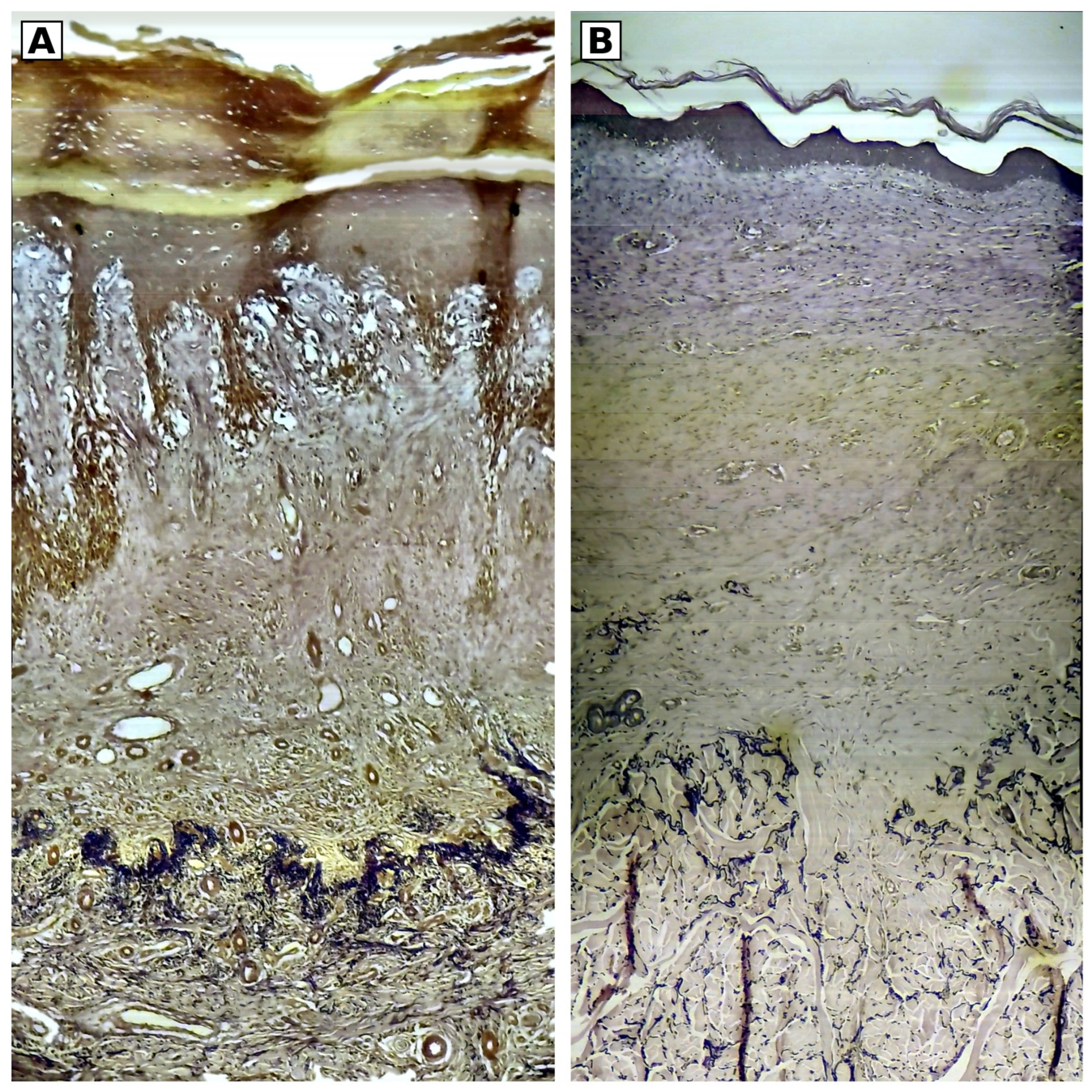
- Melanoma with regression: Look for compressed elastic layer at base of fibrosis (displaced papillary dermal elastic layer [5]), displaced elastosis, melanophages, lamellar fibrosis, and immunophenotype clues if needed (CD4 predominance and lower regulatory T cell markers compared to halo nevi) [5,9,11].
- Halo nevus (Sutton nevus): Dense lymphocytic infiltrates obscure nevus cells, with a higher CD8/CD3 ratio and greater PD1/FOXP3/CD25 expression compared to regressing melanoma; these are usually symmetric and circumscribed when visible [11].
6.6. Ancillary Studies (Step 3)
7. Key Pitfalls
- Thick invasive melanoma can obliterate elastosis centrally and mimic an umbrella-like clearing; check periphery for displacement/compression and integrate overall architectural features (asymmetry, invasion, high-grade cytology) [3].
- Small lentiginous junctional nevi on sun-damaged skin may lack the umbrella sign due to small size or recent development; absence of the umbrella sign is not diagnostic of melanoma in isolation [3].
- Assess the umbrella sign in the central one-third of the lesion, not the periphery, to avoid false-negative interpretation from the shoulder phenomenon [3].
- Purple fiber sign depends on H&E staining characteristics; treat it as a specific supportive clue when present, but its absence does not imply melanoma [3].
- Regression vs. scar: elastin histostain is often decisive—regression shows a compressed layer of thin papillary dermal elastic fibers displaced to the base of fibrosis, whereas scars lack this layer and show an abrupt transition to thick reticular dermal elastic fibers; scars <3 months can lack elastic fibers, and older scars may show regenerated thin, fragmented fibers [5].
- Features like poor circumscription, lentiginous proliferation, occasional suprabasal melanocytes, and mild atypia are not specific for melanoma, particularly on sun-damaged skin or in irritated/recurrent nevi; absence of maturation and true dermal mitotic activity are more specific [33].
- With exuberant PEH (especially SCC-like pattern), search deliberately for a melanocytic component before signing out SCC; liberal use of melanocytic immunostains is warranted [25].
- Epidermal effacement is more common in melanoma than in nevi but is not specific; interpret it in full context [29].
- Early-stage regression (dense lymphocytic infiltrate obscuring melanocytes) overlaps with brisk TILs and is subjective; many pathologists emphasize late-stage regression features for reproducibility [9].
8. Discussion
9. Conclusions
Funding
Institutional Review Board Statement
Informed Consent Statement
Data Availability Statement
Conflicts of Interest
References
- Massi, G.; LeBoit, P.E. Histological Diagnosis of Nevi and Melanoma, 2nd ed.; Springer: Berlin/Heidelberg, Germany, 2014; pp. 418–419. [Google Scholar]
- Ackerman, A.B.; Jacobson, M.; Vitale, P. Clues to Diagnosis in Dermatopathology I; ASCP Press: Chicago, IL, USA, 1991; pp. 226–227. [Google Scholar]
- Wood, B.A.; Harvey, N.T. The “Umbrella Sign”: A useful clue in the diagnosis of melanocytic lesions in sun damaged skin. Am. J. Dermatopathol. 2016, 38, 504–509. [Google Scholar] [CrossRef] [Scilit] [PubMed]
- Horenstein, M.G.; Norton, C.L.; Evans, T.N. Displacement of dermal solar elastosis in malignant melanoma. J. Cutan. Pathol. 2007, 34, 376–380. [Google Scholar] [CrossRef] [Scilit] [PubMed]
- Kamino, H.; Tam, S.; Roses, D.; Toussaint, S. Elastic fiber pattern in regressing melanoma: A histochemical and immunohistochemical study. J. Cutan. Pathol. 2010, 37, 723–729. [Google Scholar] [CrossRef] [Scilit] [PubMed]
- Harvey, N.T.; Wood, B.A. A practical approach to the diagnosis of melanocytic lesions. Arch. Pathol. Lab. Med. 2019, 143, 789–810. [Google Scholar] [CrossRef] [Scilit] [PubMed]
- Boer-Auer, A.; Kittler, H.; Tschandl, P. Pattern Analysis for Histopathologic Diagnosis of Melanocytic Lesions: A Guide to Practical Dermatopathology; Springer International Publishing: Cham, Switzerland, 2022. [Google Scholar] [CrossRef] [Scilit]
- Requena, C.; Manrique, E.; Nagore, E. Update on lentigo maligna: Diagnostic signs and treatment. Actas Dermosifiliogr. 2023, 114, 413–424. [Google Scholar] [CrossRef] [Scilit]
- Aung, P.P.; Nagarajan, P.; Prieto, V.G. Regression in primary cutaneous melanoma: Etiopathogenesis and clinical significance. Lab. Investig. 2017, 97, 657–668. [Google Scholar] [CrossRef] [Scilit] [PubMed]
- Kiszluk, A.; Alhalaseh, Y.; Asar, M.; Chen, F.; Speiser, J. Verhoeff-van Gieson may distinguish desmoplastic melanoma from re-excision scar. Am. J. Dermatopathol. 2025, 48, 193–196. [Google Scholar] [CrossRef] [Scilit] [PubMed]
- Brugués, A.; Roccuzzo, G.; Garcia-Herrera, A.; Alós, L.; Malvehy, J.; Puig, S.; Senetta, R.; Ribero, S.; Carrera, C. Inflammatory cell immunophenotypes in regressing melanomas and halo nevi: Possible keys to distinguish intensely inflamed tumors. Actas Dermosifiliogr. 2025, 116, 967–973. [Google Scholar] [CrossRef] [Scilit] [PubMed]
- Cioplea, M.; Nichita, L.; Georgescu, D.; Sticlaru, L.; Cioroianu, A.; Nedelcu, R.; Turcu, G.; Rauta, A.; Mogodici, C.; Zurac, S.; et al. FOXP3 in melanoma with regression: Between tumoral expression and regulatory T cell upregulation. J. Immunol. Res. 2020, 2020, 5416843. [Google Scholar] [CrossRef] [Scilit] [PubMed]
- Baumgartner, J.; Wilson, C.; Palmer, B.; Richter, D.; Banerjee, A.; McCarter, M. Melanoma induces immunosuppression by up-regulating FOXP3+ regulatory T cells. J. Surg. Res. 2007, 141, 72–77. [Google Scholar] [CrossRef] [Scilit] [PubMed]
- Găta, V.; Paşca, A.; Roman, A.; Muntean, M.; Morariu, D.; Bonci, E.; Dina, C.; Ungureanu, L. The expression of Forkhead Box P3 T regulatory lymphocytes as a prognostic factor in malignant melanomas. Int. J. Mol. Sci. 2024, 25, 6377. [Google Scholar] [CrossRef] [Scilit] [PubMed]
- Qiu, Y.; Ke, S.; Chen, J.; Qin, Z.; Zhang, W.; Yuan, Y.; Meng, D.; Zhao, G.; Wu, K.; Li, B.; et al. FOXP3+ regulatory T cells and the immune escape in solid tumours. Front. Immunol. 2022, 13, 982986. [Google Scholar] [CrossRef] [Scilit] [PubMed]
- Jarosz-Biej, M.; Kamińska, N.; Matuszczak, S.; Cichoń, T.; Pamuła-Piłat, J.; Czapla, J.; Smolarczyk, R.; Skwarzyńska, D.; Kulik, K.; Szala, S. M1-like macrophages change tumor blood vessels and microenvironment in murine melanoma. PLoS ONE 2018, 13, e0191012. [Google Scholar] [CrossRef] [Scilit] [PubMed]
- Gray, A.; Grushchak, S.; Mudaliar, K.; Kliethermes, S.; Carey, K.; Hutchens, K.A. The microenvironment in primary cutaneous melanoma with associated spontaneous tumor regression: Evaluation for T-regulatory cells and the presence of an immunosuppressive microenvironment. Melanoma Res. 2017, 27, 104–109. [Google Scholar] [CrossRef] [Scilit] [PubMed]
- Cabrita, R.; Lauss, M.; Sanna, A.; Donia, M.; Larsen, M.S.; Mitra, S.; Johansson, I.; Phung, B.; Harbst, K.; Vallon-Christersson, J.; et al. Tertiary lymphoid structures improve immunotherapy and survival in melanoma. Nature 2020, 577, 561–565. [Google Scholar] [CrossRef] [Scilit] [PubMed]
- Sautès-Fridman, C.; Petitprez, F.; Calderaro, J.; Fridman, W.H. Tertiary lymphoid structures in the era of cancer immunotherapy. Nat. Rev. Cancer 2019, 19, 307–325. [Google Scholar] [CrossRef] [Scilit] [PubMed]
- Baxevanis, C.N.; Sofopoulos, M.; Tsitsilonis, O.E.; Gritzapis, A.D. Exploring the pivotal functions of tertiary lymphoid structures in cancer prognosis and immunotherapy outcomes. Cancers 2025, 17, 3754. [Google Scholar] [CrossRef] [Scilit] [PubMed]
- Sofopoulos, M.; Fortis, S.P.; Vaxevanis, C.K.; Sotiriadou, N.N.; Arnogiannaki, N.; Ardavanis, A.; Vlachodimitropoulos, D.; Perez, S.A.; Baxevanis, C.N. The prognostic significance of peritumoral tertiary lymphoid structures in breast cancer. Cancer Immunol. Immunother. 2019, 68, 1733–1745. [Google Scholar] [CrossRef] [Scilit] [PubMed]
- Ensslin, C.J.; Hibler, B.P.; Lee, E.H.; Nehal, K.S.; Busam, K.J.; Rossi, A.M. Atypical melanocytic proliferations: A review of the literature. Dermatol. Surg. 2018, 44, 159–174. [Google Scholar] [CrossRef] [Scilit] [PubMed]
- McCarty, M.F.; Bielenberg, D.R.; Nilsson, M.B.; Gershenwald, J.E.; Barnhill, R.L.; Ahearne, P.; Bucana, C.D.; Fidler, I.J. Epidermal hyperplasia overlying human melanoma correlates with tumour depth and angiogenesis. Melanoma Res. 2003, 13, 379–387. [Google Scholar] [CrossRef] [Scilit] [PubMed]
- Grichnik, J.M.; Rhodes, A.R.; Sober, A.J. Benign neoplasias and hyperplasias of melanocytes. In Fitzpatrick’s Dermatology in General Medicine, 8th ed.; Goldsmith, L.A., Katz, S.I., Gilchrest, B.A., Paller, A.S., Leffell, D.J., Wolff, K., Eds.; McGraw-Hill: New York, NY, USA, 2012. [Google Scholar]
- Mott, R.T.; Rosenberg, A.; Livingston, S.; Morgan, M.B. Melanoma associated with pseudoepitheliomatous hyperplasia: A case series and investigation into the role of epidermal growth factor receptor. J. Cutan. Pathol. 2002, 29, 490–497. [Google Scholar] [CrossRef] [Scilit] [PubMed]
- Al Omoush, T.M.M.; Michal, M.; Konstantinova, A.M.; Michal, M.; Kutzner, H.; Kazakov, D.V. Melanocytic hyperplasia in the epidermis overlying trichoblastomas in 100 randomly selected cases. Am. J. Dermatopathol. 2016, 38, 297–299. [Google Scholar] [CrossRef] [Scilit] [PubMed]
- Weyers, W.; Bonczkowitz, M.; Weyers, I.; Bittinger, A.; Schill, W.B. Melanoma in situ versus melanocytic hyperplasia in sun-damaged skin: Assessment of the significance of histopathologic criteria for differential diagnosis. Am. J. Dermatopathol. 1996, 18, 560–566. [Google Scholar] [CrossRef] [Scilit] [PubMed]
- Filosa, A.; Filosa, G. Melanoma diagnosis: The importance of histopathological report. Dermatopathology 2018, 5, 41–43. [Google Scholar] [CrossRef] [PubMed]
- Hantschke, M.; Bastian, B.C.; LeBoit, P.E. Consumption of the epidermis: A diagnostic criterion for the differential diagnosis of melanoma and Spitz nevus. Am. J. Surg. Pathol. 2004, 28, 1621–1625. [Google Scholar] [CrossRef] [Scilit] [PubMed]
- Corbalán-Vélez, R.; Oviedo-Ramírez, I.; Martínez-Barba, E.; Clemente-Ruiz de Almirón, A. Epidermal effacement in malignant melanoma. Actas Dermosifiliogr. 2011, 102, 634–635. [Google Scholar] [CrossRef] [Scilit] [PubMed]
- Walters, R.F.; Groben, P.A.; Busam, K.; Millikan, R.C.; Rabinovitz, H.; Cognetta, A.; Mihm, M.C., Jr.; Prieto, V.G.; Googe, P.B.; King, R.; et al. Consumption of the epidermis: A criterion in the differential diagnosis of melanoma and dysplastic nevi that is associated with increasing Breslow depth and ulceration. Am. J. Dermatopathol. 2007, 29, 527–533. [Google Scholar] [CrossRef] [Scilit] [PubMed]
- Waqar, S.; George, S.; Jean-Baptiste, W.; Yusuf Ali, A.; Inyang, B.; Koshy, F.S.; George, K.; Poudel, P.; Chalasani, R.; Goonathilake, M.R.; et al. Recognizing histopathological simulators of melanoma to avoid misdiagnosis. Cureus 2022, 14, e26127. [Google Scholar] [CrossRef] [Scilit] [PubMed]
- Urso, C.; Rongioletti, F.; Innocenzi, D.; Batolo, D.; Chimenti, S.; Fanti, P.L.; Filotico, R.; Gianotti, R.; Lentini, M.; Tomasini, C.; et al. Histological features used in the diagnosis of melanoma are frequently found in benign melanocytic naevi. J. Clin. Pathol. 2005, 58, 409–412. [Google Scholar] [CrossRef] [Scilit] [PubMed]
- Lezcano, C.; Luo, L.; Shen, R.; Orlow, I.; Thomas, N.E.; Berwick, M.; Busam, K.J. Solar elastosis correlates with high tumor mutation burden and better 5-year disease-specific survival in patients with stage II/III melanoma. EJC Skin Cancer 2024, 2, 100274. [Google Scholar] [CrossRef] [Scilit] [PubMed]


Disclaimer/Publisher’s Note: The statements, opinions and data contained in all publications are solely those of the individual author(s) and contributor(s) and not of MDPI and/or the editor(s). MDPI and/or the editor(s) disclaim responsibility for any injury to people or property resulting from any ideas, methods, instructions or products referred to in the content. |
© 2026 by the author. Published by MDPI on behalf of the European Society of Dermatopathology. Licensee MDPI, Basel, Switzerland. This article is an open access article distributed under the terms and conditions of the Creative Commons Attribution (CC BY) license.
Share and Cite
Sofopoulos, M. Non-Melanocytic Histopathological Clues for Melanoma Diagnosis: A Practical Review of Solar Elastosis, Stromal Regression, and Epidermal Reaction Patterns. Do Old-School Clues Still Matter? Dermatopathology 2026, 13, 32. https://doi.org/10.3390/dermatopathology13030032
Sofopoulos M. Non-Melanocytic Histopathological Clues for Melanoma Diagnosis: A Practical Review of Solar Elastosis, Stromal Regression, and Epidermal Reaction Patterns. Do Old-School Clues Still Matter? Dermatopathology. 2026; 13(3):32. https://doi.org/10.3390/dermatopathology13030032
Chicago/Turabian StyleSofopoulos, Michail. 2026. "Non-Melanocytic Histopathological Clues for Melanoma Diagnosis: A Practical Review of Solar Elastosis, Stromal Regression, and Epidermal Reaction Patterns. Do Old-School Clues Still Matter?" Dermatopathology 13, no. 3: 32. https://doi.org/10.3390/dermatopathology13030032
APA StyleSofopoulos, M. (2026). Non-Melanocytic Histopathological Clues for Melanoma Diagnosis: A Practical Review of Solar Elastosis, Stromal Regression, and Epidermal Reaction Patterns. Do Old-School Clues Still Matter? Dermatopathology, 13(3), 32. https://doi.org/10.3390/dermatopathology13030032