Article

Phylogenetic Analysis of Escherichia coli Isolated from Australian Feedlot Cattle in Comparison to Pig Faecal and Poultry/Human Extraintestinal Isolates

1
The Davies Livestock Research Centre, The University of Adelaide, Adelaide, SA 5371, Australia
2
The Australian Centre for Antimicrobial Resistance Ecology, The University of Adelaide, Adelaide, SA 5005, Australia
3
Meat & Livestock Australia, Level 1, 40 Mount Street, North Sydney, NSW 2060, Australia
4
Research Centre for Infectious Disease, School of Biological Sciences, University of Adelaide, Adelaide, SA 5005, Australia
5
Australian Institute for Microbiology & Infection, University of Technology Sydney, Ultimo, NSW 2007, Australia
*
Author to whom correspondence should be addressed.
Equal contribution.
Antibiotics 2023, 12(5), 895; https://doi.org/10.3390/antibiotics12050895
Submission received: 6 April 2023 / Revised: 6 May 2023 / Accepted: 8 May 2023 / Published: 11 May 2023
(This article belongs to the Special Issue Antibiotics Resistance in Animals and the Environment)

Abstract

:
The similarity of commensal Escherichia coli isolated from healthy cattle to antimicrobial-resistant bacteria causing extraintestinal infections in humans is not fully understood. In this study, we used a bioinformatics approach based on whole genome sequencing data to determine the genetic characteristics and phylogenetic relationships among faecal Escherichia coli isolates from beef cattle (n = 37) from a single feedlot in comparison to previously analysed pig faecal (n = 45), poultry extraintestinal (n = 19), and human extraintestinal E. coli isolates (n = 40) from three previous Australian studies. Most beef cattle and pig isolates belonged to E. coli phylogroups A and B1, whereas most avian and human isolates belonged to B2 and D, although a single human extraintestinal isolate belonged to phylogenetic group A and sequence type (ST) 10. The most common E. coli sequence types (STs) included ST10 for beef cattle, ST361 for pig, ST117 for poultry, and ST73 for human isolates. Extended-spectrum and AmpC β-lactamase genes were identified in seven out of thirty-seven (18.9%) beef cattle isolates. The most common plasmid replicons identified were IncFIB (AP001918), followed by IncFII, Col156, and IncX1. The results confirm that feedlot cattle isolates examined in this study represent a reduced risk to human and environmental health with regard to being a source of antimicrobial-resistant E. coli of clinical importance.

1. Introduction

Escherichia coli is a Gram-negative, facultative anaerobic, rod-shaped bacterium commonly isolated as a normal component of the autochthonous microbiota of the animal and human gut [1]. However, many different E. coli pathotypes containing a diverse array of virulence genes (VGs) also exist, and these are capable of causing both intestinal and extraintestinal infections in both animals and humans [2]. Although many E. coli sub-types are relatively host-specific [3,4], some lineages have a broad host range and can potentially spread between humans, animals and their products, and the environment [5,6,7]. The ability of E. coli to spread to various hosts and environments whilst causing different infection types (i.e., both intestinal and extraintestinal infections) emphasizes the importance of monitoring spread and taking measures to regulate it. The most common methods to regulate the spread of specific E. coli pathotypes include the judicious use of antimicrobial agents, following good hygiene practices, and limiting the movement of infected animals and/or their contaminated products through strict biosecurity [8].
Antimicrobials have played a crucial role in the treatment and management of both intestinal and extraintestinal E. coli infection in animals and humans. However, selective pressure exerted by antimicrobials is assumed to be one of the main factors in the emergence and spread of antimicrobial resistance (AMR) among both pathogenic and commensal E. coli [9]. Although there is a clear link between the use of antimicrobials and the development of resistance among E. coli, there are also other factors in play such as the transmission rate of pathogenic subtypes to different hosts, the rates at which pathogenic lineages mutate and/or exchange accessory genes with commensals, and the selection of co-resistance to unrelated antimicrobials [10,11].
Both antimicrobial resistance genes (ARGs) and VGs may be easily transferred between E. coli strains because they are often located within transferable genetic elements such as bacteriophages, genomic islands, insertion sequences (ISs), integrons, transposons, and plasmids [12]. E. coli can also easily acquire mobile genetic elements containing these genes from other closely related bacteria. Pan genome analyses of E. coli isolates from multiple hosts have shown the ability of this species to rapidly evolve through gene acquisition and genetic modification whilst largely retaining a clonal population structure [5]. Additionally, a genetic similarity between certain broad host range bacterial clonal lineages of E. coli (such as ST10, ST58 and ST648) isolated from humans and animals suggests that frequent cross-species transmission is probable for these subtypes [13,14,15].
Whilst many studies have identified virulence and ARGs in E. coli isolated from various animal and human populations [16,17,18,19,20,21,22,23], few studies have undertaken valid comparisons between these diverse groups. Hence, little is known to what extent different livestock-associated commensal E. coli seed the environment and contribute to the AMR burden relevant to human health. Therefore, regular surveys are crucial for the effective management of AMR and infection control for both animal and human health. Australia has strict registration and regulation protocols for antimicrobial use in livestock production systems [24,25], and represents a unique study site given that there are also bans in place on the importation of live, food-producing animals, strict quarantine laws, and no land borders with other countries. Although Australia has implemented measures to prevent AMR, the spread of AMR is a complex issue that requires ongoing monitoring throughout the food chain. Regular monitoring and surveillance of animal production systems aims to identify AMR hot spots that may need immediate attention to implement effective prevention strategies [26,27].
The monitoring of AMR in E. coli can take place at various levels, including at the farm, environment, retail, and consumer level. In our previous study, tetracycline resistance increased by 17.8% in faecal-origin E. coli isolated from healthy cattle at feedlot entry compared to exit [28]. The prevalence of extended-spectrum β-lactamase (ESBL)-producing E. coli also rose from 0.7% to 4.0% between entry and exit samples. However, any possible links between these isolates and the E. coli isolated from other food animals and humans in Australia has not yet been determined. Therefore, this study compared the genetic similarity, ARG, plasmid, and VG profile of these cattle-origin E. coli isolates to contemporaneous isolates from other food animals and humans in Australia. This was undertaken to assess both the risk of animal-to-human isolate transfer via direct contact, food, and/or the environment, and the potential for horizontal gene transmission and spread among the various hosts.

2. Results

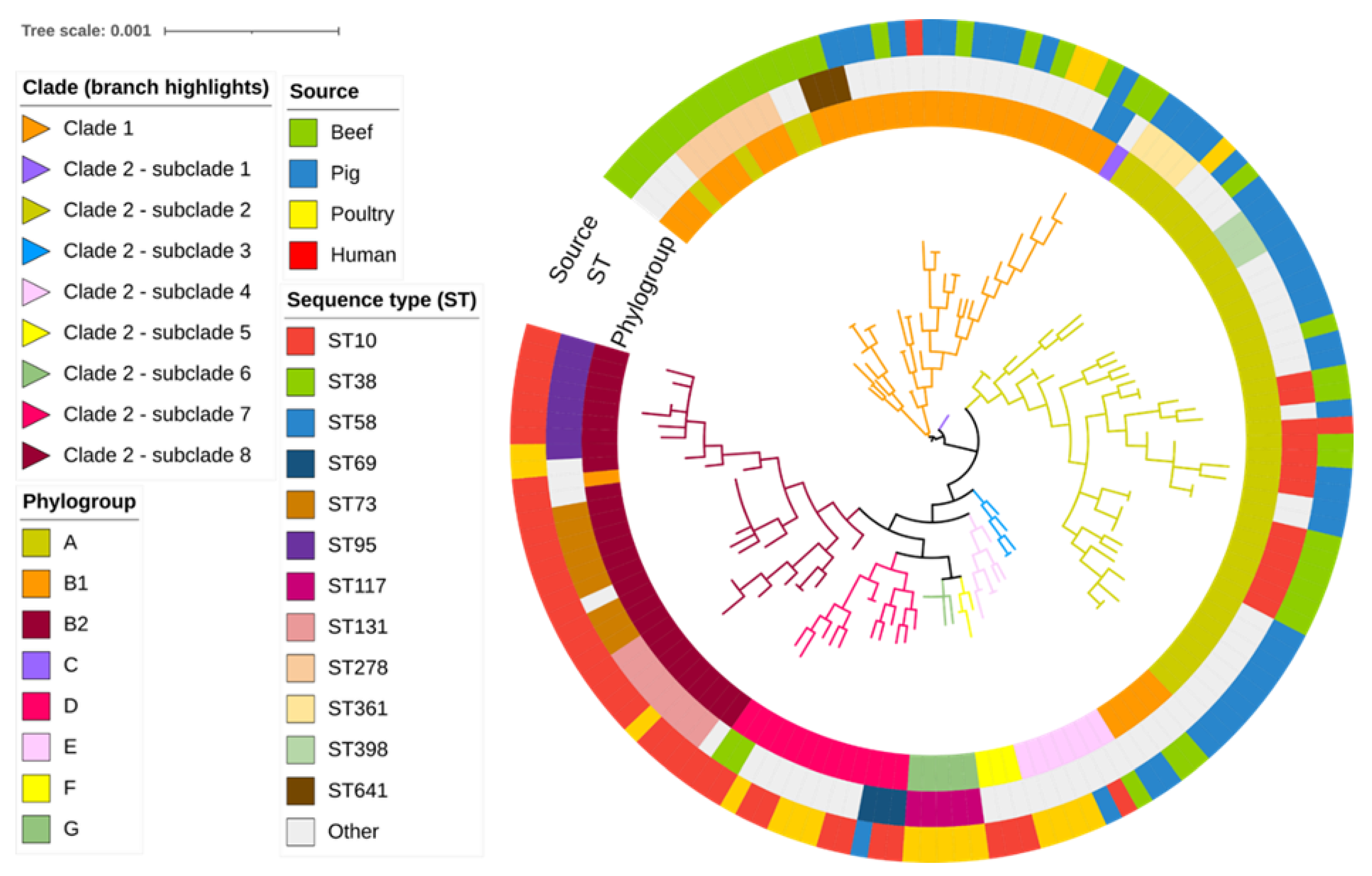
2.1. Phylogroup, Sequence Types and SNP Analyses

The E. coli isolates were distributed into phylogroups predominantly according to their host species of origin, with most cattle and pig isolates belonging to A and B1 and most poultry and human isolates belonging to B2 and D (Table 1).
Further genomic analyses revealed a high diversity of sequence types (STs) among the beef cattle E. coli isolates (18 known STs), the most frequently identified being broad host range ST10 (27.0%), ST278 (18.9%), and ST109, ST515, ST641, and ST2035 (5.4% each). The remaining 12 cattle STs were represented by single isolates only. The most common STs identified among the pig (faecal), poultry (extraintestinal), and human (extraintestinal) E. coli isolates were ST361, ST117, and ST73, respectively. As expected for extraintestinal isolates, most poultry and human isolates belonged to STs located within phylogroups B2 and D. (Figure 1; Table S1). By comparison, most cattle and pig faecal isolates belonged to STs associated with commensal phylogroups A and B1. In this way, most cattle and pig isolates were distributed into two clades (Clade 1 containing phylogroup B1 isolates, including those belonging to ST278, and Clade 2 containing phylogroup A isolates, including those belonging to ST10). Clade 2 ST10 isolates were more genetically diverse compared to Clade 1 ST278 isolates. Additionally, occasional human or poultry extraintestinal clinical isolates were found in Clade 1 (three isolates, one human and two poultry, all representing single STs) or Clade 2 (a single human ST10 isolate that was genetically distinct from cattle isolates).

2.2. Antimicrobial Resistance Genes

Overall, 60 ARGs were identified in the 141 E. coli isolates, encoding resistance to aminoglycosides (fourteen ARGs), amphenicols (four ARGs), ß-lactams (twenty-one ARGs), fluoroquinolones (three ARGs), folate synthesis inhibitors (ten ARGs), macrolides (five ARGs), and tetracyclines (three ARGs). The mean number of ARGs present in isolates obtained from each host species were: beef cattle (n = 3), poultry and human (n = 4), and pig (n = 7). Most pig E. coli isolates were MDR (extremely high: 42/45; 93.3%) compared to humans (very high: 24/40; 60.0%), poultry (high: 9/19; 47.4%), and beef cattle isolates (high: 14/37; 37.8%) (Figure 2). The study investigated pig and poultry isolates known to have the Class 1 integrase gene incl1, but the gene was also found in 10.8% (4/37) of the beef cattle isolates and 67.5% (27/40) of the human extraintestinal isolates. Among the intI1-containing E. coli isolated from beef cattle, three were originally isolated from ESBL selective agar. The study found that beef cattle E. coli isolates carrying the intI1 gene were more likely to contain multiple ARGs, including those encoding resistance to extended-spectrum ß-lactams, and were identified as ST88, ST1102, ST9967, and ST540 (Table S1).

β-Lactam Resistance Genes

A high proportion of the total isolate collection possessed the aminopenicillin resistance gene blaTEM-1B (70/141; 49.6%), with the next most commonly identified genes being blaCTX-M-15 (low prevalence: 6/141; 4.2%) and blaCTX-M-27 (low prevalence: 5/141; 3.5%), both imparting resistance to expanded-spectrum cephalosporins (Table 2). These were also the most prevalent β-lactam resistance genes identified among the beef cattle isolates (blaTEM-1B [8/37, high prevalence: 21.6%], blaCTX-M-15 and blaCTX-M-27 [low prevalence: 3/37; 8.1% each]). The blaCMY-2 plasmid-mediated AmpC β-lactamase gene was also identified in two bovine isolates.

2.3. Disinfectant Resistance Genes

Only Quaternary ammonium compound (qacE) and hydrogen peroxide (sitABCD) resistance genes were detected among the 141 E. coli isolates (Figure 3). Both disinfectants are commonly used in hospitals, healthcare facilities, and the food industry. The highest proportions of disinfectant resistance genes were detected among poultry clinical isolates (qacE [12/19, very high: 63.1%] and sitABCD [10/19, very high: 52.6%], respectively) and human clinical isolates (qacE [18/40, high: 45%]; sitABCD [28/40, very high: 70.0%]). By comparison, the prevalence of disinfectant resistance genes was much lower among pig commensal isolates (both 9/45; moderate: 20.0%) and beef cattle commensal isolates (qacE [1/37, low: 2.7%] and sitABCD [2/37, low: 5.4%]).

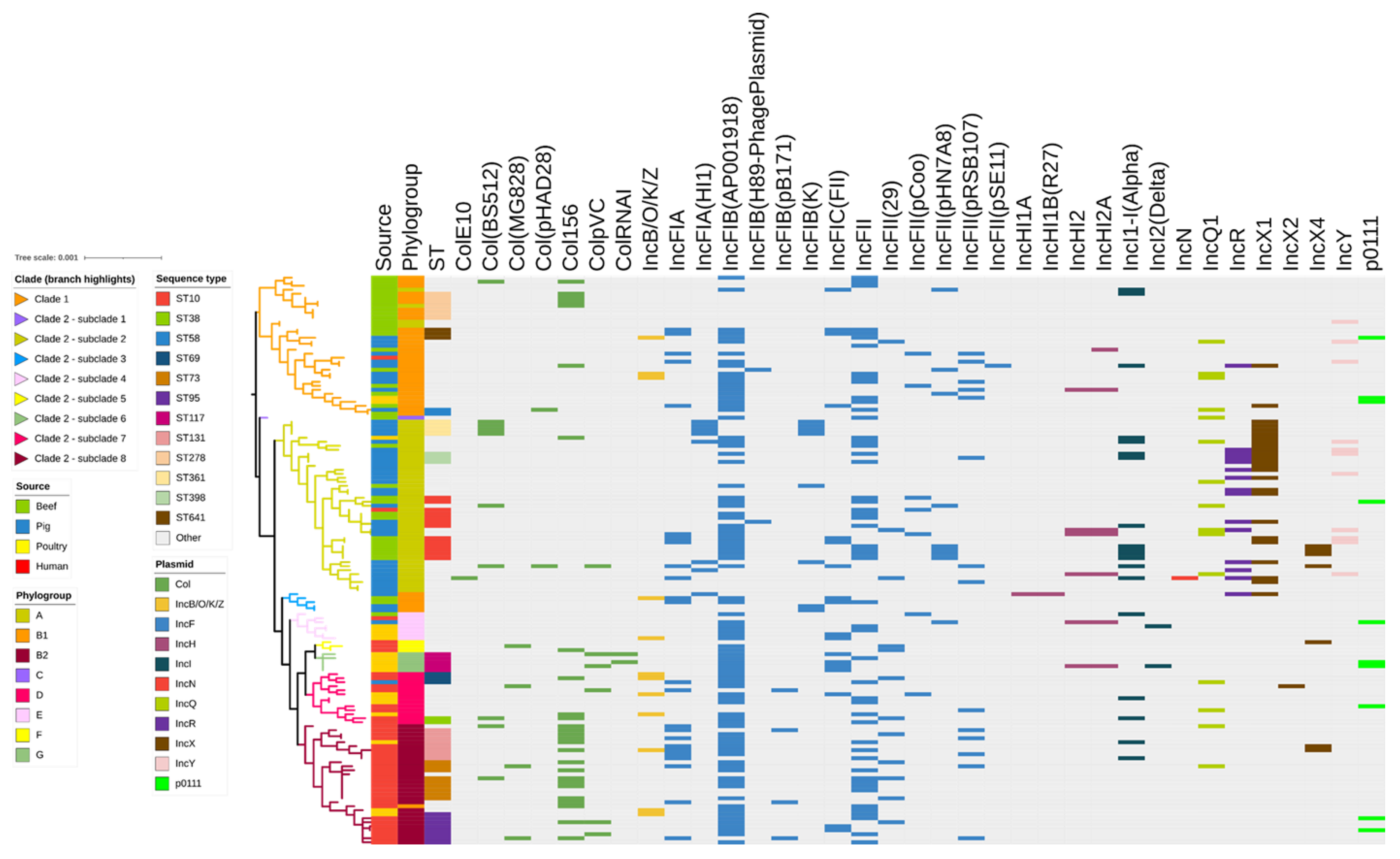
2.4. Identification of Plasmid Replicons

Overall, the PlasmidFinder analysis identified 35 different plasmid replicons in the 141 studied isolates (Figure 4). Twenty-seven Incompatibility (Inc) plasmid types were identified, the most common being IncFIB (AP001918) (89/141; 63.1%), IncFII (52/141; 36.9%) and IncX1 (25/141; 17.7%). Overall, 114/141 (80.8%) of the isolates contained more than two different plasmid replicons, with a high proportion of isolates (46/141; 32.6%) harbouring both IncFIB (AP001918) and IncFII replicons, whilst 5/141 isolates (3.5%) harboured IncFIB (AP001918), IncFII, and IncX1 replicons. Isolates that harboured two or more plasmid replicons included 24/37 beef cattle (64.8%), 37/45 pig (82%), 19/19 poultry (100%), and 34/40 human (85%) isolates. Seven Col plasmids containing genes responsible for the production of bacteriocins or colicins were also found in the isolate collection. These included Col156 (27/141; 19%), Col (BS512) (10/141; 7.1%), and ColpVC (6/141; 4.2%). Only 13/141 isolates (9.2%) did not possess any of the tested Inc replicons or colicin-associated genes. A total of 18 different plasmid types were identified among the beef cattle isolates, of which the most common were IncFIB (AP001918) (23/37; 62.2%), IncFII (16/37; 43.2%), IncFIC (FII) (8/37; 21.6%), and IncFIA, IncFII (pHN7A8), and IncI1-I (Alpha) (7/37; 18.9%, each). Among the IncF plasmids found in beef cattle, a total of 14 distinct pMLSTs were detected. A total of 8/37 beef cattle isolates (21.6%) did not contain any plasmids. The most frequently occurring pMLST was F89:A-:B- (5/14; 35.7%), followed by F89:A-:B-, F95:A-:B-, and F104:A-:B16 (3/14, 21.4% each). Among human isolates, the most commonly observed pMLST were F29:A-:B10 (8/40; 20%), F-:A-:B- (5/40; 12.5%), and F51:A-:B10 (4/40; 10%) (Table S1).

2.5. Agreement between Plasmid Replicons and ARG Arrays

The most common plasmid replicons, IncFIB (AP001918) and IncFII, were co-associated with high proportions of multiple ARGs representing several classes, indicating the likelihood that these genes are co-located on specific dominant plasmids. These included the aminoglycoside ARGs aph(3'')-Ib and aph(6)-Id, ß-lactam ARG blaTEM-1B, tetracycline ARGs tet(A) and tet(B), and folate synthesis inhibitor ARGs dfrA5, sul2 and sul1 (Figure 5).

2.6. Virulence Genes

A total of 103 VGs were identified in the 141 isolates; each isolate carried between 1 and 37 VGs (Figure 6). Eighty isolates (56.7%) had ≥10 VGs, which were distributed by phylogenetic group as follows: phylogroup B2 (29/141; 36.2%), B1 (13/141; 16.2%), D (12/141, 15.0%), A (11/141; 13.7%), E (6/141; 7.5%), G (5/141; 6.2%), F (3/141; 3.7%), and C (1/141; 1.2%). Isolates with the least VGs were mostly obtained from cattle and pigs and distributed into phylogenetic groups A and B1, whereas isolates with the most VGs associated with extraintestinal infection (as expected) were distributed into phylogenetic groups B2 and D, predominantly sourced from humans and poultry.

3. Discussion

Whilst some international studies have suggested that food-producing animals can be an important reservoir for virulent pathogens and their ARGs [29,30,31], other studies are more equivocal in their findings [32,33]. Given Australia’s unique animal production systems and their potential impact on the evolution of animal microbes in isolation from the rest of the world, the aim of this study was to compare the antimicrobial/disinfectant resistance, plasmid, and VG repertoires of commensal E. coli isolated from healthy beef cattle (n = 37) from a single feedlot, in comparison to previously characterised pig faecal (n = 45), poultry (n = 19), and human extraintestinal isolates (n = 40). This study had three major findings. First, the phylogenic analyses revealed that most beef cattle isolates clustered together with pig isolates in two main clades of related STs (within phylogroups A and B1), while most poultry and human isolates clustered together in phylogroups B2 and D. Second, while a significant proportion of beef cattle isolates carried plasmid-encoded extended-spectrum cephalosporin resistance genes (7/37; 18.9%), further analysis in comparison to human extraintestinal isolates suggested that they are of limited impact to human health. Third, a significant number of the beef cattle E. coli isolates (10/37; 27%) belonged to broad-host range ST10, including three isolates (30%) carrying genes encoding resistance to extended-spectrum cephalosporins.
The phylogenetic tree derived from the SNPs showed that isolates from beef cattle and pigs clustered together in phylogenetic groups (A and B1) not typically associated with highly virulent extraintestinal infection, whereas most poultry and human isolates were intermixed in separate clades in phylogroups B2 and D. This is consistent with previous research on poultry and human E. coli isolates, which suggested that clinical isolates from these sources share a high degree of genetic and pathotypic similarity, suggesting frequent anthropozoonotic exchange [34,35,36]. Additionally, studies conducted in Europe also confirmed that phylogroups A and B1 were most commonly represented by isolates obtained from healthy chicken, cattle and pig faeces [37,38]. In the present study, a single human extraintestinal isolate was identified as ST10; however, it was distinct from beef cattle and pig isolates within this genetically diverse, broad host range clonal lineage, which can be of clinical significance to human health [39].
The spread of pathogens that are resistant to third-generation cephalosporins is a critical concern that has extended beyond hospital environments. In this study, the plasmid-encoded resistance gene blaTEM-1B, which confers resistance to ß-lactam antimicrobials such as amoxicillin, was detected in 8/37 (21.6%) beef cattle isolates. Furthermore, the study found plasmid-mediated blaCTX-M expanded-spectrum cephalosporin ARGs in 6/37 beef cattle isolates (16.2%) and a blaCMY-2 AmpC ß-lactamase gene in an additional isolate. Contrary to this, a study conducted in South Africa, which analysed isolates obtained from cattle faeces and raw beef samples, found a significantly higher prevalence of β-lactam resistance genes, including blaTEM (85.5%), blaSHV (69.6%), and blaCTX-M (58.0%) [40]. In the present study, the amplification of beef cattle-origin isolates carrying ESBL or AmpC ß-lactamase ARGs in exit (4%) compared to entry (0.7%) samples may indicate that a proportion of the beef cattle population acquire these ARGs during the feeding period, which may be related to the reserve use of ceftiofur for bovine respiratory disease cases not responding to first or second line therapy, or those occurring late in the feeding period [25]. A previous study conducted in the United States found that administering ceftiofur to feedlot cattle can result in a selective pressure that increases the level of blaCMY-2 carriage [41].
There is widespread evidence from various studies conducted globally documenting the infrequent isolation of ESBL-producing E. coli in food animal populations such as beef cattle, pigs, and poultry [42,43,44]. However, there is currently no concrete evidence documenting the direct transmission of these bacteria from animals to humans via the food chain [45]. However, previous studies conducted in pig and poultry industries have reported a high level of ARG similarity between farmer-derived and animal-origin isolates, indicating close and direct contact as an important transmission vector [46,47,48,49,50,51,52]. Although the present study found blaCTX-M-15 in 3/40 (7.5%) human E. coli isolates, these appeared to be unrelated to the contemporaneous animal-origin ESBL genes, especially given the widespread use of ß-lactam antimicrobials in human medicine and the resulting selection pressure for the evolution of antimicrobial-resistant bacteria.
In this study, 29.7% of beef cattle isolates were MDR, compared to 93.3% for pig, 47.4% for poultry, and 60.0% for human isolates. Even though the pig isolates were selected based on carriage of intl1, the higher proportion of multidrug resistance observed may be due to the increased use of prophylactic antimicrobial agents in pig farms and the management practices that facilitate the maintenance and dissemination of antimicrobial resistance determinants in both the hosts and the farm environment compared to poultry and cattle [53]. In this study, poultry isolates containing the intl1 gene were also selected. Unlike the surveillance-derived pig isolates, these pathogenic poultry isolates showed a lower level of multidrug resistance. This discrepancy might be attributed to the possibility that pathogenic isolates could shed resistance genes in favour of virulence genes [54], the clonal nature of some isolates in different hosts [55], and variations in management practices and sample sizes [56]. By comparison, relatively low rates of multidrug resistance were observed among the cattle isolates given that the prophylactic administration of antimicrobial agents for bovine respiratory disease to large numbers of animals is rarely practiced in Australia and did not occur during this study at the host feedlot [25].
The majority of ARGs detected in the present study were likely to be located on conjugative plasmids belonging to different incompatibility groups [57,58,59]. Plasmids in E. coli can contain multiple replicons that are known to mediate resistance to antimicrobials and disinfectants as well as imparting increased virulence [60,61]. In this study, 64% of beef isolates harboured more than two plasmid replicons compared to pig isolates (82%), poultry (100%), and human (85%) isolates. The plasmid replicons IncFIB (AP001918) and IncFII were found to be strongly associated with multiple classes of ARGs. These included genes that confer resistance to ß-lactams (blaTEM-1B), aminoglycosides (aph(3'')-Ib and aph(6)-Id), tetracycline (tet(A) and tet(B)), trimethoprim (dfrA5), and sulfamethoxazole (sul2 and sul1). The RepFIB/FIIA plasmids are known to contain and transfer multidrug resistance-encoding islands and VGs [62]. This highlights the potential of these plasmids to spread AMR across bacterial populations and contribute to the increasing threat of AMR. The plasmid IncFIB (AP001918) was also reported to be the most common replicon in E. coli isolated from pig, poultry, and human isolates [31,63,64]. The IncF plasmids (IB and II) are known to contain ARGs, such as the ß lactamase genes blaTEM-1B, blaCMY-2, and blaOXA-1, and those encoding resistance to sulfonamides and trimethoprim (dfrA8, strA, strB, and sul2), as well as the tetracycline resistance genes tet(A) and tet(B) [65,66,67,68]. Whilst previous studies have shown that plasmid replicons can be shared among E. coli isolated from humans and food animals [65], the present study showed that both the frequency and heterogeneity of ARGs and plasmids were much lower in beef cattle isolates compared to pig, poultry, and human isolates, thus confirming that feedlot beef cattle from this study population represent reduced AMR risk to public health and the environment within Australia.
The increased detection of E. coli that are resistant to extended-spectrum β-lactam antimicrobials has been linked to the dissemination of international high-risk clones, such as ST10, ST38, ST69, ST73, ST115, ST117, ST131, ST354, ST410, ST457, ST517, ST648, ST711, and ST1193, in animals and humans [69,70]. In this study, the three most common STs found in beef cattle isolates were ST10 (10/37; 27.0%), ST278 (7/37; 19.0%), and ST109 (2/37; 5.4%). As a cause of human blood stream infections, ST73 (9/40; 22.5%), ST95, and ST131 (7/40; 17.5, each) predominated, whereas among pig isolates ST361 (4/45; 8.9%) and ST48 and ST398 (3/45; 7.5% each) predominated, while among poultry isolates ST 117 (5/19; 26.3%) and ST57 and ST350 (2/19; 10.5%) were most frequently observed. The clonally diverse group ST10, which belongs to phylogenetic group A, is known to have a broad host range and can be found in a variety of hosts, including humans, animals, and the environment [14]. In the present study, the clonal complex ST10 was identified as the most common lineage among the beef cattle isolates, with a prevalence of 27.0% compared to 4.4% in pigs and 2.5% in humans. Similarly, ST10 was the most commonly identified isolate from commercial beef cattle farms in the United States and in isolates from the Australian beef cattle population at slaughter [44,71]. The CC10 E. coli has been reported to carry ARGs and can sometimes cause infections in dogs, humans, and pigs [39,72]. However, the transmission of AMR from food animals to humans (and vice versa) is still not fully understood. Some research has reported direct contact with animals as a potential source of transmission for zoonotic bacteria, such as ESBL-producing E. coli, while other studies suggest the transmission is more likely to occur from humans to animals [73,74,75]. However, the fact that only a single human clinical isolate in the present study belonged to ST10 indicates that in Australia it is not a significant opportunistic human pathogen regardless of host source.
This study had some limitations. Firstly, the study included seven ESBL-producing E. coli isolated from beef cattle using selective media. However, it is important to note that the use of selective media could have introduced a selection bias in amplifying these ESBL-producing E. coli isolates, which would not normally be selected as the most common colony type on less inhibitory agar. Secondly, the E. coli data from pigs, poultry, and humans used in the study were obtained from a universal database as secondary information; ideally, the studies should have been prospective in design and more synchronised. Thirdly, E. coli isolates obtained from pigs and poultry were selected on the basis of possessing Class 1 integrons, which is considered as an indication of having an antimicrobial resistance genotype, whereas most of the beef cattle isolates were selected on the basis of being resistant to one or more antimicrobials from a previous study [26]. Fourthly, the limited sample size, particularly the availability of only 19 poultry isolates that met the selection criteria for comparison, may not be as representative of larger populations; ideally more isolates would have provided firmer conclusions. Lastly, the short-read sequencing data analysed in this study did not allow for the identified ARGs and virulence genes to be mapped to specific plasmids, which requires long read sequencing [76,77].

4. Materials and Methods

4.1. Whole Genome Sequencing and Phylogenetic Analysis

The current study was a follow-up to previous work focused on determining the prevalence of antimicrobial resistance among rectal E. coli isolates obtained at entry to and exit from an Australian beef cattle feedlot [28]. A total of thirty-three E. coli isolates determined to be resistant to at least one antimicrobial (including seven that were resistant to extended-spectrum β-lactams isolated from ESBL selective agar rather than MacConkey agar), together with four E. coli isolates that were susceptible to all tested agents, were randomly selected in the study. DNA extraction, whole genome sequencing (WGS), and genomic assembly have been described in our previously published research [28]. Raw genomic sequences were deposited in the NCBI under BioProject PRJNA844571. The whole-genome sequenced commensal E. coli isolated from beef cattle (n = 37; this study) were compared with retrieved genomic data from three previous studies undertaken in Australia. These included: (i) 45 E. coli isolates carrying the Class 1 integrase gene (intI1) isolated from sows and their offspring at a commercial pig breeding operation (accession number PRJNA509690) [78]; (ii) 19 avian pathogenic E. coli isolates that also carried intI1, which were originally obtained from post-mortem lesions from deceased or culled birds with signs of an APEC infection at different poultry operations in Australia (accession number PRJNA479542) [79]; and (iii) 40 E. coli isolates from bloodstream infections in humans in Australia (accession number PRJNA480723) [80]. Assembled sequences with less than 30x coverage and fewer than 25,000 SNPs were excluded from further analysis.
Genetic relationships between isolates were examined using single nucleotide polymorphism (SNPs) found from cleaned WGS reads mapped to an E. coli complete genome (NCBI Assembly Accession: BA000007.3). The software Snippy v4.6.0 (https://github.com/tseemann/snippy (accessed on 11 September 2022)) was used to call core SNPs, i.e., SNPs that can be determined in all isolates. A maximum likelihood (ML) tree was constructed with RAxML v8.2.10 using the model GTRCAT and a rapid bootstrap analysis with 100 bootstraps for the best scoring ML tree [81]. This was followed by recombination removal using ClonalFrameML v1.12 [82]. The final phylogenetic tree and heat map were manipulated with iTOL (https://itol.embl.de/ (accessed on 11 September 2022)) for display [83]. A heat map illustrating the presence or absence of each trait for each isolate was created to assess all data elements for all isolates.

4.2. Determination of Genotypes, Antimicrobial Resistance Genes, and Virulence Genes

Multilocus sequence type (MLST) and phylogenetic group were determined using MLST 2.0 [84] and ClermonTyping [85], respectively. To identify ARGs, we used ResFinder 4.0 [86]. To further pinpoint the chromosomal point mutations associated with AMR, we used PointFinder [87]. The input to start search in ResFinder and PointFinder was the assembled E. coli genome. Additionally, AMR genes were predicted using the Antibiotic Resistance Genes Database (ARDB) and the Comprehensive Antibiotic Resistance Database (CARD) [88]. PlasmidFinder was used with a minimum identity of 95% and coverage of 60% to detect plasmid replicons, while pMLST 2.0 was employed to perform plasmid multi-locus sequence typing [89]. Virulence genes were identified using VirulenceFinder 2.0 [90,91]. IntegronFinder was utilized to identify integrons and their unique components (accessed on 28 March 2023) [92] and confirmed using BLAST with a minimum identity of 95% (https://blast.ncbi.nlm.nih.gov/Blast/ (accessed on 28 March 2023)) [93].

4.3. Statistical Analyses

Categorical measured traits including ARGs, disinfectant resistance genes, the presence of plasmid replicons, and VGs were converted into numerical code with 1 indicating presence and 0 indicating absence. The resistance profile was categorised as MDR if the isolate exhibited resistance to one or more antimicrobials in three or more antimicrobial classes [94]. AMR and disinfectant gene frequencies were described as rare: <0.1%; very low: 0.1% to 1.0%; low: >1% to 10.0%; moderate: >10.0% to 20.0%; high: >20.0% to 50.0%; very high: >50.0% to 70.0%; and extremely high: >70.0%, according to the European Food Safety Authority (EFSA) and the European Centre for Disease Prevention and Control (ECDC) [95].

5. Conclusions

This study highlighted the relative abundance of ARGs (to both antimicrobials and disinfectants), plasmid replicons, and virulence-associated genes in commensal E. coli isolated from Australian beef cattle in comparison to additional commensals and extraintestinal pathogens isolated from food-producing animals (pigs and poultry, respectively) and extraintestinal pathogens from humans. The results confirmed that beef cattle represent a reduced risk to public health and the environment given that most isolates belonged to phylogenetic groups typically associated with commensal bacteria. Whilst a significant proportion of beef cattle isolates were ESBL/AmpC producing E. coli (some of which belonged to broad host range ST10), there was limited evidence that these higher risk clones are problematic in human medicine in Australia.

Supplementary Materials

The following supporting information can be downloaded at: https://www.mdpi.com/article/10.3390/antibiotics12050895/s1, Table S1. Prevalence of antimicrobial resistance genes, pMLST, and Class 1 integrase gene in E. coli isolated from beef cattle, pig, poultry, and human.

Author Contributions

Conceptualization, D.J.T. and K.R.P.; data curation, Y.E.M., M.F.H., K.R.P., W.Y.L. and T.V.; formal analysis, Y.E.M. and W.Y.L.; Funding acquisition, K.R.P., D.J.T. and J.P.M.; investigation, Y.E.M., M.F.H. and T.V.; project administration, K.R.P.; methodology, Y.E.M., M.F.H., T.V., S.P.D., W.Y.L., D.J.T. and K.R.P.; resources, T.V., W.Y.L., S.P.K., D.J.T. and K.R.P.; software, Y.E.M. and W.Y.L.; supervision T.V., D.J.T., J.P.M., S.P.K., W.Y.L. and K.R.P.; validation Y.E.M., M.F.H., T.V. and K.R.P.; visualization, Y.E.M., D.J.T., K.R.P. and W.Y.L.; writing—original draft, Y.E.M.; writing—review editing, Y.E.M., J.P.M., S.P.K., S.P.D., W.Y.L., K.R.P. and D.J.T. All authors have read and agreed to the published version of the manuscript.

Funding

This research was funded by Meat and Livestock Australia (MLA), grant number B.FLT.3003.

Institutional Review Board Statement

The sample collection protocols and procedures were approved by the University of Adelaide Animal Ethics Committee (AEC number S2019-072).

Informed Consent Statement

Not applicable.

Data Availability Statement

WGS reads are available in the SRA under BioProject; beef (PRJNA844571), pig (PRJNA509690), poultry (PRJNA479542), and human (PRJNA480723) isolates.

Acknowledgments

We thank Meat and Livestock Australia for the funding of this project. We would like to thank the beef feedlot farm and abattoir staff for their participation in the study. We are also grateful for the contribution made by ACARE and the Davies Livestock Research Centre laboratory members for assisting during the bench work. We also thank Mandi Carr and Manouchehr Khazandi for their initial contribution to the study design. We are thankful to Ethan Wyrsch for contributing to the integron analysis.

Conflicts of Interest

The authors declare that there are no conflict of interest.

References

  1. Jang, J.; Hur, H.G.; Sadowsky, M.J.; Byappanahalli, M.N.; Yan, T.; Ishii, S. Environmental Escherichia coli: Ecology and public health implications—A review. J. Appl. Microbiol. 2017, 123, 570–581. [Google Scholar] [CrossRef]
  2. Sora, V.M.; Meroni, G.; Martino, P.A.; Soggiu, A.; Bonizzi, L.; Zecconi, A. Extraintestinal pathogenic Escherichia coli: Virulence factors and antibiotic resistance. Pathogens 2021, 10, 1355. [Google Scholar] [CrossRef] [PubMed]
  3. Yu, D.; Banting, G.; Neumann, N.F. A review of the taxonomy, genetics, and biology of the genus Escherichia and the type species Escherichia coli . Can. J. Microbiol. 2021, 67, 553–571. [Google Scholar] [CrossRef] [PubMed]
  4. Escobar-Páramo, P.; Le Menac’h, A.; Le Gall, T.; Amorin, C.; Gouriou, S.; Picard, B.; Skurnik, D.; Denamur, E. Identification of forces shaping the commensal Escherichia coli genetic structure by comparing animal and human isolates. Environ. Microbiol. 2006, 8, 1975–1984. [Google Scholar] [CrossRef] [PubMed]
  5. Braz, V.S.; Melchior, K.; Moreira, C.G. Escherichia coli as a multifaceted pathogenic and versatile bacterium. Front. Cell. Infect. Microbiol. 2020, 10, 548492. [Google Scholar] [CrossRef]
  6. Anderson, M.A.; Whitlock, J.E.; Harwood, V.J. Diversity and distribution of Escherichia coli genotypes and antibiotic resistance phenotypes in feces of humans, cattle, and horses. Appl. Environ. Microbiol. 2006, 72, 6914–6922. [Google Scholar] [CrossRef]
  7. Clermont, O.; Olier, M.; Hoede, C.; Diancourt, L.; Brisse, S.; Keroudean, M.; Glodt, J.; Picard, B.; Oswald, E.; Denamur, E. Animal and human pathogenic Escherichia coli strains share common genetic backgrounds. Infect. Genet. Evol. 2011, 11, 654–662. [Google Scholar] [CrossRef]
  8. Collignon, P.J.; McEwen, S.A. One health-its importance in helping to better control antimicrobial resistance. Trop. Med. Infect. Dis. 2019, 4, 22. [Google Scholar] [CrossRef]
  9. Harada, K.; Asai, T. Role of antimicrobial selective pressure and secondary factors on antimicrobial resistance prevalence in Escherichia coli from food-producing animals in Japan. J. Biomed. Biotechnol. 2010, 2010, 180682. [Google Scholar] [CrossRef]
  10. Tantoso, E.; Eisenhaber, B.; Kirsch, M.; Shitov, V.; Zhao, Z.; Eisenhaber, F. To kill or to be killed: Pangenome analysis of Escherichia coli strains reveals a tailocin specific for pandemic ST131. BMC Biol. 2022, 20, 146. [Google Scholar] [CrossRef]
  11. Kaper, J.B.; Nataro, J.P.; Mobley, H.L.T. Pathogenic Escherichia coli . Nat. Rev. Microbiol. 2004, 2, 123–140. [Google Scholar] [CrossRef] [PubMed]
  12. Dobrindt, U.; Chowdary, M.G.; Krumbholz, G.; Hacker, J. Genome dynamics and its impact on evolution of Escherichia coli . Med. Microbiol. Immunol. 2010, 199, 145–154. [Google Scholar] [CrossRef] [PubMed]
  13. Massella, E.; Reid, C.J.; Cummins, M.L.; Anantanawat, K.; Zingali, T.; Serraino, A.; Piva, S.; Giacometti, F.; Djordjevic, S.P. Snapshot study of whole genome sequences of Escherichia coli from healthy companion animals, livestock, wildlife, humans and food in Italy. Antibiotics 2020, 9, 782. [Google Scholar] [CrossRef]
  14. Reid, C.J.; DeMaere, M.Z.; Djordjevic, S.P. Australian porcine clonal complex 10 (CC10) Escherichia coli belong to multiple sublineages of a highly diverse global CC10 phylogeny. Microb. Genom. 2019, 5, e000225. [Google Scholar] [CrossRef]
  15. Matamoros, S.; van Hattem, J.M.; Arcilla, M.S.; Willemse, N.; Melles, D.C.; Penders, J.; Vinh, T.N.; Thi Hoa, N.; Bootsma, M.C.J.; van Genderen, P.J.; et al. Global phylogenetic analysis of Escherichia coli and plasmids carrying the mcr-1 gene indicates bacterial diversity but plasmid restriction. Sci. Rep. 2017, 7, 15364. [Google Scholar] [CrossRef] [PubMed]
  16. Bryan, A.; Shapir, N.; Sadowsky, M.J. Frequency and distribution of tetracycline resistance genes in genetically diverse, nonselected, and nonclinical Escherichia coli strains isolated from diverse human and animal sources. Appl. Environ. Microbiol. 2004, 70, 2503–2507. [Google Scholar] [CrossRef] [PubMed]
  17. Guerra, B.; Junker, E.; Schroeter, A.; Malorny, B.; Lehmann, S.; Helmuth, R. Phenotypic and genotypic characterization of antimicrobial resistance in German Escherichia coli isolates from cattle, swine and poultry. J. Antimicrob. Chemother. 2003, 52, 489–492. [Google Scholar] [CrossRef]
  18. Adator, E.H.; Narvaez-Bravo, C.; Zaheer, R.; Cook, S.R.; Tymensen, L.; Hannon, S.J.; Booker, C.W.; Church, D.; Read, R.R.; McAllister, T.A. A one Health comparative assessment of antimicrobial resistance in generic and Extended-spectrum cephalosporin-resistant Escherichia coli from beef production, sewage and clinical settings. Microorganisms 2020, 8, 885. [Google Scholar] [CrossRef]
  19. Nhung, N.T.; Cuong, N.V.; Campbell, J.; Hoa, N.T.; Bryant, J.E.; Truc, V.N.; Kiet, B.T.; Jombart, T.; Trung, N.V.; Hien, V.B.; et al. High levels of antimicrobial resistance among Escherichia coli isolates from livestock farms and synanthropic rats and shrews in the Mekong Delta of Vietnam. Appl. Environ. Microbiol. 2015, 81, 812–820. [Google Scholar] [CrossRef]
  20. Athanasakopoulou, Z.; Reinicke, M.; Diezel, C.; Sofia, M.; Chatzopoulos, D.C.; Braun, S.D.; Reissig, A.; Spyrou, V.; Monecke, S.; Ehricht, R.; et al. Antimicrobial resistance genes in ESBL-producing Escherichia coli isolates from animals in Greece. Antibiotics 2021, 10, 389. [Google Scholar] [CrossRef]
  21. Massella, E.; Giacometti, F.; Bonilauri, P.; Reid, C.J.; Djordjevic, S.P.; Merialdi, G.; Bacci, C.; Fiorentini, L.; Massi, P.; Bardasi, L.; et al. Antimicrobial resistance profile and ExPEC virulence potential in commensal Escherichia coli of multiple sources. Antibiotics 2021, 10, 351. [Google Scholar] [CrossRef] [PubMed]
  22. Beattie, R.E.; Bakke, E.; Konopek, N.; Thill, R.; Munson, E.; Hristova, K.R. Antimicrobial resistance traits of Escherichia coli isolated from dairy manure and freshwater ecosystems are similar to one another but differ from associated clinical isolates. Microorganisms 2020, 8, 747. [Google Scholar] [CrossRef] [PubMed]
  23. Ibekwe, A.; Durso, L.; Ducey, T.F.; Oladeinde, A.; Jackson, C.R.; Frye, J.G.; Dungan, R.; Moorman, T.; Brooks, J.P.; Obayiuwana, A.; et al. Diversity of plasmids and genes encoding resistance to Extended-spectrum β-lactamase in Escherichia coli from different animal sources. Microorganisms 2021, 9, 1057. [Google Scholar] [CrossRef] [PubMed]
  24. Turnidge, J.D.; Meleady, K.T. Antimicrobial Use and Resistance in Australia (AURA) surveillance system: Coordinating national data on antimicrobial use and resistance for Australia. Aust. Health Rev. 2018, 42, 272–276. [Google Scholar] [CrossRef] [PubMed]
  25. Badger, S.; Sullivan, K.; Jordan, D.; Caraguel, C.; Page, S.; Cusack, P.; Frith, D.; Trott, D. Antimicrobial use and stewardship practices on Australian beef feedlots. Aust. Vet. J. 2020, 98, 37–47. [Google Scholar] [CrossRef] [PubMed]
  26. Silley, P.; Simjee, S.; Schwarz, S. Surveillance and monitoring of antimicrobial resistance and antibiotic consumption in humans and animals. Rev. Sci. Tech. 2012, 31, 105–120. [Google Scholar] [CrossRef]
  27. Torres, R.T.; Carvalho, J.; Fernandes, J.; Palmeira, J.D.; Cunha, M.V.; Fonseca, C. Mapping the scientific knowledge of antimicrobial resistance in food-producing animals. One Health 2021, 13, 100324. [Google Scholar] [CrossRef]
  28. Messele, Y.E.; Alkhallawi, M.; Veltman, T.; Trott, D.J.; McMeniman, J.P.; Kidd, S.P.; Low, W.Y.; Petrovski, K.R. Phenotypic and genotypic analysis of antimicrobial resistance in Escherichia coli recovered from feedlot beef cattle in Australia. Animals 2022, 12, 2256. [Google Scholar] [CrossRef]
  29. Manyi-Loh, C.; Mamphweli, S.; Meyer, E.; Okoh, A. Antibiotic use in agriculture and its consequential resistance in environmental sources: Potential public health implications. Molecules 2018, 23, 795. [Google Scholar] [CrossRef]
  30. Argudín, M.A.; Deplano, A.; Meghraoui, A.; Dodémont, M.; Heinrichs, A.; Denis, O.; Nonhoff, C.; Roisin, S. Bacteria from animals as a pool of antimicrobial resistance genes. Antibiotics 2017, 6, 12. [Google Scholar] [CrossRef]
  31. Aworh, M.K.; Kwaga, J.K.P.; Hendriksen, R.S.; Okolocha, E.C.; Thakur, S. Genetic relatedness of multidrug resistant Escherichia coli isolated from humans, chickens and poultry environments. Antimicrob. Resist. Infect. Control 2021, 10, 58. [Google Scholar] [CrossRef] [PubMed]
  32. Schwaiger, K.; Hölzel, C.; Bauer, J. Resistance gene patterns of tetracycline resistant Escherichia coli of human and porcine origin. Vet. Microbiol. 2010, 142, 329–336. [Google Scholar] [CrossRef] [PubMed]
  33. Maynard, C.; Bekal, S.; Sanschagrin, F.; Levesque, R.C.; Brousseau, R.; Masson, L.; Larivière, S.; Harel, J. Heterogeneity among virulence and antimicrobial resistance gene profiles of extraintestinal Escherichia coli isolates of animal and human origin. J. Clin. Microbiol. 2004, 42, 5444–5452. [Google Scholar] [CrossRef] [PubMed]
  34. Liu, C.M.; Stegger, M.; Aziz, M.; Johnson, T.J.; Waits, K.; Nordstrom, L.; Gauld, L.; Weaver, B.; Rolland, D.; Statham, S. Escherichia coli ST131-H 22 as a foodborne uropathogen. MBio 2018, 9, e00470-18. [Google Scholar] [CrossRef]
  35. Jakobsen, L.; Kurbasic, A.; Skjøt-Rasmussen, L.; Ejrnaes, K.; Porsbo, L.J.; Pedersen, K.; Jensen, L.B.; Emborg, H.D.; Agersø, Y.; Olsen, K.E.; et al. Escherichia coli isolates from broiler chicken meat, broiler chickens, pork, and pigs share phylogroups and antimicrobial resistance with community-dwelling humans and patients with urinary tract infection. Foodborne Pathog. Dis. 2010, 7, 537–547. [Google Scholar] [CrossRef]
  36. Overdevest, I.; Willemsen, I.; Rijnsburger, M.; Eustace, A.; Xu, L.; Hawkey, P.; Heck, M.; Savelkoul, P.; Vandenbroucke-Grauls, C.; van der Zwaluw, K.; et al. Extended-spectrum β-lactamase genes of Escherichia coli in chicken meat and humans, The Netherlands. Emerg. Infect. Dis. 2011, 17, 1216–1222. [Google Scholar] [CrossRef]
  37. Ewers, C.; de Jong, A.; Prenger-Berninghoff, E.; El Garch, F.; Leidner, U.; Tiwari, S.K.; Semmler, T. Genomic diversity and virulence potential of ESBL- and AmpC-β-lactamase-producing Escherichia coli strains from healthy food animals across Europe. Front. Microbiol. 2021, 12, 626774. [Google Scholar] [CrossRef]
  38. Shin, S.W.; Shin, M.K.; Jung, M.; Belaynehe, K.M.; Yoo, H.S. Prevalence of antimicrobial resistance and transfer of tetracycline resistance genes in Escherichia coli isolates from beef cattle. Appl. Environ. Microbiol. 2015, 81, 5560–5566. [Google Scholar] [CrossRef]
  39. Manges, A.R.; Harel, J.; Masson, L.; Edens, T.J.; Portt, A.; Reid-Smith, R.J.; Zhanel, G.G.; Kropinski, A.M.; Boerlin, P. Multilocus sequence typing and virulence gene profiles associated with Escherichia coli from human and animal Sources. Foodborne Pathog. Dis. 2015, 12, 302–310. [Google Scholar] [CrossRef]
  40. Montso, K.P.; Dlamini, S.B.; Kumar, A.; Ateba, C.N. Antimicrobial resistance factors of Extended-spectrum beta-lactamases producing Escherichia coli and Klebsiella pneumoniae isolated from cattle farms and raw beef in North-West province, South Africa. BioMed Res. Int. 2019, 2019, 4318306. [Google Scholar] [CrossRef]
  41. Alali, W.Q.; Scott, H.M.; Norby, B.; Gebreyes, W.; Loneragan, G. Quantification of the BlaCMY-2 in feces from beef feedlot cattle administered three different doses of ceftiofur in a longitudinal controlled field trial. Foodborne Pathog. Dis. 2009, 6, 917–924. [Google Scholar] [CrossRef] [PubMed]
  42. Schmid, A.; Hörmansdorfer, S.; Messelhäusser, U.; Käsbohrer, A.; Sauter-Louis, C.; Mansfeld, R. Prevalence of extended-spectrum β-lactamase-producing Escherichia coli on Bavarian dairy and beef cattle farms. Appl. Environ. Microbiol. 2013, 79, 3027–3032. [Google Scholar] [CrossRef] [PubMed]
  43. Huijbers, P.M.C.; Graat, E.A.M.; Haenen, A.P.J.; van Santen, M.G.; van Essen-Zandbergen, A.; Mevius, D.J.; van Duijkeren, E.; van Hoek, A.H.A.M. Extended-spectrum and AmpC β-lactamase-producing Escherichia coli in broilers and people living and/or working on broiler farms: Prevalence, risk factors and molecular characteristics. J. Antimicrob. Chemother. 2014, 69, 2669–2675. [Google Scholar] [CrossRef] [PubMed]
  44. Lee, S.; Mir, R.A.; Park, S.H.; Kim, D.; Kim, H.Y.; Boughton, R.K.; Morris, J.G., Jr.; Jeong, K.C. Prevalence of extended-spectrum beta-lactamases in the local farm environment and livestock: Challenges to mitigate antimicrobial resistance. Crit. Rev. Microbiol. 2020, 46, 1–14. [Google Scholar] [CrossRef] [PubMed]
  45. Madec, J.Y.; Haenni, M.; Nordmann, P.; Poirel, L. Extended-spectrum β-lactamase/AmpC- and carbapenemase-producing Enterobacteriaceae in animals: A threat for humans? Clin. Microbiol. Infect. 2017, 23, 826–833. [Google Scholar] [CrossRef]
  46. de Been, M.; Lanza, V.F.; de Toro, M.; Scharringa, J.; Dohmen, W.; Du, Y.; Hu, J.; Lei, Y.; Li, N.; Tooming-Klunderud, A.; et al. Dissemination of cephalosporin resistance genes between Escherichia coli strains from farm animals and humans by specific plasmid lineages. PLOS Genet. 2014, 10, e1004776. [Google Scholar] [CrossRef]
  47. Dierikx, C.; van der Goot, J.; Fabri, T.; van Essen-Zandbergen, A.; Smith, H.; Mevius, D. Extended-spectrum-β-lactamase- and AmpC-β-lactamase-producing Escherichia coli in Dutch broilers and broiler farmers. J. Antimicrob. Chemother. 2012, 68, 60–67. [Google Scholar] [CrossRef]
  48. Dohmen, W.; Bonten, M.J.M.; Bos, M.E.H.; van Marm, S.; Scharringa, J.; Wagenaar, J.A.; Heederik, D.J.J. Carriage of extended-spectrum β-lactamases in pig farmers is associated with occurrence in pigs. Clin. Microbiol. Infect. 2015, 21, 917–923. [Google Scholar] [CrossRef]
  49. Huijbers, P.M.C.; van Hoek, A.H.A.M.; Graat, E.A.M.; Haenen, A.P.J.; Florijn, A.; Hengeveld, P.D.; van Duijkeren, E. Methicillin-resistant Staphylococcus aureus and extended-spectrum and AmpC β-lactamase-producing Escherichia coli in broilers and in people living and/or working on organic broiler farms. Vet. Microbiol. 2015, 176, 120–125. [Google Scholar] [CrossRef]
  50. Dang, S.T.T.; Bortolaia, V.; Tran, N.T.; Le, H.Q.; Dalsgaard, A. Cephalosporin-resistant Escherichia coli isolated from farm workers and pigs in northern Vietnam. Trop. Med. Int. Health 2018, 23, 415–424. [Google Scholar] [CrossRef]
  51. Dahms, C.; Hübner, N.O.; Kossow, A.; Mellmann, A.; Dittmann, K.; Kramer, A. Occurrence of ESBL-Producing Escherichia coli in livestock and farm workers in Mecklenburg-Western Pomerania, Germany. PLoS ONE 2015, 10, e0143326. [Google Scholar] [CrossRef] [PubMed]
  52. Dohmen, W.; Liakopoulos, A.; Bonten, M.J.M.; Mevius, D.J.; Heederik, D.J.J. Longitudinal study of dynamic epidemiology of Extended-spectrum beta-lactamase-producing Escherichia coli in pigs and humans living and/or working on pig farms. Microbiol. Spectr. 2023, 11, e0294722. [Google Scholar] [CrossRef] [PubMed]
  53. Reid, C.J.; Wyrsch, E.R.; Roy Chowdhury, P.; Zingali, T.; Liu, M.; Darling, A.E.; Chapman, T.A.; Djordjevic, S.P. Porcine commensal Escherichia coli: A reservoir for class 1 integrons associated with IS26. Microb. Genom. 2017, 3, e000143. [Google Scholar] [CrossRef]
  54. Tadrowski, A.C.; Evans, M.R.; Waclaw, B. Phenotypic switching can speed up microbial evolution. Sci. Rep. 2018, 8, 8941. [Google Scholar] [CrossRef] [PubMed]
  55. Cvijović, I.; Nguyen Ba, A.N.; Desai, M.M. Experimental studies of evolutionary dynamics in microbes. Trends Genet. 2018, 34, 693–703. [Google Scholar] [CrossRef]
  56. Boerlin, P.; Travis, R.; Gyles, C.L.; Reid-Smith, R.; Janecko, N.; Lim, H.; Nicholson, V.; McEwen, S.A.; Friendship, R.; Archambault, M. Antimicrobial resistance and virulence genes of Escherichia coli isolates from swine in Ontario. Appl. Environ. Microbiol. 2005, 71, 6753–6761. [Google Scholar] [CrossRef]
  57. Neffe, L.; Abendroth, L.; Bautsch, W.; Häussler, S.; Tomasch, J. High plasmidome diversity of extended-spectrum beta-lactam-resistant Escherichia coli isolates collected during one year in one community hospital. Genomics 2022, 114, 110368. [Google Scholar] [CrossRef]
  58. Carattoli, A. Resistance plasmid families in Enterobacteriaceae. Antimicrob. Agents Chemother. 2009, 53, 2227–2238. [Google Scholar] [CrossRef]
  59. Carattoli, A. Plasmids in Gram negatives: Molecular typing of resistance plasmids. Int. J. Med. Microbiol. 2011, 301, 654–658. [Google Scholar] [CrossRef]
  60. Vrancianu, C.O.; Popa, L.I.; Bleotu, C.; Chifiriuc, M.C. Targeting plasmids to limit acquisition and transmission of antimicrobial resistance. Front. Microbiol. 2020, 11, 761. [Google Scholar] [CrossRef]
  61. Balbuena-Alonso, M.G.; Cortés-Cortés, G.; Kim, J.W.; Lozano-Zarain, P.; Camps, M.; del Carmen Rocha-Gracia, R. Genomic analysis of plasmid content in food isolates of E. coli strongly supports its role as a reservoir for the horizontal transfer of virulence and antibiotic resistance genes. Plasmid 2022, 123, 102650. [Google Scholar] [CrossRef] [PubMed]
  62. Johnson, T.J.; Nolan, L.K. Pathogenomics of the virulence plasmids of Escherichia coli . Microbiol. Mol. Biol. Rev. 2009, 73, 750–774. [Google Scholar] [CrossRef] [PubMed]
  63. Truong, D.T.Q.; Hounmanou, Y.M.G.; Dang, S.T.T.; Olsen, J.E.; Truong, G.T.H.; Tran, N.T.; Scheutz, F.; Dalsgaard, A. Genetic comparison of ESBL-Producing Escherichia coli from workers and pigs at Vietnamese pig farms. Antibiotics 2021, 10, 1165. [Google Scholar] [CrossRef] [PubMed]
  64. Azam, M.; Mohsin, M.; Johnson, T.J.; Smith, E.A.; Johnson, A.; Umair, M.; Saleemi, M.K.; Sajjad ur, R. Genomic landscape of multi-drug resistant avian pathogenic Escherichia coli recovered from broilers. Vet. Microbiol. 2020, 247, 108766. [Google Scholar] [CrossRef] [PubMed]
  65. Salinas, L.; Cárdenas, P.; Johnson, T.J.; Vasco, K.; Graham, J.; Trueba, G.; Castanheira, M. Diverse commensal Escherichia coli clones and plasmids disseminate antimicrobial resistance genes in domestic animals and children in a semirural community in Ecuador. mSphere 2019, 4, e00316-19. [Google Scholar] [CrossRef] [PubMed]
  66. Yasir, M.; Farman, M.; Shah, M.W.; Jiman-Fatani, A.A.; Othman, N.A.; Almasaudi, S.B.; Alawi, M.; Shakil, S.; Al-Abdullah, N.; Ismaeel, N.A. Genomic and antimicrobial resistance genes diversity in multidrug-resistant CTX-M-positive isolates of Escherichia coli at a health care facility in Jeddah. J. Infect. Public Health 2020, 13, 94–100. [Google Scholar] [CrossRef]
  67. Zhang, X.-Z.; Lei, C.-W.; Zeng, J.-X.; Chen, Y.-P.; Kang, Z.-Z.; Wang, Y.-L.; Ye, X.-L.; Zhai, X.-W.; Wang, H.-N. An IncX1 plasmid isolated from Salmonella enterica subsp. enterica serovar Pullorum carrying blaTEM-1B, sul2, arsenic resistant operons. Plasmid 2018, 100, 14–21. [Google Scholar] [CrossRef]
  68. Shin, S.W.; Jung, M.; Shin, M.-K.; Yoo, H.S. Profiling of antimicrobial resistance and plasmid replicon types in β-lactamase producing Escherichia coli isolated from Korean beef cattle. J. Vet. Sci. 2015, 16, 483–489. [Google Scholar] [CrossRef]
  69. Kocsis, B.; Gulyás, D.; Szabó, D. Emergence and dissemination of extraintestinal pathogenic high-risk international clones of Escherichia coli . Life 2022, 12, 2077. [Google Scholar] [CrossRef]
  70. Ludden, C.; Raven, K.E.; Jamrozy, D.; Gouliouris, T.; Blane, B.; Coll, F.; Goffau, M.d.; Naydenova, P.; Horner, C.; Hernandez-Garcia, J.; et al. One health genomic surveillance of Escherichia coli demonstrates distinct lineages and mobile genetic elements in isolates from humans versus livestock. mBio 2019, 10, e02693-18. [Google Scholar] [CrossRef]
  71. Barlow, R.; Mcmillan, K.; Mellor, G.; Duffy, L.; Jordan, D.; Abraham, R.; O’dea, M.; Sahibzada, S.; Abraham, S. Phenotypic and genotypic assessment of antimicrobial resistance in Escherichia coli from Australian cattle populations at slaughter. J. Food Prot. 2022, 85, 563–570. [Google Scholar] [CrossRef] [PubMed]
  72. Liu, X.; Liu, H.; Li, Y.; Hao, C. High prevalence of β-lactamase and plasmid-mediated quinolone resistance genes in extended-spectrum cephalosporin-resistant Escherichia coli from dogs in Shaanxi, China. Front. Microbiol. 2016, 7, 1843. [Google Scholar] [CrossRef] [PubMed]
  73. Muloi, D.; Ward, M.J.; Pedersen, A.B.; Fevre, E.M.; Woolhouse, M.E.; van Bunnik, B.A. Are food animals responsible for transfer of antimicrobial-resistant Escherichia coli or their resistance determinants to human populations? A systematic review. Foodborne Pathog. Dis. 2018, 15, 467–474. [Google Scholar] [CrossRef]
  74. García, A.; Fox, J.G.; Besser, T.E. Zoonotic enterohemorrhagic Escherichia coli: A one health perspective. Ilar J. 2010, 51, 221–232. [Google Scholar] [CrossRef] [PubMed]
  75. Crump, J.A.; Sulka, A.C.; Langer, A.J.; Schaben, C.; Crielly, A.S.; Gage, R.; Baysinger, M.; Moll, M.; Withers, G.; Toney, D.M. An outbreak of Escherichia coli O157: H7 infections among visitors to a dairy farm. N. Engl. J. Med. 2002, 347, 555–560. [Google Scholar] [CrossRef] [PubMed]
  76. Paganini, J.A.; Plantinga, N.L.; Arredondo-Alonso, S.; Willems, R.J.L.; Schürch, A.C. Recovering Escherichia coli plasmids in the absence of long-read sequencing data. Microorganisms 2021, 9, 1613. [Google Scholar] [CrossRef]
  77. Juraschek, K.; Borowiak, M.; Tausch, S.H.; Malorny, B.; Käsbohrer, A.; Otani, S.; Schwarz, S.; Meemken, D.; Deneke, C.; Hammerl, J.A. Outcome of different sequencing and assembly approaches on the detection of plasmids and localization of antimicrobial resistance genes in commensal Escherichia coli . Microorganisms 2021, 9, 598. [Google Scholar] [CrossRef]
  78. Zingali, T.; Reid, C.J.; Chapman, T.A.; Gaio, D.; Liu, M.; Darling, A.E.; Djordjevic, S.P. Whole genome sequencing analysis of porcine faecal commensal Escherichia coli carrying class 1 integrons from sows and their offspring. Microorganisms 2020, 8, 843. [Google Scholar] [CrossRef]
  79. Cummins, M.L.; Reid, C.J.; Roy Chowdhury, P.; Bushell, R.N.; Esbert, N.; Tivendale, K.A.; Noormohammadi, A.H.; Islam, S.; Marenda, M.S.; Browning, G.F.; et al. Whole genome sequence analysis of Australian avian pathogenic Escherichia coli that carry the class 1 integrase gene. Microb. Genom. 2019, 5, 250. [Google Scholar] [CrossRef]
  80. Hastak, P.; Cummins, M.L.; Gottlieb, T.; Cheong, E.; Merlino, J.; Myers, G.S.A.; Djordjevic, S.P.; Roy Chowdhury, P. Genomic profiling of Escherichia coli isolates from bacteraemia patients: A 3-year cohort study of isolates collected at a Sydney teaching hospital. Microb. Genom. 2020, 6, 371. [Google Scholar] [CrossRef]
  81. Stamatakis, A. RAxML version 8: A tool for phylogenetic analysis and post-analysis of large phylogenies. Bioinformatics 2014, 30, 1312–1313. [Google Scholar] [CrossRef] [PubMed]
  82. Didelot, X.; Wilson, D.J. ClonalFrameML: Efficient inference of recombination in whole bacterial genomes. PLoS Comput. Biol. 2015, 11, e1004041. [Google Scholar] [CrossRef] [PubMed]
  83. Letunic, I.; Bork, P. Interactive Tree Of Life (iTOL) v5: An online tool for phylogenetic tree display and annotation. Nucleic Acids Res. 2021, 49, W293–W296. [Google Scholar] [CrossRef]
  84. Larsen, M.V.; Cosentino, S.; Rasmussen, S.; Friis, C.; Hasman, H.; Marvig, R.L.; Jelsbak, L.; Sicheritz-Pontén, T.; Ussery, D.W.; Aarestrup, F.M.; et al. Multilocus sequence typing of total-genome-sequenced bacteria. J. Clin. Microbiol. 2012, 50, 1355–1361. [Google Scholar] [CrossRef] [PubMed]
  85. Beghain, J.; Bridier-Nahmias, A.; Le Nagard, H.; Denamur, E.; Clermont, O. ClermonTyping: An easy-to-use and accurate in silico method for Escherichia genus strain phylotyping. Microb. Genom. 2018, 4, 192. [Google Scholar] [CrossRef]
  86. Bortolaia, V.; Kaas, R.S.; Ruppe, E.; Roberts, M.C.; Schwarz, S.; Cattoir, V.; Philippon, A.; Allesoe, R.L.; Rebelo, A.R.; Florensa, A.F.; et al. ResFinder 4.0 for predictions of phenotypes from genotypes. J. Antimicrob. Chemother. 2020, 75, 3491–3500. [Google Scholar] [CrossRef]
  87. Zankari, E.; Allesøe, R.; Joensen, K.G.; Cavaco, L.M.; Lund, O.; Aarestrup, F.M. PointFinder: A novel web tool for WGS-based detection of antimicrobial resistance associated with chromosomal point mutations in bacterial pathogens. J. Antimicrob. Chemother. 2017, 72, 2764–2768. [Google Scholar] [CrossRef]
  88. Alcock, B.P.; Raphenya, A.R.; Lau, T.T.Y.; Tsang, K.K.; Bouchard, M.; Edalatmand, A.; Huynh, W.; Nguyen, A.V.; Cheng, A.A.; Liu, S.; et al. CARD 2020: Antibiotic resistome surveillance with the comprehensive antibiotic resistance database. Nucleic Acids Res. 2020, 48, D517–D525. [Google Scholar] [CrossRef]
  89. Carattoli, A.; Zankari, E.; García-Fernández, A.; Voldby Larsen, M.; Lund, O.; Villa, L.; Møller Aarestrup, F.; Hasman, H. In silico detection and typing of plasmids using PlasmidFinder and plasmid multilocus sequence typing. Antimicrob. Agents Chemother. 2014, 58, 3895–3903. [Google Scholar] [CrossRef]
  90. Joensen, K.G.; Scheutz, F.; Lund, O.; Hasman, H.; Kaas, R.S.; Nielsen, E.M.; Aarestrup, F.M. Real-time whole-genome sequencing for routine typing, surveillance, and outbreak detection of verotoxigenic Escherichia coli . J. Clin. Microbiol. 2014, 52, 1501–1510. [Google Scholar] [CrossRef]
  91. Tetzschner, A.M.M.; Johnson, J.R.; Johnston, B.D.; Lund, O.; Scheutz, F.; Dekker, J.P. In Silico genotyping of Escherichia coli isolates for extraintestinal virulence genes by use of whole-genome sequencing data. J. Clin. Microbiol. 2020, 58, e01269-20. [Google Scholar] [CrossRef]
  92. Néron, B.; Littner, E.; Haudiquet, M.; Perrin, A.; Cury, J.; Rocha, E.P.C. IntegronFinder 2.0: Identification and analysis of integrons across bacteria, with a focus on antibiotic resistance in Klebsiella. Microorganisms 2022, 10, 700. [Google Scholar] [CrossRef] [PubMed]
  93. Altschul, S.F.; Gish, W.; Miller, W.; Myers, E.W.; Lipman, D.J. Basic local alignment search tool. J. Mol. Biol. 1990, 215, 403–410. [Google Scholar] [CrossRef] [PubMed]
  94. Magiorakos, A.P.; Srinivasan, A.; Carey, R.B.; Carmeli, Y.; Falagas, M.E.; Giske, C.G.; Harbarth, S.; Hindler, J.F.; Kahlmeter, G.; Olsson-Liljequist, B.; et al. Multidrug-resistant, extensively drug-resistant and pandrug-resistant bacteria: An international expert proposal for interim standard definitions for acquired resistance. Clin. Microbiol. Infect. 2012, 18, 268–281. [Google Scholar] [CrossRef] [PubMed]
  95. EFSA. The European Union summary report on antimicrobial resistance in zoonotic and indicator bacteria from humans, animals and food in 2016. EFSA J. 2018, 16, e05182. [Google Scholar] [CrossRef]
Figure 1. A mid-point rooted, maximum-likelihood phylogenetic tree constructed based on analysis of single-nucleotide polymorphisms (SNPs) of the core SNPs of 141 E. coli genomes isolated from beef (n = 37), pig (n = 45), poultry (n = 19), and human (n = 40) sources. Branches are coloured by clade and subclade according to the legend. Phylogroups (inner ring), sequence types (STs) (middle ring), and sources of the isolates (outer ring) are annotated according to the legend.
Antibiotics 12 00895 g001
Figure 2. Antimicrobial resistance gene (ARG) profiles clustered on the basis of the Figure 1 SNP-based phylogenetic tree composed of 141 Escherichia coli genomes isolated from beef cattle (n = 37), pig (n = 45), poultry (n = 19), and human (n = 40) sources. Branches are coloured by clade and subclade, according to the legend. The remaining columns indicate: (1) isolate source, (2) isolate phylogroup, (3) isolate sequence type, (4) ARGs detected in the isolate, and (5) multidrug resistance profile of the isolate. The detected ARGs are clustered according to their respective antimicrobial classes, as follows: aminoglycosides (14 ARGs shown in dark green), amphenicols (4 ARGs shown in light green), β-lactams (21 ARGs shown in red), fluoroquinolones (3 ARGs shown in purple), folate synthesis inhibitors (10 ARGs shown in blue), macrolides (5 ARGs shown in magenta), and tetracyclines (3 ARGs shown in brown).
Antibiotics 12 00895 g002
Figure 3. SNP-based phylogeny of 141 Escherichia coli isolates isolated from beef cattle (green branches, n = 37), pigs (blue branches, n = 45), poultry (orange branches, n = 19), and human sources (red branches, n = 40). Purple circles indicate the isolates shown to contain the qacE disinfectant resistance gene (imparting resistance to quaternary ammonium compounds), whereas the blue circles indicate that the isolates shown contain the sitABCD gene (imparting resistance to hydrogen peroxide). Most beef and pig isolates are located within phylogroups A and B1, which are encompassed within the square box.
Antibiotics 12 00895 g003
Figure 4. Plasmid replicon profiles clustered on the basis of the Figure 1 SNP-based phylogenetic tree composed of 141 Escherichia coli genomes isolated from beef cattle (n = 37), pig (n = 45), poultry (n = 19), and human (n = 40) sources. Branches are coloured by clade and subclade according to the legend. The remaining columns indicate: (1) isolate source, (2) isolate phylogroup, (3) isolate sequence type, and (4) plasmid replicon detected in the isolate. The detected plasmids are clustered according to their respective types, as follows: Col (7 plasmid replicons shown in dark green), IncB/O/K/Z (1 plasmid replicon shown in yellow), IncF (13 plasmid replicons shown in blue), IncH (4 plasmid replicons shown in light purple), IncL (2 plasmid replicons shown in dark blue), IncN (1 plasmid replicon shown in dark red), IncQ (1 plasmid replicon shown in light green), IncR (1 plasmid replicon shown in purple), IncX (3 plasmid replicons shown in dark brown), IncY (1 plasmid replicon shown in light red), and p0111 (1 plasmid replicon shown in green).
Antibiotics 12 00895 g004
Figure 5. Cross-tabulation heat map showing the degree of correlation between plasmid replicon types and the presence of an antimicrobial resistance gene (ARG) for 141 isolates of Escherichia coli isolated from beef cattle (n = 37), pig (n = 45), poultry (n = 19), and human (n = 40) sources. Plasmid replicons are listed horizontally (total number identified in the 141 isolates is also indicated), whereas the ARGs are listed vertically in their classes. The colour strips indicate the number of isolates (in multiples of 5) exhibiting each particular plasmid/ARG match.
Antibiotics 12 00895 g005
Figure 6. Virulence gene (VG) profiles clustered on the basis of the Figure 1 SNP-based phylogenetic tree composed of 141 Escherichia coli genomes isolated from beef cattle (n = 37), pig (n = 45), poultry (n = 19), and human (n = 40) sources. Branches are coloured by clade and subclade according to the legend. The remaining columns indicate: (1) isolate source, (2) isolate phylogroup, and (3) isolate sequence type. The remaining columns refer to the presence (black) or absence (grey) of 103 specific VGs identified in the collection.
Antibiotics 12 00895 g006
Table 1. The frequency of different phylogroups in E. coli isolated from beef cattle, pig, poultry, and human sources.
Sample SourcePhylogroup (%)
AB1B2CDEFG
Beef (n = 37)16 (43.2)19 (51.3)01 (2.7)01 (2.7)00
Pig (n = 45)29 (64.4)14 (31.1)001 (2.2)1 (2.2)00
Poultry (n = 19)1 (5.3)2 (10.5)3 (15.8)04 (21.0)4 (21.0)05 (26.3)
Human (n = 40)1 (2.5)2 (5.0)26 (65.0)08 (20.0)03 (7.5)0
Table 2. The diversity of ß-lactam resistance genes identified among 141 E. coli isolated from beef cattle, pig, poultry, and human sources.
β-Lactam Sample Source
Resistance GenesBeef Cattle (n = 37)Pig (n = 45)Poultry (n = 19)Human (n = 40)
Frequency (%)Phylogroup (n)Frequency (%)Phylogroup (n)Frequency (%)Phylogroup (n)Frequency (%)Phylogroup (n)
ampC------1 (2.5)D (1)
blaCARB-2--1 (2.2)A (1)1 (5.3)G (1)--
blaCMY-22 (5.4)A (1), E (1)------
blaCTX-M-153 (8.1)A (1), B1 (2) ----3 (7.5)B2 (1), D (2)
blaCTX-M-273 (8.1)A (2), C (1)----2 (5.0)B2 (1), D (1)
blaOXA-1------2 (5.0)A (1), B2 (1)
blaSHV-1------1 (2.5)B2 (1)
blaSHV-48------1 (2.5)B2 (1)
blaSHV-102------1 (2.5)B2 (1)
blaTEM-1A----3 (15.8)B2 (2), E (1)--
blaTEM-1B8 (21.6)A (6), B1 (2)42 (93.3)A (26), B1 (14), D (1), E (1)3 (15.8)A (1), B1 (1), D(1)17 (42.5)B2 (12), D (5)
blaTEM-1C2 (5.4)A (1), B1 (1)--1 (5.3)G (1)1 (2.5)B2 (1)
blaTEM-33------1 (2.5)B2 (1)
blaTEM-34------2 (5.0)B1 (1), B2 (1)
blaTEM-57------1 (2.5)B2 (1)
blaTEM-141--1 (2.2)A (1)--1 (2.5)B2 (1)
blaTEM-206--2 (4.4)A (2)1 (5.3)D (1)1 (2.5)B2 (1)
blaTEM-209--1 (2.2)A (1)----
blaTEM-213------1 (2.5)B2 (1)
blaTEM-214--2 (4.4)A (2)1 (5.3)D (1)--
blaTEM-216--1 (2.2)A (1)----
Disclaimer/Publisher’s Note: The statements, opinions and data contained in all publications are solely those of the individual author(s) and contributor(s) and not of MDPI and/or the editor(s). MDPI and/or the editor(s) disclaim responsibility for any injury to people or property resulting from any ideas, methods, instructions or products referred to in the content.

