Article

Streptococcus salivarius as an Important Factor in Dental Biofilm Homeostasis: Influence on Streptococcus mutans and Aggregatibacter actinomycetemcomitans in Mixed Biofilm

1
Department of Microbiology and Parasitology, Faculty of Medicine, University of Rijeka, 51000 Rijeka, Croatia
2
Faculty of Physics and Centre for Micro- and Nanosciences and Technologies, University of Rijeka, 51000 Rijeka, Croatia
3
Department of Physiology and Immunology, Faculty of Medicine, University of Rijeka, 51000 Rijeka, Croatia
4
Department of Medical Statistics and Medical Informatics, Faculty of Medicine, Josip Juraj Strossmayer University of Osijek, 31000 Osijek, Croatia
5
Department of Anatomy, Faculty of Medicine, University of Rijeka, 51000 Rijeka, Croatia
6
Department of Periodontology, Faculty of Dental Medicine, University of Rijeka, 51000 Rijeka, Croatia
7
Department of Dental Medicine, Faculty of Dental Medicine and Health, Josip Juraj Strossmayer University of Osijek, 31000 Osijek, Croatia
8
Clinical Hospital Centre, 51000 Rijeka, Croatia
*
Author to whom correspondence should be addressed.
Int. J. Mol. Sci. 2023, 24(8), 7249; https://doi.org/10.3390/ijms24087249
Submission received: 13 March 2023 / Revised: 11 April 2023 / Accepted: 12 April 2023 / Published: 14 April 2023

Abstract

:
A disturbed balance within the dental biofilm can result in the dominance of cariogenic and periodontopathogenic species and disease development. Due to the failure of pharmacological treatment of biofilm infection, a preventive approach to promoting healthy oral microbiota is necessary. This study analyzed the influence of Streptococcus salivarius K12 on the development of a multispecies biofilm composed of Streptococcus mutans, S. oralis and Aggregatibacter actinomycetemcomitans. Four different materials were used: hydroxyapatite, dentin and two dense polytetrafluoroethylene (d-PTFE) membranes. Total bacteria, individual species and their proportions in the mixed biofilm were quantified. A qualitative analysis of the mixed biofilm was performed using scanning electron microscopy (SEM) and confocal laser scanning microscopy (CLSM). The results showed that in the presence of S. salivarius K 12 in the initial stage of biofilm development, the proportion of S. mutans was reduced, which resulted in the inhibition of microcolony development and the complex three-dimensional structure of the biofilm. In the mature biofilm, a significantly lower proportion of the periodontopathogenic species A. actinomycetemcomitans was found in the salivarius biofilm. Our results show that S. salivarius K 12 can inhibit the growth of pathogens in the dental biofilm and help maintain the physiological balance in the oral microbiome.

1. Introduction

The oral microbiome is a community consisting of about 500 commensal, symbiotic and pathogenic bacterial species [1,2]. A change in the balance of the oral microbiota leads to the development of oral diseases such as dental caries and periodontal and perioimplant diseases and conditions [3,4,5]. It is also associated with the development of systemic diseases, bacterial endocarditis, pneumonia and stroke [6,7,8,9,10].
Microorganisms from the oral cavity create a biofilm on all biotic and abiotic surfaces. The initial colonizers of the dental biofilm are streptococci, actinomycetes and veilonellae, which create attachment sites for other bacterial species [11,12]. The proportion of a particular species will depend on its ability to grow and outgrow neighboring cells. If there is a change in regulatory parameters such as nutrition, oral hygiene and host defense, favorable conditions can be created for pathogenic species within the biofilm and disease development [12,13].
As these diseases are not caused by a single pathogen but by biofilm microbial communities, classical pharmacological treatment has limitations. Bacteria in the biofilm are significantly more resistant to antimicrobial agents, and long-term use can cause suppression of healthy oral microbiota [14,15,16]. Numerous research studies have focused on inhibiting biofilm formation in terms of finding new materials with anti-adhesive properties [17,18,19]. The modern concept of combating biofilm infections is based on an ecological–bacterial approach, which aims to maintain physiological homeostasis within the biofilm. The measures of this preventive approach refer to promoting the growth of oral microbiota associated with health and reducing the virulent properties of biofilms [20].
Inspired by the promotion of healthy oral microbiota, this study aimed to examine whether Streptococcus salivarius K12 can influence the colonization and growth of other oral bacteria in a mixed biofilm. S. salivarius is a commensal bacterium, dominant in the oral cavity of healthy people. It has been proven that some strains have anti-inflammatory properties, produce bacteriocins and are antagonists to different bacterial species, including some with cariogenic potential [21,22,23]. Moreover, in recent clinical studies, it has been established that the administration of S. salivarius affects the improvement of clinical aspects in patients with COVID-19 [24,25].
In addition to S. salivarius, strains of Streptococcus oralis, Streptococcus mutans and Aggregatibacter actinomycetemcomitans—species commonly present in subgingival biofilms— were used in this study [26]. S. mutans is a species attributed with a highly cariogenic potential. This streptococcus is resistant to environmental stress and extremely resistant to low pH, which potentiates the breakdown of carbohydrates, thus stimulating other acidogenic species [27,28]. S. oralis is a member of the commensal oral microbiota, an opportunistic pathogen dominant in the initial stage of plaque formation, which can enhance the growth of potential pathogens in the plaque [29,30,31]. A. actinomycetemcomitans is a pathobiont that can play a crucial role in developing an infection by suppressing the host’s response. It is associated with periodontitis [9,32].
The study was conducted on four different materials: hydroxyapatite and dentin, which are most often used for in vitro studies of dental biofilm, and two d-PTFE membranes, used as barrier membranes in the alveolus preservation process. Results of this study could contribute to the development of an ecological–bacterial approach in the struggle against biofilm infections.

2. Results

2.1. Mixed Biofilm

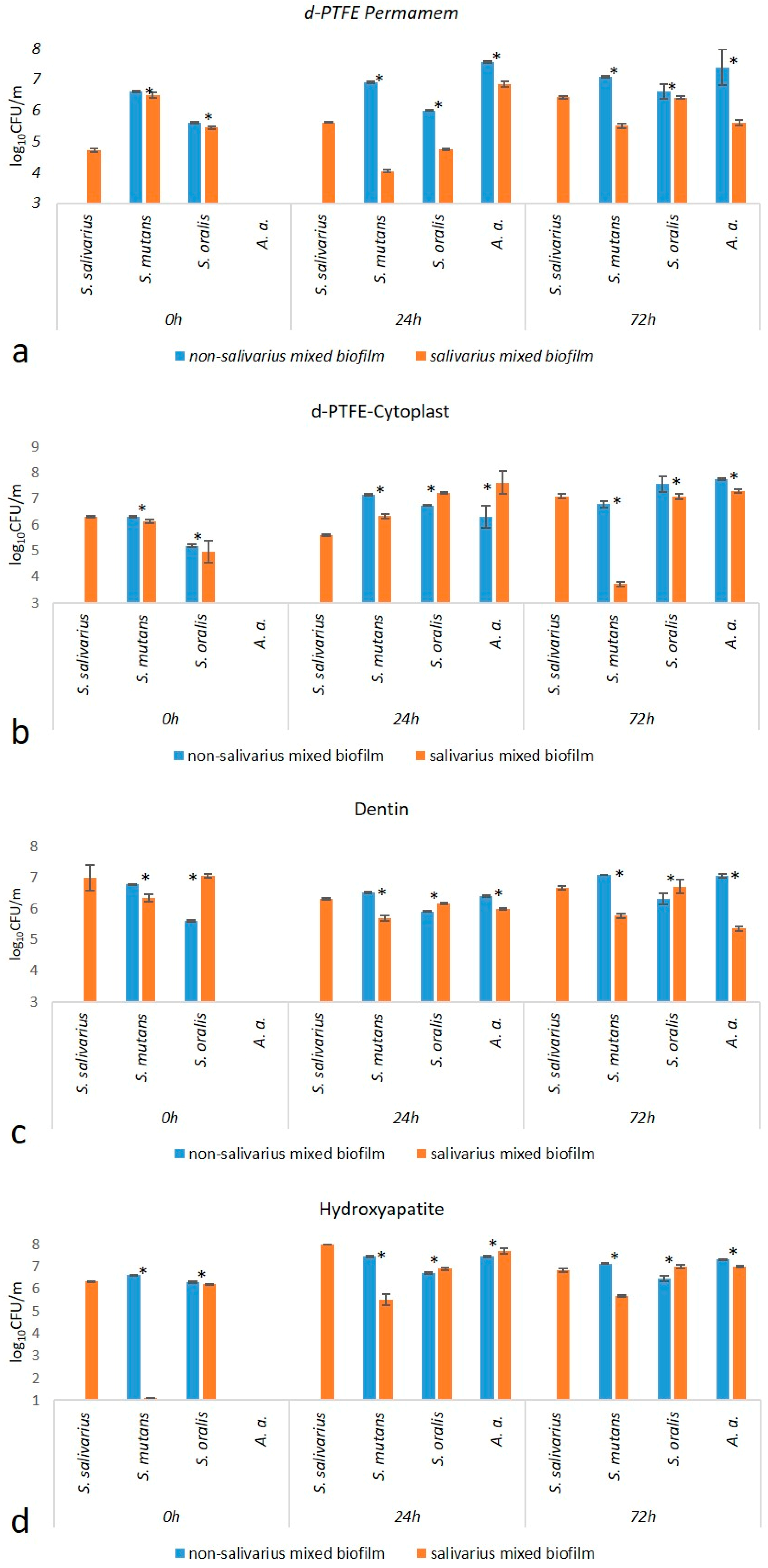
2.1.1. Total Number of Bacteria

In order to compare non-salivarius and salivarius mixed biofilms, the total number of bacteria was measured during biofilm formation. To see the difference, the number of bacteria in the stable biofilm 72 h after the addition of A. actinomycetemcomitans to the initial 24-h streptococcal biofilm is shown (Table 1). The total number of bacteria on d-PTFE membranes showed an increasing trend in the salivarius mixed biofilm, with a significant difference on the Permamem membrane. However, on dentin and hydroxyapatite, the number of bacteria in the salivarius mixed biofilm was significantly lower compared to the non-salivarius mixed biofilm.

2.1.2. Number of Individual Bacterial Species

In order to investigate the difference between non-salivarius and salivarius mixed biofilms, the accompanying growth of certain bacterial species during biofilm formation was determined. The results are shown in Table S1 and Figure 1. The following observations can be drawn: (a) In the 24-h mixed salivarius streptococcal biofilm, S. mutans was represented in a significantly lower number than in the non-salivarius mixed biofilm. (b) In the 72-h mixed biofilm, the representation of S. mutans differed depending on the material on which the biofilm was formed. Lower values of CFU/mL were present in the salivarius mixed biofilm created on dentin and hydroxyapatite. In contrast, on d-PTFE membranes, lower values of CFU/mL were present in the mixed biofilm without S. salivarius. (c) In the 72-h biofilm, on all materials, the number of A. actinomycetemcomitans was significantly lower in the salivarius mixed biofilm.

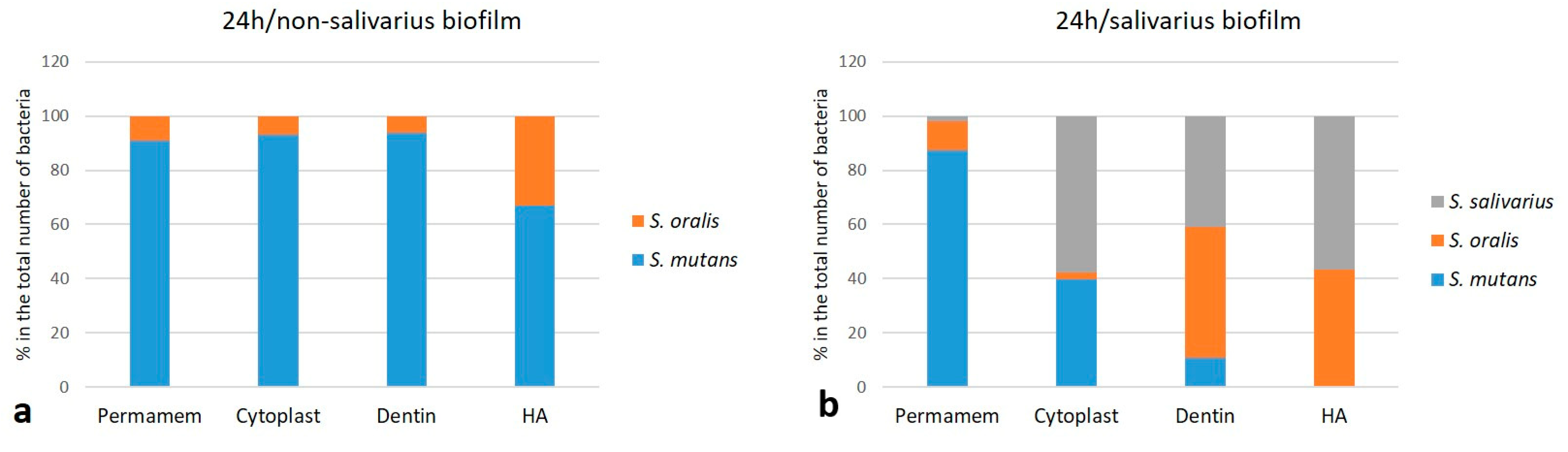
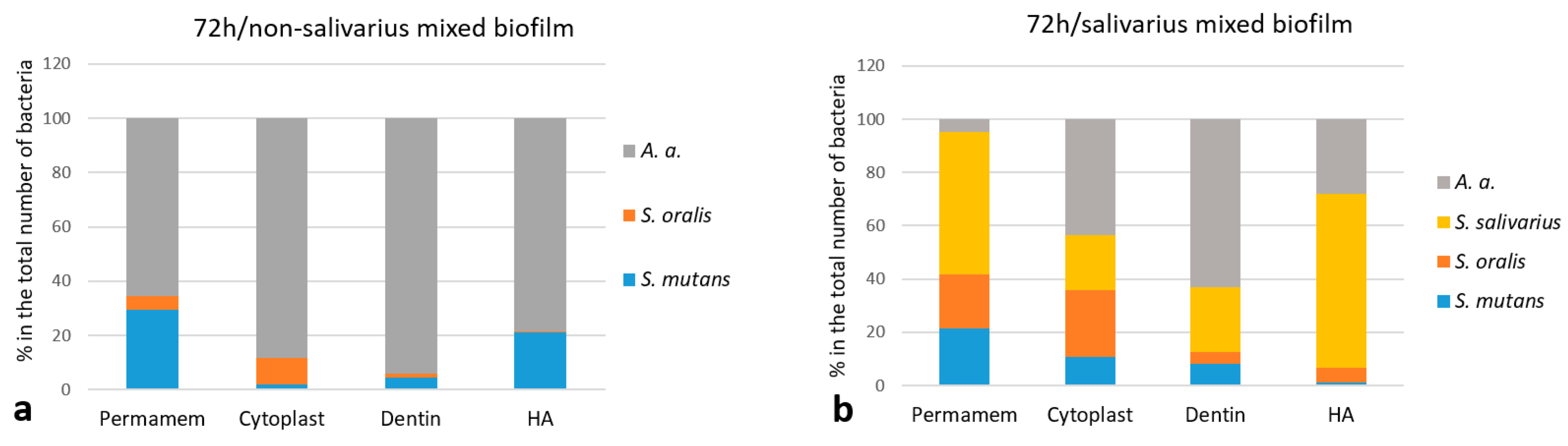
2.1.3. The Proportion of Individual Bacterial Species

To better understand the differences between non-salivarius and salivarius mixed biofilms, the proportion of individual species within the 24 and 72-h biofilms was calculated. The results are presented graphically. The initial streptococcal mixed biofilm created in the presence of S. salivarius K12 already showed a reduced proportion of S. mutans species after 24 h. The difference, although not equal, was observed in all materials (Figure 2). In the 72-h mixed biofilm, the most pronounced difference was observed in the proportion of A. actinomycetemcomitans in the biofilm with or without S. salivarius. The difference was determined on all materials. The proportion of non-salivarius biofilm ranged from 65% on Permamem d-PTFE membrane to 94% on dentin. A lower proportion of this bacterial species was recorded in the salivarius mixed biofilm, ranging from 5% on Permamem d-PTFE membrane to 63% on dentin (Figure 3).

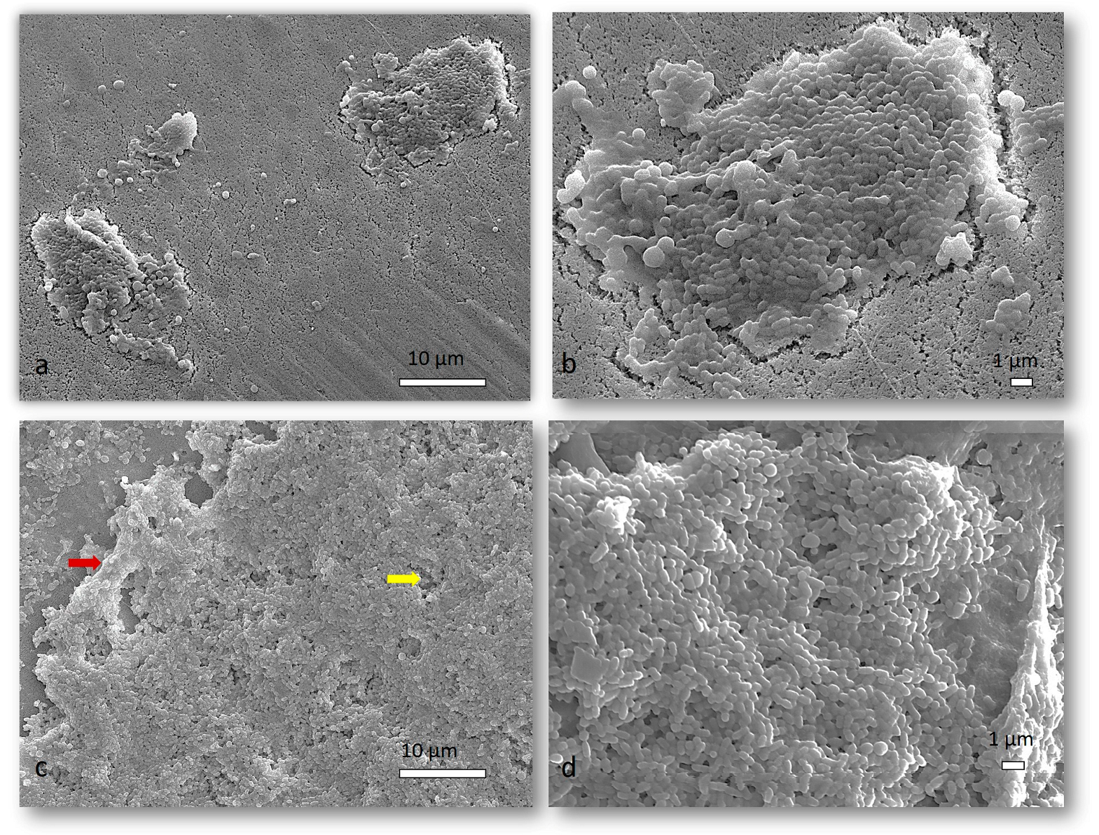
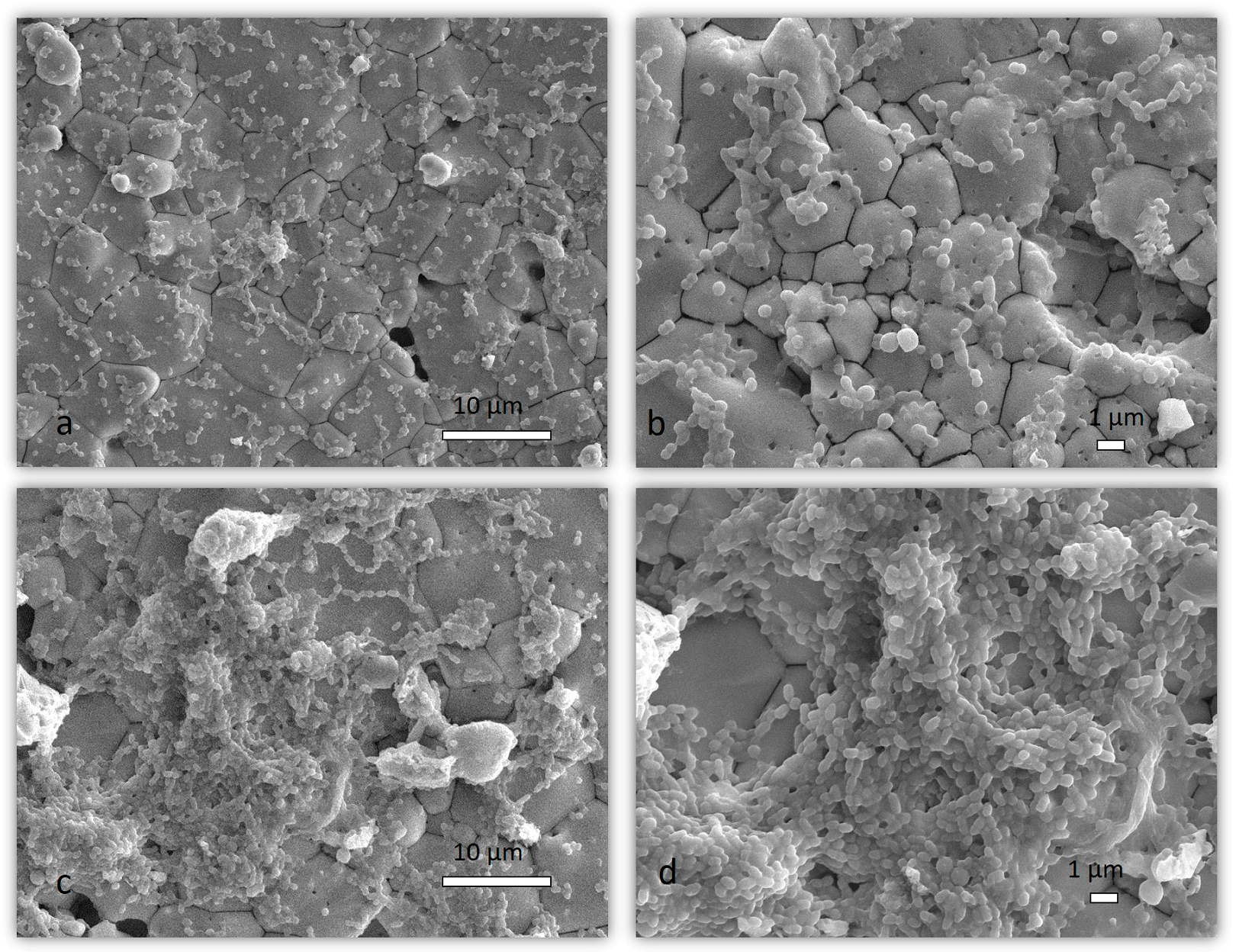
2.1.4. SEM Analysis of Mixed Biofilm

After examining the SEM micrographs, representative images were selected to show the difference in the structure of the 72-h mixed biofilm with and without the presence of S. salivarius K12 (Figure 4 and Figure 5). Figure 4a,b and Figure 5a,b show the salivarius mixed biofilm on Cytoplast d-PTFE and hydroxyapatite, respectively, structured as a disjointed organization of cells covered by a thin slime of Extracellular polymeric substances (EPS). In the non-salivarius mixed biofilm (Figure 4c,d and Figure 5c,d), a thicker biofilm layer was immediately observed. Biofilm has a complex three-dimensional structure consisting of microcolonies filled with EPS. EPS bridging between microcolonies (indicated by a red arrow) is also visible, leading to the formation of aggregates between microcolonies connected by channels (indicated by a yellow arrow) and the development of a complex biofilm. Similar results were obtained on the Permamem d-PTFE membrane and dentin.

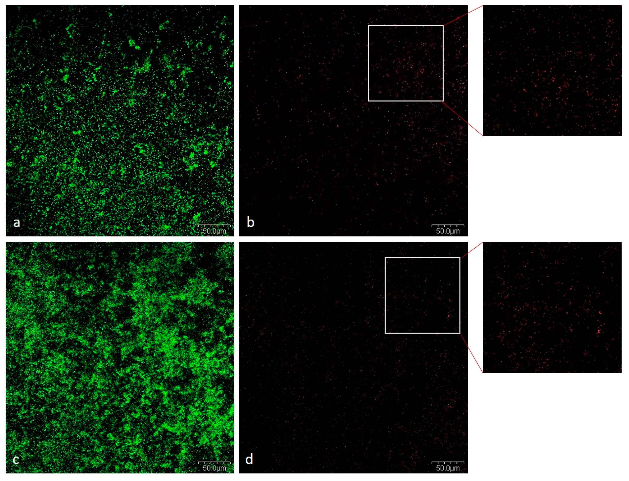
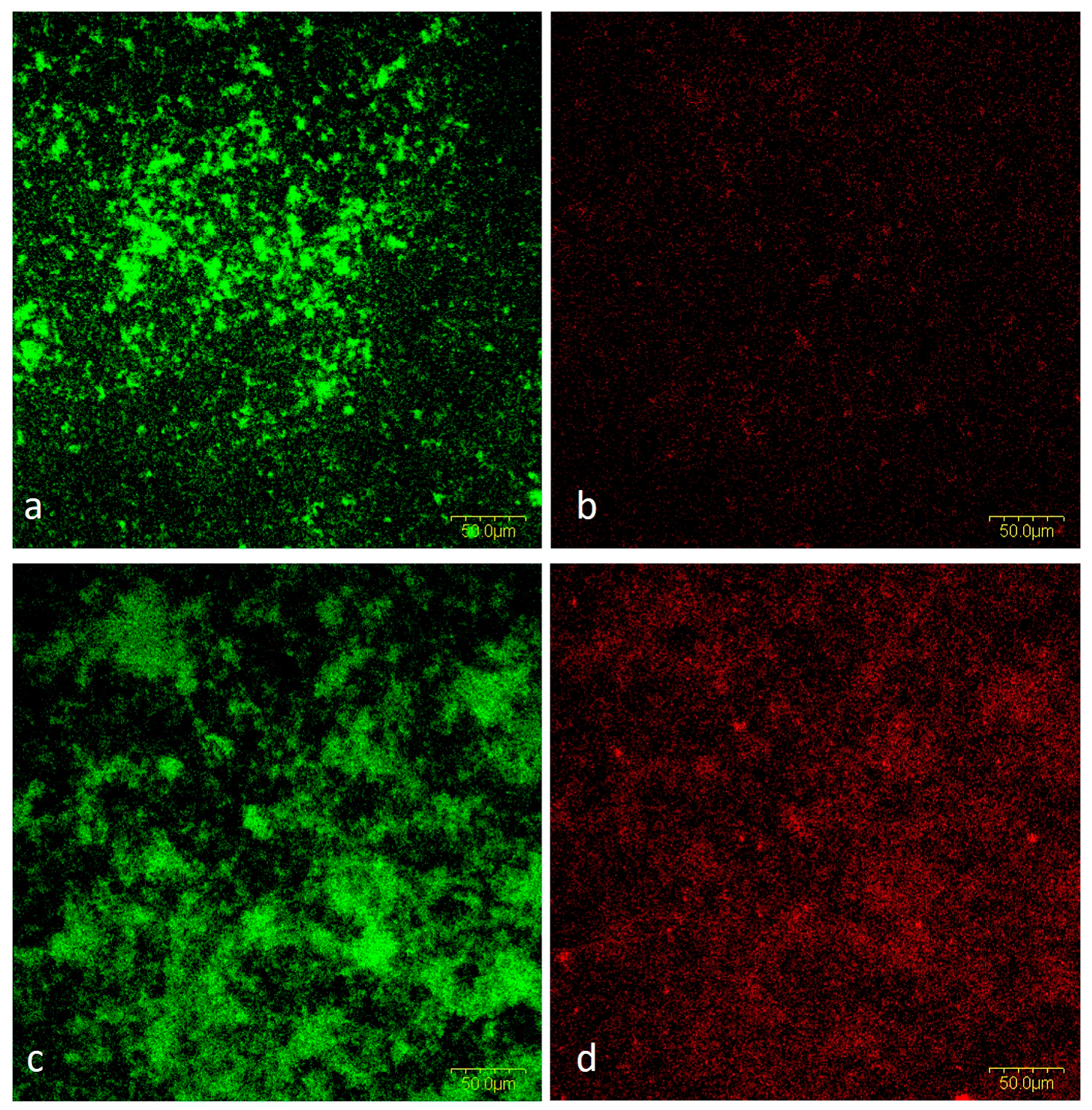
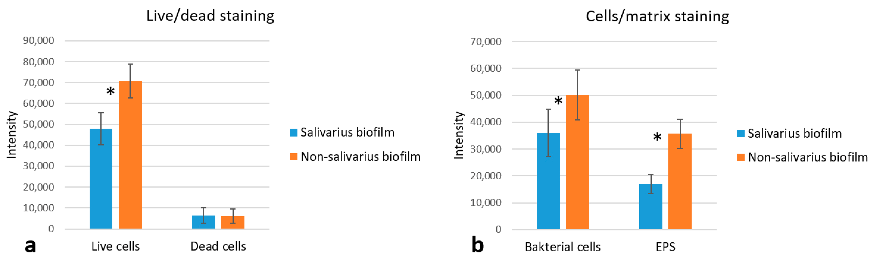
2.1.5. CLSM Analysis

Viability of Bacteria in Mixed Biofilm

Cell viability was assessed by examining the 72-h mixed biofilm after LIVE/DEAD staining. More bacterial biomass was observed in the non-salivarius mixed biofilm. However, biofilms were formed with predominantly living cells in the biofilm with and without S. salivarius K12 (Figure 6).

Cells and Biofilm Matrix in Mixed Biofilm

By staining cells and EPS within the 72-h mixed biofilm, we confirmed the results obtained by SEM analysis. The presence of S. salivarius K12 increased the biofilm’s biomass due to the creation of an EPS-rich matrix and the development of a complex biofilm structure (Figure 7). The average fluorescence intensity of the different experimental groups (for both the dead and live bacterial cells group and the EPS and live bacterial group) can be found in Figure 8.

3. Discussion

The literature points out that the most important step in controlling and preventing periodontal diseases is the inhibition of opportunistic pathogens in dental biofilm. The main role could be commensal bacteria, which antagonistically affect potential periodontal pathogens [4,33,34]. This study aimed to analyze whether the presence of S. salivarius K12 species in a mixed streptococci biofilm affects the colonization and growth of the periodontopathogenic species A. actinomycetemcomitans. Numerous studies confirm that this strain produces bacteriocins, salivaricin A2 and salivaricin B, which inhibit the growth of S. pyogenes [23,35,36]. In addition, it is an antagonist of bacterial species that cause halitosis [37,38,39]. The antagonistic effect of S. salivarius K12 on periodontopathogenic bacteria has been proven in a previous study (or studies). However, these tests were conducted in bacterial suspensions [40]. Since the action of pathogens causes periodontal diseases within a dysbiotically displaced biofilm, we wanted to examine the interactions within the biofilm in this study. Previously, the anti-adhesive and antibiofilm activity of another strain of S. salivarius, TOVE-R, was documented [41,42,43].
In this study, a mixed biofilm model with or without S. salivarius K12 was used to evaluate the antagonistic effect and to compare the number (CFU/mL) and the proportion (%) of individual bacterial species within the 72-h mixed biofilm. It was found that there was a difference that was observed in all tested materials. In the biofilm with the presence of S. salivarius K12, a different total number of bacteria was determined, depending on the material on which the biofilm was created. On dentin and hydroxyapatite, the total number of bacteria was lower in the presence of S. salivarius, while on d-PTFE membranes, the number increased (Table 1). It was also observed that in the 24-h salivarius–streptococcal mixed biofilm on the aforementioned membranes, there was a significantly higher proportion of S. mutans species (40–90%) compared to dentin and hydroxyapatite (10% and lower) (Figure 2). We believe that the physicochemical characteristics of the material’s surface and the surface of the bacterial cell itself influenced the degree of adhesion. D-PTFE membranes are hydrophobic polymers, dentin is a combined inorganic-organic material, and hydroxyapatite is a pure inorganic material. Due to their highly different chemical structures and compositions, they have different surface free energy (SFE) and surface roughness. The SFE surface is one of the main factors on which bacterial adhesion depends. Our previously published research determined the SFE of the tested bacterial species, and we proved that the difference between the SFE of bacteria and the surface they colonize affected adhesion [44,45,46,47,48]. Our results showed that in the presence of S. salivarius, in the initial 24-h streptococcal mixed biofilm, S. mutans was represented in smaller numbers and was a smaller proportion of the total number of bacteria. Events in this early phase of biofilm formation can influence population development over time and shift the balance from a non-virulent to a virulent degree.
S. mutans uses glucosyltransferase (Gtfs) to synthesize glucans from sucrose, contributing to bacterial attachment and subsequent colonization. The presence of Gtfs in the early stages of biofilm formation promotes the formation of a matrix rich in exopolysaccharides (EPS), which provides structural support for constructing three-dimensional structures of microcolonies in the biofilm [49,50,51]. The gtfB and gtfC genes that encode the production of Gtfs can be induced by autoinducers of other oral species in a mixed biofilm [52,53]. Furthermore, sucrose fermentation acidifies the matrix, favoring growth of acid-tolerant microorganisms [52]. The presence of 1% sucrose, as in the medium used in this study, increases biomass and the development of a three-dimensional structure mediated by S. mutans. Within these structures, niches with low pH favor the growth of acidogenic and aciduric pathogens protected by EPS [54]. This is consistent with our results. A thicker and more complex biofilm was observed in SEM micrographs showing non-salivarius mixed biofilm. The bacteria aggregated into microcolonies covered with EPS that filled the spaces between them, thus forming a 3D biofilm architecture. On the other hand, in the mixed biofilm with the presence of S. salivarius, the structural organization of accumulated cells covered by a thin layer of EPS was visible (Figure 4 and Figure 5). These results were confirmed by fluorescence microscopy. In the non-salivarius mixed biofilm, more bacterial biomass interwoven with a richer EPS matrix was observed (Figure 6 and Figure 7). S. salivarius shifted the balance by reducing the proportion of S. mutans already at the initial stage of biofilm development and inhibiting the formation of EPS-entangled microcolonies. In addition, weaker colonization of the periodontopathogenic species A. actinomycetemcomitans was observed in the salivarius mixed biofilm, resulting in a significantly lower number and proportion of these species in the 72-h biofilm (Table 1, Figure 3). It was previously confirmed that S. salivarius isolated from the oral cavity inhibits the growth of A. actinomycetemcomitans [55]. A. actinomycetemcomitans is a slow-growing species competing with fast-growing streptococci for the carbon substrate. This species prefers an alternative substrate, lactate, which allows it to survive in a competitive community [56,57]. However, it does not tolerate low pH levels. S. salivarius has a urease system and can lower pH by creating an alkaline microenvironment [58,59]. In addition, the EPS matrix created by S. mutans in non-salivarius mixed biofilm contains “pockets” within the microcolonies in which acids accumulate, resulting in spatially heterogeneous pH microenvironments and, ultimately, the heterogeneity of the microbial population within the biofilm [54,60]. Furthermore, S. salivarius produces bacteriocins that have an antagonistic effect on many, primarily Gram-positive bacteria [37,61,62]. The outer membrane of Gram-negative bacteria, such as A. actinomycetemcomitans, is impermeable to antibiotics [63]. However, the lactic acid in the biofilm can increase membrane permeability and sensitivity to antibiotics [64,65]. The results of this study show that S. salivarius K12 interacts with other species to have an antagonistic effect on the growth and dominance of opportunistic pathogens in dental biofilm.

4. Materials and Methods

4.1. Tested Materials

Materials independently evaluated in this study are d-PTFE membranes from different manufacturers, Permamem (Botiss biomaterials, Zossen, Germany) and Cytoplast (Osteogenics Biomedical, Lubbock, TX, USA), dentin (Immunodiagnostic Systems Holdings Ltd., Boldon, UK) and hydroxyapatite discs (Clarkson Chromatography Products Inc., South Williamsport, PA, USA). The membranes were aseptically cut to 5 × 5 mm, and t discs were 5 mm in diameter.

4.2. Bacterial Strains and Cultivation Conditions

Reference strains Streptococcus mutans ATCC 25175, Streptococcus oralis ATCC 6249, Streptococcus salivarius K12 ATCC BAA-1024, and Aggregatibacter actinomycetemcomitans ATCC 29522 (Microbiologics, St Cloud, MN, USA) were used. Bacteria were grown on blood agar plates (Biolife, Milan, Italy) supplemented with 5% sheep blood (Biognost, Zagreb, Croatia) in anaerobic conditions at 37 °C for 24–48 h.

4.3. Biofilm Development

An in vitro biofilm model with several bacterial species was developed. Pure bacterial cultures were grown to early stationary growth phase anaerobically in a modified protein-rich BHI liquid medium (Brain Heart Infusion, Becton, Dickinson and Company; Sparks, MD, USA) supplemented with 2.5 g/L mucin (Oxoid, Basingstoke, UK), 1.0 g/L yeast extract (Oxoid, Basingstoke, UK), 0.1 g/L cysteine (Sigma-Aldrich, Burlington, MA, USA), 2.0 g/L sodium bicarbonate (Merck, Darmstad, Germany), 5.0 mg/mL hemin (Sigma-Aldrich, Burlington, MA, USA), 1.0 mg/mL menadione (Merck, Darmstad, Germany) [26]. By measuring the optical density (OD 600), bacterial suspensions of selected streptococci with a concentration of 107 CFU/mL were prepared and mixed in equal proportions. A suspension was prepared to form a mixed biofilm without the presence of S. salivarius—“non-salivarius mixed biofilm” with Streptococcus mutans and S. oralis and with the presence of S. salivarius—“salivarius mixed biofilm” (with Streptococcus mutans, S. oralis and S. salivarius).
Sterile materials, d-PTFE membranes, dentin discs and HA discs were placed in 96-well microtiter plate wells and conditioned for four hours at 30 °C, with 50% artificial saliva whose composition was described earlier [66].
Saliva was removed, and 200 µL of prepared mixed bacterial suspensions were added to all materials. The microtiter plates were incubated for 24 h at 37 °C under anaerobic conditions. After incubation, supernatant with planktonic bacteria was removed, and pure A. actinomycetemcomitans suspension was added to previously formed mixed streptococci biofilm on different materials. Microtiter plates with mixed biofilm formed on different materials were cultured for 72 h, with medium change every 24 h. The plates used to assess the sterility of the culture medium were used as controls.

4.4. Biofilm Quantification

In order to establish the development of individual species present in the mixed biofilm, the 24-h streptococcal biofilm (0 h) and the final mixed biofilm after 24 and 72 h were analyzed.
Bacteria in the mixed biofilm were detached by treatment in an ultrasonic bath (Bactosonic, Bandelin, Germany) at 40 kHz for 1 min. To quantify the bacteria, tenfold dilutions were plated on blood agar plates and CFU/mL was determined. For differentiation of individual species, dilutions were plated on Difco Mitis Salivarius agar with the addition of BBL Chapman Tellurite solution (Becton, Dickinson and Company; Sparks, MD, USA). All measurements were performed three times in triplicate.

4.5. Emission Scanning Electron Microscope (SEM) Analysis

The analysis of mixed bacterial biofilms on various materials was performed using a field emission scanning electron microscope—SEM (Jeol JSM-7800F), with a beam acceleration voltage of 7 kV and a working distance of 10 mm. Prior to SEM analyses, 72-h-old biofilms were washed in sterile PBS and air-dried in a sterile high-current chamber. They were then fixed with 4% glutaraldehyde and 0.5% paraformaldehyde (Sigma-Aldrich) prepared at 4 °C in 0.1 M phosphate buffer (Sigma-Aldrich) (pH 7.2) and then dehydrated by immersion in a series of increasing ethanol concentrations (50, 70, 80, 90, and 100%, Sigma-Aldrich). The samples under study were attached to a sample holder with conductive carbon tape. To prevent surface charging during electron beam irradiation, the samples were coated with a 5 nm thin layer of Au-Pd using the precision etching and coating system PECS II (Gatan Inc., Pleasanton, CA, USA).

4.6. Confocal Laser Scanning Microscope (CLSM) Analysis

4.6.1. Testing the Viability of Bacteria in Biofilm

The viability of bacteria in the 72-h biofilm was tested using the LIVE/DEAD staining technique. LIVE/DEAD BacLight Bacterial Viability Kit L—7012 (Thermo Fisher Scientific, Waltham, MA, USA) was used in the staining procedure. Biofilms were grown as described in Section 4.3. on d-PTFE membranes. Biofilms were gently washed and covered with 5 µL of Propidium iodide and 5 µL of SYTO 9 in 990 µL of sterile distilled water. This was followed by incubation for 15 min at RT in the dark. After washing out the dye, an epifluorescence microscope with GFP/FITC (ex: 480 nm and em: 500 nm) and rhodamine (ex: 490 nm and em: 635 nm) filters were used for observation. All preparations were prepared in triplicate, and five visual fields of the prepared preparations were examined.

4.6.2. Simultaneous Staining of Cells and Biofilm Matrix

FilmTracer SYPRO Ruby Biofilm Matrix Stain and SYTO 9 (Thermo Fisher Scientific, Waltham, MA, USA) were used to stain cells and biofilm matrix simultaneously. Biofilms were grown as described in Section 4.3. on d-PTFE membranes. Biofilms were gently washed and covered with 200 µL of SYPRO Ruby Biofilm Matrix Stain solution for matrix staining. The sample was incubated for 30 min at room temperature and protected from light. After gentle washing with sterile distilled water, bacterial cells were stained with diluted SYTO 9 dye for 3 min, followed by washing and observation using an epifluorescence microscope with GFP/FITC (ex: 480 nm and em: 500 nm) and rhodamine (ex: 580 nm and em: 700 nm) filters.

4.7. Statistical Analysis

The normality of the distribution of continuous variables was tested by the Shapiro-Wilk test. Continuous data were described by the median and 95% confidence interval for the median (95% CI). The Mann–Whitney U test was used to compare the median between two groups. All P values were two-sided. The level of significance was set at Alpha of 0.05. The statistical analysis was performed using MedCalc® Statistical Software version 20.218 (MedCalc Software Ltd., Ostend, Belgium; https://www.medcalc.org (accessed on 1 April 2023).

5. Conclusions

The presence of S. salivarius K12 at the initial stage of streptococcal mixed biofilm formation inhibited the development of an EPS-rich matrix and three-dimensional biofilm structure mediated by S. mutans. There is less colonization by A. actimomycetemcomitans in the salivarius mixed streptococcal biofilm. It is present in significantly smaller numbers in its more mature phase than in the biofilm without S. salivarius K12.
Our results suggest that S. salivarius K 12 can maintain the biofilm’s physiological balance and reduce the growth of periodontal pathogens such as A. actinomycetemcomitans. In addition, our results could improve the preventive approach in the fight against biofilm infections.

Supplementary Materials

The following supporting information can be downloaded at: https://www.mdpi.com/article/10.3390/ijms24087249/s1.

Author Contributions

Conceptualization, G.B. and I.G.; methodology, G.B. and L.K.; investigation, G.B., L.K. and I.J.B.; resources, O.C.P.; data curation, K.K.; writing—original draft preparation, G.B.; writing—review and editing, I.G., D.K. and O.C.P.; visualization, L.K., I.J.B. and I.G.; project administration, O.C.P.; funding acquisition, O.C.P. All authors have read and agreed to the published version of the manuscript.

Funding

This research was funded by the Croatian Science Foundation, grant number IP-2020-02-7875, and by the University of Rijeka, grant number UNIRI-biomed-18-280 and UNIRI-biomed-18-171.

Institutional Review Board Statement

Not applicable.

Informed Consent Statement

Not applicable.

Data Availability Statement

Data sharing is not applicable to this article.

Conflicts of Interest

The authors declare no conflict of interest.

References

  1. Paster, B.J.; Boches, S.K.; Galvin, J.L.; Ericson, R.E.; Lau, C.N.; Levanos, V.A.; Sahasrabudhe, A.; Dewhirst, F.E. Bacterial Diversity in Human Subgingival Plaque. J. Bacteriol. 2001, 183, 3770–3783. [Google Scholar] [CrossRef] [Green Version]
  2. Dewhirst, F.E.; Chen, T.; Izard, J.; Paster, B.J.; Tanner, A.C.R.; Yu, W.-H.; Lakshmanan, A.; Wade, W.G. The Human Oral Microbiome. J. Bacteriol. 2010, 192, 5002–5017. [Google Scholar] [CrossRef] [Green Version]
  3. Zheng, S.; Bawazir, M.; Dhall, A.; Kim, H.-E.; He, L.; Heo, J.; Hwang, G. Implication of Surface Properties, Bacterial Motility, and Hydrodynamic Conditions on Bacterial Surface Sensing and Their Initial Adhesion. Front. Bioeng. Biotechnol. 2021, 9, 643722. [Google Scholar] [CrossRef]
  4. Jenkinson, H.F.; Lamont, R.J. Oral Microbial Communities in Sickness and in Health. Trends Microbiol. 2005, 13, 589–595. [Google Scholar] [CrossRef]
  5. Belstrøm, D.; Holmstrup, P.; Bardow, A.; Kokaras, A.; Fiehn, N.-E.; Paster, B.J. Comparative Analysis of Bacterial Profiles in Unstimulated and Stimulated Saliva Samples. J. Oral Microbiol. 2016, 8, 30112. [Google Scholar] [CrossRef]
  6. Seymour, G.J.; Ford, P.J.; Cullinan, M.P.; Leishman, S.; Yamazaki, K. Relationship between Periodontal Infections and Systemic Disease. Clin. Microbiol. Infect. 2007, 13, 3–10. [Google Scholar] [CrossRef] [Green Version]
  7. Berbari, E.F.; Cockerill, F.R.; Steckelberg, J.M. Infective Endocarditis Due to Unusual or Fastidious Microorganisms. Mayo Clin. Proc. 1997, 72, 532–542. [Google Scholar] [CrossRef]
  8. Awano, S.; Ansai, T.; Takata, Y.; Soh, I.; Akifusa, S.; Hamasaki, T.; Yoshida, A.; Sonoki, K.; Fujisawa, K.; Takehara, T. Oral Health and Mortality Risk from Pneumonia in the Elderly. J. Dent. Res. 2008, 87, 334–339. [Google Scholar] [CrossRef]
  9. Michaud, D.S.; Liu, Y.; Meyer, M.; Giovannucci, E.; Joshipura, K. Periodontal Disease, Tooth Loss, and Cancer Risk in Male Health Professionals: A Prospective Cohort Study. Lancet Oncol. 2008, 9, 550–558. [Google Scholar] [CrossRef] [Green Version]
  10. Lamont, R.J.; Koo, H.; Hajishengallis, G. The Oral Microbiota: Dynamic Communities and Host Interactions. Nat. Rev. Microbiol. 2018, 16, 745–759. [Google Scholar] [CrossRef]
  11. Flemmig, T.F.; Beikler, T. Control of Oral Biofilms: Control of Oral Biofilms. Periodontol. 2000 2011, 55, 9–15. [Google Scholar] [CrossRef]
  12. Marsh, P.D. Dental Plaque as a Biofilm and a Microbial Community—Implications for Health and Disease. BMC Oral Health 2006, 6, S14. [Google Scholar] [CrossRef] [Green Version]
  13. Heersema, L.A.; Smyth, H.D.C. A Multispecies Biofilm In Vitro Screening Model of Dental Caries for High-Throughput Susceptibility Testing. High-Throughput 2019, 8, 14. [Google Scholar] [CrossRef] [Green Version]
  14. Hall-Stoodley, L.; Costerton, J.W.; Stoodley, P. Bacterial Biofilms: From the Natural Environment to Infectious Diseases. Nat. Rev. Microbiol. 2004, 2, 95–108. [Google Scholar] [CrossRef]
  15. Nyvad, B.; Kilian, M. Microbiology of the Early Colonization of Human Enamel and Root Surfaces In Vivo. Eur. J. Oral Sci. 1987, 95, 369–380. [Google Scholar] [CrossRef]
  16. Caufield, P.W.; Dasanayake, A.P.; Li, Y. The Antimicrobial Approach to Caries Management. J. Dent. Educ. 2001, 65, 1091–1095. [Google Scholar] [CrossRef]
  17. Mhaske, A.R.; Shetty, P.C.; Sham Bhat, N.; Ramachandra, C.S.; Laxmikanth, S.M.; Nagarahalli, K.; Tekale, P.D. Antiadherent and Antibacterial Properties of Stainless Steel and NiTi Orthodontic Wires Coated with Silver against Lactobacillus Acidophilus—An In Vitro Study. Prog. Orthod. 2015, 16, 40. [Google Scholar] [CrossRef] [Green Version]
  18. Espinosa-Cristóbal, L.F.; López-Ruiz, N.; Cabada-Tarín, D.; Reyes-López, S.Y.; Zaragoza-Contreras, A.; Constandse-Cortéz, D.; Donohué-Cornejo, A.; Tovar-Carrillo, K.; Cuevas-González, J.C.; Kobayashi, T. Antiadherence and Antimicrobial Properties of Silver Nanoparticles against Streptococcus Mutans on Brackets and Wires Used for Orthodontic Treatments. J. Nanomater. 2018, 2018, 9248527. [Google Scholar] [CrossRef] [Green Version]
  19. Teughels, W.; Van Assche, N.; Sliepen, I.; Quirynen, M. Effect of Material Characteristics and/or Surface Topography on Biofilm Development. Clin. Oral Implants Res. 2006, 17 (Suppl. S2), 68–81. [Google Scholar] [CrossRef]
  20. Philip, N.; Suneja, B.; Walsh, L. Beyond Streptococcus Mutans: Clinical Implications of the Evolving Dental Caries Aetiological Paradigms and Its Associated Microbiome. Br. Dent. J. 2018, 224, 219–225. [Google Scholar] [CrossRef]
  21. Zupancic, K.; Kriksic, V.; Kovacevic, I.; Kovacevic, D. Influence of Oral Probiotic Streptococcus Salivarius K12 on Ear and Oral Cavity Health in Humans: Systematic Review. Probiotics Antimicrob. Proteins 2017, 9, 102–110. [Google Scholar] [CrossRef] [PubMed]
  22. Tanzer, J.M.; Kurasz, A.B.; Clive, J. Competitive Displacement of Mutans Streptococci and Inhibition of Tooth Decay by Streptococcus Salivarius TOVE-R. Infect. Immun. 1985, 48, 44–50. [Google Scholar] [CrossRef] [Green Version]
  23. Di Pierro, F.; Colombo, M.; Zanvit, A.; Risso, P.; Rottoli, A. Use of Streptococcus Salivarius K12 in the Prevention of Streptococcal and Viral Pharyngotonsillitis in Children. Drug Healthc. Patient Saf. 2014, 6, 15–20. [Google Scholar] [CrossRef] [Green Version]
  24. Iebba, V.; Zanotta, N.; Campisciano, G.; Zerbato, V.; Di Bella, S.; Cason, C.; Luzzati, R.; Confalonieri, M.; Palamara, A.T.; Comar, M. Profiling of Oral Microbiota and Cytokines in COVID-19 Patients. Front. Microbiol. 2021, 12, 671813. [Google Scholar] [CrossRef]
  25. Di Pierro, F.; Iqtadar, S.; Mumtaz, S.U.; Bertuccioli, A.; Recchia, M.; Zerbinati, N.; Khan, A. Clinical Effects of Streptococcus Salivarius K12 in Hospitalized COVID-19 Patients: Results of a Preliminary Study. Microorganisms 2022, 10, 1926. [Google Scholar] [CrossRef]
  26. Sánchez, M.C.; Llama-Palacios, A.; Blanc, V.; León, R.; Herrera, D.; Sanz, M. Structure, Viability and Bacterial Kinetics of an in Vitro Biofilm Model Using Six Bacteria from the Subgingival Microbiota: An in Vitro Subgingival Biofilm Model. J. Periodontal Res. 2011, 46, 252–260. [Google Scholar] [CrossRef]
  27. Lemos, J.A.; Burne, R.A. A Model of Efficiency: Stress Tolerance by Streptococcus Mutans. Microbiology 2008, 154, 3247–3255. [Google Scholar] [CrossRef] [Green Version]
  28. Lemos, J.A.; Palmer, S.R.; Zeng, L.; Wen, Z.T.; Kajfasz, J.K.; Freires, I.A.; Abranches, J.; Brady, L.J. The Biology of Streptococcus Mutans. Microbiol. Spectr. 2019, 7, 7.1.03. [Google Scholar] [CrossRef]
  29. Okahashi, N.; Nakata, M.; Kuwata, H.; Kawabata, S. Streptococcus Oralis Induces Lysosomal Impairment of Macrophages via Bacterial Hydrogen Peroxide. Infect. Immun. 2016, 84, 2042–2050. [Google Scholar] [CrossRef] [Green Version]
  30. Kolenbrander, P.E.; Palmer, R.J.; Periasamy, S.; Jakubovics, N.S. Oral Multispecies Biofilm Development and the Key Role of Cell–Cell Distance. Nat. Rev. Microbiol. 2010, 8, 471–480. [Google Scholar] [CrossRef]
  31. Whitmore, S.E.; Lamont, R.J. The Pathogenic Persona of Community-Associated Oral Streptococci: Pathogenic Streptococcal Communities. Mol. Microbiol. 2011, 81, 305–314. [Google Scholar] [CrossRef] [PubMed] [Green Version]
  32. Claesson, N.-L.; Jensen, B.; Åberg; Aggregatibacter, H. Actinomycetemcomitans: Clinical Significance of a Pathobiont Subjected to Ample Changes in Classification and Nomenclature. Pathogens 2019, 8, 243. [Google Scholar] [CrossRef] [Green Version]
  33. van Essche, M.; Loozen, G.; Godts, C.; Boon, N.; Pauwels, M.; Quirynen, M.; Teughels, W. Bacterial Antagonism Against Periodontopathogens. J. Periodontol. 2013, 84, 801–811. [Google Scholar] [CrossRef] [PubMed]
  34. Koll-Klais, P.; Mandar, R.; Leibur, E.; Marcotte, H.; Hammarstrom, L.; Mikelsaar, M. Oral Lactobacilli in Chronic Periodontitis and Periodontal Health: Species Composition and Antimicrobial Activity. Oral Microbiol. Immunol. 2005, 20, 354–361. [Google Scholar] [CrossRef] [PubMed]
  35. Sharma, S.; Verma, K.K. Skin and Soft Tissue Infection. Indian J. Pediatr. 2001, 68 (Suppl. S3), S46–S50. [Google Scholar] [PubMed]
  36. Di Pierro, F.; Adami, T.; Rapacioli, G.; Giardini, N.; Streitberger, C. Clinical Evaluation of the Oral Probiotic Streptococcus Salivarius K12 in the Prevention of Recurrent Pharyngitis and/or Tonsillitis Caused by Streptococcus Pyogenes in Adults. Expert Opin. Biol. Ther. 2013, 13, 339–343. [Google Scholar] [CrossRef]
  37. Masdea, L.; Kulik, E.M.; Hauser-Gerspach, I.; Ramseier, A.M.; Filippi, A.; Waltimo, T. Antimicrobial Activity of Streptococcus Salivarius K12 on Bacteria Involved in Oral Malodour. Arch. Oral Biol. 2012, 57, 1041–1047. [Google Scholar] [CrossRef]
  38. Burton, J.P.; Chilcott, C.N.; Moore, C.J.; Speiser, G.; Tagg, J.R. A Preliminary Study of the Effect of Probiotic Streptococcus Salivarius K12 on Oral Malodour Parameters. J. Appl. Microbiol. 2006, 100, 754–764. [Google Scholar] [CrossRef]
  39. Burton, J.; Chilcott, C.; Tagg, J. The Rationale and Potential for the Reduction of Oral Malodour Using Streptococcus Salivarius Probiotics. Oral Dis. 2005, 11, 29–31. [Google Scholar] [CrossRef]
  40. Jansen, P.M.; Abdelbary, M.M.H.; Conrads, G. A Concerted Probiotic Activity to Inhibit Periodontitis-Associated Bacteria. PLoS ONE 2021, 16, e0248308. [Google Scholar] [CrossRef]
  41. Van Hoogmoed, C.G.; Geertsema-doornbusch, G.I.; Teughels, W.; Quirynen, M.; Busscher, H.J.; Van der Mei, H.C. Reduction of Periodontal Pathogens Adhesion by Antagonistic Strains: Reduction of Periodontopathogens. Oral Microbiol. Immunol. 2007, 23, 43–48. [Google Scholar] [CrossRef] [PubMed]
  42. Sliepen, I.; Essche, M.V.; Loozen, G.; Eldere, J.V.; Quirynen, M.; Teughels, W. Interference with Aggregatibacter Actinomycetemcomitans: Colonization of Epithelial Cells. J. Dent. Res. 2007, 86, 611–617. [Google Scholar]
  43. Inhibition of Ecological Emergence of Mutans Streptococci Naturally Transmitted between Rats and Consequent Caries Inhibition by Streptococcus Salivarius TOVE-R Infection—PMC. Available online: https://www.ncbi.nlm.nih.gov/pmc/articles/PMC262061/ (accessed on 7 February 2023).
  44. Lazzini, G.; Lutey, A.H.A.; Romoli, L.; Fuso, F. Molecular Dynamics Model for the Antibactericity of Textured Surfaces. Colloids Surf. B Biointerfaces 2021, 199, 111504. [Google Scholar] [CrossRef] [PubMed]
  45. Begić, G.; Petković Didović, M.; Lučić Blagojević, S.; Jelovica Badovinac, I.; Žigon, J.; Perčić, M.; Cvijanović Peloza, O.; Gobin, I. Adhesion of Oral Bacteria to Commercial D-PTFE Membranes: Polymer Microstructure Makes a Difference. Int. J. Mol. Sci. 2022, 23, 2983. [Google Scholar] [CrossRef] [PubMed]
  46. Zhang, X.; Zhang, Q.; Yan, T.; Jiang, Z.; Zhang, X.; Zuo, Y.Y. Quantitatively Predicting Bacterial Adhesion Using Surface Free Energy Determined with a Spectrophotometric Method. Environ. Sci. Technol. 2015, 49, 6164–6171. [Google Scholar] [CrossRef] [Green Version]
  47. Zhang, X.; Zhang, Q.; Yan, T.; Jiang, Z.; Zhang, X.; Zuo, Y.Y. Surface Free Energy Activated High-Throughput Cell Sorting. Anal. Chem. 2014, 86, 9350–9355. [Google Scholar] [CrossRef] [Green Version]
  48. Okulus, Z.; Strzemiecka, B.; Czarnecka, B.; Buchwald, T.; Voelkel, A. Surface Energy of Bovine Dentin and Enamel by Means of Inverse Gas Chromatography. Mater. Sci. Eng. C 2015, 49, 382–389. [Google Scholar] [CrossRef]
  49. Lei, L.; Long, L.; Yang, X.; Qiu, Y.; Zeng, Y.; Hu, T.; Wang, S.; Li, Y. The VicRK Two-Component System Regulates Streptococcus Mutans Virulence. Curr. Issues Mol. Biol. 2019, 32, 167–200. [Google Scholar] [CrossRef]
  50. Steinberg, D.; Beeman, D.; Bowen, W.H. Kinetic Properties of Glucosyltransferase Adsorbed Onto Saliva-Coated Hydroxyapatite. Artif. Cells Blood Substit. Biotechnol. 1996, 24, 553–566. [Google Scholar] [CrossRef]
  51. Venkitaraman, A.R.; Vacca-Smith, A.M.; Kopec, L.K.; Bowen, W.H. Characterization of GlucosyltransferaseB, GtfC, and GtfD in Solution and on the Surface of Hydroxyapatite. J. Dent. Res. 1995, 74, 1695–1701. [Google Scholar] [CrossRef]
  52. Waters, C.M.; Bassler, B.L. QUORUM SENSING: Cell-to-Cell Communication in Bacteria. Annu. Rev. Cell Dev. Biol. 2005, 21, 319–346. [Google Scholar] [CrossRef] [PubMed] [Green Version]
  53. Yoshida, A.; Ansai, T.; Takehara, T.; Kuramitsu, H.K. LuxS-Based Signaling Affects Streptococcus Mutans Biofilm Formation. Appl. Environ. Microbiol. 2005, 71, 2372–2380. [Google Scholar] [CrossRef] [PubMed] [Green Version]
  54. Koo, H.; Xiao, J.; Klein, M.I.; Jeon, J.G. Exopolysaccharides Produced by Streptococcus Mutans Glucosyltransferases Modulate the Establishment of Microcolonies within Multispecies Biofilms. J. Bacteriol. 2010, 192, 3024–3032. [Google Scholar] [CrossRef] [Green Version]
  55. Vaniabella, R.; Joenoes, H.; Bachtiar, B.M. Analysis on the Inhibition of Aggregatibacter Actinomycetemcomitans by Streptococcus Salivarius Isolated from Saliva and Tongue Dorsum of Adults. Asian J. Pharm. Clin. Res. 2017, 10, 6. [Google Scholar] [CrossRef] [Green Version]
  56. Staat, R.H.; Gawronski, T.H.; Cressey, D.E.; Harris, R.S.; Folke, L.E.A. Effects of Dietary Sucrose Levels on the Quantity and Microbial Composition of Human Dental Plaque. J. Dent. Res. 1975, 54, 872–880. [Google Scholar] [CrossRef]
  57. Brown, S.A.; Whiteley, M. A Novel Exclusion Mechanism for Carbon Resource Partitioning in Aggregatibacter Actinomycetemcomitans. J. Bacteriol. 2007, 189, 6407–6414. [Google Scholar] [CrossRef] [Green Version]
  58. Sissons, C.H.; Hancock, E.M. Urease Activity in Streptococcus Salivarius at Low PH. Arch. Oral Biol. 1993, 38, 507–516. [Google Scholar] [CrossRef]
  59. Burne, R.A.; Marquis, R.E. Alkali Production by Oral Bacteria and Protection against Dental Caries. FEMS Microbiol. Lett. 2000, 193, 1–6. [Google Scholar] [CrossRef]
  60. Jakubovics, N.S. Intermicrobial Interactions as a Driver for Community Composition and Stratification of Oral Biofilms. J. Mol. Biol. 2015, 427, 3662–3675. [Google Scholar] [CrossRef]
  61. Wescombe, P.A.; Heng, N.C.; Burton, J.P.; Chilcott, C.N.; Tagg, J.R. Streptococcal Bacteriocins and the Case for Streptococcus Salivarius as Model Oral Probiotics. Future Microbiol. 2009, 4, 819–835. [Google Scholar] [CrossRef] [Green Version]
  62. Wescombe, P.A.; Upton, M.; Renault, P.; Wirawan, R.E.; Power, D.; Burton, J.P.; Chilcott, C.N.; Tagg, J.R. Salivaricin 9, a New Lantibiotic Produced by Streptococcus Salivarius. Microbiology 2011, 157, 1290–1299. [Google Scholar] [CrossRef] [PubMed] [Green Version]
  63. Lantibiotics: Structure, Biosynthesis and Mode of Action|FEMS Microbiology Reviews|Oxford Academic. Available online: https://academic.oup.com/femsre/article/25/3/285/624045 (accessed on 28 February 2023).
  64. Alakomi, H.-L.; Skyttä, E.; Saarela, M.; Mattila-Sandholm, T.; Latva-Kala, K.; Helander, I.M. Lactic Acid Permeabilizes Gram-Negative Bacteria by Disrupting the Outer Membrane. Appl. Environ. Microbiol. 2000, 66, 2001–2005. [Google Scholar] [CrossRef] [PubMed] [Green Version]
  65. Stanojević-Nikolić, S.; Dimić, G.R.; Mojović, L.; Pejin, J.D.; Djukić-Vuković, A.; Kocić-Tanackov, S.D. Antimicrobial Activity of Lactic Acid against Pathogen and Spoilage Microorganisms. J. Food Process. Preserv. 2016, 40, 990–998. [Google Scholar] [CrossRef]
  66. Pratten, J.; Smith, A.W.; Wilson, M. Response of Single Species Biofilms and Microcosm Dental Plaques to Pulsing with Chlorhexidine. J. Antimicrob. Chemother. 1998, 42, 453–459. [Google Scholar] [CrossRef] [PubMed]
Figure 1. Comparison of the number (log10CFU/mL) of individual bacterial species in non-salivarius and salivarius mixed biofilms on: (a) Permamem, (b) Cytoplast, (c) Dentin and (d) Hydroxyapatite. 95% CI—95% Confidence interval: * Mann–Whitney U test.
Ijms 24 07249 g001
Figure 2. Distribution (%) of individual bacterial species in initial streptococcal biofilm (n = 9); (a) non-salivarius streptococcal mixed biofilm; (b) salivarius streptococcal mixed biofilm.
Ijms 24 07249 g002
Figure 3. Distribution (%) of individual bacterial species on Permamem and Cytoplast d-PTFE membrane, dentin and hydroxyapatite (n = 9); (a) 72-h non-salivarius mixed biofilm; (b) salivarius mixed biofilm.
Ijms 24 07249 g003
Figure 4. Representative SEM micrographs of a 72-h biofilm on Cytoplast d-PTFE. Different magnifications of salivarius mixed biofilm are presented in (a,b), while different magnifications of non-salivarius mixed biofilm are presented in (c,d); Red arrow—EPS bridging between microcolonies; Yellow arrow—aggregates between microcolonies connected by channels; Magnification of 2000× and 5000×.
Ijms 24 07249 g004
Figure 5. Representative SEM micrographs of a 72-h biofilm on hydroxyapatite. Different magnifications of salivarius mixed biofilm are presented in (a,b), while different magnifications of non-salivarius mixed biofilm are presented in (c,d); Magnification of 2000× and 5000×.
Ijms 24 07249 g005
Figure 6. Illustration of dead (red) and live bacterial cells (green); (a) SYTO-9 stain in salivarius mixed biofilm; (b) Propidium iodide in salivarius mixed biofilm (c) SYTO-9 in non-salivarius mixed biofilm; (d) Propidium iodide in non-salivarius mixed biofilm; Magnification 20×.
Ijms 24 07249 g006
Figure 7. Illustration of the structural organization of EPS (red) and live bacterial cells (green); (a) SYTO-9 stain in salivarius mixed biofilm; (b) SYPRO Ruby Biofilm Matrix Stain in salivarius mixed biofilm; (c) SYTO-9 stain in non-salivarius mixed biofilm; (d) SYPRO Ruby Biofilm Matrix Stain in non-salivarius mixed biofilm; Magnification 20×.
Ijms 24 07249 g007
Figure 8. The average fluorescence intensity of the different experimental groups (n = 15). The asterisk (*) indicates statistically significant differences between the groups (p < 0.05); (a) Live/dead staining; (b) Cell/matrix staining.
Ijms 24 07249 g008
Table 1. Comparison of the total number of bacteria (log CFU/mL) in a 72-h mixed biofilm.
72 hp *
Non-Salivarius Mixed BiofilmSalivarius Mixed Biofilm
Median (95% CI for Median)
d-PTFE-Permamem6.70 (6.12–6.94)7.09 (6.95–7.15)0.005
d-PTFE-Cytoplast6.19 (5.73–7.13)6.45 (6.29–6.53)0.27
Dentin6.89 (6.48–8.16)5.75 (5.47–6.01)<0.001
Hydroxyapatite7.29 (5.39–7.83)6.39 (5.95–6.77)0.04
95% CI—95% Confidence interval: * Mann–Whitney U test; Bold denotes significant, (n = 9).
Disclaimer/Publisher’s Note: The statements, opinions and data contained in all publications are solely those of the individual author(s) and contributor(s) and not of MDPI and/or the editor(s). MDPI and/or the editor(s) disclaim responsibility for any injury to people or property resulting from any ideas, methods, instructions or products referred to in the content.

