
Antioxidant Activity of Spiranthes sinensis and Its Protective Effect against UVB-Induced Skin Fibroblast Damage

1
Biotechnology Center, National Chung Hsing University, Taichung 40227, Taiwan
2
Department of Chemical Engineering and Biotechnology, Tatung University, Taipei 10452, Taiwan
*
Author to whom correspondence should be addressed.
Processes 2021, 9(9), 1564; https://doi.org/10.3390/pr9091564
Submission received: 17 August 2021
/
Revised: 27 August 2021
/
Accepted: 30 August 2021
/
Published: 1 September 2021
(This article belongs to the Special Issue Biological Activity Evaluation Process of Natural Antioxidants)
Abstract
:Spiranthes sinensis (S. sinensis), a species of orchid, is a well-known herb medicine used to treat disorders such as stomachache, diabetes, shingles, and certain inflammatory diseases. Presently, the antioxidant activity of S. sinensis as well as its protective effect on UVB-induced skin injury are unclear. In this study, S. sinensis was extracted with boiling water or 75% (v/v) ethanol, and then its antioxidant composition and antioxidant activity were determined. The protective effects of S. sinensis against UVB-induced damage in human skin fibroblasts (CCD-966SK) were also investigated. Our data showed that the extraction yield of boiling water was higher than that of 75% ethanol. However, compared to the aqueous extracts, the ethanol extracts not only had higher phenolic, flavonoid, and condensed tannin contents, but also exhibited higher free radical scavenging activity, higher reducing power, and higher ferrous ion-chelating capacity. When fibroblasts were pre-cultured with the water or ethanol extracts of S. sinensis (1500 μg/mL) for 24 h before applying UVB irradiation, the S. sinensis extracts restored 17% to 27% of cell viability compared to a control only irradiated with UVB. Overall, our study suggests that S. sinensis extracts can be used as effective antioxidants and have the potential to protect skin fibroblasts from UVB irradiation.
1. Introduction
It is well known that the genotoxic and mutagenic effects of long-term exposure to sunlight are mainly caused by ultraviolet radiation (UVR) in the solar spectrum. Ultraviolet rays can be classified into three categories according to their wavelength range: UVA (315–400 nm), UVB (280–315 nm), and UVC (100–280 nm) [1]. UVA accounts for 95% of the UVR reaching the earth, while all UVC and 90% of UVB are absorbed by the earth’s atmosphere. Despite the fact that only a small fraction of UVB reaches the earth, UVB is more likely to cause sunburn than UVA. In contrast to UVA, which can penetrate the dermis, most of the high-energy UVB can be absorbed by the epidermis [2]. UVB is a main mutagenic factor because UVB photons absorbed by DNA cause structural damage to DNA itself and a deficiency of nucleotide repair proteins [3]. Although both UVA and UVB induce the formation of reactive oxygen species (ROS), which is the major cause of DNA damage in epidermal stem cells [1,4], UVA is generally less cytotoxic and mutagenic than UVB [5]. Based on the previous studies, we chose UVB to induce damage to skin fibroblasts in the present study. However, it is important to highlight that the combination of UVA and UVB may lead to photoaging, immunosuppression, and skin cancer [1,6].
Spiranthes sinensis (Pers.) Ames, a species of orchid, is a well-known herb growing in eastern and southeastern Asia. S. sinensis (Pers.) Ames can be used to treat stomachache, diabetes, and shingles. The species S. mauritianum from the same genus has been used to treat bacterial infections and inflammatory diseases [7]. A dihydroflavanoid compound isolated from the roots of S. australis showed anticancer activity in vitro [8]. Phenanthrenes, flavonoids, phenolics, and a bibenzyl compound have been identified in petroleum ether and ethyl acetate extracts of S. sinensis [9]. These compounds may explain the biological activity of S. sinensis.
Natural plant extracts have been demonstrated to possess antioxidant activity and protect the skin from UV damage. For instance, a lemon balm extract has been shown to significantly inhibit UVB-induced intracellular ROS production and reduce UV-induced DNA damage [10]. An Agastache rugosa Kuntze extract could restore UVB-induced decreases in glutathione and superoxide dismutase levels in HaCaT keratinocytes [11]; the extract also inhibited the activity and reduced the protein level of pro-matrix metalloproteinases-2 and -9 elevated due to UVB. Water extracts of Galinsoga herb also showed protective effects against UVB, partly due to their capacity to inhibit the production of ROS [12].
Overall, the increase in UVB radiation on earth poses a significant threat to the skin, increasing health risks with long-term consequences, such as photoaging and photocarcinogenesis of the skin. The market of cosmetic products based on natural compounds with anti-aging properties and for photoprotection is continuing to grow due to increasing concerns related to quality of life and one’s appearance, and there is a growing demand for expanding research in this area. Presently, the protective potential of the antioxidant activity of natural S. sinensis against UVB-induced skin damage remains unclear. This study aimed to evaluate the antioxidant capacity of natural S. sinensis and to investigate the protective potential of S. sinensis against UV-induced skin damage. In this work, natural S. sinensis was extracted with boiling water or 75% (v/v) ethanol, and the antioxidant capacity of the extracts was determined. Finally, the protective ability of S. sinensis against UVB irradiation damage to human skin fibroblasts was confirmed.
2. Materials and Methods
2.1. Extraction of S. sinensis
The dried whole plant of S. sinensis was obtained from an herbal medicine store in Taipei, Taiwan. Before extraction, the raw materials were dried in an oven at 40 °C for one week. For extraction with boiling water, 10 g of the sample was ground to a powder (18 mesh) using a mechanical grinder and then extracted with 400 mL of boiling water for 30 min. The extract mixture was cooled to room temperature, centrifuged at 8000× g for 20 min, and then filtered through filter paper (Whatman No. 1). The filtered residue was further extracted twice with 400 mL of boiling water. For the extraction with 75% (v/v) ethanol, the only difference was that the extraction was performed at room temperature with a rotary shaking machine at 120 rpm for 24 h. The extracts were light brown; the background resulting from the color was corrected against a blank in subsequent analyses. Regardless of the extraction solvent used, the combined extracts were rotary-evaporated (Rotavapor R-210, Büchi, Flawil, Switzerland) and then lyophilized (UNITOP 600SL Freezer Dryer, Virtis, Gardiner, NY, USA). The lyophilized powder was stored in a dry box.
2.2. Determination of Antioxidants Content
2.2.1. Total Phenols
The content of total phenols was evaluated using the Folin–Ciocalteu reagent [13]. Aliquots (100 μL) of the extracts or standards were mixed with 2 mL of 2% Na2CO3 and incubated for 2 min at room temperature, followed by the addition of 100 μL of 50% Folin–Ciocalteu reagent. After reaction at 30 °C for 30 min, the absorbance against the blank was determined at 750 nm using a spectrophotometer (Multiskan Spectrum, Thermo Fisher Scientific, Waltham, MA, USA). The concentration of phenol was calculated from a standard curve constructed with gallic acid (y = 1.74x + 0.012; y is the absorbance, and x is the concentration of gallic acid in μg/mL; R2 = 0.9998). Results were expressed as milligrams of gallic acid equivalents (mg GAE) per gram of extract. Ascorbic acid and quercetin were used as positive controls.
2.2.2. Total Flavonoid Content
The total flavonoid content was evaluated by a Dowd method similar to that described by Gursoy et al. [14]. In brief, 100 μL of the extract or standard was mixed with 2% aluminum trichloride (AlCl3) in the same volume of methanol. After incubation in the dark for 10 min, the absorbance was determined at 430 nm against a blank control. The concentration of flavonoids was calculated from the standard curve constructed with quercetin (y = 83.4x + 0.003; y is the absorbance and, x is the concentration of quercetin in μg/mL; R2 = 0.9997). Results were expressed as milligrams of quercetin equivalents (mg QE) per gram of extract. Ascorbic acid and quercetin were used as positive controls.
2.2.3. Condensed Tannins
Colorimetric analysis of condensed tannins was carried out by the vanillin–HCl assay described by Broadhurst and Jones (1978) [15]. To the extract or standard (50 μL), 300 μL of 4% vanillin reagent prepared in methanol and 300 μL of 37% HCl were added. After incubation in the dark for 20 min at room temperature, the absorbance of the sample against the blank was measured at 500 nm. The content of condensed tannins was calculated from the standard curve constructed with (+)-catechin standards (y = 1.7x + 0.027; y is the absorbance, and x is the concentration of (+)-catechin in μg/mL; R2 = 0.995). Results were expressed as milligrams of catechin equivalents (mg CE) per gram of extract. Ascorbic acid and quercetin were used as positive controls.
2.3. Evaluation of Antioxidant Activity
2.3.1. DPPH Assay of Free Radical Scavenging Activity
DPPH free radical scavenging activity was measured in 96-well microplates according to the procedure described by Shimada et al. with slight modifications [16]. In total, 50 μL of extract and 150 μL of 200 μM DPPH in methanol were mixed in a microwell, then reacted in the dark at 25 °C for 90 min, and then the absorbance was measured at 517 nm with a multi-well plate reader (Molecular Devices, SpectraMax M2, Sunnyvale, CA, USA). Quercetin and ascorbic acid were used as positive controls. The scavenging activity (%) was obtained by the following equation:
where ADPPH is the absorbance of DPPH only, As-DPPH is the absorbance of DPPH in the presence of the sample, and As-blank is the absorbance of the sample without DPPH. The compound 6-hydroxy-2,5,7,8-tetramethylchromane-2-carboxylic acid (Trolox) was used as a standard, and the activity was also expressed as μmoles of Trolox equivalents (TE) per gram of extract. EC50 was defined as the concentration required to scavenge 50% of the free radicals; EC50 was interpolated from the linear region of the standard curve (R2 values were 0.9416, 0.9672, 0.9694, and 0.9863 for water extract, 75% ethanol extract, ascorbic acid, and quercetin, respectively).
Scavenging activity (%) = [ADPPH − (As-DPPH − As-blank)]/ADPPH × 100
2.3.2. ABTS Assay of Free Radical Scavenging Activity
ABTS free radical scavenging activity was determined in 96-well microplates based on the procedure described by Loziene et al. with slight modifications [17]. ABTS radicals were generated by mixing 50 mL of ABTS (2 mM) with 200 μL of potassium persulfate (70 mM); both solutions were prepared in a potassium phosphate buffer (pH 7.4). The mixture was incubated in the dark for 15–16 h to produce the ABTS radical solution, which was further diluted with phosphate buffer until the absorbance at 734 nm reached the value of 0.800 ± 0.030. Then, 10 μL of the extract was mixed with 1.0 mL of the diluted ABTS solution, and the absorbance at 734 nm was determined after incubation in the dark for 10 min. Quercetin and ascorbic acid were used as positive controls. The scavenging activity (%) was obtained by the following equation:
where AABTS is the absorbance of ABTS, As-ABTS is the absorbance of ABTS in the sample, and As-blank is the absorbance of the sample without ABTS. Trolox was used as a standard, and the activity was also expressed as μmoles of Trolox equivalents (TE) per gram of extract. The EC50 was interpolated from the linear region of the standard curve (R2 values were 0.9928, 0.9856, 0.9925, and 0.9813 for water extract, 75% ethanol extract, ascorbic acid, and quercetin, respectively).
Scavenging activity (%) = [AABTS − (As-ABTS − As-blank)]/AABTS × 100
2.3.3. Ferric Reducing Ability of Plasma (FRAP)
The reducing ability of the extracts was analyzed with modifications based on Benzie and Strain [18]. The FRAP solution was prepared by mixing 2.5 mL of 10 mM 2,4,6-tripyridyl-s-triazine (TPTZ) in 40 mM HCl, 2.5 mL of 20 mM FeCl3 · 6H2O, and 25 mL of 300 mM acetate buffer (pH 3.6). The fresh FRAP reagent stock was preheated to 37 °C. Then, 30 μL of standard or extracts and 90 μL of deionized water were added to 900 μL of FRAP solution. The mixture was reacted at 37 °C for 4 min, and then the absorbance was measured at 593 nm against the blank. A standard curve was constructed using FeSO4·7H2O (y = 0.0051x + 0.069; y is the absorbance, and x is the concentration of FeSO4·7H2O in μM; R2 = 0.9997), and the reducing activity was expressed as μmoles Fe2+ per gram of extract.
2.3.4. Ferrous Ion (Fe2+) Chelating Activity
The iron (II) chelating activity of the extracts was estimated using a slightly modified ferrozine method similar to that described by Zhao et al. [19]. The working solution contained 100 μL of the extract and 10 μL of 2 mM ferrous chloride. The reaction was started by adding 20 μL of 5 mM ferrozine, and the total volume was adjusted to 500 μL with deionized water or 75% ethanol, depending on the solvent used to prepare the extract, and then the mixture was incubated for 10 min at room temperature with thorough mixing. The absorbance of this solution against the blank was measured at 562 nm. A standard curve was created using EDTA (y = 0.5129x + 1.0319; y is the absorbance, and x is the concentration of EDTA in μM; R2 = 0.9972), and the Fe2+ chelating activity was expressed as μmoles of EDTA equivalents (EDTAE) per gram of extract.
2.3.5. Oxygen Radical Absorbance Capacity (ORAC)
The ORAC was based on the method described by Zulueta et al. with some modifications [20]. Extracts and a 48 nM fluorescein solution were preheated to 37 °C for 15 min in a water bath. Then, 50 μL of the extract was mixed with 50 μL of 221 mM 2,2′-azobis(2-amidinopropane) dihydrochloride (AAPH) and 48 nM fluorescein in the wells of a 96-well plate. The fluorescence level was measured every minute, at λex = 485 nm and λem = 535 nm, for 1 h with a microplate reader (Molecular Devices, SpectraMax M2). The net area under the fluorescence–time curve (AUC) was calculated by the following equation:
where AUCtest is the area of the test sample and AUCblank is the area of the blank. A standard curve was constructed using Trolox (y = 10017x + 24031; y is the net AUC, and x is the concentration of Trolox in μM; R2 = 0.994), and the ORAC values were expressed as μmoles of Trolox equivalents (TE) per gram of extract.
net AUC = AUCtest − AUCblank
2.4. Cell Line and Cell Culture
The human skin fibroblast cell line CCD-966SK was obtained from the Bioresource Collection and Research Center (Hsinchu, Taiwan). The cell line was maintained using a medium consisting of 90% (v/v) Dulbecco’s Modified Eagle’s medium (DMEM) and 10% (v/v) fetal bovine serum; this medium also contained 100 IU/mL penicillin and 100 μg/mL streptomycin. Cells were routinely cultured at 37 °C and 5% CO2.
2.5. MTT Cell Viability Assay and Cytotoxicity of S. sinensis Extracts
CCD-966SK cells at the log phase of growth were treated with trypsin, then the cell concentration was determined by a hemocytometer, and 20,000 cells/well were seeded into 96-well plates. The medium was removed after 24 h of incubation, and the monolayer of cells was washed three times with PBS, and then the same volume of S. sinensis extract dissolved in 0.5% DMSO at the indicated concentrations was added. The culture was incubated for another 24 h, washed with PBS, and then further incubated with 180 μL of DMEM and 20 μL of 5 mg/mL 3-(4,5-dimethylthiazol-2-yl)-2,5-diphenyltetrazolium bromide (MTT) for 4 h. After removal of the supernatant, 200 μL of 0.5% DMSO was added to each well and then reacted for 30 min. The absorbance at 550 nm was then measured with an absorbance meter. Cell viability was calculated by the following equation:
where Acontrol is the absorbance of the control (200 μL of 0.5% DMSO without extracts), and Atest is the absorbance of the test sample (200 μL of S. sinensis extract dissolved in 0.5% DMSO).
Viability (%) = [(Atest/Acontrol)] × 100%
2.6. Protective Effects of S. sinensis Extracts against UVB
A monolayer of CCD-966SK cells was prepared as described in Section 2.5. The medium was removed after 24 h of incubation, and the cells were washed three times with PBS, and then the same volume of S. sinensis extract dissolved in 0.5% DMSO at the indicated concentrations was added. Monolayers of cells were cultured for 24 h, then washed three times with PBS, and covered with 200 μL of the same buffer. The monolayer of cells was irradiated with UVB at intensities of 50 and 75 mJ/cm2 in a Vilber Lourmat BLX-312 crosslinker (Torcy, France). PBS was substituted with a fresh medium, and the cells were further incubated for 24 h. Cell viability was then determined by MTT analysis as described in Section 2.5.
2.7. Determination of Intracellular GSH
The determination of GSH was performed according to the method by Stevenson et al. [21]. Cell culture and UVB treatment were identical to those described in Section 2.5 and Section 2.6, respectively. After exposure to UVB, PBS was substituted with fresh medium, and the cells were further incubated for 24 h, followed by removal of the medium and two washes twice PBS. Cells were trypsinized and transferred to a centrifuge tube, then collected by centrifugation at 3000× g for 5 min at 4 °C. The supernatant was removed before adding 1 mL of 0.5% trichloroacetic acid, 1 mL of 1 mM EDTA, and 1 mL of 0.1 M HCl to the cells. After reaction for 10 min, the cells were centrifuged at 10,000× g for 30 min at 4 °C. Then, 200 μL of supernatant was mixed with 3.6 mL of 0.1 M sodium phosphate buffer containing 1 mM EDTA (pH 7.5) and 0.2 mL of 0.1 mg/mL o-phthalaldehyde. The reaction was carried out in the dark for 20 min at room temperature, and then fluorescence was measured at λex = 350 nm and λem = 420 nm, using a microplate reader.
2.8. Statistical Analysis
All tests were performed in triplicate. The data are reported as mean ± SD. Determination of variance and Duncan’s multiple range tests were performed using SAS software (SASInstitute Inc., Cary, NC, USA) to assess differences between means. Pearson correlation analysis was carried out using SPSS software (SPSS Inc., Chicago, IL, USA) to analyze the correlations between the results.
3. Results and Discussion
3.1. Extraction Yields and Antioxidants Content in Different Extracts
As shown in Table 1, the extraction yield of water was almost twice as high as that of 75% ethanol. The higher extraction efficiency of water was attributed to its higher polarity and the higher extraction temperature applied. However, the 75% ethanol extract had a higher content of phenols, flavonoids, and condensed tannins. Our results indicated that 75% ethanol extracted antioxidants from S. sinensis more efficiently than boiling water. Liang et al. indicated that the amount of ferulic acid and total flavonoids extracted from S. sinensis with 75% ethanol was also higher than that extracted with water at the same extraction temperature (60 °C) [22]. The total flavonoid content obtained in this work was at least three times higher than that reported in the same reference [22], regardless of the solvent used. The total phenolic content of the 75% ethanol extract was much higher than that of 7.95–16.62 mg GAE/g in a 75% methanol extract obtained from Bletilla formosana (a well-known traditional Chinese herb) [23]. Compared to the phenolic content of 44 herbs investigated by Ravipati et al. [24], the total phenolic content of water and 75% ethanolic extracts of S. sinensis was higher than that of 36 and 41 species, respectively; as for the total flavonoid content, the aqueous and 75% ethanolic extracts of S. sinensis contained higher amounts than those of only 5 and 6 of the 44 species. Our results indicated that S. sinensis is rich in polyphenols but not in flavonoids.
3.2. Radical Scavenging Activity
The antioxidant capacity of S. sinensis extracts was evaluated by DPPH and ABTS free radical scavenging activities. For both radicals, the scavenging activity increased almost linearly with increasing extract concentration (data not shown). For the DPPH radical, the EC50 values were 660 and 447 μg/mL for water and ethanol extracts, respectively (Table 2). For the ABTS radical, the EC50 values of the ethanol and water extracts were 3031 and 5167 μg/mL, respectively (Table 2). Our results showed that the ethanol extract had a higher scavenging activity against both radicals. The EC50 values of the ethanol extract against DPPH free radicals were similar to those of extracts of Rhizoma Bletillae (also known as Bletilla) [23]. The DPPH free radical scavenging activity of the water extract was significantly higher than that of a water-soluble crude polysaccharide in the aqueous extract of Dendrobium stem [25].
3.3. Ferric Reducing Antioxidant Power (FRAP)
The reducing power can be considered an important indicator of the antioxidant capacity of natural extracts. Our data herein showed a significant difference in FRAP values between the two extracts (Table 2); the reducing power of the ethanol extract was 2.5-fold higher than that of the water extract. The difference in the reducing power of the extracts is consistent with the results of free radical scavenging studies (Section 3.2). However, the FRAP values of both extracts were significantly lower than those of ascorbic acid and quercetin. The FRAP of hot water extracts of both Mimosa pudica [26] and Bischofia javanica [27] was lower than that of their methanol extracts, which is similar to our results.
3.4. Ferrous Ion-Chelating Activity
Transition metal ions, especially copper and iron, are important factors for the generation of free radicals through Fenton reactions, which mediate redox or lipid peroxidation reactions [28]. Chelating agents, such as certain phenolic compounds, can stabilize the transition metal ions in the organism and inhibit the production of free radicals, thus rescuing the damage caused by free radicals. As shown in Table 2, the ethanol extract exhibited a higher metal chelating activity of 38 μmol EDTAE/g, compared to 25.1 EDTAE/g for the water extract. The metal chelating activity of the boiling water extract of S. sinensis was similar to that of B. javanica and almost twice that of Polygonum chinensis [27].
3.5. Oxygen Radical Absorbance Capacity
The DPPH and ABTS free radical scavenging assays are relatively simple and rapid; however, these two radicals are not physiologically present, and assays based on them may underestimate some antioxidant components that exhibit relatively slow response mechanisms [20]. On the other hand, the ORAC assay utilizes the AAPH radical initiator, which spontaneously decomposes at 37 °C to form two carbon-centered radicals that react with oxygen to produce peroxyl radicals, which are common in human biology.
As shown in Table 2, the ethanol extract exhibited a higher ORAC value of 2011 μmol TE/g, almost three times higher than that of the water extract of 683 μmol TE/g. The ORAC value of the water extract of S. sinensis was higher than those of 9 of the 30 plants studied by others [29]. However, in this study, the ORAC values of the water and ethanolic extracts were significantly lower than those of ascorbic acid, which is quite different from the results for the DPPH and ABTS radicals, that showed that both water and ethanolic extracts had greatly higher scavenging activity than ascorbic acid and quercetin. The ORAC-based observations suggest that the antioxidant components of S. Sinensis extracts may have a lower ability to scavenge reactive free radicals such as hydroxyl, peroxide, or alkoxy radicals [30] in vivo, compared to ascorbic acid and quercetin.
3.6. Cytotoxicity of the Extracts in Human Fibroblasts
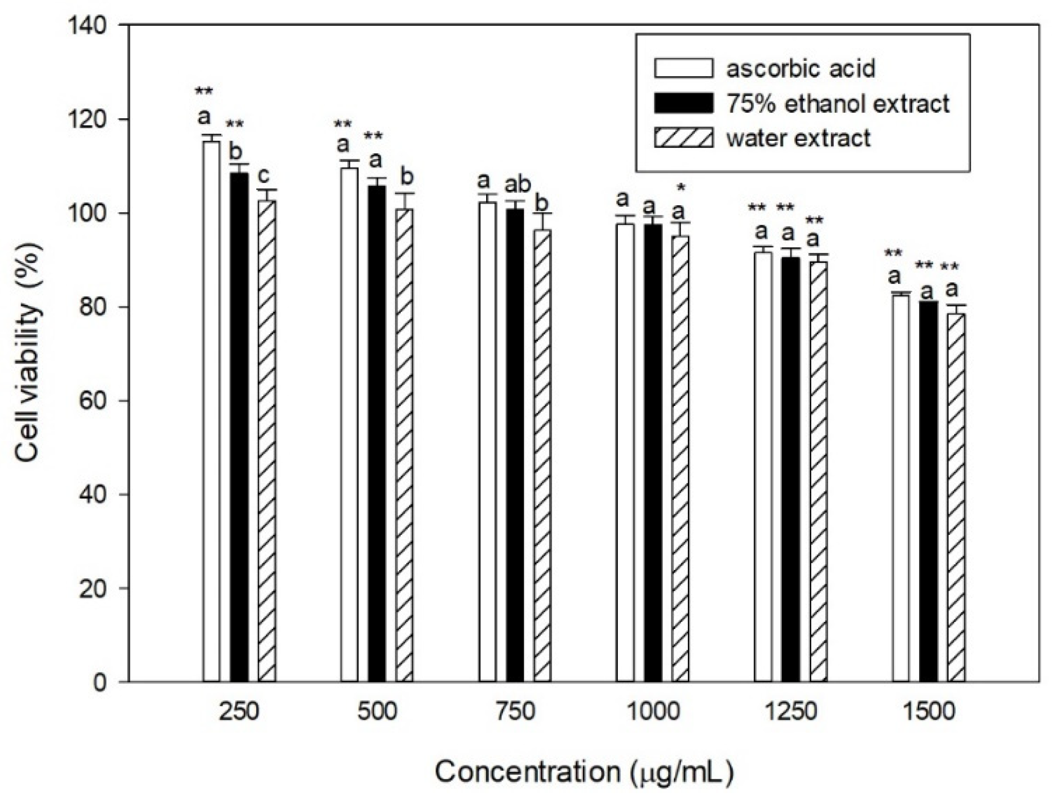
The cytotoxicity of the extracts in human fibroblasts was examined using a standard MTT assay (Figure 1). Cell viability decreased gradually with increasing concentrations of the extracts; for both extracts and ascorbic acid, cell viability was greater than 90% when the concentration was increased to 1250 μg/mL, indicating minimal cytotoxicity at this concentration. At 1500 μg/mL, cell viability was 82%, 81%, and 78% for ascorbic acid, ethanol, and water extracts, respectively; the cytotoxicity of each extract did not differ significantly. Compared to the boiling water extract of M. pudica, the cytotoxicity of the S. sinensis extract in the same cell line was much lower, as the M. pudica extract was able to achieve 80% cell viability at only 100 μg/mL [31]. Although our results indicate that S. sinensis extracts below 1500 μg/mL have limited cytotoxicity in human fibroblasts, it is still observed that there seems to be a decreasing trend in cell viability with increasing extract concentration. The MTT assay is based on the reduction of MTT (3-[4,5-dimethylthiazol-2-yl]-2,5 diphenyl tetrazolium bromide) into formazan crystals by living cells. The intracellular reducing power is mainly provided by NAD(P)H, which comes from dehydrogenase activity in the plasma membrane, endoplasmic reticulum, and mitochondria. One of the possible reasons for the false-negative results is the chemical interaction between the extracts and the MTT indicator. This may be due to the presence of some unknown components with reducing properties in the plant extracts, which can be further explored in future studies.
3.7. Effect of UVB on the Viability of Human Fibroblasts
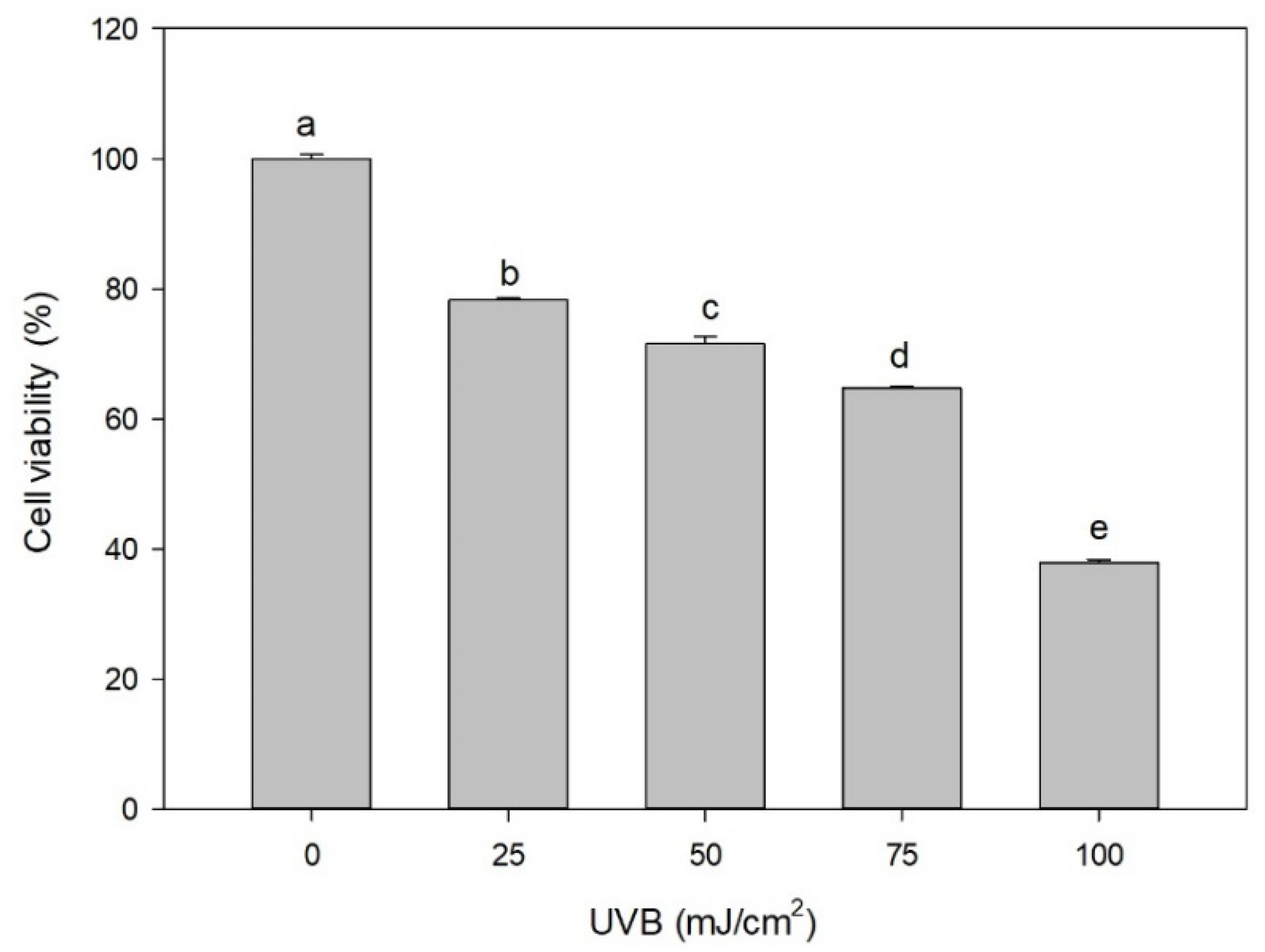
Figure 2 shows the effects of UVB intensity on the viability of human fibroblasts. The viability of human fibroblasts decreased with increasing UVB intensity. Cell viability was 37% when UVB intensity was 100 mJ/cm2, clearly indicating that UVB irradiation is detrimental to human fibroblasts. Similar results were obtained by others using the same cell line [32], where 25% and 50% loss of cell viability were observed at UVB doses of 17 and 70 mJ/cm2, respectively; the loss of cell viability was accompanied by morphological alternations. In later experiments, UVB intensities of 50 and 75 mJ/cm2 were selected, and the viability was 64% and 47%, respectively.
3.8. Effect of the Extracts on the Viability of Human Fibroblasts Exposed to UVB
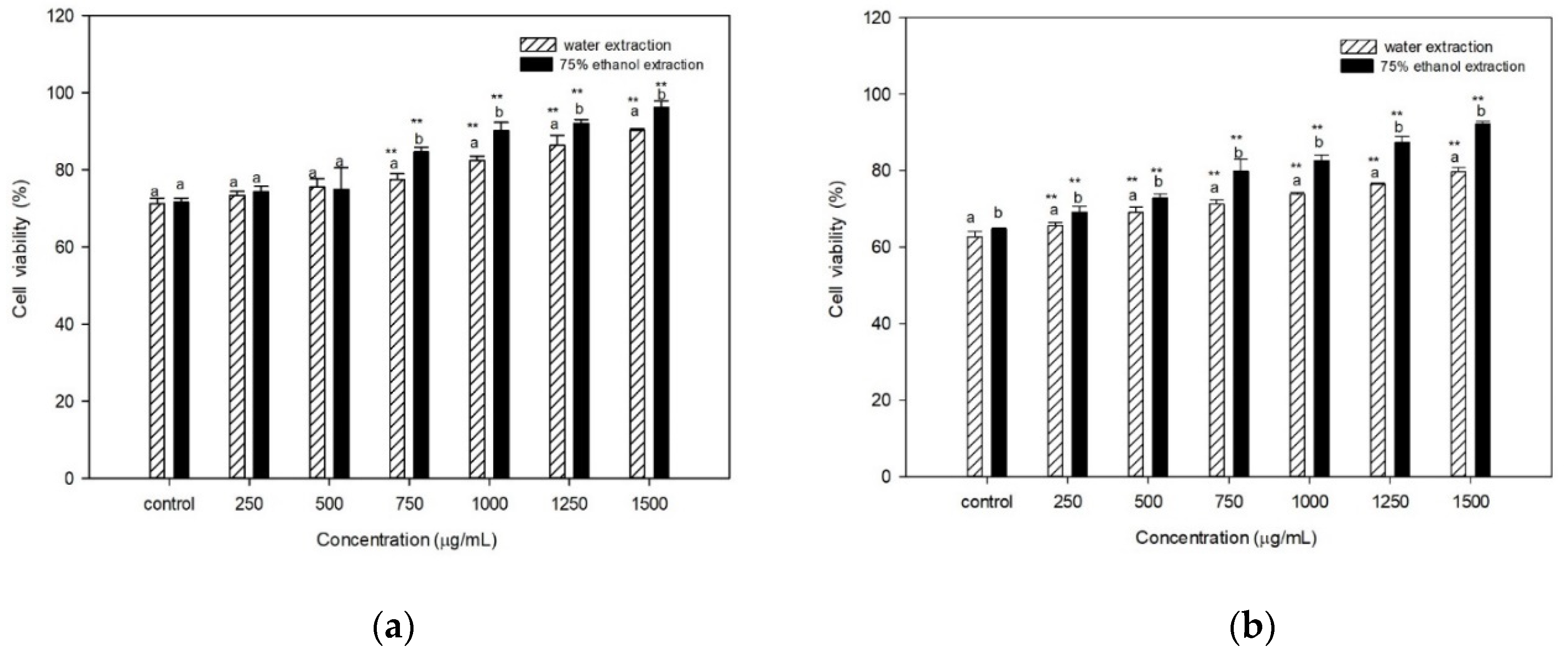
Figure 3 shows that, regardless of the UVB dose used, the survival of fibroblasts increased with increasing concentrations of the extracts. When the cells were exposed to UVB irradiation at 50 mJ/cm2 (Figure 3a), the viability recovered from 71% and 72% (without the addition of the extracts) to 90% and 96% after the addition of 1500 μg/mL of the water and ethanol extracts, respectively. In the presence of 75 mJ/cm2 UVB (Figure 3b), the viability recovered from 63% and 65% (without the addition of the extracts) to 80% and 92%, respectively, after the addition of 1500 μg/mL of the water and ethanol extracts. Our results demonstrated a protective effect of the extracts against UVB-induced fibroblast death. The 75% ethanol extract of S. sinensis showed stronger protective effects against UVB-induced fibroblast injury than the water extract, which may be partly due to its higher content of phenols, flavonoids, and condensed tannins. However, aqueous extracts of G. quadriradiata and Galinsoga parviflora showed higher UV-protecting activity against NHDF human skin fibroblasts than their ethanolic extracts [12]. The M. pudica [31], G. parviflora, and G. quadriradiata extracts have higher UV-protecting activity than those of S. sinensis, as similar cell viability was restored at lower concentrations of the extracts [12].
3.9. Effect of the Extracts on the GSH Content of Human Fibroblasts Exposed to UVB
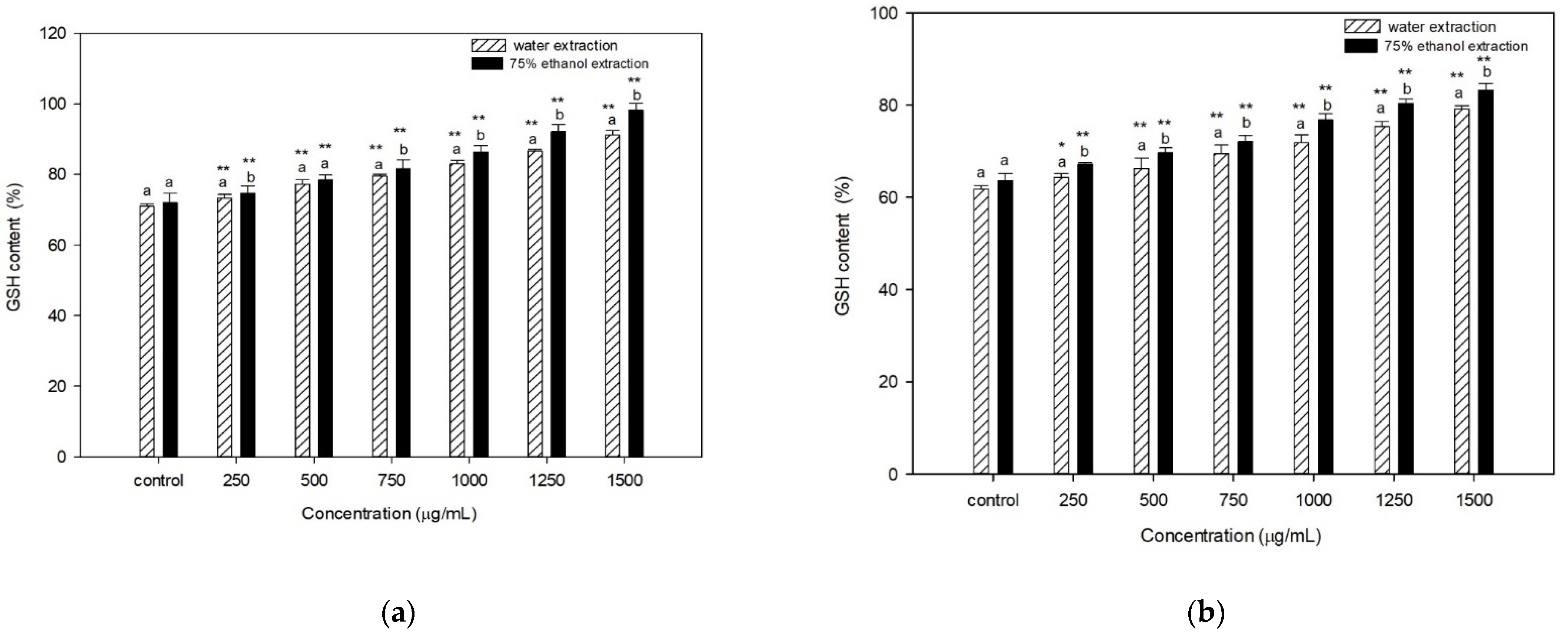
GSH is a major non-protein antioxidant that plays a key role in defending against oxidative stress and maintaining redox homeostasis. To investigate the possible role of GSH in improving the survival of UVB-irradiated human fibroblasts, the effect of extracts on the total GSH content was studied. When cells were exposed to UVB irradiation at 50 mJ/cm2 (Figure 4a), the GSH content increased from 71% and 72% (without the addition of the extracts) to 91% and 98% with the addition of 1500 μg/mL of the water and ethanol extracts, respectively. In the presence of 75 mJ/cm2 UVB (Figure 4b), the GSH content increased from 62% and 64% (without the addition of the extracts) to 79% and 83%, respectively, after the addition of 1500 μg/mL of the water and ethanol extracts. The hot water extract of A. rugosa leaves has been reported to have a GSH-enhancing effect on HaCaT keratinocytes [11]. In this study, there was little difference between the two different extracts regardless of the UVB intensity applied. Observing the results of Figure 3 and Figure 4 together, the increase in cell viability and GSH was maintained with increasing concentrations of S. sinensis extracts, indicating that S. sinensis could continuously protect fibroblasts from death caused by UVB irradiation. Similar to our results, flavonoid antioxidants such as quercetin restored the GSH levels that were reduced by UVB exposure, leading to DNA damage and cell death, and protected the skin from inflammation induced by UVB exposure [33]. Furthermore, the enhancement of GSH by the antioxidant properties of S. sinensis extracts shown here may play an important role in protecting skin cells from oxidative damage caused by UVB irradiation.
4. Conclusions
In this work, the extraction yield of boiling water was much higher than that of 75% ethanol. However, the ethanol extract contained more phenols, flavonoids, and condensed tannins. The ethanol extract also exhibited higher free radical scavenging activity, higher reducing power, and higher ferrous ion-chelating capacity compared to the aqueous extract. The stronger antioxidant activity of the ethanol extract may be due to its higher antioxidant content. Both extracts protected human skin fibroblasts from UVB irradiation and had limited cytotoxicity in the concentration range tested. The restoration of intracellular GSH may be an important mechanism underlying the protective effects of these extracts. In conclusion, our data demonstrate that the water and ethanol extracts of S. sinensis have a high antioxidant capacity and are also effective in protecting human skin fibroblasts from UVB irradiation.
Author Contributions
Conceptualization, C.-J.S.; methodology, C.-J.S., Y.-L.W. and C.-Y.Y.; investigation, Y.-L.W. and C.-Y.Y.; resources, C.-Y.Y.; data curation, Y.-L.W., Y.-Z.P. and C.-Y.Y.; writing—original draft preparation, S.-M.H. and C.-Y.Y.; writing—review and editing, S.-M.H., C.-J.S. and C.-Y.Y.; funding acquisition, C.-Y.Y. All authors have read and agreed to the published version of the manuscript.
Funding
This research was funded by the Ministry of Science and Technology, Taiwan, grant number MOST 108-2221-E-036-011-MY2.
Acknowledgments
We thank Shiow-Ling Lee (Department of Chemical Engineering and Biotechnology, Tatung University, Taipei, Taiwan) for suggestions on the analytical methods used in this work.
Conflicts of Interest
The authors declare no conflict of interest.
References
- Panich, U.; Sittithumcharee, G.; Rathviboon, N.; Jirawatnotai, S. Ultraviolet radiation-induced skin aging: The role of DNA damage and oxidative stress in epidermal stem cell damage mediated skin aging. Stem Cells Int. 2016, 2016, 7370642. [Google Scholar] [CrossRef] [PubMed] [Green Version]
- Maverakis, E.; Miyamura, Y.; Bowen, M.P.; Correa, G.; Ono, Y.; Goodarzi, H. Light, including ultraviolet. J. Autoimmun. 2010, 34, J247–J257. [Google Scholar] [CrossRef] [Green Version]
- Kamileri, I.; Karakasilioti, I.; Garinis, G.A. Nucleotide excision repair: New tricks with old bricks. Trends Genet. 2012, 28, 566–573. [Google Scholar] [CrossRef] [PubMed]
- Kong, Y.H.; Xu, S.P. Juglanin administration protects skin against UVB-induced injury by reducing Nrf2-dependent ROS generation. Int. J. Mol. Med. 2020, 46, 67–82. [Google Scholar] [CrossRef] [Green Version]
- Ravanat, J.-L.; Douki, T.; Cadet, J. Direct and indirect effects of UV radiation on DNA and its components. J. Photochem. Photobiol. B. 2001, 63, 88–102. [Google Scholar] [CrossRef]
- Mimeault, M.; Batra, S.K. Recent advances on skin-resident stem/progenitor cell functions in skin regeneration, aging and cancers and novel anti-aging and cancer therapies. J. Cell. Mol. Med. 2010, 14, 116–134. [Google Scholar] [CrossRef] [PubMed] [Green Version]
- Matu, E.N.; van Staden, J. Antibacterial and anti-inflammatory activities of some plants used for medicinal purposes in Kenya. J. Ethnopharmacol. 2003, 87, 35–41. [Google Scholar] [CrossRef]
- Yan, J.; Huang, Y.; Miao, Y.-E.; Tjiu, W.W.; Liu, T. Polydopamine-coated electrospun poly(vinyl alcohol)/poly(acrylic acid) membranes as efficient dye adsorbent with good recyclability. J. Hazard. Mater. 2015, 283, 730–739. [Google Scholar] [CrossRef]
- Liu, L.; Yin, Q.M.; Yan, X.; Hu, C.; Wang, W.; Wang, R.K.; Luo, X.; Zhang, X.W. Bioactivity-guided isolation of cytotoxic phenanthrenes from Spiranthes sinensis. J. Agric. Food Chem. 2019, 67, 7274–7280. [Google Scholar] [CrossRef]
- Pérez-Sánchez, A.; Barrajón-Catalán, E.; Herranz-López, M.; Castillo, J.; Micol, V. Lemon balm extract (Melissa officinalis, L.) promotes melanogenesis and prevents UVB-induced oxidative stress and DNA damage in a skin cell model. J. Dermatol. Sci. 2016, 84, 169–177. [Google Scholar] [CrossRef] [PubMed]
- Oh, Y.; Lim, H.W.; Huang, Y.H.; Kwon, H.S.; Jin, C.D.; Kim, K.; Lim, C.J. Attenuating properties of Agastache rugosa leaf extract against ultraviolet-B-induced photoaging via up-regulating glutathione and superoxide dismutase in a human keratinocyte cell line. J. Photochem. Photobiol. B 2016, 163, 170–176. [Google Scholar] [CrossRef]
- Bazylko, A.; Borzym, J.; Parzonko, A. Determination of in vitro antioxidant and UV-protecting activity of aqueous and ethanolic extracts from Galinsoga parviflora and Galinsoga quadriradiata herb. J. Photochem. Photobiol. B 2015, 149, 189–195. [Google Scholar] [CrossRef]
- Huang, D.; Ou, B.; Prior, R.L. The Chemistry behind antioxidant capacity assays. J. Agric. Food Chem. 2005, 53, 1841–1856. [Google Scholar] [CrossRef]
- Gursoy, N.; Sarikurkcu, C.; Cengiz, M.; Solak, M.H. Antioxidant activities, metal contents, total phenolics and flavonoids of seven Morchella species. Food Chem. Toxicol. 2009, 47, 2381–2388. [Google Scholar] [CrossRef]
- Broadhurst, R.B.; Jones, W.T. Analysis of condensed tannins using acidified vanillin. J. Sci. Food Agric. 1978, 29, 788–794. [Google Scholar] [CrossRef]
- Shimada, K.; Fujikawa, K.; Yahara, K.; Nakamura, T. Antioxidative properties of xanthan on the autoxidation of soybean oil in cyclodextrin emulsion. J. Agric. Food Chem. 1992, 40, 945–948. [Google Scholar] [CrossRef]
- Ložienė, K.; Venskutonis, P.R.; Šipailienė, A.; Labokas, J. Radical scavenging and antibacterial properties of the extracts from different Thymus pulegioides L. chemotypes. Food Chem. 2007, 103, 546–559. [Google Scholar] [CrossRef]
- Benzie, I.F.F.; Strain, J.J. The ferric reducing ability of plasma (FRAP) as a measure of “antioxidant power”: The FRAP assay. Anal. Biochem. 1996, 239, 70–76. [Google Scholar] [CrossRef] [PubMed] [Green Version]
- Zhao, H.; Fan, W.; Dong, J.; Lu, J.; Chen, J.; Shan, L.; Lin, Y.; Kong, W. Evaluation of antioxidant activities and total phenolic contents of typical malting barley varieties. Food Chem. 2008, 107, 296–304. [Google Scholar] [CrossRef]
- Zulueta, A.; Esteve, M.J.; Frígola, A. ORAC and TEAC assays comparison to measure the antioxidant capacity of food products. Food Chem. 2009, 114, 310–316. [Google Scholar] [CrossRef]
- Stevenson, D.; Wokosin, D.; Girkin, J.; Grant, M.H. Measurement of the intracellular distribution of reduced glutathione in cultured rat hepatocytes using monochlorobimane and confocal laser scanning microscopy. Toxicol. Vitr. 2002, 16, 609–619. [Google Scholar] [CrossRef]
- Liang, C.P.; Chang, C.H.; Liang, C.C.; Hung, K.Y.; Hsieh, C.W. In vitro antioxidant activities, free radical scavenging capacity, and tyrosinase inhibitory of flavonoid compounds and ferulic acid from Spiranthes sinensis (Pers.) Ames. Molecules 2014, 19, 4681–4694. [Google Scholar] [CrossRef] [Green Version]
- Wu, T.-Y.; Chen, C.-C.; Lay, H.-L. Study on the components and antioxidant activity of the Bletilla plant in Taiwan. J. Food Drug Anal. 2010, 18, 279–289. [Google Scholar] [CrossRef]
- Ravipati, A.S.; Zhang, L.; Koyyalamudi, S.R.; Jeong, S.C.; Reddy, N.; Bartlett, J.; Smith, P.T.; Shanmugam, K.; Münch, G.; Wu, M.J.; et al. Antioxidant and anti-inflammatory activities of selected Chinese medicinal plants and their relation with antioxidant content. BMC Complement. Altern. Med. 2012, 12, 173. [Google Scholar] [CrossRef] [PubMed] [Green Version]
- Luo, A.; Fan, Y. In vitro antioxidant of a water-soluble polysaccharide from Dendrobium fimhriatum Hook. var. oculatum Hook. Int. J. Mol. Sci. 2011, 12, 4068–4079. [Google Scholar] [CrossRef] [PubMed] [Green Version]
- Tsai, C.-F. Studies on the Biological Activities of Mimosa pudica; Tatung University: Taipei, Taiwan, 2012. [Google Scholar]
- Liu, Y.-C. Antioxidant Activity of Methanol Extracts and Subfractions from Bischofia javanica and Polygonum chinensis; Tatung University: Taipei, Taiwan, 2008. [Google Scholar]
- Valko, M.; Morris, H.; Cronin, M.T.D. Metals, toxicity and oxidative stress. Curr. Med. Chem. 2005, 12, 1161–1208. [Google Scholar] [CrossRef] [PubMed] [Green Version]
- Dudonné, S.; Vitrac, X.; Coutière, P.; Woillez, M.; Mérillon, J.-M. Comparative study of antioxidant properties and total phenolic content of 30 plant extracts of industrial interest using DPPH, ABTS, FRAP, SOD, and ORAC assays. J. Agric. Food Chem. 2009, 57, 1768–1774. [Google Scholar] [CrossRef]
- Prior, R.L. Oxygen radical absorbance capacity (ORAC): New horizons in relating dietary antioxidants/bioactives and health benefits. J. Funct. Foods 2015, 18, 797–810. [Google Scholar] [CrossRef]
- Chou, C.-J. Protective Effects of Mimosa pudica against UVB-Induced Damage of Human Fibroblasts; Tatung University: Taipei, Taiwan, 2015. [Google Scholar]
- Wu, C.-L.; Chou, H.-C.; Cheng, C.-S.; Li, J.-M.; Lin, S.-T.; Chen, Y.-W.; Chan, H.-L. Proteomic analysis of UVB-induced protein expression- and redox-dependent changes in skin fibroblasts using lysine- and cysteine-labeling two-dimensional difference gel electrophoresis. J. Proteom. 2012, 75, 1991–2014. [Google Scholar] [CrossRef]
- Yin, Y.; Li, W.; Son, Y.-O.; Sun, L.; Lu, J.; Kim, D.; Wang, X.; Yao, H.; Wang, L.; Pratheeshkumar, P.; et al. Quercitrin protects skin from UVB-induced oxidative damage. Toxicol. Appl. Pharmacol. 2013, 269, 89–99. [Google Scholar] [CrossRef] [Green Version]
Figure 1.
Effect of the concentration of S. sinensis extracts on the viability of CCD-966SK cells. The cell viability of the control (without ascorbic acid and extracts) was set to 100%. Different letters in the same group indicate significant difference at p < 0.05; * and ** indicate a significant difference in comparison to the control at p < 0.05 and p < 0.01, respectively.

Figure 2.
Effect of UVB dose on the viability of CCD-966SK cell. Different letters indicate a significant difference at p < 0.05.
Figure 2.
Effect of UVB dose on the viability of CCD-966SK cell. Different letters indicate a significant difference at p < 0.05.

Figure 3.
Effects of different S. sinensis extracts on the viability of CCD-966SK cells exposed to (a) 50 mJ/cm2 and (b) 75 mJ/cm2 UVB. Cell viability before exposure was set to 100%; cell viability after exposure but without the extracts was the control. Different letters in the same group indicate a significant difference at p < 0.05; ** indicates a significant difference in comparison to control at p < 0.01.

Figure 4.
Effects of different extracts of S. sinensis on the GSH content of CCD-966SK cells exposed to (a) 50 mJ/cm2 and (b) 75 mJ/cm2 UVB. The GSH content before exposure was set to 100%; the GSH content after exposure but without the extracts was the control. Different letters in the same group indicate a significant difference at p < 0.05; * and ** indicate a significant difference in comparison to the control at p < 0.05 and p < 0.01, respectively.


Table 1.
Extraction yields, total phenols, total flavonoids, and condensed tannins in extracts of S. sinensis.
Table 1.
Extraction yields, total phenols, total flavonoids, and condensed tannins in extracts of S. sinensis.
| Extraction Solvent | Extraction Yield (%) 1,2 | Content 1 | ||
|---|---|---|---|---|
| Total Phenols (mg GAE 3/g Extract) | Total Flavonoids (mg QE 4/g Extract) | Condensed Tannins (mg CE 5/g Extract) | ||
| water | 25.11 ± 2.83 b | 95.12 ± 0.24 b | 7.77 ± 0.40 b | 194.41 ± 12.84 b |
| 75% ethanol | 13.50 ± 2.21 a | 161.86 ± 1.23 a | 15.89 ± 1.60 a | 439.11 ± 16.78 a |
1 Values (mean ± SD; n = 3) followed by different letters (superscript Latin lowercase alphabet) in the same column are significantly different (p < 0.05). 2 Extraction yield (%) = (weight of extract/dry weight of S. sinensis) × 100%. 3 GAE, gallic acid equivalents. 4 QE, quercetin equivalents. 5 CE, catechin equivalents.
Table 2.
Antioxidant activity of S. sinensis.
| Radical Scavenging EC50 1,2 (μg/mL) | FRAP 1,3 (μmol Fe2+/g Extract) | Metal Chelating 1 (μmol EDTAE 4/g Extract) | ORAC 1,5 (μmol TE 6/g Extract) | ||
|---|---|---|---|---|---|
| DPPH | ABTS | ||||
| Water extract | 659.38 ± 9.30 a | 5167.33 ± 207.81 a | 15.14 ± 0.34 d | 25.13 ± 0.60 d | 683.38 ± 131.64 d |
| Ethanol extract | 446.90 ± 5.23 b | 3031.15 ± 105.92 b | 38.43 ± 0.80 c | 38.04 ± 0.40 c | 2010.69 ± 106.58 c |
| Ascorbic acid | 42.52 ± 0.24 c | 451.54 ± 8.63 c | 968.08 ± 8.54 b | 54.62 ± 0.71 b | 2556.49 ± 127.76 b |
| Quercetin | 19.72 ± 1.44 d | 126.84 ± 3.42 d | 1356.75 ± 58.69 a | 174.63 ± 4.03 a | 3187.04 ± 100.44 a |
1 Values (mean ± SD; n = 3) followed by different letters (superscript Latin lowercase alphabet) in the same column are significantly different (p < 0.05). 2 EC50 is the concentration required to scavenge 50% of the radical. 3 FRAP, ferric reducing ability of plasma. 4 EDTAE, ethylenediaminetetraacetic acid equivalents. 5 ORAC, oxygen radical absorbance capacity. 6 TE, Trolox equivalents.
Publisher’s Note: MDPI stays neutral with regard to jurisdictional claims in published maps and institutional affiliations. |
© 2021 by the authors. Licensee MDPI, Basel, Switzerland. This article is an open access article distributed under the terms and conditions of the Creative Commons Attribution (CC BY) license (https://creativecommons.org/licenses/by/4.0/).
Share and Cite
MDPI and ACS Style
Huang, S.-M.; Shieh, C.-J.; Wu, Y.-L.; Pan, Y.-Z.; Yu, C.-Y. Antioxidant Activity of Spiranthes sinensis and Its Protective Effect against UVB-Induced Skin Fibroblast Damage. Processes 2021, 9, 1564. https://doi.org/10.3390/pr9091564
AMA Style
Huang S-M, Shieh C-J, Wu Y-L, Pan Y-Z, Yu C-Y. Antioxidant Activity of Spiranthes sinensis and Its Protective Effect against UVB-Induced Skin Fibroblast Damage. Processes. 2021; 9(9):1564. https://doi.org/10.3390/pr9091564
Chicago/Turabian StyleHuang, Shang-Ming, Chwen-Jen Shieh, Yu-Lin Wu, Yang-Zing Pan, and Chi-Yang Yu. 2021. "Antioxidant Activity of Spiranthes sinensis and Its Protective Effect against UVB-Induced Skin Fibroblast Damage" Processes 9, no. 9: 1564. https://doi.org/10.3390/pr9091564
Note that from the first issue of 2016, this journal uses article numbers instead of page numbers. See further details here.