Article

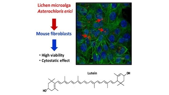
Chemical Characterization of the Lichen-Symbiont Microalga Asterochloris erici and Study of Its Cytostatic Effect on the L929 Murine Fibrosarcoma Cell Line

by
M. Concepción Matesanz
1,
Mercedes Villa-Carvajal
2,
Javier Linares
1,
Sonia Morante-Zarcero
3,
Isabel Sierra
3,
Eva Barreno
2,
Myriam Catalá
4,*,† and
M. Teresa Portolés
1,5,†
1
Department of Biochemistry and Molecular Biology, Faculty of Chemistry, Institute of Health Research San Carlos IdISSC, Complutense University, 28040 Madrid, Madrid, Spain
2
ICBIBE, Botany, Faculty of Biology, Valencia University, C/Dr. Moliner 50, 46100 Burjassot, Valencia, Spain
3
Departament of Chemical and Environmental Chemistry, ESCET, Rey Juan Carlos University, C/Tulipán s/n, 28933 Móstoles, Madrid, Spain
4
Department of Biology and Geology, Physics and Inorganic Chemistry, ESCET, Rey Juan Carlos University, C/Tulipán s/n, 28933 Móstoles, Madrid, Spain
5
CIBER of Bioengineering, Biomaterials and Nanomedicine, CIBER-BBN, 28040 Madrid, Madrid, Spain
*
Author to whom correspondence should be addressed.
Both authors have contributed to the same extent.
Processes 2021, 9(9), 1509; https://doi.org/10.3390/pr9091509
Submission received: 18 May 2021 / Revised: 15 August 2021 / Accepted: 23 August 2021 / Published: 26 August 2021

Abstract

:
New resources of food, pharmaceuticals or biotechnological products are needed. The huge biodiversity of aero-terrestrial lichen-symbiont microalgae belonging to the Chlorophyta group remains unexplored despite they present interesting features such as extreme stress tolerance and growth in water shortage. Appropriateness for human consumption demands the demonstration of the absence of toxic effects. In vitro biocompatibility of crude homogenates of axenic microalga Asterochloris erici, isolated from the lichen Cladonia cristatella, was analyzed after treatment of cultured L929 fibroblasts with different concentrations of microalgal homogenates. The microalgal protein content (37%) was similar to spirulina or soybean. Antioxidant capacity (10.6 ± 0.6 µmol TE/g WW) or phenolic content (7.5 ± 0.5 mg GAE/g DW) were high compared to Chlorella. The results show that crude homogenates of A. erici do not induce cytotoxicity but seem to have some cytostatic effect inducing slight cell cycle alterations and intracellular reactive oxygen species (ROS) increase at the highest concentration. Carotenoid analysis demonstrates high contents of lutein (1211 µg/g microalga DW), a xanthophyll with antioxidant and cytostatic properties in vivo and high commercial added value. These findings confirm that Asterochloris erici can be suitable for the development of alimentary or pharmaceutical applications and further in vivo animal testing. The cytostatic effects should be further investigated for antitumor agents.

Graphical Abstract

1. Introduction

Lichen thalli are the outcome (holobiont) of close and cyclical symbiotic associations involving, at least, two different organisms, a heterotrophic fungus (mycobiont) and photoautotrophic partners (photobionts) [1,2], such photobionts being unicellular green algae (phycobionts, chlorobionts) or/and cyanobacteria (cyanobionts). Lichenization allows the symbionts to colonize diverse terrestrial habitats from the seashores to the high mountains, reaching a large distribution from the tropics to the polar regions [3,4]. Their long life and adaptation to extreme environmental conditions are supported by the production of numerous protective virtually unexplored compounds against different physical and biological stresses [5]. Isolation, characterization and culture methods of lichen microalgae have greatly improved in the last decades [6,7,8].
Green algae, or Chlorophyta, are a huge and diverse phylum of eukaryotic microorganisms. These eukaryotic photoautotrophs should not be confused with the prokaryotic cyanobacteria, also known as blue-green algae. Microalgal biotechnology has been developed for various commercial applications; as photosynthetic organisms, algae contain pigments such as chlorophylls together with high contents of antioxidants, proteins, vitamins and polysaccharides, which can be applied for nutrient supplements, cosmetic purposes or human or animal consumption [9,10,11,12,13].
Some species of microalgae and lichens are natural sources of bioactive compounds such as antibiotics, antioxidants or toxins with remarkable biotechnological potential [14,15,16,17,18,19]. Moreover, many of these microalgal compounds may act as antiviral, antitumor, anti-inflammatory and antimicrobial agents with a marked selectivity in a variety of molecular targets, making them attractive for the pharmaceutical industry [20,21,22,23,24]. They have also garnered attention for their potential in the food industry as antioxidants and antimicrobial additives in a scenario where additives such as parabens or alkylphenols are in question due to the concern on their endocrine disruption potential [25]. Several of the aforementioned compounds are polysaccharides, carotenoids or unsaturated long chain fatty acids bearing chemical structures that are not found elsewhere or that are present at much higher concentrations than in other natural sources [26]. In particular, the biotechnological importance of lipid compounds is beginning to attract strong attention in applied research (i.e., biofuels) [27].
Likewise, it has being established that some lichen microalgae show original and differential combinations of polysaccharides in their cell walls and extracellular polymers with immunostimulant capabilities of biotechnological interest [22,28], as well as extraordinary antioxidant defences [29,30,31]. However, the huge biodiversity of Chlorophyta microalgae remains, to date, largely unexplored and unexploited [32]; thus, they represent a unique opportunity for obtaining novel or known metabolites at low cost. At present, the industrial biotechnological potential of free-living aquatic green algae as Chlorella, Chlamydomonas and Dunaliella spp. is being developed [33]. Nevertheless, the potential of using aero-terrestrial green algae associated with lichens has never been studied.
Asterochloris erici (formerly known as Trebouxia erici Ahmadjian, 1960) is a phycobiont isolated from the north American endemic lichen Cladonia cristatella Tuck., belonging to the class Trebouxiophyceae (Chlorophyta), that has been molecularly and phylogenetically well described by Škaloud and Peksa [34]. The possible use of this species for biomedical, cosmetic, and alimentary purposes requires the development of biocompatibility studies in order to demonstrate the absence of toxic or injurious effects on mammalian cells. The toxicity of a new agent can be analyzed by in vitro tests with cell cultures, by in vivo experiments with animal models and by clinical studies in humans. In vitro studies with different cell types are the first step performed in evaluating the possible damage induced by a new compound in contact with cells. These in vitro biocompatibility assays involve maximum standardization and control of the experimental conditions while reducing costs and ethical concerns [35].
The objective of this study is to determine the nutritional properties, the biocompatibility and the effects on cell viability in vitro of one strain (SAG 32.85 = UTEX 911) of the lichen microalga Asterochloris erici (Ahmadjian) Škaloud and Peksa.

2. Experimental Section

2.1. Instruments

A Qiagen Tissuelyser was used for the homogenization of microalgae suspensions. Optical microscopy was performed with a Leitz Labovert FS inverted microscope, equipped with a Leica DC 300 digital camera. Cells were examined by a LEICA SP2 confocal laser scanning microscope. A FACScalibur Becton Dickinson flow cytometer was used for Intracellular reactive oxygen species (ROS) content and cell viability. Cell cycle analysis and apoptosis detection were performed with a LSR Becton Dickinson flow cytometer, and calculations were conducted with the CellQuest Program of Becton Dickinson. Chromatographic analysis of pigments was performed by ultra-high-performance liquid chromatography (Shimadzu Corp., Kyoto, Japan) with photodiode-array detection (UHPLC-PDA). A Rotofix 32 centrifuge (Hettich zentrifugen, Tuttlingen, Germany) was used for the extraction procedure, and a Cary 60 UV-Vis spectrophotometer (Agilent, Las Rozas, Spain) was used for all spectrophotometrical methods.

2.2. Chemicals and Biochemicals

Dulbecco’s Modified Eagle’s Medium (DMEM) and propidium iodide (PI) were purchased from the Sigma Chemical Company (St. Louis, MO, USA). Fetal Bovine Serum (FBS) was purchased from Gibco (BRL). L-glutamine, penicillin and streptomycin were from BioWhittaker Europe (Verviers, Belgium). FITC-phalloidin and DAPI (4′-6-diamidino-2′-phenylindole) were from Molecular Probes, while Hoechst 33258 was purchased from Polysciences, Inc. (Warrington, PA, USA). 2′,7′-dichlorofluorescein diacetate (DCFH2-DA) was obtained from Serva (Heidelberg, Germany). Carotenoid and chlorophyll standards were purchased from DHI LASB Products (Hørsholm, Denmark). Acetonitrile and methanol HPLC grade were from Fisher Scientific (New Hampshire, USA). Gallic acid, Folin Ciocalteu Reagent (FCR), 2,2-diphenyl-1-picrylhydrazyl (DPPH•) and 6-hydroxy-2,5,7,8-tetramethylchromane-2-carboxylic acid (Trolox) were purchased from Sigma-Aldrich (Madrid, Spain). Ethanol and methanol were purchased from Scharlau (Barcelone, Spain), while sodium carbonate was obtained from Panreac (Barcelone, Spain). Water (resistance 18.2 MΩ cm) was obtained from a Millipore Milli-Q-System (Billerica, MA, USA). Other chemicals used were of analytical grade.

2.3. Strain Maintenance, Culture Conditions and Preparation of Microalgae Homogenates

An axenic strain of the lichen photobiont Asterochloris erici (Ahmadjian) Škaloud and Peksa (SAG 32.85 = UTEX 911) was used for this study. In order to obtain material for in vitro biocompatibility studies, microalgal cultures were carried out for 15 days in BBM3N, as previously described [36]. The cultures were then centrifuged at 15,557g for 10 min, and the cell pellets were frozen and stored at −20 °C. The day before biocompatibility testing, microalgae were homogenated at 1 mg/mL in complete DMEM (DMEM supplemented with 10% FBS, 1 mM L-glutamine, 200 μg/mL of penicillin and 200 μg/mL of streptomycin) with a tissuelyser (1 mm glass beads, 40 Hz, 15 min), sterlised in an autoclave and subsequent frozen-stored. Working dilutions in complete DMEM containing 0.1, 0.01 and 0.001 mg/mL were prepared just before their application to mammalian cells.

2.4. Proximate Chemical Composition Determination

Proteins, ash, moisture and nitrogen contents in fresh Asterochloris erici (Ahmadjian) Škaloud and Peksa were determined by standard AOAC [37] methods. The moisture content was determined by the oven method at 105 °C until a constant weight was obtained. The ash content was gravimetrically determined after heating at 550 °C for 24 h in a muffle furnace. The protein content was calculated by converting the nitrogen content, determined by the Kjeldahl method (6.25 × N). Total lipids were extracted with chloroform/methanol (2:1, v/v), according to Sánchez-Machado et al. [38]. Total carbohydrate content was determined as the weight difference using protein, lipid, moisture and ash content data. All measurements were performed in triplicate.

2.5. Antioxidant Activity and Total Phenolic Compounds

2.5.1. Extraction Procedure

The extraction procedure was accomplished from freeze-dried microalgae, according to Goiris et al. [39] with some modifications. Extractions were performed in the dark at room temperature (which was usually between 23 and 25 °C). The amount of 100 mg biomass was weighed by triplicate in falcon tubes where the extraction process took place. For this purpose, 2 mL of the extraction solvent (ethanol/water, 3/1, v/v) was added, and the mixture was crushed in a mortar to break the cell walls and stirred for 20 min. Then, the samples were centrifuged at 6000 rpm for 10 min. After that, a second extraction of the resulting pellets was completed by using another 2 mL of ethanol/water mixture (3/1, v/v), and the combined supernatants for each sample were filtered through nylon membrane filters (0.45 μm pore size) and, finally, maintained at −18 °C until the beginning of the analysis (within a day).

2.5.2. Radical Scavenging Activity (RSA)

The free radical DPPH• scavenging activity (RSA) of the extract was estimated by the method of Brand-Williams et al. [40] that was slightly modified. Firstly, 3.9 mL of a 40 ppm (w/v) DPPH• solution, freshly prepared in methanol, was mixed in a vortex with 0.1 mL of the sample extract or of the extraction solvent (blank sample). After an incubation period (60 min at room temperature in the dark), the absorbance was measured at 517 nm in order to estimate the remaining DPPH• concentration. The following equation was used to calculate the RSA (%):
RSA   ( % ) = ( A DPPH   A sample ) A DPPH × 100
where ADPPH is the absorbance of the DPPH• radical in blank sample, and Asample is the absorbance in the hydroalcoholic extract (sample). A standard calibration curve was prepared with Trolox at a concentration of 0.5–400 ppm (w/v). Finally, the RSA of the samples was expressed as Trolox equivalents (TE) in µmol/g of sample (dry weight basis, DW).

2.5.3. Total Phenolic Content

Total phenolics compound (TPC) concentration in the extracts was determined according to the method of Folin-Ciocalteu [41], with some modifications. Firstly, an aliquot of the sample extract or extraction solvent (0.75 μL) was mixed with of Milli-Q water (645 μL) and FCR (30 μL). Then, 20% (w/v) sodium carbonate (75 μL) and Milli-Q water (675 μL) were added and mixed in a vortex. After incubation for 60 min at room temperature in darkness, the absorbance was measured in a UV-Vis spectrophotometer at 725 nm. Gallic acid was used to prepare the standard calibration curve (10–500 ppm, w/v), and TPCs were expressed as milligram gallic acid equivalents (GAE)/g of sample (DW).

2.6. Carotenoids and Chlorophylls Extraction and Chromatographic Analysis

Microalgae lyophilized powder was frozen with liquid nitrogen and then homogenized in a Qiagen Tissuelyser with stainless steel beads (2 min, 40 Hz). Pigments were extracted from 5 mg of microalgae powder using 1 mL of acetone (95%). Extraction was performed in an ultrasonic bath for 5 min, twice or up to the complete extraction of pigments. After extraction, samples were centrifuged at 16,200× g for 5 min, and then the extracts were filtered through nylon membrane filters (0.22 μm pore size).
The carotenoid composition was determined according to García-Plazaola and Becerril (2001) [42], with some modifications. The separation was achieved on an Agilent 46 x 10 mm, 2.7 mm on a 150 mm × 4.6 mm, 5 µm particle size, Poroshell HPH-C18 column (Tecknokroma, Barcelona, Spain). The mobile phase consisted of two components: solvent A, acetonitrile:methanol:tris buffer (0.1 M, pH 8) (83:2:15, v:v:v), and solvent B, methanol:ethyl acetate (68:32, v:v). The pigments were eluted using a linear gradient from 100% A to 100% B for the first 7 min, followed by an isocratic elution with 100% B for the next 4 min. The mobile phase flow rate was 1 mL/min. The injection volume was 10 µL. The on-line UV-Vis spectra were recorded from 350 to 800 nm. The absorbance was measured at 445 nm for all carotenoids (with exception of astaxantin measured at 477 nm) and at 631 nm and 664 nm for chlorophyll b and a, respectively. Carotenoids (β-carotene, neoxanthin, fucoxanthin, violaxanthin, anteraxanthin, astaxanthin, zeaxanthin and lutein), chlorophyll a and chlorophyll b were quantified in the microalgae extract using molar absorptivity. The results were expressed in µg/g of sample (DW), and all measurements were performed in triplicate. Detection and quantification limits were well below 0.5 µg/L, and 1.0 µg/L, respectively, for all the pigments analysed.

2.7. Cell Culture for the In Vitro Biocompatibility Studies

L929 mouse fibroblasts were seeded on 6-well culture plates (105 cells/mL) in complete Dulbecco’s Modified Eagle’s Medium (DMEM) containing 10% fetal bovine serum, 200 μg/mL antibiotics (penicillin and streptomycin) and 1 mM L-glutamine. Cells were cultured for 24 h at 37 °C under a CO2 (5%) atmosphere. L929 mouse fibroblasts were gently gifted by Dr. M.A. Lizarbe and Dr. N. Olmo (Department of Biochemistry and Molecular Biology, Universidad Complutense, Madrid, Spain). The effect of different concentrations (0.1, 0.01 and 0.001 mg/mL) of A. erici on fibroblasts was assessed after adding the lichen microalga crude homogenates to the culture medium and maintaining the cells during 24 h in the presence of these extracts. After each treatment, the fibroblasts were detached with 0.25% trypsin-EDTA solution for 15 min, and cell proliferation was measured with a Neubauer hemocytometer. On the other hand, fibroblasts were centrifuged at 310 g for 10 min and resuspended in fresh medium in order to carry out multiparametric analysis by flow cytometry including cell viability, cell cycle, apoptosis and intracellular content of reactive oxygen species (ROS). Control cultures without extracts were performed in parallel.

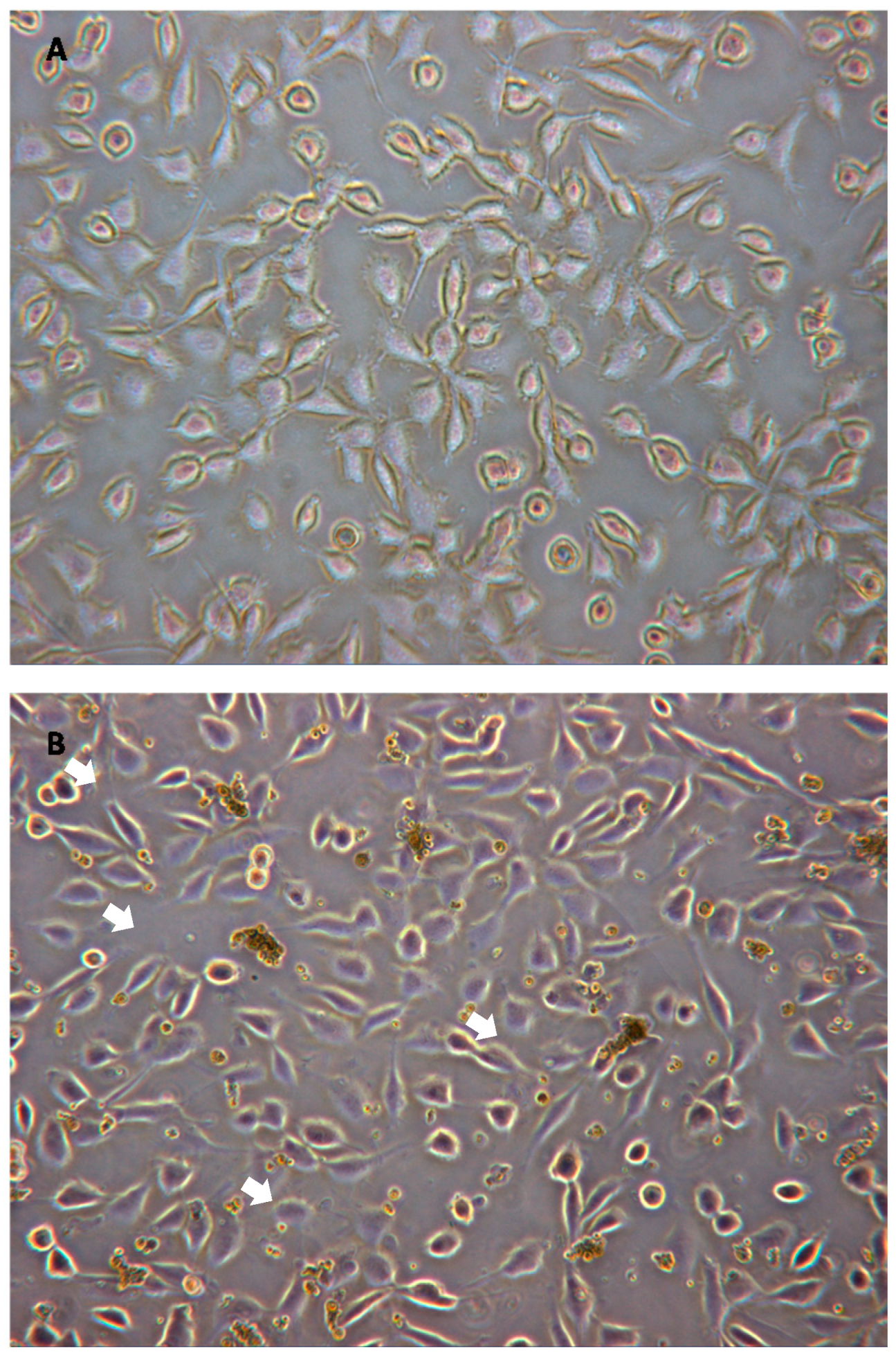
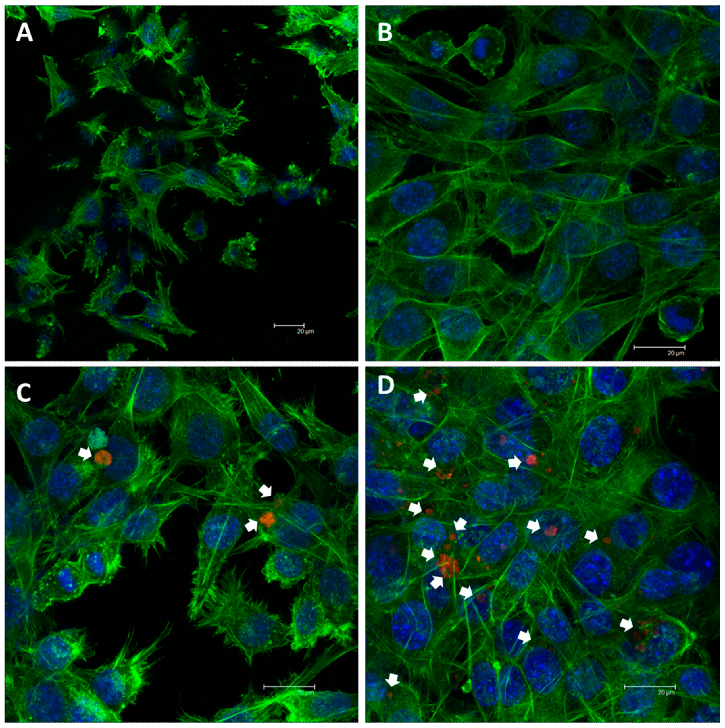
2.8. Cell Morphology Studies

2.8.1. Optical Microscopy

Morphological studies were performed by optical microscopy with L929 mouse fibroblasts after 24 h of culture with different concentrations (0.1, 0.01 and 0.001 mg/mL) of A. erici extracts. Cells were examined under an inverted microscope equipped with a digital camera. Controls without these extracts were observed in parallel.

2.8.2. Confocal Microscopy

The confocal microscopy studies were performed as described elsewhere [43]. Briefly, L929 fibroblasts were cultured for 24 h on glass coverslips in the presence of different concentrations (0.1, 0.01 and 0.001 mg/mL) of A. erici crude homogenates. Controls without extracts of microalgal homogenates were carried out in parallel. Cells were fixed with 3.7% paraformaldehyde, permeabilized with 0.1% Triton X-100 and preincubated with 1% BSA in PBS. Then, the cells were treated with FITC-phalloidin (1:40, v:v) in order to stain green the cytoskeleton F-actin filaments. Cell nuclei were stained in blue with 3 µM DAPI, and the cells were examined by a confocal laser scanning microscope, exciting the fluorescence of FITC at 488 nm and measuring the emitted fluorescence at 491–586 nm. The DAPI fluorescence was excited at 405 nm and detected at 420–480 nm.

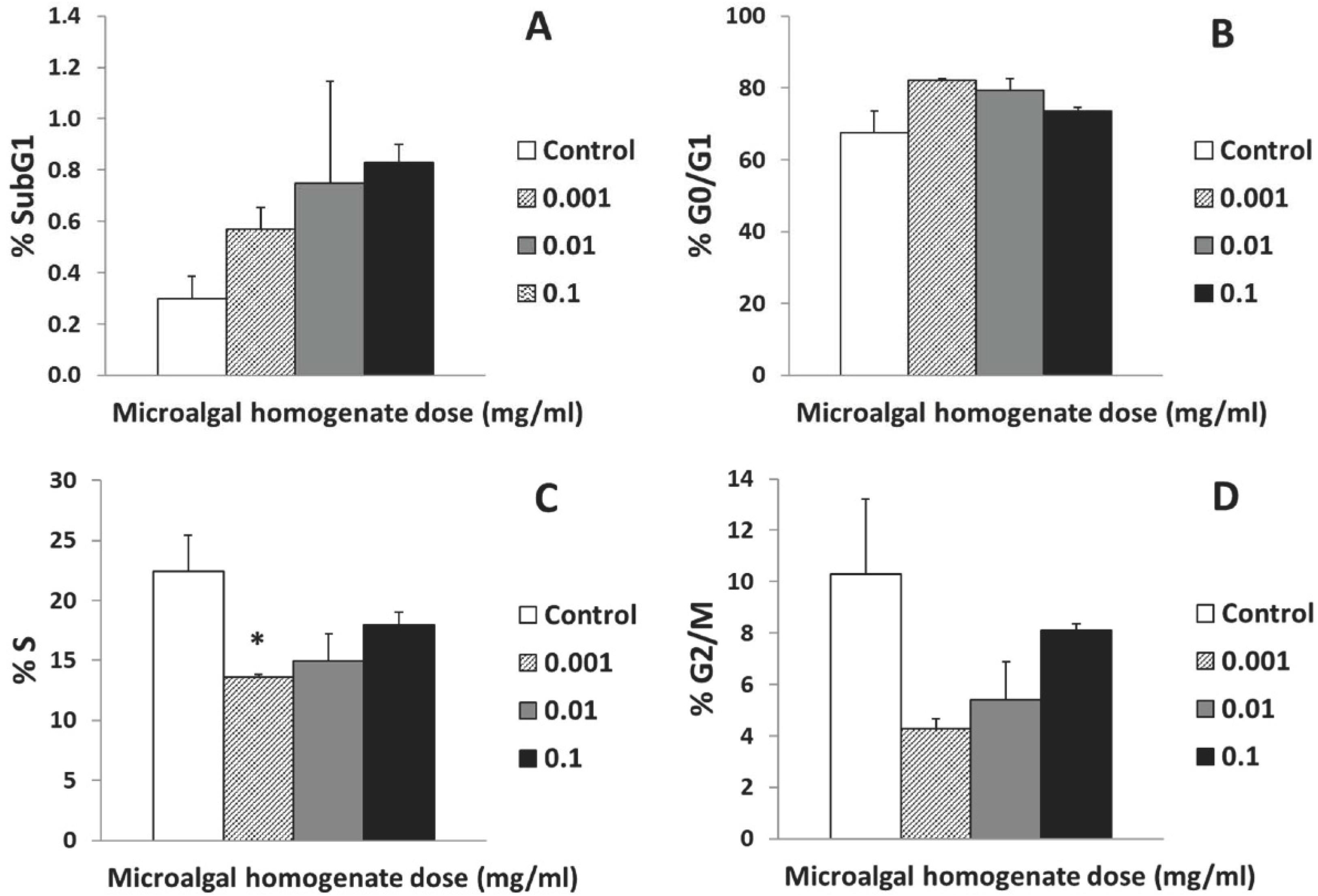
2.9. Cell Cycle Analysis and Apoptosis Detection

The analyses of the cell cycle and the detection of apoptosis were carried out by flow cytometry, as described previously by Matesanz et al. [43]. Briefly, the cells were treated with ethanol (30%), Hoechst 33258 (5 µg/mL) and BSA (1%) in PBS for 30 min in darkness and at room temperature. The Hoechst fluorescence was detected at 450 nm after excitation at 350 nm in a flow cytometer. The CellQuest Program of Becton Dickinson was used to calculate the cell percentage in each cycle phase: G0/G1, S and G2/M. Apoptosis was evaluated by using the SubG1 fraction.

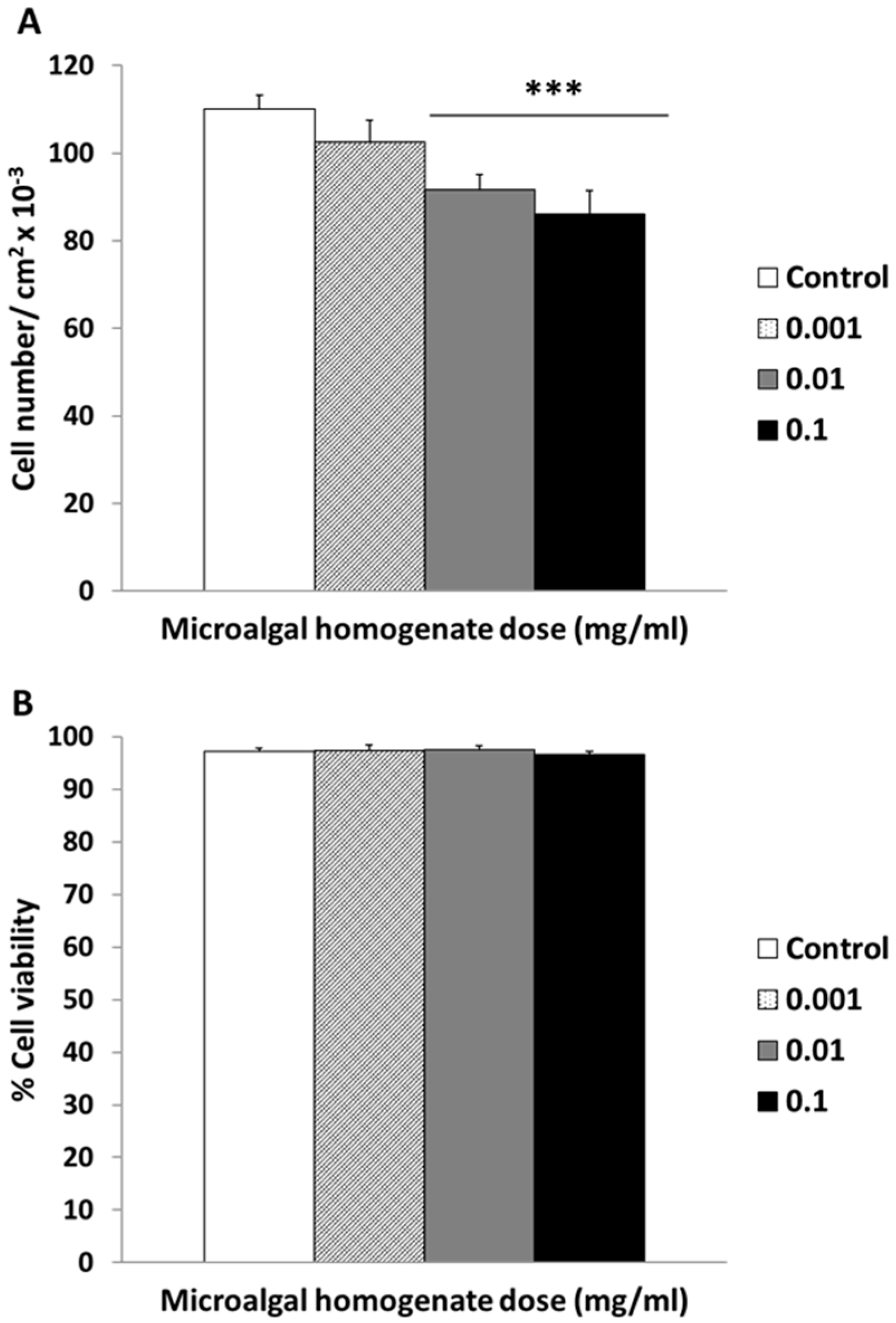
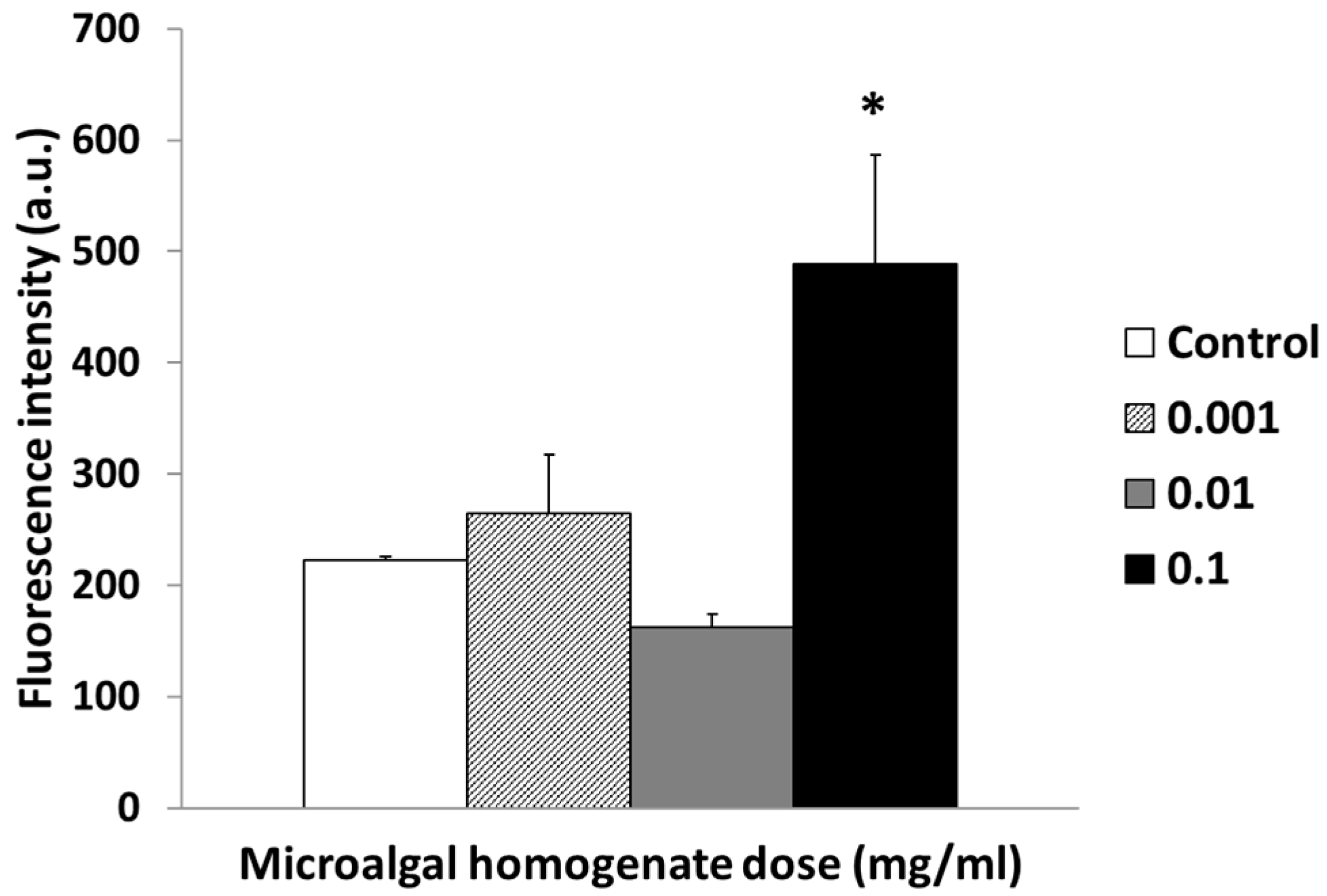
2.10. Intracellular Reactive Oxygen Species (ROS) Content and Cell Viability

The quantification of both intracellular ROS content and cell viability has been described in detail [43]. Briefly, the cells were incubated at 37 °C with 100 µM DCFH2-DA for 30 min, and the fluorescence of DCF (excited at 488 nm and detected with a 530/30 band pass filter in a flow cytometer) was used as measurement of intracellular ROS. Propidium iodide (PI) was used as probe to quantify the cell viability by PI exclusion test and flow cytometry.

2.11. Statistics

The Statistical Package for the Social Sciences (SPSS) version 19 software was used to perform the statistical analysis of data, expressed as means ± standard deviations of a representative of three experiments carried out in triplicate. Statistical comparisons were performed through analysis of variance (ANOVA). Scheffé’s test was employed for post hoc assessments of differences among groups. The value p < 0.05 was regarded as statistically significant.

3. Results

3.1. Proximate Composition, Antioxidant Activity and Total Phenolic Compounds

The proximate chemical composition of fresh A. erici is shown in Table 1. The moisture content was 75 ± 1% WW (wet weight). The ash content was 5 ± 1% DW and was a value of the same order of magnitude as those reported for the green microalgae chlorella (5.8%) and Botryococcus braunii (5.4%) [44]. Nevertheless, it is important to point out that the low ash content found in A. erici is similar to that of land plants (5–10%) [45], but interestingly it seems very low when compared to the data reported for some red macroalgae such as seaweed Gracilaria changii (42% WW) [46]. The protein content was 37 ± 1% DW, being higher than in the green microalgae such as Chlorella vulgaris and Haematococcus pluvialis and several seaweeds such as as Sargassum vulgare or Gracilaria changii [46,47], but is in the same order of magnitude as those reported for the microalgae spirulina, Tetraselmis chuii and Chlorella sp. by other authors [44]. This protein content was similar than other proteinic vegetable foods, such as soybeans, indicating that A. erici may become a potential source of this nutrient. The total lipid content was 10 ± 2% DW, which is higher than these values observed for other algae where the value is, in general, always less than 6% [38,46,48,49,50]. However, this lipid value can be considered low, considering that some microalgae have lipid content near 41% WW (Haematococcus pluvialis) or 49% WW (Nannochloropsis granulata). Finally, the total carbohydrate content (determined as the weight difference using protein, lipid, moisture and ash content data) was 48% DW; that is, in the range of 4–76% (carbohydrate plus fiber) reported for various authors for different seaweed species [51].
The TPC of ethanolic extracts A. erici (see Section 2.5.1) resulted in 7.5 ± 0.5 mg GAE/g DW. The value of TPC was high in comparison with other green microalgae such as Chlorella, between 0.75–2.21 mg GAE/g DW [39], measured in a similar extraction procedure. The antioxidant activity was analyzed by the DPPH method that measures free radical scavenging ability. High DPPH values might be attributed to high levels of phenolic compounds [39]. In this sense, the antioxidant values exhibited by A. erici in the present study (10.6 ± 0.6 µmol TE/g WW) are in accordance with TPC value, suggesting that the antioxidant activity of A. erici may be related to its phenolic content.

3.2. Carotenoid and Chlorophyll Content

The values of chlorophyll a and b in A. erici are shown in Table 2. The value of chlorophyll b is higher than that of chlorophyll a. In relation with carotenoids, neoxanthin, violaxanthin, antheraxanthin and lutein were detected with a clear predominance of lutein (Table 2). The quantification of this pigment rendered values of 1.2 mg/g WW, in the same order of magnitude as other chlorophytes with commercial interest for cancer and retinal degeneration prevention [52]. This predominance of lutein has also been observed in Chlorella vulgaris [53] where it has been found to be bioavailable when administered as whole algal dietary supplements, increasing the erythrocyte lutein content [54].

3.3. Cellular Cytocompatibility

Cell viability, morphology, proliferation, cell cycle, apoptosis and intracellular free radicals of L929 mouse fibroblasts cultured for 24 h in the presence of different concentrations of A. erici crude homogenates were analyzed in order to evaluate the absence of toxic effects on mammalian cells.
Figure 1 shows optical microscopy images of L929 cells after 24 h culture with 0.1 mg/mL of A. erici homogenate. Due to mechanical resistance of the microalgal cell walls, the envelopes of some algae can still be observed on the L929 monolayer. Using non-filtered homogenates allows studying both the effects of soluble algal compound as well as non-soluble wall components (i.e., polysaccharides) for which its bioactivity is frequently disregarded. Fibroblast membranes seemed intact, and no signs of necrosis were observed in cultures. In addition, cell counts were performed to confirm these observations. Percent viability of cells was the same with respect to untreated control regardless of applied concentration (Figure 2B). Instead, a concentration dependent effect was observed when studying the cell proliferation as the number of cells was progressively reduced, becoming a significant effect at higher concentrations (Figure 2A). In summary, these studies revealed that the crude homogenates of A. erici do not affect cell viability causing cell death, but they have a significant effect on cell proliferation as higher concentrations result in higher cell counts.
In order to assess the potential cytotoxicity of A. erici, intracellular free radicals were measured by flow cytometry in L929 cells from cultures treated with different concentrations of microalgal crude homogenates. The results are shown in Figure 3, where a significant increase in free radicals with the highest concentration applied is observed.
The studies of cell cycle and apoptosis detection (Figure 4) showed that the levels of L929 fibroblasts in G0/G1 phase increased very slightly, but the differences are not significant. However, the levels of cells in S phase decreased with all the concentrations of crude microalgae homogenate, which is the lowest concentration achieving a statistically significant decrease. The levels of cells in subG1 fraction, usually taken as an apoptosis indicator, slightly increased in a concentration dependent manner.
An assessment of the cell morphology and actin cytoskeleton architecture was performed by confocal microscopy (Figure 5). The photographs show that there is no apparent alteration of nuclear chromatin or actin filaments of the cytoskeleton at any of the concentrations studied (Figure 5B–D) compared with controls (Figure 5A). At higher concentrations, the red autofluorescence of chlorophyll can be observed (indicated with white arrows in Figure 5C,D).

4. Discussion

Addressing the second Sustainable Development Goal declared by the UN (Zero Hunger) in a scenario of global warming, needs the identification of new alimentary sources adapted to water shortage. The composition of A. erici compares with Chlorella and proteinic seeds such as soybean regarding protein content (37%). This lichen microalga, differently from aquatic species, is adapted to grow with low water availability and even survives complete desiccation for considerable periods. Moreover, it shows high phenolic content and tenfold antioxidant capacity compared to Chlorella [39], which suggests that the applications of A. erici could go beyond alimentary purposes, providing nutraceutical utilities.
To our best knowledge, this is the first study on the biocompatibility of a lichen microalga (Chlorophyta) on mammalian cells using a systematic standardized in vitro approach. Our results demonstrate that whole lichen microalgal homogenates do not induce cytotoxicity or morphological alterations. However, they induce an intracellular ROS increase at the highest concentration, and they can slow down the rate of cell proliferation preventing their entrance in S phase and, therefore, the fraction of cells attaining G2/M phases. Chlorella carotenoids, more specifically xanthophylls, have been found to inhibit cancer cells proliferation [53,55,56]. Chromatographic analysis of A. erici carotenoid composition revealed the presence of an important amount of lutein (Table 2).
Xanthophylls are a group of carotenoids containing at least one O atom known to play a crucial role in photoprotection of photosystem II (PSII) from ROS under excess light. Lutein is a tetraterpenoid (C40H56O2) alternating single and double carbon–carbon bonds with attached methyl side groups with a predominant function in the deactivation of 3Chl* [57]. The presence of a hydroxyl group at both ends of the molecule distinguishes lutein (and its stereoisomer zeaxanthin) from other carotenoids and is responsible for its high chemical reactivity [58]. Since xanthophylls biosynthesis does not occur in animal cells, their primary intake depends on diet (or supplementation), being lutein intake from dietary sources strongly associated with plasma concentrations (reviewed in [59]).
Apart from its best-known pivotal role against oxidative damage of retinal tissues, lutein has anti-inflammatory, immunomodulatory, neuroprotective and antiangiogenic properties. It exerts several antioxidant activities from the upregulation of antioxidant enzymes in order to direct antioxidant action such as the inhibition of membrane lipids peroxidation. In lipopolysaccharide (LPS)-stimulated macrophages, lutein has been found to decrease intracellular hydrogen peroxide (H2O2) accumulation by scavenging superoxide anion and H2O2. The administration of lutein affords neuroprotective effects against cerebral ischemic injury, increasing reduced/oxidized GSH ratio as well as the enzymatic activities of superoxide dismutase, GSH-peroxidase and catalase. It has been shown to inhibit the expression of proinflammatory genes by suppressing nuclear factor NF-κB translocation and reducing the secretion of cytokines (TNF, IL-1) and arachidonic acid metabolites [60]. Nonetheless, specific fractionation studies are needed to obtain insights into the responsibility of lutein, or other lichen microalgal compounds, in this relevant cytostatic feature of A. erici. Moreover, other procedures of sample homogenisation, such as liquid nitrogen grinding or freeze-drying, should be implemented in order to improve algal substances bioavailability to test their full potential.
Cladonia lichens present an important biotechnological potential [61]; thus, Cladonia’s phycobiont Asterochloris erici is also likely to possess important bioactivity. This species has been relatively well characterized at the genetic and physiological levels and is readily available from various international collections [34]. Although experiments with algal-free mycobionts have demonstrated that many lichen secondary metabolites are synthesized mainly by the fungal component, and phycobionts belonging to specific species of green microalgae may contribute to a lichen’s secondary metabolite profile. Furthermore, lichenized fungi produce biologically active metabolites only in association with suitable algal partners [62]. These substances show bioactive properties being considered as promising sources of antibiotic, antioxidant and anticancer drugs [21,22,63,64].
Distinct from free living, symbiont eukaryotic microalgae have been shown to produce a high number of new substances with high biological activity on various mammalian cells. For example, maitotoxin causes the release of neurotransmitters from pheochromocytoma cells or muscle contraction in a Ca-dependent manner [65,66]. Zooxanthellatoxins derived from Symbiodinium (coral phycobionts) have been shown to cause platelet aggregation and aorta contraction through the regulation of Ca2+ levels and permeability and the enhancement of tyrosine phosphorylation of p42 mitogen-activated protein kinase [67,68]. Albeit the best studied symbiotic microalgae are freshwater and marine organisms and lichen microalgae are aero-terrestrial, all symbiotic organisms seemingly share certain biological traits, such as their ability to grow in low nutrient habitats, and to exchange substances with other organisms that are very phylogenetically distant, such as animals, bacteria or archaea [69,70], rendering them exceptional sources of bioactive products. In this sense, testing crude homogenates including cell wall compounds is of particular relevance given its function as physical communications interface among neighbors and constitutes a proxy for digestion studies.
The results presented here indicate, for the first time, that a lichen microalgal species is not toxic nor induces acute damage on mammalian cells in culture, although they induce a slight cytostatic effect together with a moderate increase in intracellular free radicals at the highest concentration applied. The use of crude and non-filtered homogenates allows the assessment of the bioactivity of non-soluble compounds of cell walls (i.e., polysaccharides) together with soluble cytosolic substances. Free microalgal compounds such as antioxidants and polysaccharides have been demonstrated to modulate carcinogenesis in the gastrointestinal tract [71] and augment antitumor resistance [72]. Alcoholic extracts of Chlorella sorokiniana decreased cell viability and induced apoptosis and oxidative stress in a human hepatoma cell line [55]. Polysaccharides from Chlorella pyrenoidosa presented significant antitumor activity against A549 human lung adenocarcinoma cells [73]. Therefore, this slight cytostatic activity should be further studied in tumor cells to test algal components potential as preventive nutraceuticals or even for anticancer drugs.
These results indicate that the lichen microalga Asterochloris erici is a potential candidate for various biotechnological applications. Perhaps the most immediate one could be the use of lichen microalgae in dietary supplements. The food supplements containing microalgae known as Blue-Green Algae Supplements (BGAS) are mainly derived from prokaryotic organisms such as spirulina (reclassified as Arthrospira platensis), Arthrospira maxima and Aphanizomenon flos-aquae species (cyanobacteria). Whereas spirulina is “Generally Recognized As Safe (GRAS)” by the Food and Drug Administration of the USA; both spirulina and A. flos-aquae are listed in Annex B of EFSA “Compendium of botanicals” reported to contain naturally occurring substances of possible concern. In natural environments (e.g., lakes), they can coexist with other potentially toxic strains of cyanobacteria, such as Microcystis sp. which share the same habitat, causing BGAS product contamination as evidenced in different countries [74]. In addition, A. flos-aquae has been found to produce neurotoxins [75].
On the other hand, studies on microalgae and microalgal compounds safety have been frequently performed in vivo using murine models. There is a growing social and ethical concern for the use and sacrifice of animals for biological tests, and it is mandatory to find and apply alternative methods to limit animal experimentation to a minimum. An in vitro approach is in better accordance with this new scenario since it avoids animal testing and constitutes a rapid, reliable and cost-effective method for the assessment of natural products bioactivity providing, at the same time, insights into the mechanisms of action at the cellular level. Nonetheless, once the absence of in vitro cytotoxicity is demonstrated, further studies in more complex models, including in vivo tests at the final step, may be necessary to ascertain safety for human or animal applications.

5. Conclusions

These results taken together present the first experimental evidence that the crude homogenates of the lichen microalga Asterochloris erici do not induce acute toxicity in mammalian L929 murine fibrosarcoma cells. This microalga seems to possess a slight cytostatic effect, delaying cell proliferation. The high content of the xanthophyll lutein, a well-known pigment with potent antioxidant and cancer prevention properties, could be involved in this cytostatic effect, as demonstrated for other chlorophytes. In addition, Asterochloris erici has appreciable protein content and low total lipid content, together with high phenolic content and antioxidant capacity, making it a potential important low-fat functional ingredient in the food industry.

Author Contributions

M.T.P., E.B. and M.C. designed the study. M.T.P. and E.B. were responsible for funding acquisition. E.B., M.V.-C. and M.C. obtained microalgal biomass. I.S. and S.M.-Z. designed and performed the analytical and nutrient studies of A. erici. M.C.M. and J.L. performed the cellular assays, cytometric and confocal studies under the direction and supervision of M.T.P. The manuscript was written and revised by all the authors. All authors have read and agreed to the published version of the manuscript.

Funding

This study was funded by grants from the following: the Comunidad de Madrid (project S2009/MAT-1472); the Ministerio de Economía y Competitividad, MINECO and FEDER: MAT2013-43299-R and CGL2016-79158-P); and PROMETEO/2017/039 Excellence in Research (Generalitat Valenciana, Spain). M.C. Matesanz is greatly indebted to Spanish Ministry of Education (MEC) for predoctoral fellowship.

Institutional Review Board Statement

Not applicable.

Informed Consent Statement

Not applicable.

Acknowledgments

The authors wish to thank the staff of the Cytometry and Fluorescence Microscopy Center of the Universidad Complutense de Madrid (Spain) for their assistance in flow cytometry and confocal microscopy studies.

Conflicts of Interest

The authors declare no conflict of interest. The funders had no role in the design of the study; in the collection, analyses or interpretation of data; in the writing of the manuscript or in the decision to publish the results.

References

  1. Margulis, L.; Barreno, E. Looking at lichens. Bioscience 2003, 53, 776. [Google Scholar] [CrossRef]
  2. Aschenbrenner, I.A.; Cernava, T.; Berg, G.; Grube, M. Understanding microbial multi-species symbioses. Front. Microbiol. 2016, 18, 180. [Google Scholar] [CrossRef]
  3. Seckbach, J.; Grube, M. Die Hard: Lichens Stress and developmental strategies in lichens. In Symbioses and Stress: Joint Ventures in Biology; Seckbach, J., Grube, M., Eds.; Springer: Dordrecht, Germany, 2010; pp. 509–523. [Google Scholar]
  4. del Campo, E.M.; Catalá, S.; Gimeno, J.; del Hoyo, A.; Martínez-Alberola, F.; Casano, L.M.; Grube, M.; Barreno, E. The genetic structure of the cosmopolitan three-partner lichen Ramalina farinacea evidences the concerted diversification of symbionts. FEMS Microbiol. Ecol. 2013, 83, 310–323. [Google Scholar] [CrossRef] [PubMed] [Green Version]
  5. Mitrović, T.; Stamenković, S.; Cvetković, V.; Nikolic, M.; Tosic, S.; Stojicic, D. Lichens as source of versatile bioactive compounds A brief history of lichen utilization. Biol. Nyssana 2011, 2, 1–6. [Google Scholar]
  6. Gasulla, F.; Guera, A.; Barreno, E.; Guéra, A.; Barreno, E. A simple and rapid method for isolating lichen photobionts. Symbiosis 2010, 51, 175–179. [Google Scholar] [CrossRef]
  7. Molins, A.; Moya, P.; García-Breijo, F.J.; Reig-Armiñana, J.; Barreno, E. Lichens as micro-ecosystems: Novel approaches to efficiently reveal the hidden diversity of phycobionts in a single thallus. In Book of Abstracts, Proceedings: 8th Congress of the International Symbiosis Society (Symbiotic Lifestyle), Oral Com., Lisbon, Portugal, 12–18 July 2015; Munzi, S., Ulm, F., Eds.; Faculdade de Ciências, Universidade de Lisboa: Lisbon, Portugal, 2015; p. 205. [Google Scholar]
  8. Molins, A.; Moya, P.; García-Breijo, F.J.; Reig-Armiñana, J.; Barreno, E. A multi-tool approach to assess microalgal diversity in lichens: Isolation, Sanger sequencing, HTS and ultrastructural correlations. Lichenologist 2018, 50, 123–138. [Google Scholar] [CrossRef] [Green Version]
  9. Carballo-Cárdenas, E.C.; Tuan, P.M.; Janssen, M.; Wijffels, R.H. Vitamin E (α-tocopherol) production by the marine microalgae Dunaliella tertiolecta and Tetraselmis suecica in batch cultivation. Biomol. Eng. 2003, 20, 139–147. [Google Scholar] [CrossRef]
  10. Spolaore, P.; Joannis-Cassan, C.; Duran, E.; Isambert, A. Commercial applications of microalgae. J. Biosci. Bioeng. 2006, 101, 87–96. [Google Scholar] [CrossRef] [Green Version]
  11. García-Casal, M.N.; Ramírez, J.; Leets, I.; Pereira, A.C.; Quiroga, M.F. Antioxidant capacity, polyphenol content and iron bioavailability from algae (Ulva sp., Sargassum sp. and Porphyra sp.) in human subjects. Br. J. Nutr. 2009, 101, 79–85. [Google Scholar] [CrossRef] [Green Version]
  12. Michalak, I.; Górka, B.; Wieczorek, P.P.; Rój, E.; Lipok, J.; Łęska, B.; Messyasz, B.; Wilk, R.; Schroeder, G.; Dobrzyńska-Inger, A.; et al. Supercritical fluid extraction of algae enhances levels of biologically active compounds promoting plant growth. Eur. J. Phycol. 2016, 51, 243–252. [Google Scholar] [CrossRef]
  13. Saadaoui, I.; Rasheed, R.; Aguilar, A.; Cherif, M.; Al Jabri, H.; Sayadi, S.; Manning, S.R. Microalgal-based feed: Promising alternative feedstocks for livestock and poultry production. J. Anim. Sci. Biotechnol. 2021, 12, 1–15. [Google Scholar] [CrossRef]
  14. Guedes, A.C.; Giao, M.S.; Seabra, R.; Ferreira, A.C.; Tamagnini, P.; Moradas-Ferreira, P.; Malcata, F.X. Evaluation of the antioxidant activity of cell extracts from microalgae. Mar. Drugs 2013, 11, 1256–1270. [Google Scholar] [CrossRef] [Green Version]
  15. Zong, A.; Cao, H.; Wang, F. Anticancer polysaccharides from natural resources: A review of recent research. Carbohydr. Polym. 2012, 90, 1395–1410. [Google Scholar] [CrossRef] [PubMed]
  16. Maksimova, I.V.; Bratkovskaia, L.B.; Plekhanov, S.E. Extracellular carbohydrates and polysaccharides of the algae Chlorella pyrenoidosa Chick S-39. Biol. Bull. 2004, 312, 217–224. [Google Scholar]
  17. Rumin, J.; Gonçalves de Oliveira Junior, R.; Bérard, J.B.; Picot, L. Improving Microalgae Research and Marketing in the European Atlantic Area: Analysis of Major Gaps and Barriers Limiting Sector Development. Mar. Drugs 2021, 19, 319. [Google Scholar] [CrossRef] [PubMed]
  18. Pandey, A.; Dikshit, A. Lichens: Fungal symbionts and their secondary metabolites. In New and Future Developments in Microbial Biotechnology and Bioengineering; Elsevier: Amsterdam, The Netherlands, 2021; pp. 107–115. [Google Scholar]
  19. Muggia, B.L.; Schmitt, I.; Grube, M. Lichens as treasure chests of natural products. SIM News 2009, 59, 85–97. [Google Scholar]
  20. Amaro, H.; Guedes, A.; Malcata, F. Antimicrobial activities of microalgae: An invited review. In Science Against Microbial Pathogens: Communicating Current Research and Technological Advances; Formatex Microbiology Book Series; Méndez-Vilas, A., Ed.; Formatex Research Center: Badajoz, Spain, 2011; pp. 1272–1280. [Google Scholar]
  21. Ranković, B. Lichen Secondary Metabolites: Bioactive Properties and Pharmaceutical Potential; Ranković, B., Ed.; Springer International Publishing: Cham, Switzerland, 2015; ISBN 9783319133744. [Google Scholar]
  22. Shrestha, G.; St. Clair, L.L.; O’Neill, K.L. The immunostimulating role of lichen polysaccharides: A review. Phyther. Res. 2015, 29, 317–322. [Google Scholar] [CrossRef] [PubMed]
  23. Ganguly, R.; Pierce, G.N. The toxicity of dietary trans fats. Food Chem. Toxicol. 2015, 78, 170–176. [Google Scholar] [CrossRef]
  24. Rumin, J.; Nicolau, E.; Gonçalves de Oliveira Junior, R.; Fuentes-Grünewald, C.; Picot, L. Analysis of Scientific Research Driving Microalgae Market Opportunities in Europe. Mar. Drugs 2020, 18, 264. [Google Scholar] [CrossRef]
  25. Petersen, G.; Rasmussen, D.; Gustavson, K. Study on Enhancing the Endocrine Disrupter Priority List with A Focus on Low Production Volume Chemicals; DG Environment (European Commision): Hørsholm, Denmark, 2007. [Google Scholar]
  26. Newman, D.J.; Cragg, G.M. Natural products from marine invertebrates and microbes as modulators of antitumor targets. Curr. Drug Targets 2006, 7, 279–304. [Google Scholar] [CrossRef]
  27. Lang, I.; Hodac, L.; Friedl, T.; Feussner, I. Fatty acid profiles and their distribution patterns in microalgae: A comprehensive analysis of more than 2000 strains from the SAG culture collection. BMC Plant Biol. 2011, 11, 124. [Google Scholar] [CrossRef] [Green Version]
  28. Casano, L.M.; Braga, M.R.; Álvarez, R.; del Campo, E.M.; Barreno, E. Differences in the cell walls and extracellular polymers of the two Trebouxia microalgae coexisting in the lichen Ramalina farinacea are consistent with their distinct capacity to immobilize extracellular Pb. Plant Sci. 2015, 236, 195–204. [Google Scholar] [CrossRef]
  29. del Hoyo, A.; Álvarez, R.; del Campo, E.M.; Gasulla, F.; Barreno, E.; Casano, L.M.; Alvarez, R.; del Campo, E.M.; Gasulla, F.; Barreno, E.; et al. Oxidative stress induces distinct physiological responses in the two Trebouxia phycobionts of the lichen Ramalina farinacea. Ann. Bot. 2011, 107, 109–118. [Google Scholar] [CrossRef]
  30. Álvarez, R.; del Hoyo, A.; García-Breijo, F.; Reig-Armiñana, J.; del Campo, E.M.; Guéra, A.; Barreno, E.; Casano, L.M. Different strategies to achieve Pb-tolerance by the two Trebouxia algae coexisting in the lichen Ramalina farinacea. J. Plant Physiol. 2012, 169, 1797–1806. [Google Scholar] [CrossRef]
  31. Expósito, J.R.; Coello, A.J.; Barreno, E.; Casano, L.M.; Catalá, M. Endogenous NO Is Involved in Dissimilar Responses to Rehydration and Pb(NO3)2 in Ramalina farinacea Thalli and Its Isolated Phycobionts. Microb. Ecol. 2019, 72, 604–616. [Google Scholar] [CrossRef]
  32. Muggia, L.; Leavitt, S.; Barreno, E. The hidden diversity of lichenised Trebouxiophyceae (Chlorophyta). Phycologia 2018, 57, 503–524. [Google Scholar] [CrossRef] [Green Version]
  33. Cadoret, J.P.; Garnier, M.; Saint-Jean, B. Microalgae. Functional genomics and biotechnology. Adv. Bot. Res. 2012, 64, 285–341. [Google Scholar]
  34. Skaloud, P.; Peksa, O. Evolutionary inferences based on ITS rDNA and actin sequences reveal extensive diversity of the common lichen alga Asterochloris (Trebouxiophyceae, Chlorophyta). Mol. Phylogenet. Evol. 2010, 54, 36–46. [Google Scholar] [CrossRef]
  35. Krewski, D.; Andersen, M.E.; Tyshenko, M.G.; Krishnan, K.; Hartung, T.; Boekelheide, K.; Wambaugh, J.F.; Jones, D.; Whelan, M.; Thomas, R.; et al. Toxicity testing in the 21st century: Progress in the past decade and future perspectives. Arch. Toxicol. 2019, 94, 1–58. [Google Scholar] [CrossRef]
  36. Goldsmith, S.J.; Thomas, M.A.; Gries, C. A new technique for photobiont culturing and manipulation. Lichenologist 1997, 29, 559–569. [Google Scholar] [CrossRef]
  37. Horwitz, W. (Ed.) Official Methods of Analysis of AOAC International. In Association of Official Analysis Chemists International; AOAC International: Washington, DC, USA, 1995; ISBN 0935584544. [Google Scholar]
  38. Sánchez-Machado, D.I.; López-Cervantes, J.; López-Hernández, J.; Paseiro-Losada, P. Fatty acids, total lipid, protein and ash contents of processed edible seaweeds. Food Chem. 2004, 85, 439–444. [Google Scholar] [CrossRef]
  39. Goiris, K.; Muylaert, K.; Fraeye, I.; Foubert, I.; De Brabanter, J.; De Cooman, L. Antioxidant potential of microalgae in relation to their phenolic and carotenoid content. J. Appl. Phycol. 2012, 24, 1477–1486. [Google Scholar] [CrossRef]
  40. Brand-Williams, W.; Cuvelier, M.E.; Berset, C. Use of a free radical method to evaluate antioxidant activity. LWT-Food Sci. Technol. 1995, 28, 25–30. [Google Scholar] [CrossRef]
  41. Singleton, V.L.; Orthofer, R.; Lamuela-Raventós, R.M. [14] Analysis of total phenols and other oxidation substrates and antioxidants by means of folin-ciocalteu reagent. Methods Enzymol. 1999, 299, 152–178. [Google Scholar]
  42. García-Plazaola, J.I.; Becerril, J.M. A rapid high-performance liquid chromatography method to measure lipophilic antioxidants in stressed plants: Simultaneous determination of carotenoids and tocopherols. Phytochem. Anal. 1999, 10, 307–313. [Google Scholar] [CrossRef]
  43. Matesanz, M.-C.; Vila, M.; Feito, M.-J.; Linares, J.; Gonçalves, G.; Vallet-Regi, M.; Marques, P.-A.A.P.; Portolés, M.-T. The effects of graphene oxide nanosheets localized on F-actin filaments on cell-cycle alterations. Biomaterials 2013, 34, 1562–1569. [Google Scholar] [CrossRef]
  44. Tibbetts, S.M.; Milley, J.E.; Lall, S.P. Chemical composition and nutritional properties of freshwater and marine microalgal biomass cultured in photobioreactors. J. Appl. Phycol. 2015, 27, 1109–1119. [Google Scholar] [CrossRef] [Green Version]
  45. Rupérez, P. Mineral content of edible marine seaweeds. Food Chem. 2002, 79, 23–26. [Google Scholar] [CrossRef]
  46. Chan, P.T.; Matanjun, P. Chemical composition and physicochemical properties of tropical red seaweed, Gracilaria changii. Food Chem. 2017, 221, 302–310. [Google Scholar] [CrossRef]
  47. Batista, A.P.; Gouveia, L.; Bandarra, N.M.; Franco, J.M.; Raymundo, A. Comparison of microalgal biomass profiles as novel functional ingredient for food products. Algal Res. 2013, 2, 164–173. [Google Scholar] [CrossRef] [Green Version]
  48. Herbreteau, F.; Coiffard, L.J.M.; Derrien, A.; De Roeck-Holtzhauer, Y. The Fatty Acid Composition of Five Species of Macroalgae. Bot. Mar. 1997, 40, 25–28. [Google Scholar] [CrossRef]
  49. Gómez-Ordóñez, E.; Jiménez-Escrig, A.; Rupérez, P. Dietary fibre and physicochemical properties of several edible seaweeds from the northwestern Spanish coast. Food Res. Int. 2010, 43, 2289–2294. [Google Scholar] [CrossRef]
  50. Peinado, I.; Girón, J.; Koutsidis, G.; Ames, J.M. Chemical composition, antioxidant activity and sensory evaluation of five different species of brown edible seaweeds. Food Res. Int. 2014, 66, 36–44. [Google Scholar] [CrossRef] [Green Version]
  51. Marinho-Soriano, E.; Fonseca, P.C.; Carneiro, M.A.A.; Moreira, W.S.C. Seasonal variation in the chemical composition of two tropical seaweeds. Bioresour. Technol. 2006, 97, 2402–2406. [Google Scholar] [CrossRef]
  52. Del Campo, J.A.; Moreno, J.; Rodríguez, H.; Vargas, M.A.; Rivas, J.; Guerrero, M. Carotenoid content of chlorophycean microalgae: Factors determining lutein accumulation in Muriellopsis sp. (Chlorophyta). J. Biotechnol. 2000, 76, 51–59. [Google Scholar] [CrossRef]
  53. Cha, K.H.; Koo, S.Y.; Lee, D.-U. Antiproliferative effects of carotenoids extracted from Chlorella ellipsoidea and Chlorella vulgaris on human colon cancer cells. J. Agric. Food Chem. 2008, 56, 10521–10526. [Google Scholar] [CrossRef] [PubMed]
  54. Miyazawa, T.; Nakagawa, K.; Kimura, F.; Nakashima, Y.; Maruyama, I.; Higuchi, O.; Miyazawa, T. Chlorella is an effective dietary source of lutein for human erythrocytes. J. Oleo Sci. 2013, 62, 773–779. [Google Scholar] [CrossRef] [PubMed] [Green Version]
  55. Chung, J.-G.; Peng, H.-Y.; Chu, Y.-C.; Hsieh, Y.-M.; Wang, S.-D.; Chou, S.-T. Anti-invasion and apoptosis induction of chlorella (Chlorella sorokiniana) in Hep G2 human hepatocellular carcinoma cells. J. Funct. Foods 2012, 4, 302–310. [Google Scholar] [CrossRef]
  56. Jayappriyan, K.R.; Rajkumar, R.; Venkatakrishnan, V.; Nagaraj, S.; Rengasamy, R. In vitro anticancer activity of natural β-carotene from Dunaliella salina EU5891199 in PC-3 cells. Biomed. Prev. Nutr. 2013, 3, 99–105. [Google Scholar] [CrossRef]
  57. Jahns, P.; Holzwarth, A.R.A.R. The role of the xanthophyll cycle and of lutein in photoprotection of photosystem II. Biochim. Biophys. Acta-Bioenerg. 2012, 1817, 182–193. [Google Scholar] [CrossRef] [Green Version]
  58. Martin, H.D.; Ruck, C.; Schmidt, M.; Sell, S.; Beutner, S.; Mayer, B.; Walsh, R. Chemistry of carotenoid oxidation and free radical reactions. Pure Appl. Chem. 1999, 71, 2253–2262. [Google Scholar] [CrossRef]
  59. Alves-Rodrigues, A.; Shao, A. The science behind lutein. Toxicol. Lett. 2004, 150, 57–83. [Google Scholar] [CrossRef]
  60. Perrone, S.; Tei, M.; Longini, M.; Buonocore, G. The Multiple Facets of Lutein: A Call for Further Investigation in the Perinatal Period. Oxidative Med. Cell. Longev. 2016, 2016, 1–8. [Google Scholar] [CrossRef] [Green Version]
  61. Oksanen, I. Ecological and biotechnological aspects of lichens. Appl. Microbiol. Biotechnol. 2006, 73, 723–734. [Google Scholar] [CrossRef]
  62. Culberson, C.F.; Armaleo, D. Induction of a complete secondary-product pathway in a cultured lichen fungus. Exp. Mycol. 1992, 16, 52–63. [Google Scholar] [CrossRef]
  63. Shrestha, G.; St. Clair, L.L. Lichens: A promising source of antibiotic and anticancer drugs. Phytochem. Rev. 2013, 12, 229–244. [Google Scholar] [CrossRef]
  64. Zambare, V.P.; Christopher, L.P. Biopharmaceutical potential of lichens. Pharm. Biol. 2012, 50, 778–798. [Google Scholar] [CrossRef]
  65. Takahashi, M.; Tatsumi, M.; Ohizumi, Y.; Yasumoto, T. Ca2+ channel activating function of maitotoxin, the most potent marine toxin known, in clonal rat pheochromocytoma cells. J. Biol. Chem. 1983, 258, 10944–10949. [Google Scholar] [CrossRef]
  66. Ohizumi, Y.; Yasumoto, T. Contractile response of the rabbit aorta to maitotoxin, the most potent marine toxin. J. Physiol. 1983, 337, 711–721. [Google Scholar] [CrossRef] [PubMed]
  67. Rho, M.-C.; Nakahata, N.; Nakamura, H.; Murai, A.; Ohizumi, Y. Activation of rabbit platelets by Ca2+ influx and thromboxane A2 release in an external Ca2+-dependent manner by zooxanthellatoxin-A, a novel polyol. Br. J. Pharmacol. 1995, 115, 433–440. [Google Scholar] [CrossRef] [Green Version]
  68. Moriya, T.; Ishida, Y.; Nakamura, H.; Asari, T.; Murai, A.; Ohizumi, Y. Vasoconstriction induced by zooxanthellatoxin-B, a polyoxygenated long-chain product from a marine alga. Eur. J. Pharmacol. 1998, 350, 59–65. [Google Scholar] [CrossRef]
  69. Meilhoc, E.; Boscari, A.; Bruand, C.; Puppo, A.; Brouquisse, R. Nitric oxide in legume-rhizobium symbiosis. Plant Sci. 2011, 181, 573–581. [Google Scholar] [CrossRef]
  70. Gordon, B.R.; Leggat, W. Symbiodinium-invertebrate symbioses and the role of metabolomics. Mar. Drugs 2010, 8, 2546–2568. [Google Scholar] [CrossRef] [Green Version]
  71. Amaro, H.M.; Barros, R.; Guedes, A.C.; Sousa-Pinto, I.; Malcata, F.X. Microalgal compounds modulate carcinogenesis in the gastrointestinal tract. Trends Biotechnol. 2013, 31, 92–98. [Google Scholar] [CrossRef]
  72. Tanaka, K.; Konishi, F.; Himeno, K.; Taniguchi, K.; Nomoto, K. Augmentation of antitumor resistance by a strain of unicellular green algae, Chlorella vulgaris. Cancer Immunol. Immunother. 1984, 17, 90–94. [Google Scholar] [CrossRef]
  73. Sheng, J.; Yu, F.; Xin, Z.; Zhao, L.; Zhu, X.; Hu, Q. Preparation, identification and their antitumor activities in vitro of polysaccharides from Chlorella pyrenoidosa. Food Chem. 2007, 105, 533–539. [Google Scholar] [CrossRef]
  74. Vichi, S.; Lavorini, P.; Funari, E.; Scardala, S.; Testai, E. Contamination by Microcystis and microcystins of blue-green algae food supplements (BGAS) on the Italian market and possible risk for the exposed population. Food Chem. Toxicol. 2012, 50, 4493–4499. [Google Scholar] [CrossRef]
  75. Cox, P.A.; Banack, S.A.; Murch, S.J.; Rasmussen, U.; Tien, G.; Bidigare, R.R.; Metcalf, J.S.; Morrison, L.F.; Codd, G.A.; Bergman, B. Diverse taxa of cyanobacteria produce beta-N-methylamino-L-alanine, a neurotoxic amino acid. Proc. Natl. Acad. Sci. USA 2005, 102, 5074–5078. [Google Scholar] [CrossRef] [Green Version]
Figure 1. Morphology evaluation of L929 fibroblasts. (A) Control. (B) Treated with 0.1 mg/mL of the lichen microalgal crude homogenates for 24 h. White arrows indicate algal bodies. Cells were seeded at a density of 2 × 10⁵ cells/mL. Magnification 200×. Bright field.
Processes 09 01509 g001
Figure 2. Cytotoxicity. L929 fibroblasts treated with different concentrations of the lichen microalgal crude homogenates (mg/mL) for 24 h. (A) Cell proliferation. (B) Cell viability. Data are expressed as means + standard deviations. Statistical significance *** p < 0.005.
Processes 09 01509 g002
Figure 3. Intracellular ROS. Level of intracellular reactive oxygen species (ROS) in L929 fibroblasts treated with different concentrations of the lichen microalgal crude homogenates (mg/mL) for 24 h. Data are expressed as means + standard deviations. In each sample, 10,000 cells were analyzed. Statistical significance * p < 0.05.
Processes 09 01509 g003
Figure 4. Cell cycle and apoptosis. Cell cycle analysis of L929 fibroblasts treated with different concentration of the lichen microalgal crude homogenates (mg/mL) for 24 h. (A) SubG1 fraction, indicative of apoptosis; (B) G0/G1; (C) S and (D) G2/M phases of a representative of three repetitive experiments. In each sample, 10,000 cells were analyzed. Statistical significance * p < 0.05.
Processes 09 01509 g004
Figure 5. Cytoskeleton ultrastructure. Confocal microscopy images of L929 fibroblasts cultured for 24 h in the presence of different concentrations of Asterochloris erici crude homogenates. Cells were stained with DAPI for the visualization of the cell nuclei in blue and FITC-phalloidin for cytoplasmic F-actin filaments in green. Red emission corresponds to microalgal chlorophyll autofluorescence. Overlay of the three fluorescence channels. (A) Control fibroblasts. (BD) fibroblasts cultured with 0.001 mg/mL, 0.01 mg/mL and 0.1 mg/mL of microalgal homogenates, respectively (magnification: see magnitude bar). White arrows indicate chlorophyll red autofluorescence.
Processes 09 01509 g005
Table 1. The proximate composition of Asterochloris erici.
CompositionConcentration (%) *
Moisture75 ± 1
Ash5 ± 1
Protein37 ± 1
Lipids10 ± 2
* Data are the mean of three independent samples ± standard error. Moisture in % wet weight; ash, proteins and lipids in % dry weight.
Table 2. Chlorophyll and carotenoid composition of Asterochloris erici.
PigmentConcentration (µg/g Microalga Dry Weight)
Chlorophyll a507 ± 41
Chlorophyll b6105 ± 580
β-Carotene268 ± 20
Neoxanthin165 ± 12
Fucoxanthinn.d. (<0.058)
Violaxanthin267 ± 16
Antheraxanthin57 ± 6
Astaxanthin106 ± 9
Zeaxanthinn.d. (<0.079)
Lutein1211 ± 119
Data are the mean of three independent samples ± standard error. N.d.: under detection limit.
Publisher’s Note: MDPI stays neutral with regard to jurisdictional claims in published maps and institutional affiliations.

