
Technological Advancements for the Detection of Antibiotics in Food Products

 , , ,
, , ,  , , ,
, , ,  and
and
Abstract
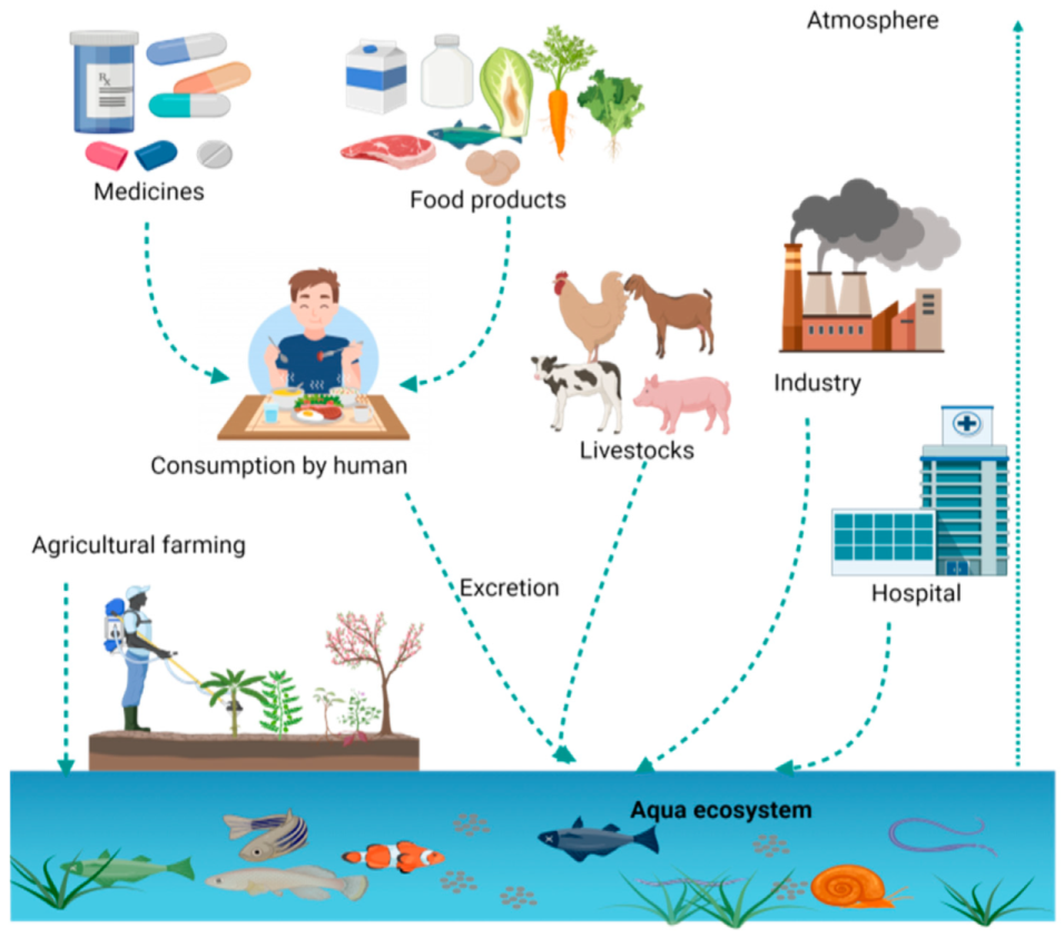
:1. Introduction
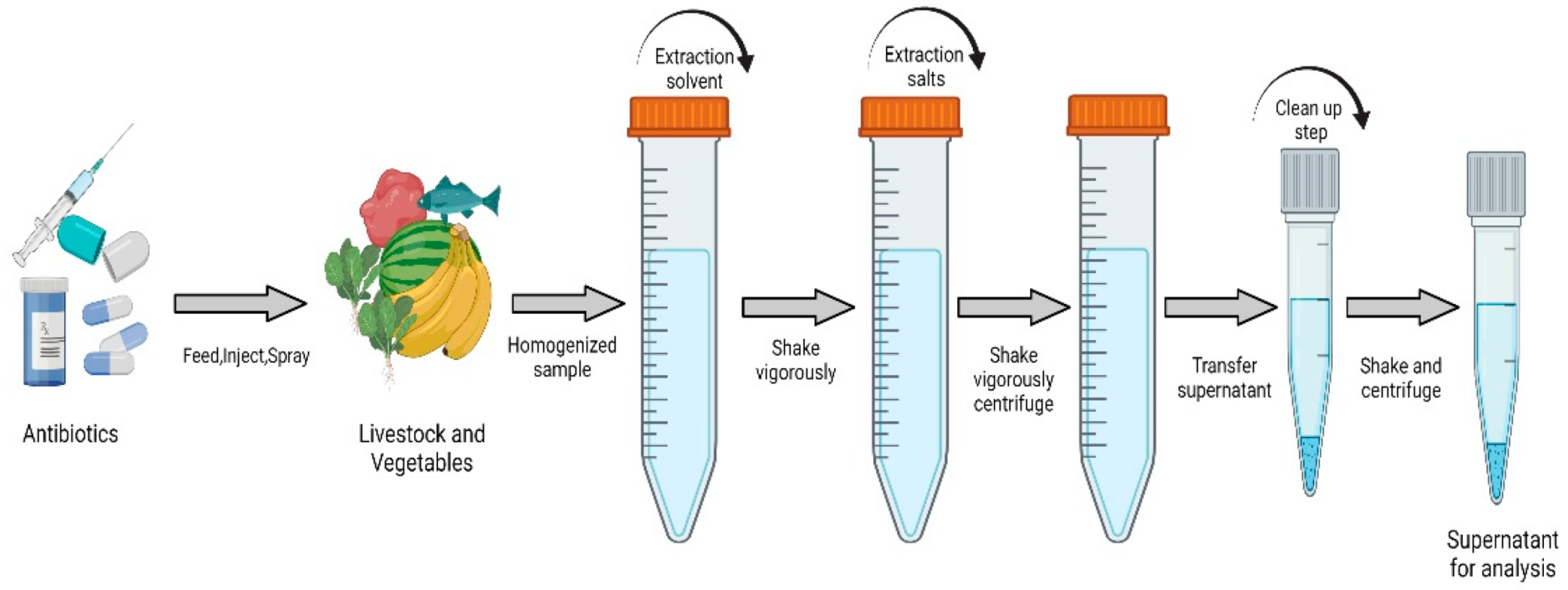
2. Extraction of Antibiotics from Food Samples
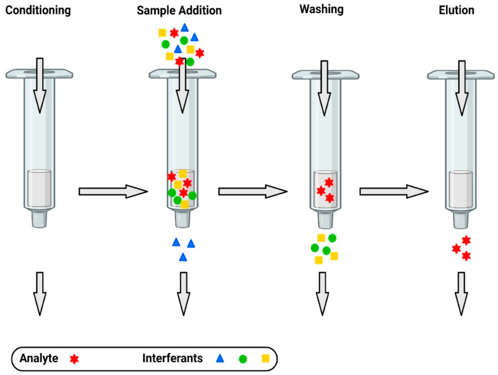
2.1. Classical SPE Sorbents
2.2. New SPE Sorbents
3. Separation and Detection of Antibiotics
3.1. Chromatography
3.1.1. Column Selection
3.1.2. Mobile Phase Selection
3.2. Capillary Electrophoresis
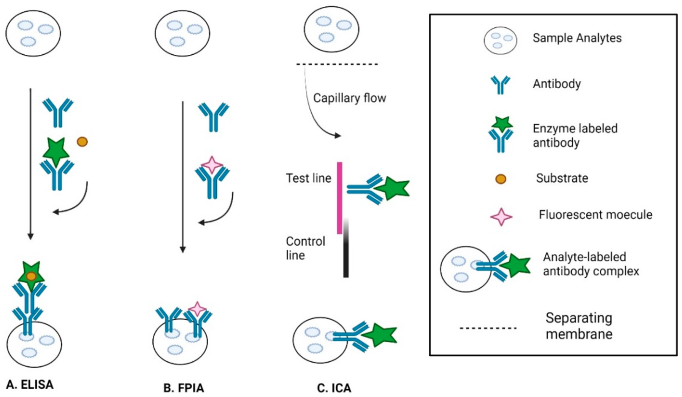
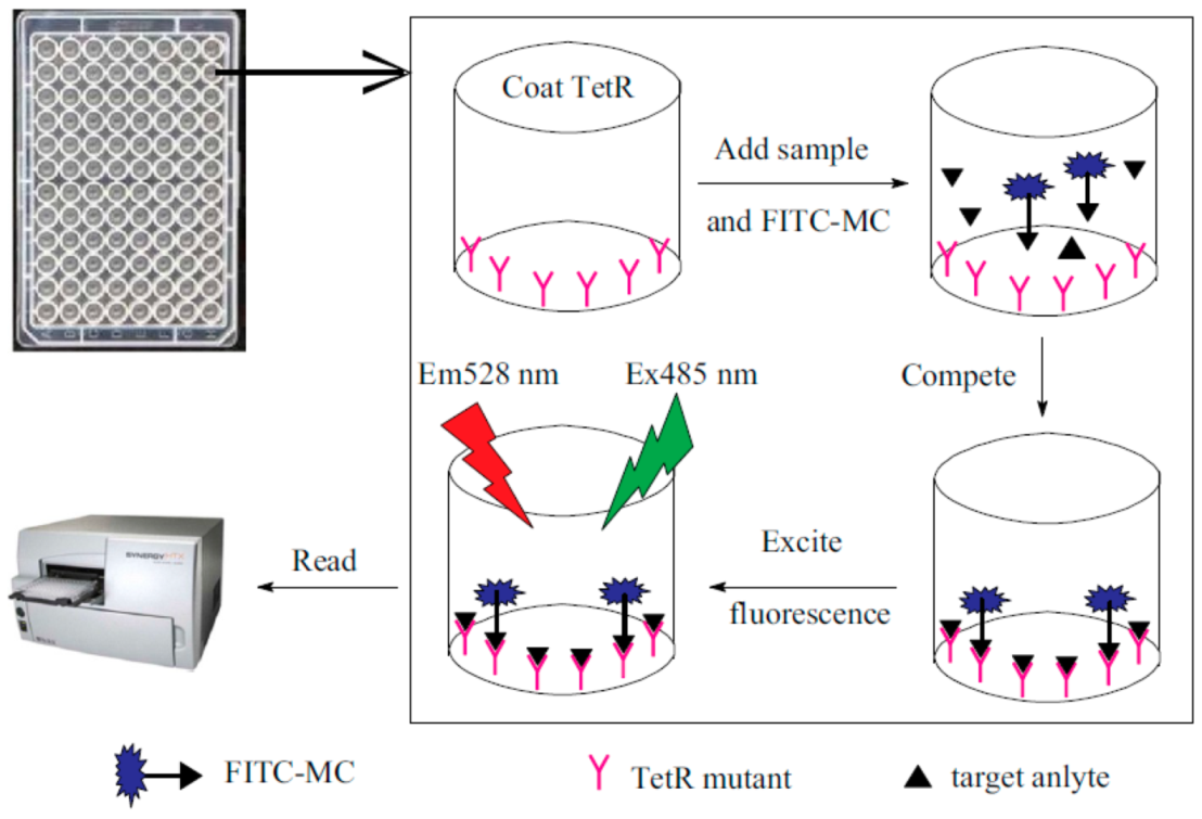
3.3. Immunological Methods
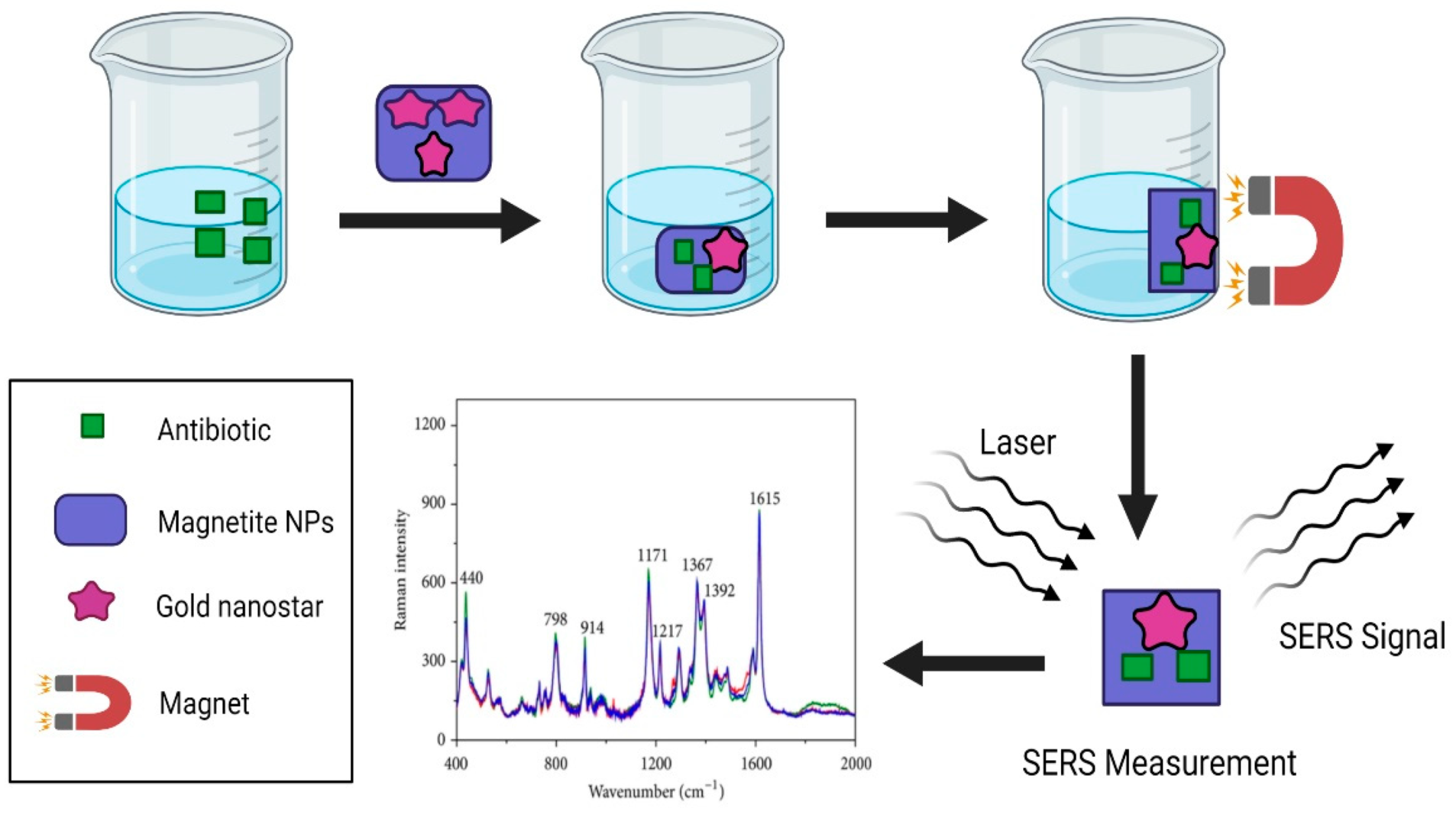
3.4. Surface-Enhanced Raman Spectroscopy (SERS)-Based Methods
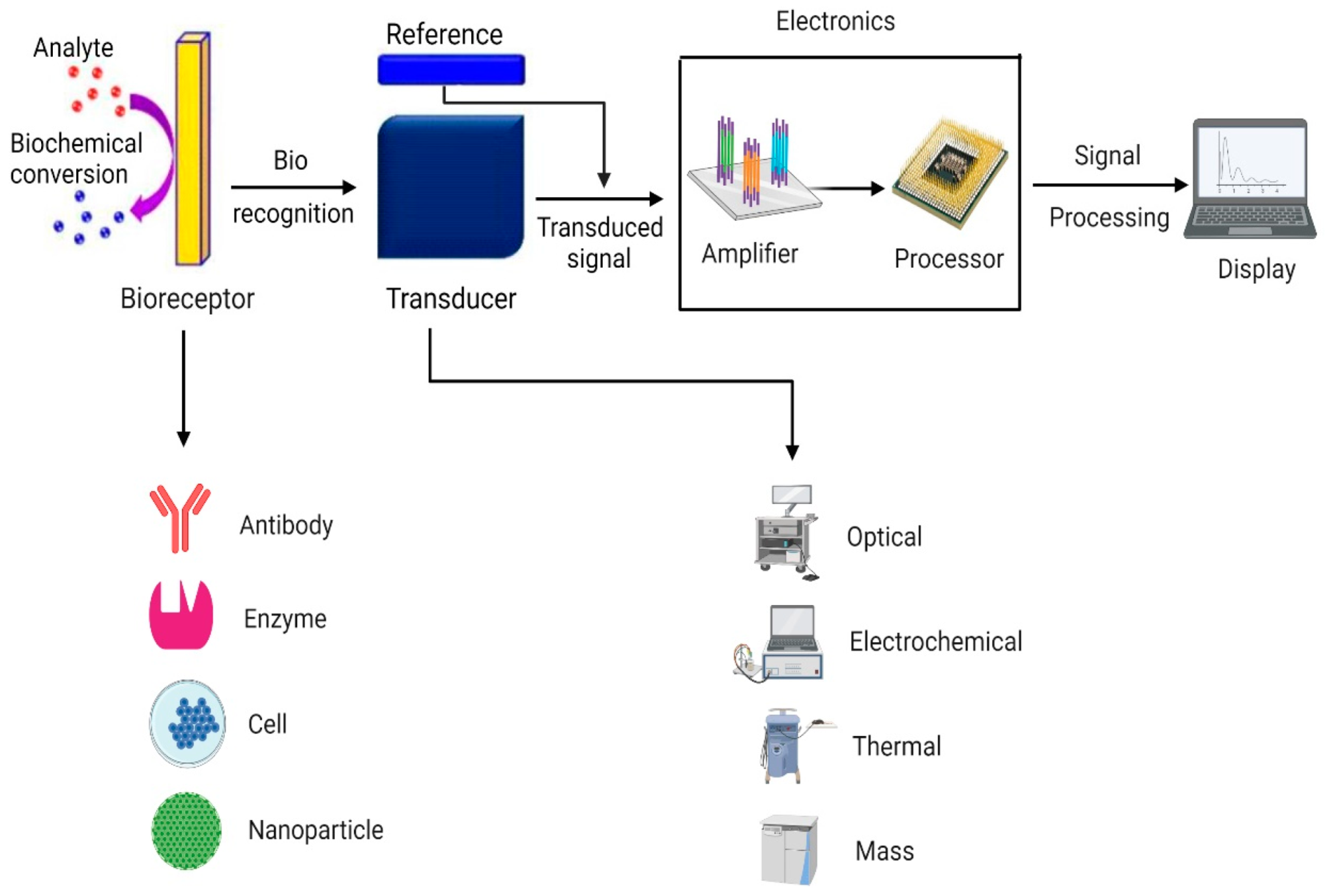
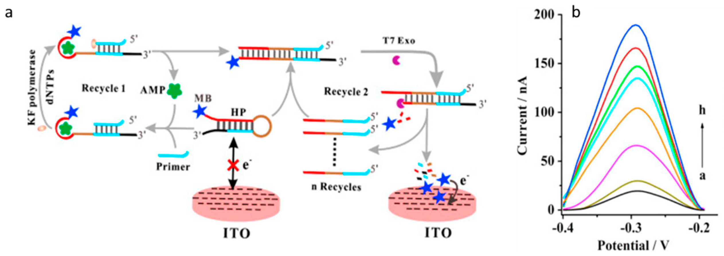
3.5. Biosensors
4. Conclusions and Future Perspectives
Author Contributions
Funding
Institutional Review Board Statement
Informed Consent Statement
Data Availability Statement
Acknowledgments
Conflicts of Interest
References
- Chandler, C.I.R. Current Accounts of Antimicrobial Resistance: Stabilisation, Individualisation and Antibiotics as Infrastructure. Palgrave Commun. 2019, 5, 53. [Google Scholar] [CrossRef] [Green Version]
- Angelakis, E.; Merhej, V.; Raoult, D. Related Actions of Probiotics and Antibiotics on Gut Microbiota and Weight Modification. Lancet Infect. Dis. 2013, 13, 889–899. [Google Scholar] [CrossRef]
- Menkem, Z.E.; Ngangom, B.L.; Tamunjoh, S.S.A.; Boyom, F.F. Antibiotic Residues in Food Animals: Public Health Concern. Acta Ecol. Sin. 2019, 39, 411–415. [Google Scholar] [CrossRef]
- Gothwal, R.; Shashidhar, T. Antibiotic Pollution in the Environment: A Review: Antibiotic Pollution in the Environment. Clean Soil Air Water 2015, 43, 479–489. [Google Scholar] [CrossRef]
- Ji, K.; Kho, Y.; Park, C.; Paek, D.; Ryu, P.; Paek, D.; Kim, M.; Kim, P.; Choi, K. Influence of Water and Food Consumption on Inadvertent Antibiotics Intake among General Population. Environ. Res. 2010, 110, 641–649. [Google Scholar] [CrossRef]
- Santos, L.; Ramos, F. Analytical Strategies for the Detection and Quantification of Antibiotic Residues in Aquaculturefishes: A Review. Trends Food Sci. Technol. 2016, 52. [Google Scholar] [CrossRef]
- Joshi, A.; Kim, K.-H. Recent Advances in Nanomaterial-Based Electrochemical Detection of Antibiotics: Challenges and Future Perspectives. Biosens. Bioelectron. 2020, 153, 112046. [Google Scholar] [CrossRef]
- Bueno, I.; Williams-Nguyen, J.; Hwang, H.; Sargeant, J.M.; Nault, A.J.; Singer, R.S. Systematic Review: Impact of Point Sources on Antibiotic-Resistant Bacteria in the Natural Environment. Zoonoses Public Health 2018, 65, e162–e184. [Google Scholar] [CrossRef]
- Rozman, U.; Duh, D.; Cimerman, M.; Turk, S.Š. Hospital Wastewater Effluent: Hot Spot for Antibiotic Resistant Bacteria. J. Water Sanit. Hyg. Dev. 2020, 10, 171–178. [Google Scholar] [CrossRef]
- Petrovich, M.L.; Zilberman, A.; Kaplan, A.; Eliraz, G.R.; Wang, Y.; Langenfeld, K.; Duhaime, M.; Wigginton, K.; Poretsky, R.; Avisar, D.; et al. Microbial and Viral Communities and Their Antibiotic Resistance Genes Throughout a Hospital Wastewater Treatment System. Front. Microbiol. 2020, 11, 153. [Google Scholar] [CrossRef] [PubMed] [Green Version]
- Kumar, A.; Pal, D. Antibiotic Resistance and Wastewater: Correlation, Impact and Critical Human Health Challenges. J. Environ. Chem. Eng. 2018, 6, 52–58. [Google Scholar] [CrossRef]
- Chen, J.; Ying, G.-G.; Deng, W.-J. Antibiotic Residues in Food: Extraction, Analysis, and Human Health Concerns. J. Agric. Food Chem. 2019, 67, 7569–7586. [Google Scholar] [CrossRef] [PubMed]
- Phillips, I. Does the Use of Antibiotics in Food Animals Pose a Risk to Human Health? A Critical Review of Published Data. J. Antimicrob. Chemother. 2003, 53, 28–52. [Google Scholar] [CrossRef]
- Kneebone, J.; Tsang, P.C.W.; Towson, D.H. Rapid Antibiotic Screening Tests Detect Antibiotic Residues in Powdered Milk Products. J. Dairy Sci. 2010, 93, 3961–3964. [Google Scholar] [CrossRef]
- Jammoul, A.; El Darra, N. Evaluation of Antibiotics Residues in Chicken Meat Samples in Lebanon. Antibiotics 2019, 8, 69. [Google Scholar] [CrossRef] [PubMed] [Green Version]
- Farouk, F.; Azzazy, H.M.E.; Niessen, W.M.A. Challenges in the Determination of Aminoglycoside Antibiotics, a Review. Anal. Chim. Acta 2015, 890, 21–43. [Google Scholar] [CrossRef] [PubMed]
- Larsson, D.G.J. Antibiotics in the Environment. Upsala J. Med. Sci. 2014, 119, 108–112. [Google Scholar] [CrossRef] [PubMed]
- Majdinasab, M.; Mishra, R.K.; Tang, X.; Marty, J.L. Detection of Antibiotics in Food: New Achievements in the Development of Biosensors. TrAC Trends Anal. Chem. 2020, 127, 115883. [Google Scholar] [CrossRef]
- Khan, M.Z.H. Recent Biosensors for Detection of Antibiotics in Animal Derived Food. Crit. Rev. Anal. Chem. 2020, 1–11. [Google Scholar] [CrossRef] [PubMed]
- Colombo, R.; Papetti, A. Advances in the Analysis of Veterinary Drug Residues in Food Matrices by Capillary Electrophoresis Techniques. Molecules 2019, 24, 4617. [Google Scholar] [CrossRef] [Green Version]
- Li, W.; Dai, X.; Pu, E.; Bian, H.; Chen, Z.; Zhang, X.; Guo, Z.; Li, P.; Li, H.; Yong, Y.; et al. HLB-MCX-Based Solid-Phase Extraction Combined with Liquid Chromatography–Tandem Mass Spectrometry for the Simultaneous Determination of Four Agricultural Antibiotics (Kasugamycin, Validamycin A, Ningnanmycin, and Polyoxin B) Residues in Plant-Origin Foods. J. Agric. Food Chem. 2020, 68, 14025–14037. [Google Scholar] [CrossRef] [PubMed]
- Di, S.; Yu, J.; Chen, P.; Zhu, G.; Zhu, S. Net-like Mesoporous Carbon Nanocomposites for Magnetic Solid-Phase Extraction of Sulfonamides Prior to Their Quantitation by UPLC-HRMS. Microchim. Acta 2020, 187, 112. [Google Scholar] [CrossRef] [PubMed]
- Alampanos, V.; Samanidou, V.; Papadoyannis, I. Trends in Sample Preparation for the HPLC Determination of Penicillins in Biofluids. J. Appl. Bioanal. 2019, 5, 9–17. [Google Scholar] [CrossRef]
- Sun, Y.; Zhao, J.; Liang, L. Recent Development of Antibiotic Detection in Food and Environment: The Combination of Sensors and Nanomaterials. Microchim. Acta 2021, 188, 21. [Google Scholar] [CrossRef]
- Zhi, S.; Zhou, J.; Liu, H.; Wu, H.; Zhang, Z.; Ding, Y.; Zhang, K. Simultaneous Extraction and Determination of 45 Veterinary Antibiotics in Swine Manure by Liquid Chromatography-Tandem Mass Spectrometry. J. Chromatogr. B 2020, 1154, 122286. [Google Scholar] [CrossRef]
- Lorenzetti, A.S.; Lista, A.G.; Domini, C.E. Reverse Ultrasound-Assisted Emulsification-Microextraction of Macrolides from Chicken Fat Followed by Electrophoretic Determination. LWT 2019, 113, 108334. [Google Scholar] [CrossRef]
- Zhao, J.; Liu, P.; Yuan, H.; Peng, Y.; Hong, Q.; Liu, M. Rapid Detection of Tetracycline Residues in Duck Meat Using Surface Enhanced Raman Spectroscopy. J. Spectrosc. 2016, 2016, 1–6. [Google Scholar] [CrossRef] [Green Version]
- Baghani, A.; Mesdaghinia, A.; Rafieiyan, M.; Soltan Dallal, M.M.; Douraghi, M. Tetracycline and Ciprofloxacin Multiresidues in Beef and Chicken Meat Samples Using Indirect Competitive ELISA. J. Immunoass. Immunochem. 2019, 40, 328–342. [Google Scholar] [CrossRef]
- Gaudin, V. Advances in Biosensor Development for the Screening of Antibiotic Residues in Food Products of Animal Origin-A Comprehensive Review. Biosens. Bioelectron. 2017, 90, 363–377. [Google Scholar] [CrossRef]
- Mungroo, N.; Neethirajan, S. Biosensors for the Detection of Antibiotics in Poultry Industry—A Review. Biosensors 2014, 4, 472–493. [Google Scholar] [CrossRef] [PubMed] [Green Version]
- Hutchings, M.I.; Truman, A.W.; Wilkinson, B. Antibiotics: Past, Present and Future. Curr. Opin. Microbiol. 2019, 51, 72–80. [Google Scholar] [CrossRef]
- Coates, A.R.; Halls, G.; Hu, Y. Novel Classes of Antibiotics or More of the Same?: New Antibiotic Classes Are Urgently Needed. Br. J. Pharmacol. 2011, 163, 184–194. [Google Scholar] [CrossRef] [Green Version]
- Ha, J.; Song, G.; Ai, L.-F.; Li, J.-C. Determination of Six Polyether Antibiotic Residues in Foods of Animal Origin by Solid Phase Extraction Combined with Liquid Chromatography-Tandem Mass Spectrometry. J. Chromatogr. B 2016, 1017–1018, 187–194. [Google Scholar] [CrossRef]
- Xu, G.; Zhang, B.; Wang, X.; Li, N.; Zhao, Y.; Liu, L.; Lin, J.-M.; Zhao, R.-S. Porous Covalent Organonitridic Frameworks for Solid-Phase Extraction of Sulfonamide Antibiotics. Mikrochim. Acta 2018, 186, 26. [Google Scholar] [CrossRef] [PubMed]
- Pérez-Rodríguez, M.; Pellerano, R.G.; Pezza, L.; Pezza, H.R. An Overview of the Main Foodstuff Sample Preparation Technologies for Tetracycline Residue Determination. Talanta 2018, 182, 1–21. [Google Scholar] [CrossRef] [Green Version]
- Wang, X.; Li, P. Rapid Screening of Mycotoxins in Liquid Milk and Milk Powder by Automated Size-Exclusion SPE-UPLC-MS/MS and Quantification of Matrix Effects over the Whole Chromatographic Run. Food Chem. 2015, 173, 897–904. [Google Scholar] [CrossRef]
- Deng, F.; Yu, H.; Pan, X.; Hu, G.; Wang, Q.; Peng, R.; Tan, L.; Yang, Z. Ultra-High Performance Liquid Chromatography Tandem Mass Spectrometry for the Determination of Five Glycopeptide Antibiotics in Food and Biological Samples Using Solid-Phase Extraction. J. Chromatogr. A 2018, 1538, 54–59. [Google Scholar] [CrossRef] [PubMed]
- Li, T.; Wang, C.; Xu, Z.; Chakraborty, A. A Coupled Method of On-Line Solid Phase Extraction with the UHPLC‒MS/MS for Detection of Sulfonamides Antibiotics Residues in Aquaculture. Chemosphere 2020, 254, 126765. [Google Scholar] [CrossRef] [PubMed]
- Du, F.; Sun, L.; Tan, W.; Wei, Z.; Nie, H.; Huang, Z.; Ruan, G.; Li, J. Magnetic Stir Cake Sorptive Extraction of Trace Tetracycline Antibiotics in Food Samples: Preparation of Metal–Organic Framework-Embedded PolyHIPE Monolithic Composites, Validation and Application. Anal. Bioanal. Chem. 2019, 411, 2239–2248. [Google Scholar] [CrossRef] [PubMed]
- Weng, R.; Sun, L.; Jiang, L.; Li, N.; Ruan, G.; Li, J.; Du, F. Electrospun Graphene Oxide–Doped Nanofiber-Based Solid Phase Extraction Followed by High-Performance Liquid Chromatography for the Determination of Tetracycline Antibiotic Residues in Food Samples. Food Anal. Methods 2019, 12, 1594–1603. [Google Scholar] [CrossRef]
- Wang, R.; Li, C.; Li, Q.; Zhang, S.; lv, F.; Yan, Z. Electrospinning Fabrication of Covalent Organic Framework Composite Nanofibers for Pipette Tip Solid Phase Extraction of Tetracycline Antibiotics in Grass Carp and Duck. J. Chromatogr. A 2020, 1622, 461098. [Google Scholar] [CrossRef]
- Figueiredo, L.; Erny, G.L.; Santos, L.; Alves, A. Applications of Molecularly Imprinted Polymers to the Analysis and Removal of Personal Care Products: A Review. Talanta 2016, 146, 754–765. [Google Scholar] [CrossRef] [PubMed] [Green Version]
- Yang, B.; Wang, L.; Luo, C.; Wang, X.; Sun, C. Simultaneous Determination of 11 Aminoglycoside Residues in Honey, Milk, and Pork by Liquid Chromatography with Tandem Mass Spectrometry and Molecularly Imprinted Polymer Solid Phase Extraction. J. AOAC Int. 2017, 100, 1869–1878. [Google Scholar] [CrossRef] [PubMed]
- Liu, X.; Xie, S.; Ni, T.; Chen, D.; Wang, X.; Pan, Y.; Wang, Y.; Huang, L.; Cheng, G.; Qu, W.; et al. Magnetic Solid-Phase Extraction Based on Carbon Nanotubes for the Determination of Polyether Antibiotic and s-Triazine Drug Residues in Animal Food with LC-MS/MS. J. Sep. Sci. 2017, 40, 2416–2430. [Google Scholar] [CrossRef] [PubMed]
- Nasir, A.N.M.; Yahaya, N.; Zain, N.N.M.; Lim, V.; Kamaruzaman, S.; Saad, B.; Nishiyama, N.; Yoshida, N.; Hirota, Y. Thiol-Functionalized Magnetic Carbon Nanotubes for Magnetic Micro-Solid Phase Extraction of Sulfonamide Antibiotics from Milks and Commercial Chicken Meat Products. Food Chem. 2019, 276, 458–466. [Google Scholar] [CrossRef]
- Arroyo-Manzanares, N.; Gámiz-Gracia, L.; García-Campaña, A.M. Alternative Sample Treatments for the Determination of Sulfonamides in Milk by HPLC with Fluorescence Detection. Food Chem. 2014, 143, 459–464. [Google Scholar] [CrossRef]
- Wu, L.; Song, Y.; Hu, M.; Xu, X.; Zhang, H.; Yu, A.; Ma, Q.; Wang, Z. Determination of Sulfonamides in Butter Samples by Ionic Liquid Magnetic Bar Liquid-Phase Microextraction High-Performance Liquid Chromatography. Anal. Bioanal. Chem. 2015, 407, 569–580. [Google Scholar] [CrossRef]
- Zhao, Y.; Wu, R.; Yu, H.; Li, J.; Liu, L.; Wang, S.; Chen, X.; Chan, T.-W.D. Magnetic Solid-Phase Extraction of Sulfonamide Antibiotics in Water and Animal-Derived Food Samples Using Core-Shell Magnetite and Molybdenum Disulfide Nanocomposite Adsorbent. J. Chromatogr. A 2020, 1610, 460543. [Google Scholar] [CrossRef]
- Xu, G.; Dong, X.; Hou, L.; Wang, X.; Liu, L.; Ma, H.; Zhao, R.-S. Room-Temperature Synthesis of Flower-Shaped Covalent Organic Frameworks for Solid-Phase Extraction of Quinolone Antibiotics. Anal. Chim. Acta 2020, 1126, 82–90. [Google Scholar] [CrossRef]
- Golzari Aqda, T.; Behkami, S.; Raoofi, M.; Bagheri, H. Graphene Oxide-Starch-Based Micro-Solid Phase Extraction of Antibiotic Residues from Milk Samples. J. Chromatogr. A 2019, 1591, 7–14. [Google Scholar] [CrossRef]
- Song, X.; Zhou, T.; Li, J.; Su, Y.; Xie, J.; He, L. Determination of Macrolide Antibiotics Residues in Pork Using Molecularly Imprinted Dispersive Solid-Phase Extraction Coupled with LC-MS/MS. J. Sep. Sci. 2018, 41, 1138–1148. [Google Scholar] [CrossRef] [PubMed]
- Xu, J.-J.; An, M.; Yang, R.; Tan, Z.; Hao, J.; Cao, J.; Peng, L.-Q.; Cao, W. Determination of Tetracycline Antibiotic Residues in Honey and Milk by Miniaturized Solid Phase Extraction Using Chitosan-Modified Graphitized Multiwalled Carbon Nanotubes. J. Agric. Food Chem. 2016, 64, 2647–2654. [Google Scholar] [CrossRef] [PubMed]
- Arabsorkhi, B.; Sereshti, H. Determination of Tetracycline and Cefotaxime Residues in Honey by Micro-Solid Phase Extraction Based on Electrospun Nanofibers Coupled with HPLC. Microchem. J. 2018, 140, 241–247. [Google Scholar] [CrossRef]
- Xie, Y.; Hu, Q.; Zhao, M.; Cheng, Y.; Guo, Y.; Qian, H.; Yao, W. Simultaneous Determination of Erythromycin, Tetracycline, and Chloramphenicol Residue in Raw Milk by Molecularly Imprinted Polymer Mixed with Solid-Phase Extraction. Food Anal. Methods 2018, 11, 374–381. [Google Scholar] [CrossRef]
- Islas, G.; Rodriguez, J.A.; Perez-Silva, I.; Miranda, J.M.; Ibarra, I.S. Solid-Phase Extraction and Large-Volume Sample Stacking-Capillary Electrophoresis for Determination of Tetracycline Residues in Milk. J. Anal. Methods Chem. 2018, 2018, e5394527. [Google Scholar] [CrossRef] [Green Version]
- Betina, V. Bioautography in Paper and Thin-Layer Chromatography and Its Scope in the Antibiotic Field. J. Chromatogr. A 1973, 78, 41–51. [Google Scholar] [CrossRef]
- Armentano, A.; Summa, S.; Lo Magro, S.; Palermo, C.; Nardiello, D.; Centonze, D.; Muscarella, M. Rapid Method for the Quantification of 13 Sulphonamides in Milk by Conventional High-Performance Liquid Chromatography with Diode Array Ultraviolet Detection Using a Column Packed with Core-Shell Particles. J. Chromatogr. A 2018, 1531, 46–52. [Google Scholar] [CrossRef]
- Hui, W.; Li, Q.; Ma, H.; Wu, M.; Feng, K.; Zhu, H.; Yang, P.; Li, J.; Chen, C.; Yan, K. Rapid Screening for 15 Sulfonamide Residues in Foods of Animal Origin by High-Performance Liquid Chromatography–UV Method. J. Chromatogr. Sci. 2018, 56, 636–643. [Google Scholar] [CrossRef]
- Łukaszewicz, P.; Białk-Bielińska, A.; Dołżonek, J.; Kumirska, J.; Caban, M.; Stepnowski, P. A New Approach for the Extraction of Tetracyclines from Soil Matrices: Application of the Microwave-Extraction Technique. Anal. Bioanal. Chem. 2018, 410, 1697–1707. [Google Scholar] [CrossRef]
- Wang, Z.; Song, X.; Zhou, T.; Bian, K.; Zhang, F.; He, L.; Liu, Q. Simultaneous Determination of Ten Macrolides Drugs in Feeds by High Performance Liquid Chromatography with Evaporation Light Scattering Detection. RSC Adv. 2015, 5, 1491–1499. [Google Scholar] [CrossRef]
- Song, X.; Xie, J.; Zhang, M.; Zhang, Y.; Li, J.; Huang, Q.; He, L. Simultaneous Determination of Eight Cyclopolypeptide Antibiotics in Feed by High Performance Liquid Chromatography Coupled with Evaporation Light Scattering Detection. J. Chromatogr. B 2018, 1076, 103–109. [Google Scholar] [CrossRef]
- Mishra, A.; Chhonker, Y.S.; Bisen, A.C.; Prasad, Y.D.; Tulsankar, S.L.; Chandasana, H.; Dey, T.; Verma, S.K.; Bala, V.; Kanojiya, S.; et al. Rapid and Simultaneous Analysis of Multiple Classes of Antimicrobial Drugs by Liquid Chromatography-Tandem Mass Spectrometry and Its Application to Routine Biomedical, Food, and Soil Analyses. ACS Omega 2020, 5, 31584–31597. [Google Scholar] [CrossRef]
- Liu, Q.; Li, J.; Song, X.; Zhang, M.; Li, E.; Gao, F.; He, L. Simultaneous Determination of Aminoglycoside Antibiotics in Feeds Using High Performance Liquid Chromatography with Evaporative Light Scattering Detection. RSC Adv. 2017, 7, 1251–1259. [Google Scholar] [CrossRef] [Green Version]
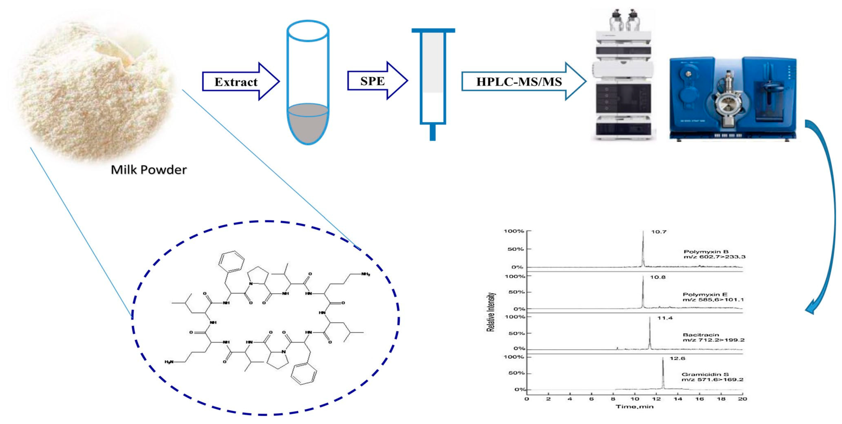
- Liu, T.; Zhang, C.; Zhang, F.; Nie, B.; Yuan, F.; Huang, H.; Li, H. Sensitive Determination of Four Polypeptide Antibiotic Residues in Milk Powder by High Performance Liquid Chromatography–Electrospray Tandem Mass Spectrometry. Chromatographia 2019, 82, 1479–1487. [Google Scholar] [CrossRef]
- Wang, C.; Li, X.; Yu, F.; Wang, Y.; Ye, D.; Hu, X.; Zhou, L.; Du, J.; Xia, X. Multi-Class Analysis of Veterinary Drugs in Eggs Using Dispersive-Solid Phase Extraction and Ultra-High Performance Liquid Chromatography-Tandem Mass Spectrometry. Food Chem. 2021, 334, 127598. [Google Scholar] [CrossRef] [PubMed]
- Lai, X.; Liu, J.; Xu, X.; Li, J.; Zhang, B.; Wei, L.; Cai, H.; Cheng, X. Ultrasensitive High-performance Liquid Chromatography Determination of Tetracycline Antibiotics and Their 4-epimer Derivatives Based on Dual Effect of Methanesulfonic Acid. J. Sep. Sci. 2020, 43, 398–405. [Google Scholar] [CrossRef]
- Lu, Z.; Deng, F.; He, R.; Tan, L.; Luo, X.; Pan, X.; Yang, Z. A Pass-through Solid-Phase Extraction Clean-up Method for the Determination of 11 Quinolone Antibiotics in Chicken Meat and Egg Samples Using Ultra-Performance Liquid Chromatography Tandem Mass Spectrometry. Microchem. J. 2019, 151, 104213. [Google Scholar] [CrossRef]
- Wang, K.; Lin, K.; Huang, X.; Chen, M. A Simple and Fast Extraction Method for the Determination of Multiclass Antibiotics in Eggs Using LC-MS/MS. J. Agric. Food Chem. 2017, 65, 5064–5073. [Google Scholar] [CrossRef] [PubMed]
- Yu, H.; Jia, Y.; Wu, R.; Chen, X.; Chan, T.-W.D. Determination of Fluoroquinolones in Food Samples by Magnetic Solid-Phase Extraction Based on a Magnetic Molecular Sieve Nanocomposite Prior to High-Performance Liquid Chromatography and Tandem Mass Spectrometry. Anal. Bioanal. Chem. 2019, 411, 2817–2826. [Google Scholar] [CrossRef] [PubMed]
- He, T.; Xu, Z.; Ren, J. Pressure-Assisted Electrokinetic Injection Stacking for Seven Typical Antibiotics in Waters to Achieve Μg/L Level Analysis by Capillary Electrophoresis with UV Detection. Microchem. J. 2019, 146, 1295–1300. [Google Scholar] [CrossRef]
- Moreno-González, D.; Krulišová, M.; Gámiz-Gracia, L.; García-Campaña, A.M. Determination of Tetracyclines in Human Urine Samples by Capillary Electrophoresis in Combination with Field Amplified Sample Injection. Electrophoresis 2018, 39, 608–615. [Google Scholar] [CrossRef] [PubMed]
- Ferreira, T.A.; Flores-Aguilar, J.F.; Santos, E.M.; Rodriguez, J.A.; Ibarra, I.S. New Poly(Ionic Liquid) Based Fiber for Determination of Oxytetracycline in Milk Samples by Application of SPME-CE Technique. Molecules 2019, 24, 430. [Google Scholar] [CrossRef] [PubMed] [Green Version]
- Moreno-González, D.; Hamed, A.M.; Gilbert-López, B.; Gámiz-Gracia, L.; García-Campaña, A.M. Evaluation of a Multiresidue Capillary Electrophoresis-Quadrupole-Time-of-Flight Mass Spectrometry Method for the Determination of Antibiotics in Milk Samples. J. Chromatogr. A 2017, 1510, 100–107. [Google Scholar] [CrossRef] [PubMed]
- Díaz-Quiroz, C.A.; Francisco Hernández-Chávez, J.; Ulloa-Mercado, G.; Gortáres-Moroyoqui, P.; Martínez-Macías, R.; Meza-Escalante, E.; Serrano-Palacios, D. Simultaneous Quantification of Antibiotics in Wastewater from Pig Farms by Capillary Electrophoresis. J. Chromatogr. B 2018, 1092, 386–393. [Google Scholar] [CrossRef] [PubMed]
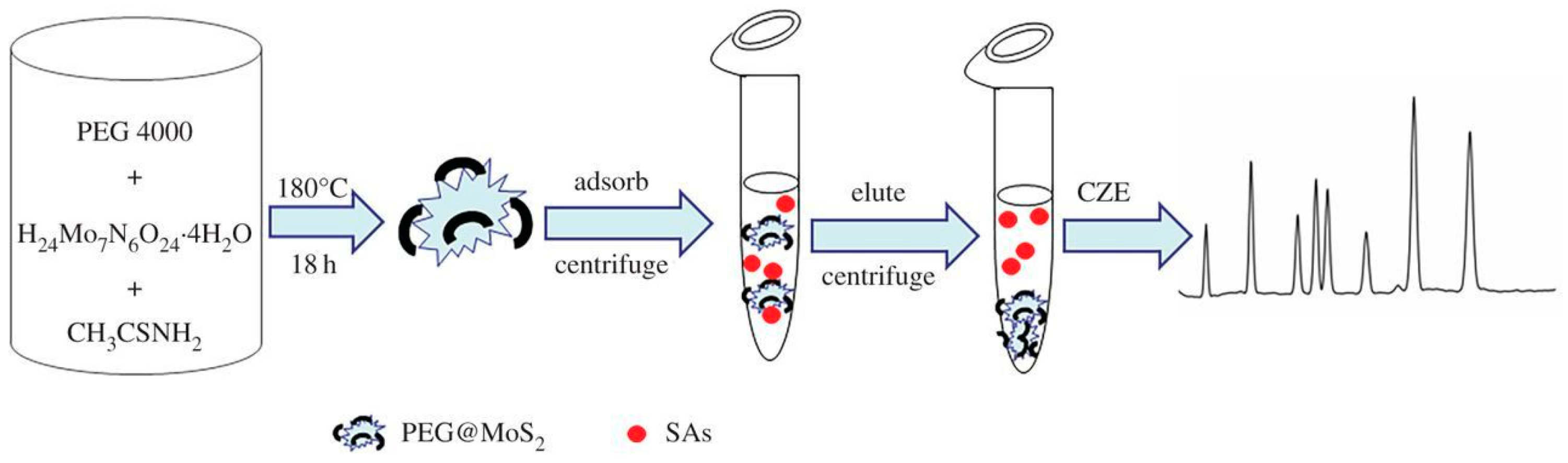
- An, J.; Wang, X.; Ming, M.; Li, J.; Ye, N. Determination of Sulfonamides in Milk by Capillary Electrophoresis with PEG@MoS2 as a Dispersive Solid-Phase Extraction Sorbent. R. Soc. Open Sci. 2018, 5, 172104. [Google Scholar] [CrossRef] [Green Version]
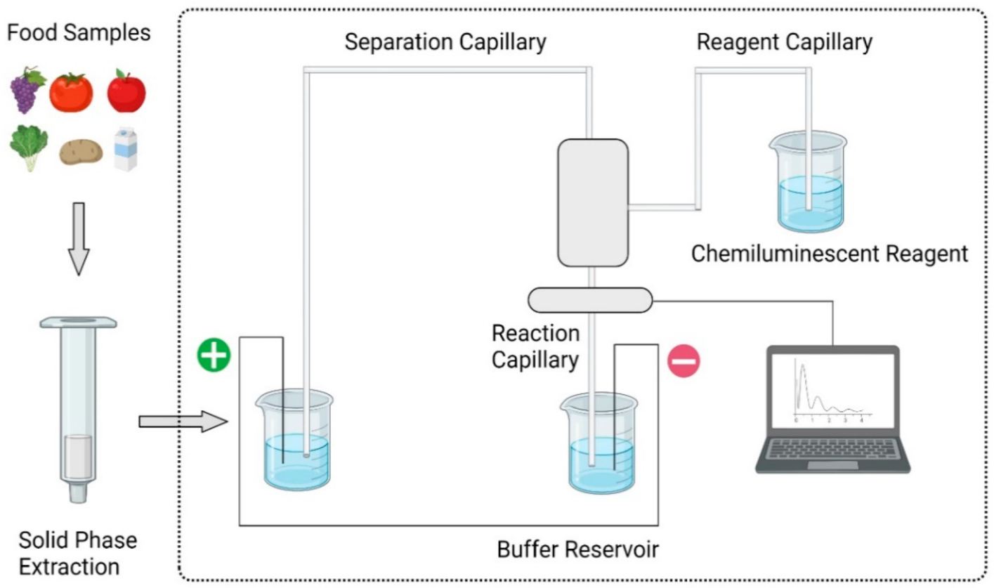
- Dai, T.; Duan, J.; Li, X.; Xu, X.; Shi, H.; Kang, W. Determination of Sulfonamide Residues in Food by Capillary Zone Electrophoresis with On-Line Chemiluminescence Detection Based on an Ag(III) Complex. Int. J. Mol. Sci. 2017, 18, 1286. [Google Scholar] [CrossRef] [Green Version]
- Hong, Y.; Guo, X.; Chen, G.; Zhou, J.; Zou, X.; Liao, X.; Hou, T. Determination of Five Macrolide Antibiotic Residues in Milk by Micellar Electrokinetic Capillary Chromatography with Field Amplified Sample Stacking. J. Food Saf. 2018, 38. [Google Scholar] [CrossRef]
- Lahouidak, S.; Soriano, M.L.; Salghi, R.; Zougagh, M.; Ríos, Á. Graphene Quantum Dots for Enhancement of Fluorimetric Detection Coupled to Capillary Electrophoresis for Detection of Ofloxacin. Electrophoresis 2019, 40, 2336–2341. [Google Scholar] [CrossRef]
- Tejada-Casado, C.; Moreno-González, D.; Lara, F.J.; García-Campaña, A.M.; del Olmo-Iruela, M. Determination of Benzimidazoles in Meat Samples by Capillary Zone Electrophoresis Tandem Mass Spectrometry Following Dispersive Liquid–Liquid Microextraction. J. Chromatogr. A 2017, 1490, 212–219. [Google Scholar] [CrossRef] [PubMed]
- Bosma, R.; Devasagayam, J.; Singh, A.; Collier, C.M. Microchip Capillary Electrophoresis Dairy Device Using Fluorescence Spectroscopy for Detection of Ciprofloxacin in Milk Samples. Sci. Rep. 2020, 10, 1–8. [Google Scholar] [CrossRef]
- Tian, Z.; Gao, J.; Qin, W. Determination of Fluoroquinolones in Milk by Ionic Liquid-Mediated Two Phase Extraction Followed by Capillary Electrophoresis Analysis. Madr. J. Anal. Sci. Instrum. 2018, 3, 62–67. [Google Scholar] [CrossRef]
- Kergaravat, S.V.; Nagel, O.G.; Althaus, R.L.; Hernández, S.R. Magneto Immunofluorescence Assay for Quinolone Detection in Bovine Milk. Food Anal. Methods 2020, 13, 1539–1547. [Google Scholar] [CrossRef] [Green Version]
- Li, C.; Zhang, Y.; Eremin, S.A.; Yakup, O.; Yao, G.; Zhang, X. Detection of Kanamycin and Gentamicin Residues in Animal-Derived Food Using IgY Antibody Based Ic-ELISA and FPIA. Food Chem. 2017, 227, 48–54. [Google Scholar] [CrossRef] [PubMed] [Green Version]
- Li, H.; Ma, S.; Zhang, X.; Li, C.; Dong, B.; Mujtaba, M.G.; Wei, Y.; Liang, X.; Yu, X.; Wen, K.; et al. Generic Hapten Synthesis, Broad-Specificity Monoclonal Antibodies Preparation, and Ultrasensitive ELISA for Five Antibacterial Synergists in Chicken and Milk. J. Agric. Food Chem. 2018, 66, 11170–11179. [Google Scholar] [CrossRef] [PubMed]
- Wang, Z.; Hu, S.; Bao, H.; Xing, K.; Liu, J.; Xia, J.; Lai, W.H.; Peng, J. Immunochromatographic Assay Based on Time-Resolved Fluorescent Nanobeads for the Rapid Detection of Sulfamethazine in Egg, Honey, and Pork. J. Sci. Food Agric. 2020. [Google Scholar] [CrossRef]
- Liang, X.; Sheng, Y.; Yu, W.; Zhao, S.; Shan, H.; Zhang, Q.; Wang, Z. Comparison of Chicken IgY and Mammalian IgG in Three Immunoassays for Detection of Sulfamethazine in Milk. Food Anal. Methods 2018, 11, 3452–3463. [Google Scholar] [CrossRef]
- Shen, X.; Chen, J.; Lv, S.; Sun, X.; Dzantiev, B.B.; Eremin, S.A.; Zherdev, A.V.; Xu, J.; Sun, Y.; Lei, H. Fluorescence Polarization Immunoassay for Determination of Enrofloxacin in Pork Liver and Chicken. Molecules 2019, 24, 4462. [Google Scholar] [CrossRef] [Green Version]
- Wang, G.; Xia, W.Q.; Liu, J.X.; Wang, J.P.; Liu, J. Directional Evolution of TetR Protein and Development of a Fluoroimmunoassay for Screening of Tetracyclines in Egg. Microchem. J. 2019, 150, 104184. [Google Scholar] [CrossRef]
- Chen, J.; Shanin, I.A.; Lv, S.; Wang, Q.; Mao, C.; Xu, Z.; Sun, Y.; Wu, Q.; Eremin, S.A.; Lei, H. Heterologous Strategy Enhancing the Sensitivity of the Fluorescence Polarization Immunoassay of Clinafloxacin in Goat Milk. J. Sci. Food Agric. 2016, 96, 1341–1346. [Google Scholar] [CrossRef] [PubMed]
- Mukunzi, D.; Suryoprabowo, S.; Song, S.; Liu, L.; Kuang, H. Development of an Indirect Enzyme-Linked Immunosorbent Assay and Lateral-Flow Test Strips for Pefloxacin and Its Analogues in Chicken Muscle Samples. Food Agric. Immunol. 2018, 29, 484–497. [Google Scholar] [CrossRef] [Green Version]
- He, J.; Hu, J.; Thirumalai, D.; Schade, R.; Du, E.; Zhang, X. Development of Indirect Competitive ELISA Using Egg Yolk-Derived Immunoglobulin (IgY) for the Detection of Gentamicin Residues. J. Environ. Sci. Health Part B 2016, 51, 8–13. [Google Scholar] [CrossRef]
- Liu, H.; Sun, Y.; Jin, Z.; Yang, L.; Liu, J. Capillarity-Constructed Reversible Hot Spots for Molecular Trapping inside Silver Nanorod Arrays Light up Ultrahigh SERS Enhancement. Chem. Sci. 2013, 4, 3490–3496. [Google Scholar] [CrossRef]
- Jiang, Y.; Sun, D.-W.; Pu, H.; Wei, Q. Surface Enhanced Raman Spectroscopy (SERS): A Novel Reliable Technique for Rapid Detection of Common Harmful Chemical Residues. Trends Food Sci. Technol. 2018, 75, 10–22. [Google Scholar] [CrossRef]
- Wang, Q.; Zhao, W.-M. Optical Methods of Antibiotic Residues Detections: A Comprehensive Review. Sens. Actuators B Chem. 2018, 269, 238–256. [Google Scholar] [CrossRef]
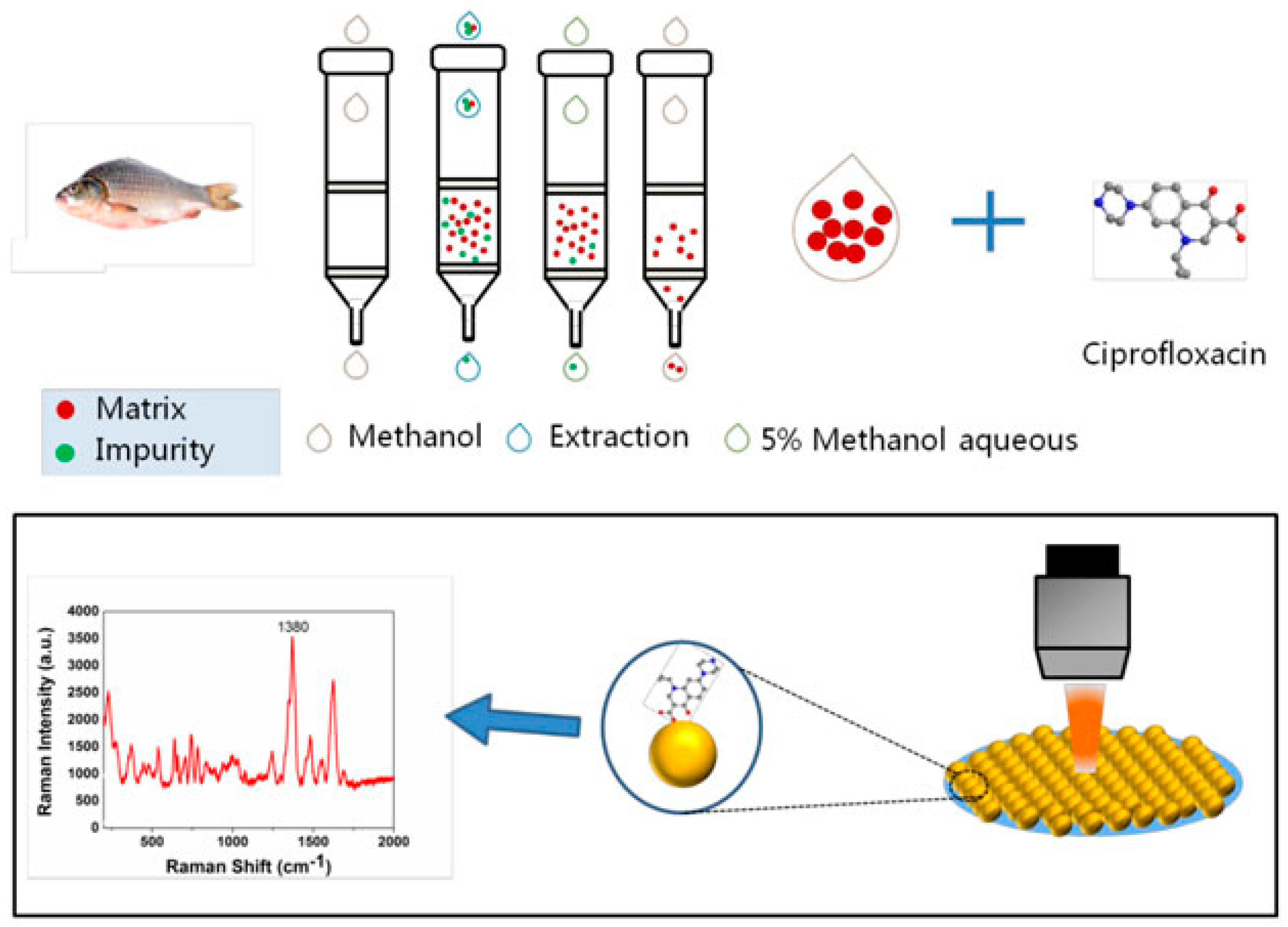
- Zhang, Y.; Teng, Y.; Qin, Y.; Ren, Z.; Wang, Z. Determination of Ciprofloxacin in Fish by Surface-Enhanced Raman Scattering Using a Liquid-Liquid Self-Assembled Gold Nanofilm. Anal. Lett. 2020, 53, 660–670. [Google Scholar] [CrossRef]
- Jiang, X.; Qin, X.; Yin, D.; Gong, M.; Yang, L.; Zhao, B.; Ruan, W. Rapid Monitoring of Benzylpenicillin Sodium Using Raman and Surface Enhanced Raman Spectroscopy. Spectrochim. Acta Part A Mol. Biomol. Spectrosc. 2015, 140, 474–478. [Google Scholar] [CrossRef]
- Peng, Y.; Liu, M.; Chen, X.; Yuan, H.; Zhao, J. Surface-Enhanced Raman Spectroscopy Coupled with Gold Nanoparticles for Rapid Detection of Amoxicillin Residues in Duck Meat. Spectrosc. Lett. 2017, 50, 579–584. [Google Scholar] [CrossRef]
- Wali, L.A.; Hasan, K.K.; Alwan, A.M. Rapid and Highly Efficient Detection of Ultra-Low Concentration of Penicillin G by Gold Nanoparticles/Porous Silicon SERS Active Substrate. Spectrochim. Acta A. Mol. Biomol. Spectrosc. 2019, 206, 31–36. [Google Scholar] [CrossRef]
- Jabbar, A.A.; Alwan, A.M.; Haider, A.J. Modifying and Fine Controlling of Silver Nanoparticle Nucleation Sites and SERS Performance by Double Silicon Etching Process. Plasmonics 2018, 13, 1171–1182. [Google Scholar] [CrossRef]
- Ali, W.H.; Dheyab, A.B.; Alwan, A.M.; Abber, Z.S. Study the Role of Mud-like Psi Morphologies on the Performance of AuNPS SERS Sensor for Efficient Detection of Amoxicillin. In AIP Conference Proceedings; AIP Publishing LLC: Melville, NY, USA, 2020; Volume 2290, p. 050061. [Google Scholar] [CrossRef]
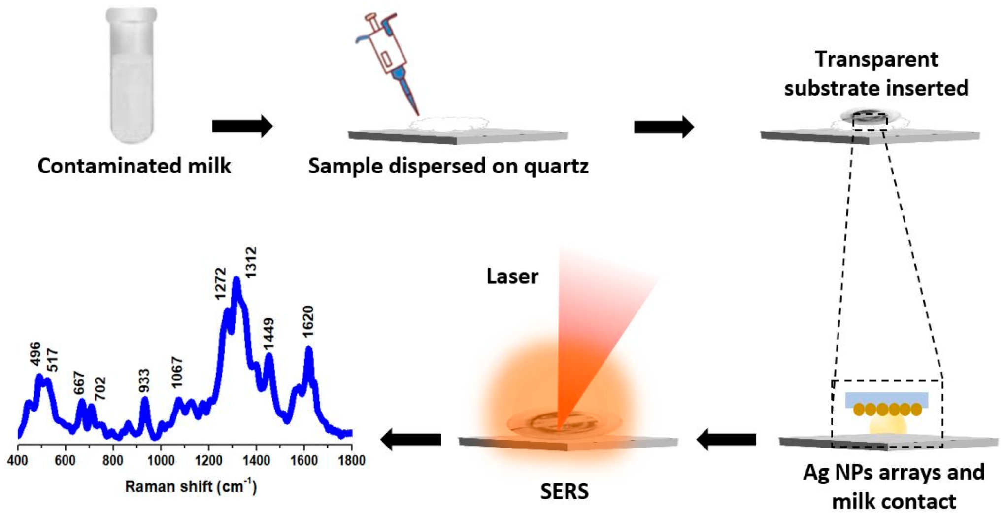
- Muhammad, M.; Yan, B.; Yao, G.; Chao, K.; Zhu, C.; Huang, Q. Surface-Enhanced Raman Spectroscopy for Trace Detection of Tetracycline and Dicyandiamide in Milk Using Transparent Substrate of Ag Nanoparticle Arrays. ACS Appl. Nano Mater. 2020, 3, 7066–7075. [Google Scholar] [CrossRef]
- Wang, X.; Shi, W.; She, G.; Mu, L. Surface-Enhanced Raman Scattering (SERS) on Transition Metal and Semiconductor Nanostructures. Phys. Chem. Chem. Phys. 2012, 14, 5891. [Google Scholar] [CrossRef] [PubMed]
- Shi, Q.; Huang, J.; Sun, Y.; Deng, R.; Teng, M.; Li, Q.; Yang, Y.; Hu, X.; Zhang, Z.; Zhang, G. A SERS-Based Multiple Immuno-Nanoprobe for Ultrasensitive Detection of Neomycin and Quinolone Antibiotics via a Lateral Flow Assay. Microchim. Acta 2018, 185, 84. [Google Scholar] [CrossRef] [PubMed]
- Fan, R.; Tang, S.; Luo, S.; Liu, H.; Zhang, W.; Yang, C.; He, L.; Chen, Y. Duplex Surface Enhanced Raman Scattering-Based Lateral Flow Immunosensor for the Low-Level Detection of Antibiotic Residues in Milk. Molecules 2020, 25, 5249. [Google Scholar] [CrossRef]
- Qu, L.; Liu, Y.-Y.; Liu, M.-K.; Yang, G.-H.; Li, D.-W.; Li, H.-T. Highly Reproducible Ag NPs/CNT-Intercalated GO Membranes for Enrichment and SERS Detection of Antibiotics. ACS Appl. Mater. Interfaces 2016, 8, 28180–28186. [Google Scholar] [CrossRef] [PubMed]
- Cui, J.; Chen, S.; Ma, X.; Shao, H.; Zhan, J. Galvanic Displacement-Induced Codeposition of Reduced-Graphene-Oxide/Silver on Alloy Fibers for Non-Destructive SPME@SERS Analysis of Antibiotics. Microchim. Acta 2019, 186, 19. [Google Scholar] [CrossRef] [PubMed]
- Zheng, Z.; Cong, S.; Gong, W.; Xuan, J.; Li, G.; Lu, W.; Geng, F.; Zhao, Z. Semiconductor SERS Enhancement Enabled by Oxygen Incorporation. Nat. Commun. 2017, 8, 1993. [Google Scholar] [CrossRef] [Green Version]
- Jiang, X.; Chen, Y.; Du, J.; Li, X.; Shen, Y.; Yang, M.; Han, X.; Yang, L.; Zhao, B. Comparative Study of Semiconductor TiO2 and Noble Metal Ag Substrates: The Differences between Chemical Enhancement and Electromagnetic Enhancement in SERS. J. Raman Spectrosc. 2018, 49. [Google Scholar] [CrossRef]
- Wang, W.; Sang, Q.; Yang, M.; Du, J.; Yang, L.; Jiang, X.; Han, X.; Zhao, B. Detection of Several Quinolone Antibiotic Residues in Water Based on Ag-TiO2 SERS Strategy. Sci. Total Environ. 2020, 702, 134956. [Google Scholar] [CrossRef] [PubMed]
- Xie, Y.; Zhao, M.; Hu, Q.; Cheng, Y.; Guo, Y.; Qian, H.; Yao, W. Selective Detection of Chloramphenicol in Milk Based on a Molecularly Imprinted Polymer-Surface-Enhanced Raman Spectroscopic Nanosensor: Selective Detection of Chloramphenicol in Milk. J. Raman Spectrosc. 2017, 48, 204–210. [Google Scholar] [CrossRef]
- Ashley, J.; Wu, K.; Hansen, M.F.; Schmidt, M.S.; Boisen, A.; Sun, Y. Quantitative Detection of Trace Level Cloxacillin in Food Samples Using Magnetic Molecularly Imprinted Polymer Extraction and Surface-Enhanced Raman Spectroscopy Nanopillars. Anal. Chem. 2017, 89, 11484–11490. [Google Scholar] [CrossRef] [Green Version]
- Pinheiro, P.; Fateixa, S.; Nogueira, H.; Trindade, T. Magnetite-Supported Gold Nanostars for the Uptake and SERS Detection of Tetracycline. Nanomaterials 2018, 9, 31. [Google Scholar] [CrossRef] [PubMed] [Green Version]
- Chen, Y.; Li, X.; Yang, M.; Yang, L.; Han, X.; Jiang, X.; Zhao, B. High Sensitive Detection of Penicillin G Residues in Milk by Surface-Enhanced Raman Scattering. Talanta 2017, 167, 236–241. [Google Scholar] [CrossRef] [PubMed]
- Li, M.; Wu, H.; Wu, Y.; Ying, Y.; Wen, Y.; Guo, X.; Yang, H. Heterostructured Cube Au-Ag Composites for Rapid Raman Detection of Antibiotic Ciprofloxacin: Rapid Raman Detection of Antibiotic Ciprofloxacin. J. Raman Spectrosc. 2017, 48, 525–529. [Google Scholar] [CrossRef]
- Bhalla, N.; Jolly, P.; Formisano, N.; Estrela, P. Introduction to Biosensors. Essays Biochem. 2016, 60, 1–8. [Google Scholar] [CrossRef] [Green Version]
- Dixon, M.C. Quartz Crystal Microbalance with Dissipation Monitoring: Enabling Real-Time Characterization of Biological Materials and Their Interactions. J. Biomol. Tech. JBT 2008, 19, 151–158. [Google Scholar]
- Shen, Y.; Xu, L.; Li, Y. Biosensors for Rapid Detection of Salmonella in Food: A Review. Compr. Rev. Food Sci. Food Saf. 2021, 20, 149–197. [Google Scholar] [CrossRef]
- Tan, B.; Zhao, H.; Du, L.; Gan, X.; Quan, X. A Versatile Fluorescent Biosensor Based on Target-Responsive Graphene Oxide Hydrogel for Antibiotic Detection. Biosens. Bioelectron. 2016, 83, 267–273. [Google Scholar] [CrossRef] [PubMed]
- Wu, S.; Zhang, H.; Shi, Z.; Duan, N.; Fang, C.; Dai, S.; Wang, Z. Aptamer-Based Fluorescence Biosensor for Chloramphenicol Determination Using Upconversion Nanoparticles. Food Control 2015, 50, 597–604. [Google Scholar] [CrossRef]
- Yue, F.; Li, F.; Kong, Q.; Guo, Y.; Sun, X. Recent Advances in Aptamer-Based Sensors for Aminoglycoside Antibiotics Detection and Their Applications. Sci. Total Environ. 2021, 762, 143129. [Google Scholar] [CrossRef] [PubMed]
- Ebralidze, I.I.; Laschuk, N.O.; Poisson, J.; Zenkina, O.V. Chapter 1-Colorimetric Sensors and Sensor Arrays. In Nanomaterials Design for Sensing Applications; Zenkina, O.V., Ed.; Micro and Nano Technologies; Elsevier: Amsterdam, The Netherlands, 2019; pp. 1–39. [Google Scholar] [CrossRef]
- Ma, Q.; Wang, Y.; Jia, J.; Xiang, Y. Colorimetric Aptasensors for Determination of Tobramycin in Milk and Chicken Eggs Based on DNA and Gold Nanoparticles. Food Chem. 2018, 249, 98–103. [Google Scholar] [CrossRef]
- Abedalwafa, M.A.; Li, Y.; Ni, C.; Wang, L. Colorimetric Sensor Arrays for the Detection and Identification of Antibiotics. Anal. Methods 2019, 11, 2836–2854. [Google Scholar] [CrossRef]
- Hao, L.; Gu, H.; Duan, N.; Wu, S.; Wang, Z. A Chemiluminescent Aptasensor for Simultaneous Detection of Three Antibiotics in Milk. Anal. Methods 2016, 8, 7929–7936. [Google Scholar] [CrossRef]
- Emrani, A.S.; Danesh, N.M.; Lavaee, P.; Ramezani, M.; Abnous, K.; Taghdisi, S.M. Colorimetric and Fluorescence Quenching Aptasensors for Detection of Streptomycin in Blood Serum and Milk Based on Double-Stranded DNA and Gold Nanoparticles. Food Chem. 2016, 190, 115–121. [Google Scholar] [CrossRef]
- Marquette, C.A.; Blum, L.J. Electro-Chemiluminescent Biosensing. Anal. Bioanal. Chem. 2008, 390, 155–168. [Google Scholar] [CrossRef]
- Yang, L.; Ni, H.; Li, C.; Zhang, X.; Wen, K.; Ke, Y.; Yang, H.; Shi, W.; Zhang, S.; Shen, J.; et al. Development of a Highly Specific Chemiluminescence Aptasensor for Sulfamethazine Detection in Milk Based on in Vitro Selected Aptamers. Sens. Actuators B Chem. 2019, 281, 801–811. [Google Scholar] [CrossRef]
- Situ, C.; Mooney, M.H.; Elliott, C.T.; Buijs, J. Advances in Surface Plasmon Resonance Biosensor Technology towards High-Throughput, Food-Safety Analysis. TrAC Trends Anal. Chem. 2010, 29, 1305–1315. [Google Scholar] [CrossRef]
- Blidar, A.; Feier, B.; Tertis, M.; Galatus, R.; Cristea, C. Electrochemical Surface Plasmon Resonance (EC-SPR) Aptasensor for Ampicillin Detection. Anal. Bioanal. Chem. 2019, 411, 1053–1065. [Google Scholar] [CrossRef] [PubMed]
- Cervera-Chiner, L.; Jiménez, Y.; Montoya, Á.; Juan-Borrás, M.; Pascual, N.; Arnau, A.; Escriche, I. High Fundamental Frequency Quartz Crystal Microbalance (HFF-QCMD) Immunosensor for Detection of Sulfathiazole in Honey. Food Control 2020, 115, 107296. [Google Scholar] [CrossRef]
- Lan, L.; Yao, Y.; Ping, J.; Ying, Y. Recent Advances in Nanomaterial-Based Biosensors for Antibiotics Detection. Biosens. Bioelectron. 2017, 91, 504–514. [Google Scholar] [CrossRef]
- Wang, X.; Dong, S.; Gai, P.; Duan, R.; Li, F. Highly Sensitive Homogeneous Electrochemical Aptasensor for Antibiotic Residues Detection Based on Dual Recycling Amplification Strategy. Biosens. Bioelectron. 2016, 82, 49–54. [Google Scholar] [CrossRef]
- Munteanu, F.-D.; Titoiu, A.; Marty, J.-L.; Vasilescu, A. Detection of Antibiotics and Evaluation of Antibacterial Activity with Screen-Printed Electrodes. Sensors 2018, 18, 901. [Google Scholar] [CrossRef] [Green Version]
- Starzec, K.; Cristea, C.; Tertis, M.; Feier, B.; Wieczorek, M.; Kościelniak, P.; Kochana, J. Employment of Electrostriction Phenomenon for Label-Free Electrochemical Immunosensing of Tetracycline. Bioelectrochemistry 2020, 132, 107405. [Google Scholar] [CrossRef] [PubMed]
- Wang, H.; Wang, Y.; Liu, S.; Yu, J.; Xu, W.; Guo, Y.; Huang, J. Target–Aptamer Binding Triggered Quadratic Recycling Amplification for Highly Specific and Ultrasensitive Detection of Antibiotics at the Attomole Level. Chem. Commun. 2015, 51, 8377–8380. [Google Scholar] [CrossRef] [PubMed]
- Pilehvar, S.; Gielkens, K.; Trashin, S.A.; Dardenne, F.; Blust, R.; De Wael, K. (Electro)Sensing of Phenicol Antibiotics—A Review. Crit. Rev. Food Sci. Nutr. 2016, 56, 2416–2429. [Google Scholar] [CrossRef]
- Pan, M.; Li, S.; Wang, J.; Sheng, W.; Wang, S. Development and Validation of a Reproducible and Label-Free Surface Plasmon Resonance Immunosensor for Enrofloxacin Detection in Animal-Derived Foods. Sensors 2017, 17, 1984. [Google Scholar] [CrossRef] [PubMed] [Green Version]
- Mishra, G.K.; Sharma, A.; Bhand, S. Ultrasensitive Detection of Streptomycin Using Flow Injection Analysis-Electrochemical Quartz Crystal Nanobalance (FIA-EQCN) Biosensor. Biosens. Bioelectron. 2015, 67, 532–539. [Google Scholar] [CrossRef]
- Fogel, R.; Limson, J.; Seshia, A.A. Acoustic Biosensors. Essays Biochem. 2016, 60, 101–110. [Google Scholar] [CrossRef] [PubMed] [Green Version]
- Liu, C.; Lu, C.; Tang, Z.; Chen, X.; Wang, G.; Sun, F. Aptamer-Functionalized Magnetic Nanoparticles for Simultaneous Fluorometric Determination of Oxytetracycline and Kanamycin. Microchim. Acta 2015, 182, 2567–2575. [Google Scholar] [CrossRef]


| Class | Examples | References |
|---|---|---|
| Glycopeptides | Vancomycin, Teicoplanin, Telavancin, Oritavancin, Dalbavancin | [31] |
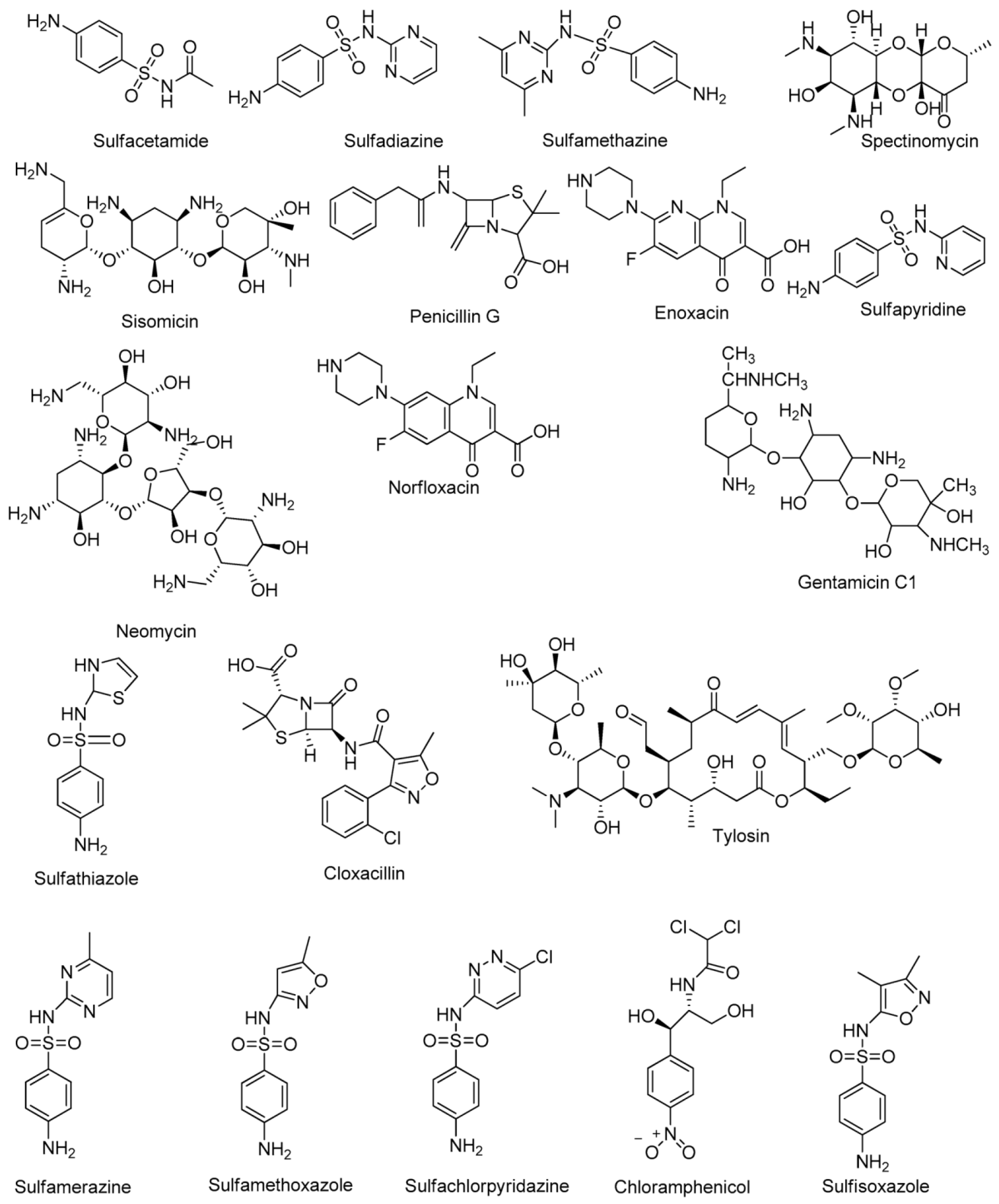
| Sulfonamides | Sulfacetamide, Sulfadiazine, Sulfathiazole, Sulfapyridine, Sulfamerazine, Sulfamethazine, Sulfamethoxazole, Sulfasoxazole, Sulfachloropyridazine | [32] |
| Tetracyclines | Tetracycline, Oxytetracycline, Doxycycline, Chlorotetracycline, Methacycline | [32] |
| Aminoglycosides | Amikacin, Paramomycin, Dihydrostreptomycin, Hygromycin, Kanamycin, Netilmycin, Spectinomycin, Sisomycin, Streptomycin, Tobramycin, Gentamicin, Neomycin | [32] |
| Β—Lactams | Amoxicillin, Ampicillin, Cloxacillin, Penicillin G | [32] |
| Macrolides | Erythromycin, Clarithromycin, Tylosin | [32] |
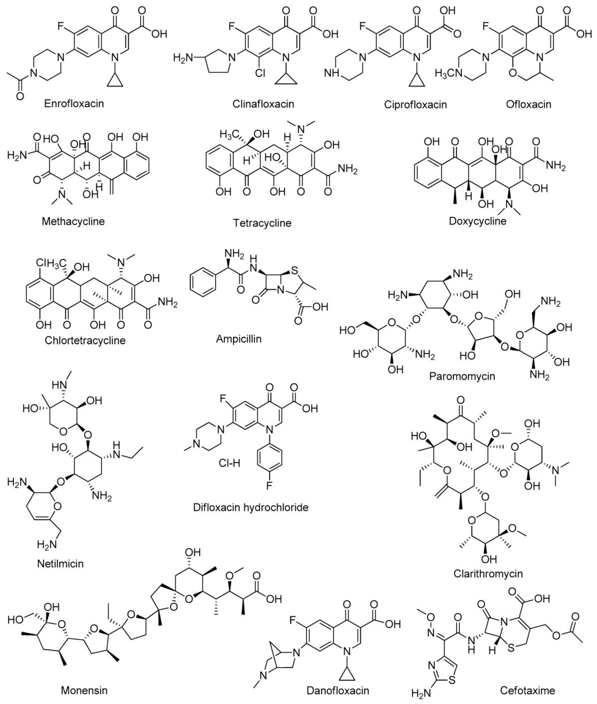
| Fluoroquinolones | Lomefloxacin, Ciprofloxacin, Enroflaxacin, Danofloxacin, Difloxacin hydrochloride, Clinafloxacin | [31,32] |
| Polyethers | Lasalocid, Salinomycin, Monensin, Narasin, Nigericin | [33] |
| Analytes | Amount | Antibiotics | Extraction Methods | Adsorbent | Elution | Separation and Detection | Year | References |
|---|---|---|---|---|---|---|---|---|
| Water, milk, pork, and fish | 1 L, 250 g, 1000 g, and 500 g | Sulfonamide antibiotics | Magnetic SPE | Fe3O4 @MoS2 | Methanol containing 1% ammonium hydroxide | HPLC–MS/MS | 2020 | [48] |
| Water and food | 5g | Quinolone antibiotics | SPE | TAPA-TFPB-COFs | Methanol and water | LC–MS | 2020 | [49] |
| Milk | 5g | Amoxicillin, Ampicillin, and Cloxacillin | Micro-SPE | Starch-based polymer | Methanol | HPLC-UV | 2019 | [50] |
| Pork meat | 1g | Macrolides | SPE | Molecularly imprinted polymer | 10% acetic acid in methanol. | LC–MS/MS | 2018 | [51] |
| Chicken | 5.0 mL | Tetracyclines | SPE | E-spun-GO-PANCMANFs | Ethanol/formic acid/dichloromethane, 40/20/40(v/v/v) | HPLC-FLD | 2019 | [40] |
| Honey and milk | 4 mg and 20 mg | Oxytetracycline, tetracycline, Doxycyclin, Cholrtetracycline, Methacycline | SPE | Multiwalled carbon nanotubes | Methanol and double-distilled water | UPLC-QTOF/MS | 2016 | [52] |
| Honey | 100 mL | Tetracycline, cefotaxime | Micro-SPE | Electrospun graphene oxide doped polyethylene terephthalate nanofibers | Acetonitrile | HPLC-UV | 2018 | [53] |
| Raw milk | 4.0 mL | Tetracyclines, Erythromycin, Chloramphenicol | SPE | Molecularly imprinted polymer | Ethanol, methanol, acetonitrile, and 0.05% ammonium acetate solution | HPLC-ELSD | 2018 | [54] |
| Milk | 1.0 mL | Tetracyclines | SPE | Sep-Pak Vac C18 cartridges | Methanol | LVSS-CE | 2018 | [55] |
| Analytes | Antibiotics | Detection Methods | Extraction Methods | Mobile Phase | Stationary Phase | LOD | Year | Recovery (%) | References |
|---|---|---|---|---|---|---|---|---|---|
| Fish, shrimp, and crab | Sulfonamides | UHPLC‒MS/MS | On-line SPE | (1) 0.1% formic acid in water | C18 pentafluorophenyl (F5 or PFP) column | 0.00146–0.0155 ng/mL | 2020 | 71.5–102 | [38] |
| (2) 0.1%formic acid in ACN | 0.00490–0.0516 ng/mL | ||||||||
| Eggs | Sulfonamides, quinolones, tetracyclines, macrolides, lincosamide, nitrofurans, β-lactams, nitromidazoles, and cloramphenicols | LC–MS/MS | HILIC-SPE | (1)H2O and ACN, | Poroshell 120 EC-C18 column | 0.005−2.00 ng/mL | 2017 | 70.8–116.1 | [68] |
| (2)H2O and ACN containing0.1% formic | 0.015−6.00 ng/mL | ||||||||
| Animal feed | Cyclopolypeptide | HPLC-ELSD | On-line SPE | Methanol and ammonium acetate aqueous solution containing formic acid (B) | Kinetex Biphenyl column | 2–5 µg/mL | 2018 | 72.0–105.4 | [61] |
| Eggs | Sulfonamides, quinolones, pleuromutilins,β- lactams | UHPLC–MS/MS | Dispersive SPE | 0.1% FA and MeOH:ACN,2:8, v/v, containing 0.1% FA | BEH C18 column | 0.1–1 ng/mL | 2021 | 70.5–119.2 | [65] |
| Animal feeds | Aminoglycosides | HPLC-ELSD | Dispersive SPE | Acetonitrile and water | Hypersil BDS C18 | 0.2–0.7 µg/mL | 2017 | 61.2–104.0 | [63] |
| Foods of animal origin | Sulfonamides | HPLC-UV | Centrifugation | Mixtures of acetonitrile, water, formic acid, and ammonium | Inertsil ODS-3 | 6.5–11.0 ng/mL | 2018 | 85–95 | [58] |
| Milk | Sulfonamides | HPLC/UV-DAD | Centrifugation | Acetate buffer solution at pH 4.50 and a mixture of methanol acetonitrile 50:50 (v/v) | C18 column | 2.7–15 ng/mL | 2018 | 55–86 | [57] |
| Analytes | Antibiotics | Extraction Methods | Buffer | pH | Limits of Detection | Voltage (kV) | Recovery (%) | Year | Reference |
|---|---|---|---|---|---|---|---|---|---|
| Milk | Oxytetracycline | SPME | Sodium phosphate + EDTA disodium salt +5% 2-propanol | 12 | 0.07 µg/mL | 14 | - | 2019 | [72] |
| Milk | Tetracyclinesand Quinolones | SPE | Mcllvaine’s buffer | 9.0 | 0.5–2.9 ng/mL | 25 | 72.6–105.8 | 2017 | [73] |
| Milk | Sulfonamides | DSPE | Phosphate buffer | 7.26 | 0.03–0.20 µg/mL | 18 | 60.52–110.91 | 2018 | [75] |
| Milk, pork, and chicken | Sulfonamide | SPE | Sodium borate buffer | 9.5 | 0.65–3.14 µg/mL | 18 | 79.5–112.4 | 2017 | [76] |
| Water | Fluoroquinolones and sulfonamides | DLLME | Borate buffer | 10 | 1.96 ng/mL | 15 | 83.3–98.7 | 2019 | [70] |
| 4.06 ng/mL | |||||||||
| Milk | Macrolides | Ultrasonic and centrifugation | Phosphate+ sodium cholate+ cetyltrimethylammonium bromide | 7.0 | 0.002–0.004 µg/mL | 10 | 72.8–93.7 | 2018 | [77] |
| 2 ng/mL | |||||||||
| 4 ng/mL | |||||||||
| Milk | Ofloxacin | MEP | sodium tetraborate+SDS+ 10% (v/v) methanol | 7.5 | 1.07 ng/mL | 20 | _ | 2019 | [78] |
| Meat | Benzimidazoles | DLLME | Formic acid | 2.2 | >0.003 µg/mL | 20 | 70.1–95.5 | 2017 | [79] |
| Analytes | Antibiotics | Detection | Extraction | IC50 | LOD | Recovery (%) | Year | References |
|---|---|---|---|---|---|---|---|---|
| Milk | Sulfamethazine | ic-ELISA FPIA | Centrifugation and dilution | 6.70 ng/mL 4.76 ng/mL 1.66 ng/mL | ---- | 86.1–131.8 | 2018 | [86] |
| 25.29 ng/mL 23.92 ng/mL 10.60 ng/mL | 81.8–120.2 | |||||||
| Animal-derived food | Gentamicins | FPIA ic-ELISA | PEG precipitation | 7.70 ± 0.6 µg/mL | 0.17 µg/mL | ---- | 2017 | [83] |
| 0.32 ± 0.06 µg/mL | 0.001 µg/mL | |||||||
| Kanamycin | FPIA ic-ELISA | 7.97 ± 0.9 µg/mL | 0.007 µg/mL | |||||
| 0.15 ± 0.01 µg/mL | 0.001 µg/mL | |||||||
| Egg yolk | Gentamicins | ic-ELISA | Centrifugation | 2.69 ng/mL | 0.01 ng/mL | ---- | 2016 | [91] |
| Pork and chicken | Enrofloxacin | FPIA | Centrifugation | 21.49 ng/mL | 1.68 ng/mL | 91.3–112.9 | 2019 | [87] |
| Egg | Tetracyclines | Fluoroimmunoassay | Centrifugation | 3.1–17.2 ng/mL | 0.3–5.8 ng/mL | ---- | 2019 | [88] |
| Goat milk | Clinafloxacin | FPIA | Centrifugation | 29.3 µg/L | 4.1 µg/L | 86.8–104.5 | 2015 | [89] |
| Egg, honey, and pork | Sulfamethazine | Immunochromatographic assay | Centrifugation | ---- | 0.016 ng/mL | 90.5–113.9 | 2020 | [85] |
| 0.049 ng/mL | 82.4–112.0 | |||||||
| 0.029 ng/mL | 79.8–93.4 | |||||||
| Chicken muscle | Fluoroquinolones | ic-ELISA Lateral Flow Test Strip | Centrifugation | 0.2 ng/mL | 0.082 ng/mL | ---- | 2017 | [90] |
| Analytes | SERs Activenanomaterial | Target Antibiotics | Extraction | LOD | Recoveries (%) | Year | References |
|---|---|---|---|---|---|---|---|
| Milk | Ag–AgNPs | Penicillin G | Centrifugation and Acetonitrile extraction | 0.85 µg/kg | 76–97 | 2017 | [113] |
| Fish | Gold nanofilm | Ciprofloxacin | Centrifugation and SPE | 0.19 µg/mL | 84.6–103.8 | 2019 | [95] |
| Duck | AuNPs | Amoxicillin | Centrifugation | 0.2 mg/L | 96–139 | 2017 | [97] |
| Milk | Au@AgNPs | Tetracyclines Penicillins | -- | 0.015 ng/mL 0.010 ng/mL | 88.8–111.3 | 2020 | [104] |
| Water | Ag–TiO2 | Danofloxacin | Centrifugation | 3.16 × 10⁻¹¹ mol/L | >80.8 | 2019 | [109] |
| Animal tissue mimics | RGO/Ag | Sulfonamide Sulfamethoxazole | -- | 1.9 ng/mL 4.4 ng/mL | ---- | 2018 | [106] |
| Chicken and water | Au–Ag composites | Ciprofloxacin | Centrifugation | 2 × 10⁻⁷ M 8 × 10⁻⁸ M | 91–105 | 2017 | [114] |
| Biosensor | Detection Method | Target Antibiotics | Bioreceptor | Sample | LOD | Year | References |
|---|---|---|---|---|---|---|---|
| Electrochemical biosensor | Amperometry | Sulfapyridine | Antibody | Milk | 2.4 ng/mL | 2018 | [133] |
| Electrochemical impedance spectroscopy (EIS) | Tetracyclines | Antibody | Water | 12.4 ng/mL | 2020 | [134] | |
| Differential pulse voltammetry (DPV) | Ampicillin | Aptamer | Milk | 3.8 × 10⁻⁴ ng/mL | 2015 | [135] | |
| Amperometry | Chloramphenicol | Antibody | Pork, chicken, beef | 0.045 ng/mL | 2016 | [136] | |
| Optical biosensor | Colorimetric | Tobramycin | Aptamer | Milk, chicken, egg | 10.89 ng/mL | 2018 | [122] |
| Chemi- luminescent (CL) | Sulfamethazine | Aptamer | Milk | 0.92 ng/mL | 2019 | [127] | |
| Surface plasmon resonance (SPR) | Enrofloxacin | Antibody | Milk | 0.07 ng/mL | 2017 | [137] | |
| Mass sensitive biosensors | Piezoelectric quartz crystal microbalance (QCM) | Streptomycin | Antibody | Milk | 0.3 ng/mL | 2015 | [138] |
| Piezoelectric surface acoustic wave (SAW) | Penicillin G | Antibody | Milk | 2.2 ng/mL | 2016 | [139] | |
| Cantilever | Oxytetracycline | Aptamer | Meat, egg | 0.85 ng/mL | 2015 | [140] |
Publisher’s Note: MDPI stays neutral with regard to jurisdictional claims in published maps and institutional affiliations. |
© 2021 by the authors. Licensee MDPI, Basel, Switzerland. This article is an open access article distributed under the terms and conditions of the Creative Commons Attribution (CC BY) license (https://creativecommons.org/licenses/by/4.0/).
Share and Cite
Dawadi, S.; Thapa, R.; Modi, B.; Bhandari, S.; Timilsina, A.P.; Yadav, R.P.; Aryal, B.; Gautam, S.; Sharma, P.; Thapa, B.B.; et al. Technological Advancements for the Detection of Antibiotics in Food Products. Processes 2021, 9, 1500. https://doi.org/10.3390/pr9091500
Dawadi S, Thapa R, Modi B, Bhandari S, Timilsina AP, Yadav RP, Aryal B, Gautam S, Sharma P, Thapa BB, et al. Technological Advancements for the Detection of Antibiotics in Food Products. Processes. 2021; 9(9):1500. https://doi.org/10.3390/pr9091500
Chicago/Turabian StyleDawadi, Sonika, Ranjita Thapa, Bindu Modi, Sobika Bhandari, Arjun Prasad Timilsina, Ram Prabodh Yadav, Babita Aryal, Sijan Gautam, Purnima Sharma, Bijaya Bahadur Thapa, and et al. 2021. "Technological Advancements for the Detection of Antibiotics in Food Products" Processes 9, no. 9: 1500. https://doi.org/10.3390/pr9091500
APA StyleDawadi, S., Thapa, R., Modi, B., Bhandari, S., Timilsina, A. P., Yadav, R. P., Aryal, B., Gautam, S., Sharma, P., Thapa, B. B., Aryal, N., Aryal, S., Regmi, B. P., & Parajuli, N. (2021). Technological Advancements for the Detection of Antibiotics in Food Products. Processes, 9(9), 1500. https://doi.org/10.3390/pr9091500