Review

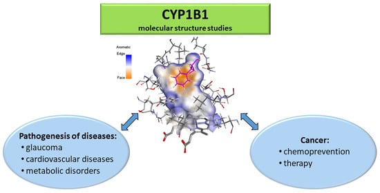
New Perspectives of CYP1B1 Inhibitors in the Light of Molecular Studies

by
Renata Mikstacka
1,* and
Zbigniew Dutkiewicz
2,*
1
Department of Inorganic and Analytical Chemistry, Collegium Medicum, Nicolaus Copernicus University in Toruń, Dr A. Jurasza 2, 85-089 Bydgoszcz, Poland
2
Department of Chemical Technology of Drugs, Poznań University of Medical Sciences, Grunwaldzka 6, 60-780 Poznań, Poland
*
Authors to whom correspondence should be addressed.
Processes 2021, 9(5), 817; https://doi.org/10.3390/pr9050817
Submission received: 28 March 2021 / Revised: 27 April 2021 / Accepted: 3 May 2021 / Published: 8 May 2021

Abstract

:
Human cytochrome P450 1B1 (CYP1B1) is an extrahepatic heme-containing monooxygenase. CYP1B1 contributes to the oxidative metabolism of xenobiotics, drugs, and endogenous substrates like melatonin, fatty acids, steroid hormones, and retinoids, which are involved in diverse critical cellular functions. CYP1B1 plays an important role in the pathogenesis of cardiovascular diseases, hormone-related cancers and is responsible for anti-cancer drug resistance. Inhibition of CYP1B1 activity is considered as an approach in cancer chemoprevention and cancer chemotherapy. CYP1B1 can activate anti-cancer prodrugs in tumor cells which display overexpression of CYP1B1 in comparison to normal cells. CYP1B1 involvement in carcinogenesis and cancer progression encourages investigation of CYP1B1 interactions with its ligands: substrates and inhibitors. Computational methods, with a simulation of molecular dynamics (MD), allow the observation of molecular interactions at the binding site of CYP1B1, which are essential in relation to the enzyme’s functions.

Graphical Abstract

1. Introduction

Cytochrome P450 enzymes (CYPs) are hemoproteins representing a superfamily of monooxygenases responsible for the metabolism of endogenous and exogenous substrates. Human cytochrome P450 family 1 (CYP1) consists of three isoforms: CYP1A1, CYP1A2, and CYP1B1. The family 1 member CYP1B1 is an extrahepatic enzyme expressed in hormone-dependent tissues like the breast, prostate, ovaries, pancreas, and testis [1]. CYP1B1 catalyzes oxidation and N- or O-dealkylation of xenobiotics including procarcinogens and environmental pollutants [2], and endogenous substrates such as fatty acids and steroids.
CYP1B1 plays an important role in the pathogenesis of hormone-induced cancers, being responsible for the metabolism of 17-alpha-estradiol (E2) to highly mutagenic and carcinogenic 4-hydroxy-E2 [3,4]. CYP1B1 overexpressed in cancer cells is partly responsible for the resistance to anti-cancer drugs such as tamoxifen, paclitaxel and docetaxel, which are inactivated by this enzyme [5,6,7]. CYP1B1 is a tumor biomarker because of its overexpression in tumor cells derived from among others: breast, colon, lung skin, brain, and testis [8]. Therefore, the inhibition of CYP1B1 activity is considered a therapeutic target for cancer chemoprevention and chemotherapy. On the other hand, the constitutively overexpressed CYP1B1 in cancer cells and the tumor microenvironment may be used to design anti-cancer prodrugs activated by this enzyme. Another pathway of CYP1B1 studies concerns the relation between mutations of the CYP1B1 gene and the occurrence of eye diseases [9,10] and cardiovascular disorders [11] (Figure 1).
CYP1s are induced by agonists of the aryl hydrocarbon receptor (AhR), including polychlorinated dioxins with strong inducer 2,3,7,8-tetrachlorodibenzo-p-dioxin (TCDD) and polycyclic aromatic hydrocarbons [12,13]. The role of AhR as a transcription factor is not limited to the metabolism of xenobiotics. The AhR transduction pathway is also activated by endogenous compounds, which include the products of metabolism of polyunsaturated fatty acids and heme, and by endobiotics—ligands which are produced in the gastrointestinal tract and skin [14,15].
For a decade, epigenetic factors affecting CYP1B1 gene expression were studied [16]. Methylation of cytosine residues of genomic DNA of the regulatory region(s) of a gene can lead to silencing or diminished expression of the gene. Studies of two cell lines: MCF7 and HepB2, indicated that DNA methylation diminished the expression of the CYP1B1 gene in HepB2 cells [17]. Differential regulation of the expression of CYP1B1 and CYP1B1 genes coding two isoforms of CYP1 was observed in mouse hepatoma and fibroblast cell lines induced by TCDD [18]. In gastric and colorectal cancers, DNA methylation in the promotor region of CYP1B1 was shown [19,20].
This review presents the multiple pharmacological roles of CYP1B1 in a human organism, emphasizing the latest achievements, focusing on the computational studies of the molecular interactions of CYP1B1 with its substrates and inhibitors, explaining CYP1B1 multiple functions at the molecular level.

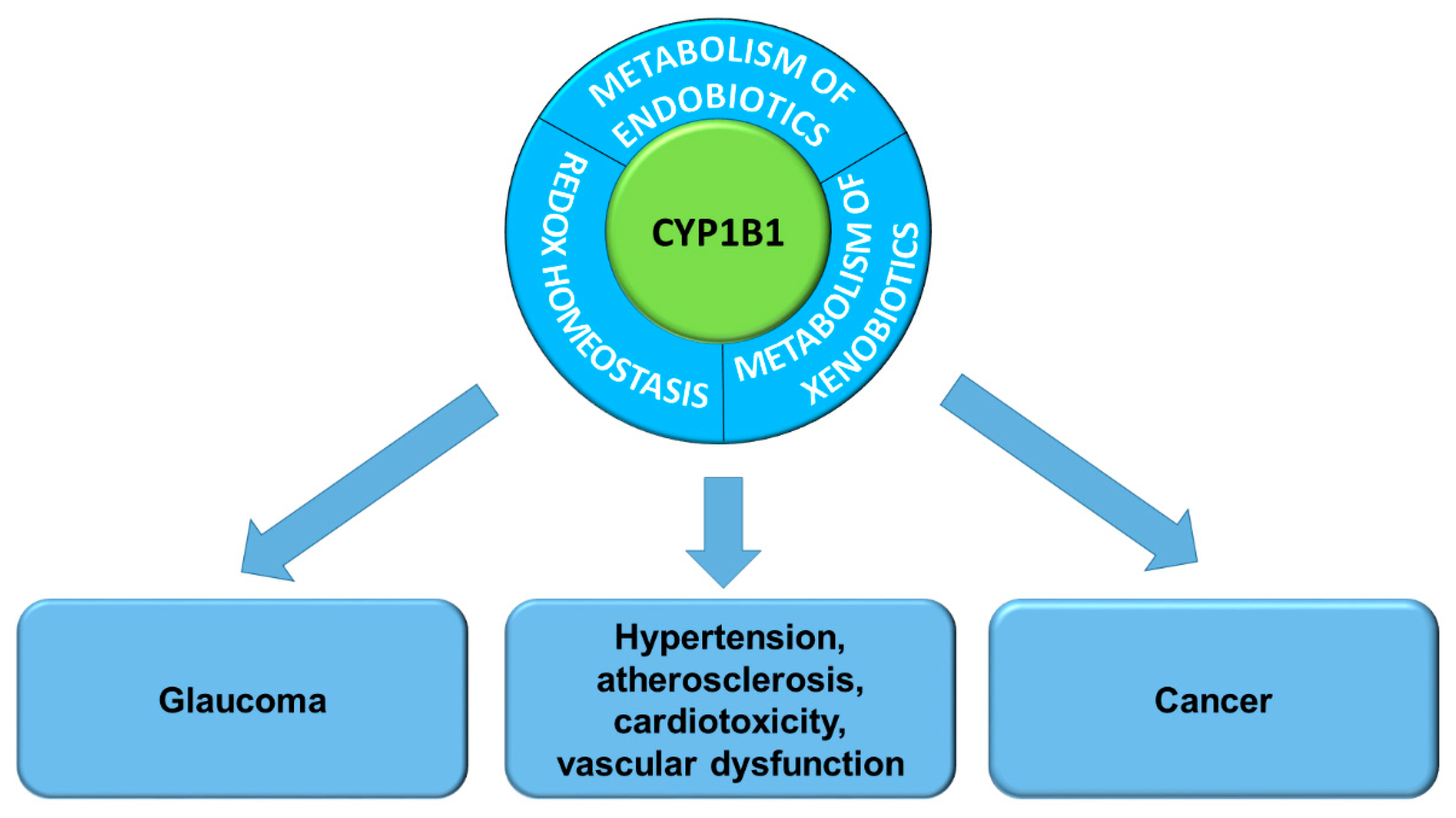
2. The Role of CYP1B1 in the Pathogenesis of Diseases

CYP450 family 1 isozymes were supposed to play an essential role in cancer initiation by transforming procarcinogens into ultimate carcinogens, which react with DNA and form DNA adducts. Recent studies indicate that CYP1B1 contributes to cancer initiation, and to a significant extent, to the following stages of carcinogenesis: promotion and progression, while the primary role of CYP1A1 and CYP1A2 is detoxification of xenobiotics leading to activation of procarcinogens.
In the Human Gene Mutation Database (http://www.hgmd.cf.ac.uk; accessed on 6 May 2021), 240 variants of the CYP1B1 gene are listed. The association of their function with the pathogenesis of diseases in humans has been recently intensively studied [21]. Molecular structures of pathogenic CYP1B1 mutant variants are analyzed in relation to their impaired function with the use of the most advanced bioinformatics tools.

2.1. CYP1B1 Contribution in Pathogenesis of Eye Diseases

Glaucoma is a group of eye diseases leading to the irreversible loss of vision. In the development of glaucoma, the CYP1B1 gene is involved: primary congenital glaucoma (PCG) constitutes 55% of inherited pediatric onset of the disease and is caused predominantly by mutations in the CYP1B1 gene [21].
The studies of CYP1B1 variants and their effect on the risk of eye diseases have been carried out—such studies for PCG and primary open angle glaucoma (POAG) have been carried out since the late nineties (see Section 3.4). In 2006, molecular structures for the eight CYP1B1 mutations occurring only in PCG patients were obtained by comparative modeling and were subjected to MD simulations (30 ns). The impact of studied mutations on the enzyme structure and function was evident [22].
In order to explain the molecular basis of the variable phenotypes resulting from the CYP1B1 gene mutations, the subclones of twenty-three CYP1B1 missense variants occurring in glaucoma patients were examined by measuring in a cell system the dual activity of the enzyme to metabolize both retinol and 17β-estradiol. Most variants linked to POAG showed low steroid metabolism, while null or very high retinol metabolism was observed in variants identified in PCG [23]. In the Pakistani population, identification of five known and three novel variants of CYP1B1 in 14 families with PCG was reported. However, in 22 of the 36 families analyzed (61%), pathogenic alleles were not found [24].
Meta-analysis of seven data sets of Chinese PCG patients revealed L107V and R390H as the most common CYP1B1 mutations, which are localized in close proximity to the active site cavity and hemoglobin binding region. Long-term MD simulations (100 ns) showed the destabilization of the mutant proteins caused the change of the molecular interaction between the enzyme and the substrate [25]. For better reliability of in silico predictions, the authors performed sequence conservation analysis, hydrogen bond network analysis, electrostatic potential analysis, and MD simulation, which all confirmed the impact of CYP1B1 mutations on the function of the enzyme [25].

2.2. Redox Homeostasis

The function of CYP1B1 depends on the oxygen supply. Deletion of CYP1B1 gene is associated with increased oxidative stress in the retinal vascular and trabecular meshwork (TM) cells in culture and retinal and TM tissue in vivo [26]. In the absence of Cyp1b1 in the hepatic sinusoidal endothelial cells prepared from Cyp1b1−/− mice, a low level of hepcidin and decreased production of the bone morphogenetic protein (BMP6) were observed [27,28].
Hepcidin is a hormone that regulates the intracellular level of iron by the interaction with the main iron transporter—ferroportin. A low level of hepcidin is associated with the increased iron level and, as a result, increased intracellular oxidative stress. The BMP6 is a critical endogenous regulator of iron metabolism; it activates hepcidin expression and reduces serum iron in mice [29]. Lack of the BMP6 leads to a high intracellular iron level [30].
The role of CYP1B1 in hyperoxia cytotoxicity in human lung endothelial cells in vitro was studied [31]. Human cells overexpressing CYP1B1 were more susceptible to oxygen toxicity, while the use of CYP1B1-siRNA attenuated the toxic effect of hyperoxia. In immortalized lung endothelial cells derived from Cyp1b1-null and wild-type mice, Cyp1b1 expression promoted apoptosis induced by oxidative stress, while in cells derived from Cyp1b1-null mice, hyperoxia cytotoxicity was significantly diminished [31]. In studies in vivo, Cyp1b1−/− mice lacking the cytochrome P450 1B1 gene were less susceptible to hyperoxic lung injury than C57BL/6 wild type mice [32].
The effect of transcription factor AhR activation on redox homeostasis in basal-like and BRCA1-related breast cancer was reported [33]. In a mouse model of BRCA1-related breast cancer, the levels of reactive oxygen species (ROS) correlated with the expression and activity of the AhR. Chemical or genetic inhibition of the AhR is proposed for maintaining cellular redox homeostasis, modulation of the tumor promoting the microenvironment, and overcoming the resistance of human breast cancer cells to erlotinib used in anti-cancer therapy.

2.3. CYP1B1 in Cardiovascular Diseases

In the last decade, the implication of CYP1B1 in cardiometabolic diseases has been recognized and intensively studied [34]. CYP1B1 is responsible for the cardiovascular side effects of anti-cancer treatment. Cardiovascular disease is the second cause of mortality in the group of cancer survivors, after secondary malignancies. Inhibition of the CYP1B1 can be a promising therapeutic strategy that has the potential to prevent firstly: chemo- and radio-resistance to cancer therapy and secondly, treatment-induced cardiovascular complications without reducing their anti-cancer effects [11]. Most chemotherapeutic agents like doxorubicin, dasatinib, sunitinib, and docetaxel induce CYP1B1 gene expression in cardiac-derived cells in vitro and in animal models in vivo [11], leading to the resistance to cancer chemotherapy. Increased CYP1B1 expression is associated with cardiac hypertrophy [35,36,37,38], hypertension [39,40,41,42,43], atherosclerosis [44], cardiotoxicity, and vascular dysfunction. 6β-Hydroxytestosterone, a metabolite of testosterone generated by CYP1B1, contributes to vascular changes in angiotensin II-induced hypertension in male mice [45].
Efficacy of CYP1B1 inhibition in protection against chemotherapy-induced cardiotoxicity and in the prevention of chemo- and radio-resistance has been demonstrated in numerous preclinical studies. Phytochemicals such as quercetin, apigenin, and other polyphenols exhibit protective activity against cancer chemotherapeutics’ side effects [11]. However, the activity of these agents is directed to many other targets and is also responsible for drug interactions. Their antioxidant activity may impair ROS-dependent cancer therapy.
2,4,3′,5′-tetramethoxy-trans-stilbene (TMS), a synthetic derivative of resveratrol as a selective CYP1B1 inhibitor protected from chronic DOX-induced cardiotoxicity in rats in vivo and RL-1 cardiomyocyte-like cells in vitro through a mid-chain hydroxyeicosatetraenoic acid (HETE)-dependent mechanism. Overexpression of CYP1B1 significantly induced cellular hypertrophy and the production of mid-chain HETE metabolites. At the same time, inhibition of CYP1B1 with the use of 2,4,3′,4′-tetramethoxy-trans-stilbene (TMS) or CYP1B1-siRNA significantly attenuated isoproterenol-induced hypertrophy [37].

2.4. Metabolic Diseases

CYP1B1 is considered a therapeutic target for the treatment of metabolic diseases [34]. CYP1B1 metabolizes endogenous substrates, including steroid hormones, fatty acids, melatonin, vitamins, and retinoids. The disorders of endogenous metabolism lead to metabolic diseases that may result in obesity, atherosclerosis, hypertension, and cancer. In the studies on Cyp1b1-knockout mice, CYP1B1 deficiency reduced obesity induced by high fat diet—when compared with their wild-type counterparts, and improved glucose tolerance [46]. Cyp1b1 deletion caused suppression of peroxisome proliferator-activated receptor γ (PPAR γ) and the genes regulated by PPARγ [47]. PPARs are nuclear receptors involved in the homeostasis of lipids and glucose. To summarize, modulation of CYP1B1 activity attenuates the processes that lead to obesity, hypertension, and atherosclerosis. CYP1B1 is expressed in vascular smooth muscle cells (VSMCs) and, to a lesser extent, in endothelial cells [48] and is involved in the development of hypertension via the regulation of arachidonic acid metabolism. Inhibition of CYP1B1 activity by TMS resulted in decreased ROS production and extracellular signal-regulated kinase ½ and p38 mitogen-activated protein kinase activity and significantly alleviated hypertension [49].

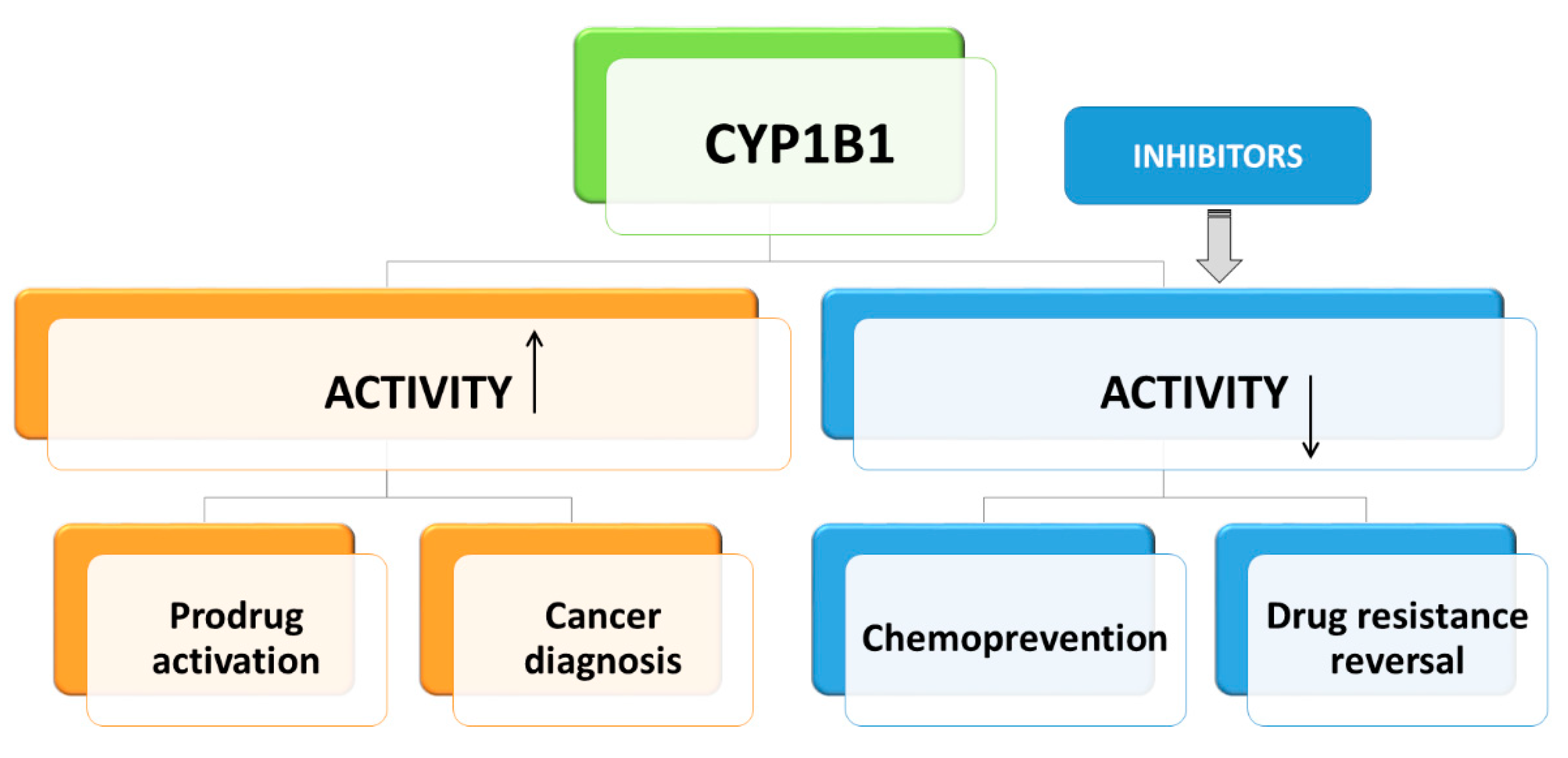
2.5. The Role of CYP1B1 in Cancer Chemoprevention and Therapy

Regarding its functions in cellular metabolism, CYP1B1 is considered as a target in cancer chemoprevention and therapy (Figure 2). Cancer chemoprevention is the strategy to prevent or delay the initiation, promotion, or progression of cancer disease with the use of natural compounds or their synthetic derivatives. Progress of cancer depends on the activation of pathways leading to inflammation and angiogenesis [50,51,52]. Many phytochemicals exert anti-inflammatory and anti-angiogenic activities [53,54,55]. Most of these compounds, like flavonoids, furanocoumarins, chalcones, stilbenoids, anthraquinones, and alkaloids, are also potent and specific inhibitors of CYP1B1 activity [56,57]. Thus, the association of CYP1B1 with the inflammatory process and angiogenesis is suggested. Therefore, the use of CYP1B1 inhibitors targeting both cancer cells and their microenvironment, including neutrophils, macrophages, lymphocytes, and fibroblasts, is supposed to be a promising strategy in chemoprevention [58]. CYP1B1 may contribute to the cytotoxicity of natural compounds against cancer cells and cancer stem cells resistant to chemotherapy [59,60].
The mechanism of CYP1B1 inhibition is analyzed with the use of computational methods to obtain safe and more efficient chemopreventive agents [61]. Since the 1990s, natural stilbenoids, trans-resveratrol (RESV), pterostilbene, piceatannol, and synthetic RESV analogs have been extensively studied in relation to chemoprevention [62] and with their interaction with CYP1s. Synthetic analogs of resveratrol were screened for their inhibitory activity towards cytochrome P450 family 1. TMS synthesized two decades ago is still supposed to be one of the most potent inhibitors of CYP1B1 activity. CYP1B1′s interactions with inhibitors and their effect on conformational changes in the enzyme structure were studied with a series of 3,4-dimethoxy-trans-stilbene derivatives docked to the crystal structure of human CYP1B1 [63].
One of the most critical problems in cancer therapy is overcoming drug resistance. Flutamide is an example of an anti-cancer chemotherapeutics metabolized by CYP1B1 to a 2-hydroxy derivative that does not exert anti-cancer activity [6]. More recently, the properties of flutamide as an agonist of AhR were shown [64]. Increased expression of CYP1B1 results in decreased flutamide efficacy as an anti-cancer agent. CYP1B1 can be a potential tumor biomarker and a target for anti-cancer therapy in renal cell carcinoma (RCC). Expression of CYP1B1 was determined in RCC cell lines: Caki-1 and 769-P, and tissue microarrays of 96 RCC and 25 normal tissues. The gene CYP1B1 in Caki-1 and 769-P cells was silenced by RNA interference, and functional analyses were performed to determine the biological significance of CYP1B1 in RCC progression. CYP1B1 promoted RCC development by inducing the cell division cycle 20 homolog (CDC20) expression and inhibiting apoptosis through the down-regulation of the death-associated protein kinase-1 (DAPK1) [65].
CYP1B1 is overexpressed in primary and metastatic loci of epithelial ovarian cancers. Treatment with α-naphthoflavone reduces paclitaxel resistance and enhances the sensitivity of ovary cells to paclitaxel in vitro and in the xenograft model of nude mice [7]. A new highly selective and extremely potent α-naphthoflavone derivative (IC50 = 0.043 nM) was synthesized. It overcomes docetaxel-resistance caused by the overexpressed CYP1B1 in MCF-7/1B1 cells [66].
To summarize, co-administration of the anti-cancer agents and CYP1B1 inhibitors is suggested to decrease drug resistance and ameliorate the outcome of anti-cancer therapy.
Investigation of the molecular mechanism of CYP1B1 contribution in oncogenesis revealed that CYP1B1 promotes cell proliferation and metastasis by inducing epithelial-mesenchymal transition (EMT) and Wnt/β-catenin signaling via Sp1 (a transcription factor involved in cell growth and metastasis) induction [67]. CYP1B1 induces the development and progression of prostate cancer cells by modulating caspase 1 (CASP1) expression. An inverse correlation between CYP1B1 and CASP1 in human prostate cancer samples was shown. In vivo lentivirus-delivered short hairpin (sh) RNA specific for CYP1B1 was used to reduce the tumor progression in the xenograft mouse model. The results support the idea that attenuation of CYP1B1 may be effective in the treatment of prostate cancer [68].
Regarding the high level of CYP1B1 activity in cancer cells, the design of prodrugs that could be activated by CYP1B1 directly in a tumor is still a developing area of study. In vitro models were proposed to screen CYP1B1- targeted anti-cancer prodrugs. In KLE cells (human endometrial carcinoma cell line), which overexpress CYP1B1 activity, the increased cytotoxicity of studied prodrugs was observed in comparison to cells with CYP1B1 inhibited by αnaphthoflavone (ANF) that is a potent inhibitor of CYP1 isozymes [69]. Recently discovered DMAKO-20 (5,8-dimethyl alkannin oxime derivative) is an anti-cancer prodrug activated by CYP1B1, cytotoxic to cancer cells in vitro and in vivo, while it is nontoxic to the human normal cells [70].
A new approach in tumor diagnosis is based on CYP1B1 expressed in tumor cells. The derivative of ANF, 3-hydroxy-3′-fluoro-6,7,10-trimethoxyANF, was earlier characterized as a very potent CYP1B1 inhibitor with IC50 equal to 0.043 nM [66]. This compound modified with a linker still potently inhibited CYP1B1 with IC50 equal to 4 nm. Near-infrared fluorescent dye was attached to ANF derivative and used as CYP1B1 targeted imaging probes in HCT-15-tumor bearing mice [71]. This non-invasive method of imaging cancer cells is a promising approach in tumor diagnosis.

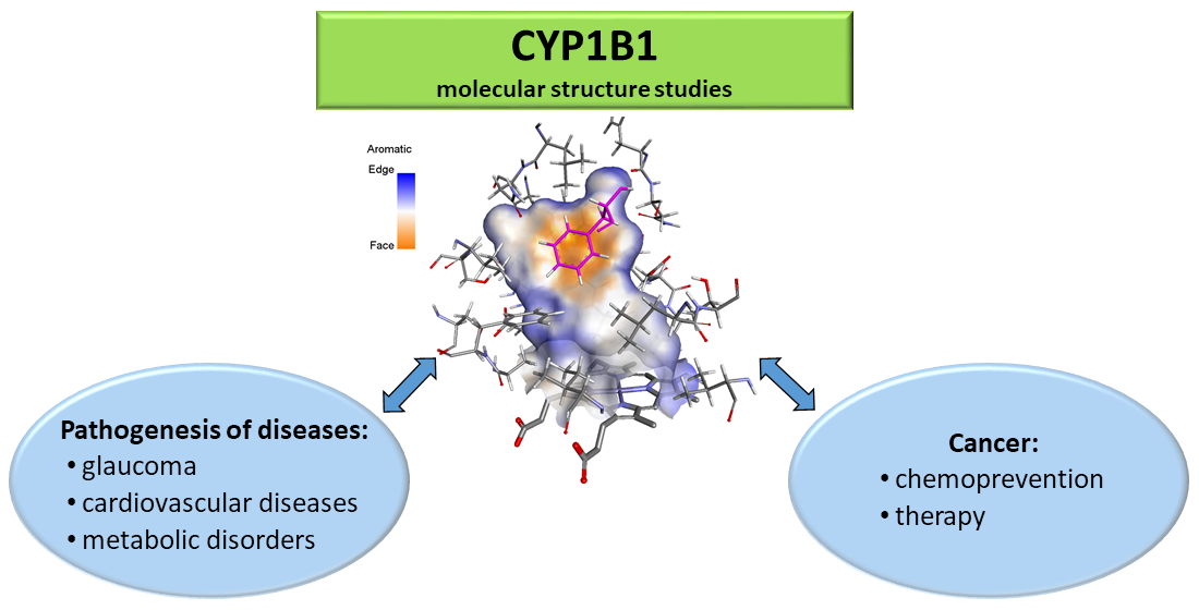
3. CYP1B1 Molecular Structure Studies

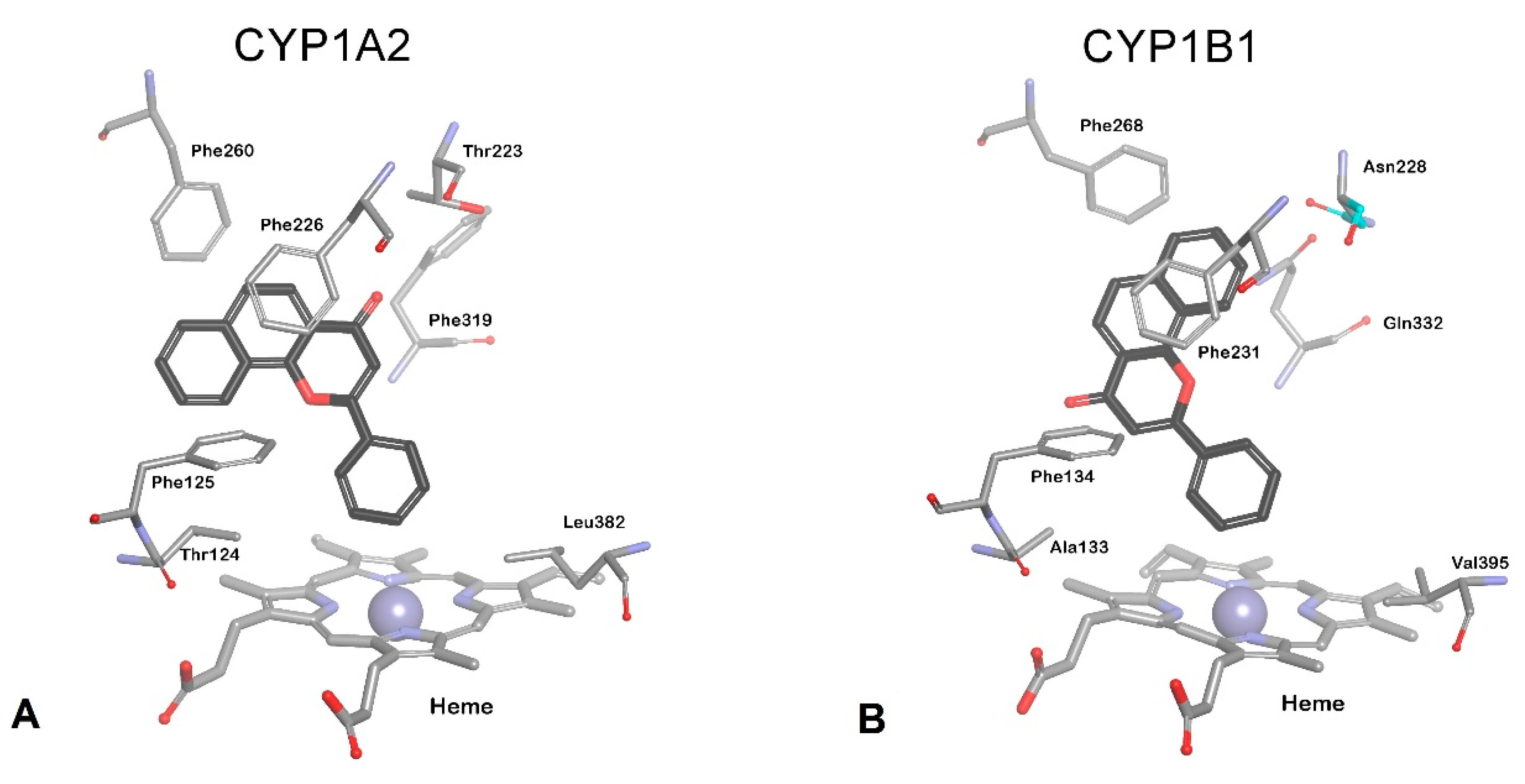
3.1. Family CYP1 Crystal Structures

The insight into molecular structures of CYP1 isozymes provides much valuable information due to the development of computational methods: molecular docking and molecular dynamics simulations. All members of the family CYP1 have flat and planar active sites. CYP1B1 protein consists of 543 amino acids and has a slightly smaller active site than CYP1A1. It has low amino acid sequence identity with CYP1A1 (38%) and CYP1A2 (37%), but it exhibits similar substrate specificity [72,73,74]. Until now, a single molecular structure of human CYP1A2, four human CYP1A1 structures, and two human CYP1B1 structures differing in the bound ligands are available. Recently, two new structures of CYP1B1 were deposited in Protein Data Base (unpublished data). Besides, two ancestral CYP1B1 structures were generated and characterized (Table 1).
According to the size and shape of the binding site cavity, relatively small and planar molecules as polycyclic aromatic hydrocarbons (PAH), stilbenes, flavonoids, and anthraquinones are substrates/inhibitors of CYPs family1 [80,81]. Structure-based drug design requires detailed knowledge of active-site interactions with potential ligands. In studies of substrate-enzyme interactions, computational methods, particularly long-term molecular dynamics simulations, that identify the subtle changes of molecular conformations provided the elucidation of how non-planar substrates might be accommodated in small CYP’s binding sites. Currently used bioinformatics tools are briefly discussed in Section 3.5. The studies on CYP1A1 molecular structures revealed that the accommodation of the substrates with higher molecular weight and non-planar structure in the enzyme binding site is possible due to conformational changes of the side chains in the enzyme cavity [75,76].
Crystallographic CYP1A1 structure with bound ANF (PDB: 4I8V) was characterized in 2013 by Walsh and coworkers [74]. For CYP1A1, two new molecular structures were solved with a furanocoumarin bergamottin (PDB: 6DWM) and tyrosine kinase inhibitor erlotinib (PDB: 6DWN) as ligands [75]. All three structures show a small planar active site. In the case of the crystallographic structure of CYP1A1 with the non-planar substrate, Pim kinase inhibitor GDC-0339 (PDB: 6O5Y) molecular docking revealed the significant changes of shape and size of CYP1A1 cavity as a result of interactions with the ligand [76]. The molecular CYP1A1 structure was controlled by residues in the active site roof with major changes in the conformation of the F helix break and relocation of Phe224 from the active site to the protein surface [76]. The structural rearrangements were observed in the CYP1A1 binding site when diverse ligands were cocrystallized with the enzyme [76]. Molecular dynamics allow discerning the conformational changes in the CYP1A1 cavity, which facilitate the accommodation of the compounds of the studied series of stilbene derivatives in the binding site. These changes included the repositioning of side chains of amino acids located in the roof of the CYP1A1 active site [76].

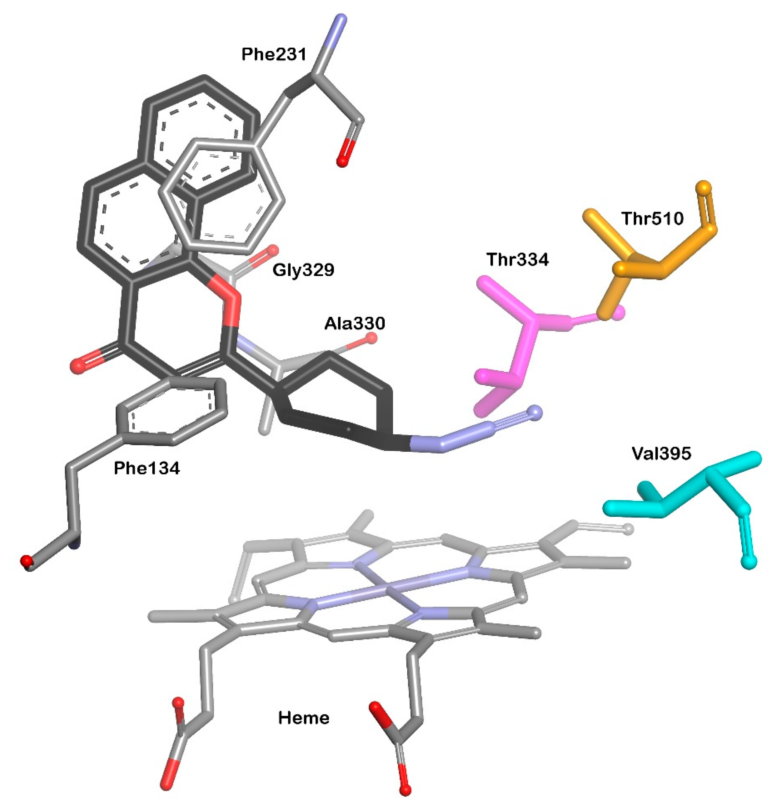
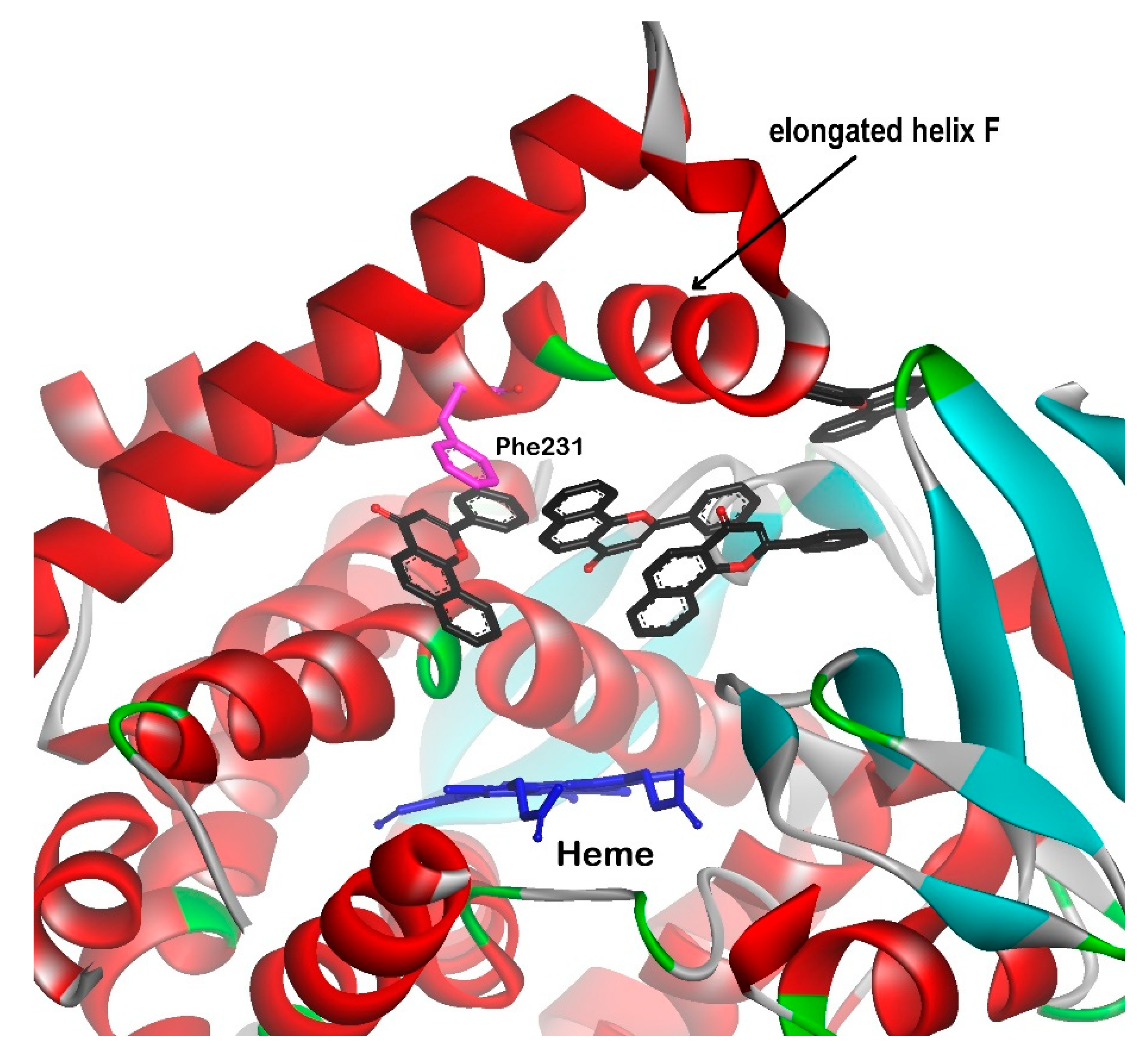
3.2. CYP1B1 Crystal Structure Studies

Since 2011, a single crystal structure of CYP1B1 with ANF as the bound ligand (PDB id: 3PM0) determined by X-ray crystallography to 2.7 resolution Å was available [73]. Previously, the interactions of natural and synthetic inhibitors with CYP1B1 catalytic site were estimated using CYP1A2 based homologous structures [61]. Both enzyme structures are generated with ANF as a ligand; however, ANF binds in a different orientation in P450 1B1 from that observed for 1A2 (Figure 3). A distortion of helix F places Phe231 in CYP1B1 and Phe226 in CYP1A2 in similar positions for π-π stacking with ANF. Hydrophobic interactions between ANF and amino acid residues are the predominant interactions that contribute to binding affinity.
More recently, the new structure of CYP1B1 with inhibitor having azide group as a ligand (PDB id: 6IQ5) was crystallized [78]. In this research, ANF was used as a lead compound to design efficient CYP1B1 inhibitors with better solubility and inhibitory potency. Synthesized benzo[h]chromone with linked cyclohexanyl B-ring exerted a strong CYP1B1 inhibitory activity with potency more than 2-times stronger than that of ANF. Moreover, the introduction of azide substituent caused a 120-fold increase of selectivity against CYP1B1 compared to CYP1A1. X ray crystal structure allowed to confirm the location of the inhibitor molecule in the CYP1B1 cavity, while the positioning of terminal nitrogen atom could not be determined due to the low resolution. The authors performed docking analysis using Surflex Dock program on Sybyl X2 software and found that the azide group was located in the hydrophobic pocket constructed by Thr334, Val395, and Thr510. The docking results exhibited the role of hydrophobic interactions affecting the binding affinity and inhibitory potency of the ANF derivative (Figure 4).
The ability of human P450s to catalyze a plethora of chemical reactions makes these enzymes particularly valuable for the commercial biosynthesis of important molecules. However, the use of cytochromes P450 in biotechnology for the synthesis of chemicals, including final drugs and drug metabolites, requires higher thermostability than that of human cytochromes. The ancestral mammalian cytochrome P450 subfamily 1B (N98_CYP1B1_Mammal) was reconstructed to investigate its thermostability, substrate selectivity, and metabolism of representative substrates. The structure of ancestral CYP1B1 was determined with ANF and 17-estradiol in the binding site [79]. Comparing the human CYP1B1 (PDB id: 3PM0) and N98_CYP1B1_Mammal (PDB id: 6OYU) complexed with ANF enabled the identification of the similarities and differences in their structures. First of all, predicted ancestral CYP1B1 has maintained a canonical cytochrome P450 fold, and the sequence differences among the variants were found far from the active site. The difference that was supposed to have critical implication to the enzyme catalytic activity concerned the F helix exhibiting characteristic break in the CYP1 isozymes. In the ancestor of CYP1B1, this break is not present; instead, the F helix is extended by eight residues comparing with the human CYP1B1. The elongation of the F helix results in the appearance of a wide channel from the active site to the protein surface, what in turn leads to the binding as many as four ANF molecules (Figure 5). This open conformation of the enzyme is also preserved in the structure of the ancestral CYP1B1 with only one molecule of 17-estradiol as a ligand. It is assumed that electrostatic and aromatic interactions between distinct secondary structure elements in the ancestral form contribute to its higher thermostability than that of the extant human CYP1B1 [79].
Crystallography and molecular dynamics (MD) simulation studies on the cytochrome P450 structures of different isozymes demonstrated the importance of gating amino acid residues associated with the opening and closing of the ligand tunnels. Lipophilicity of a ligand is another factor influencing its penetration into the enzyme active site. Cytochromes P450 are membrane bound enzymes, and the ligand can enter the active site from the lipophilic membrane and the aqueous environment. MD simulations allow the discovery of multiple ligand tunnels in cytochrome crystal structures. CYP1B1 structure (3PM0), has the most enzyme crystal structures available in the Protein Data Base, was generated in the closed conformation. The opening and closing motions of most ligand tunnels are associated with conformational changes of the flexible B–C and F–G blocks of the cytochromes. Understanding the gating mechanism can help analyze the catalytic mechanism and substrate selectivity of CYPs [82].

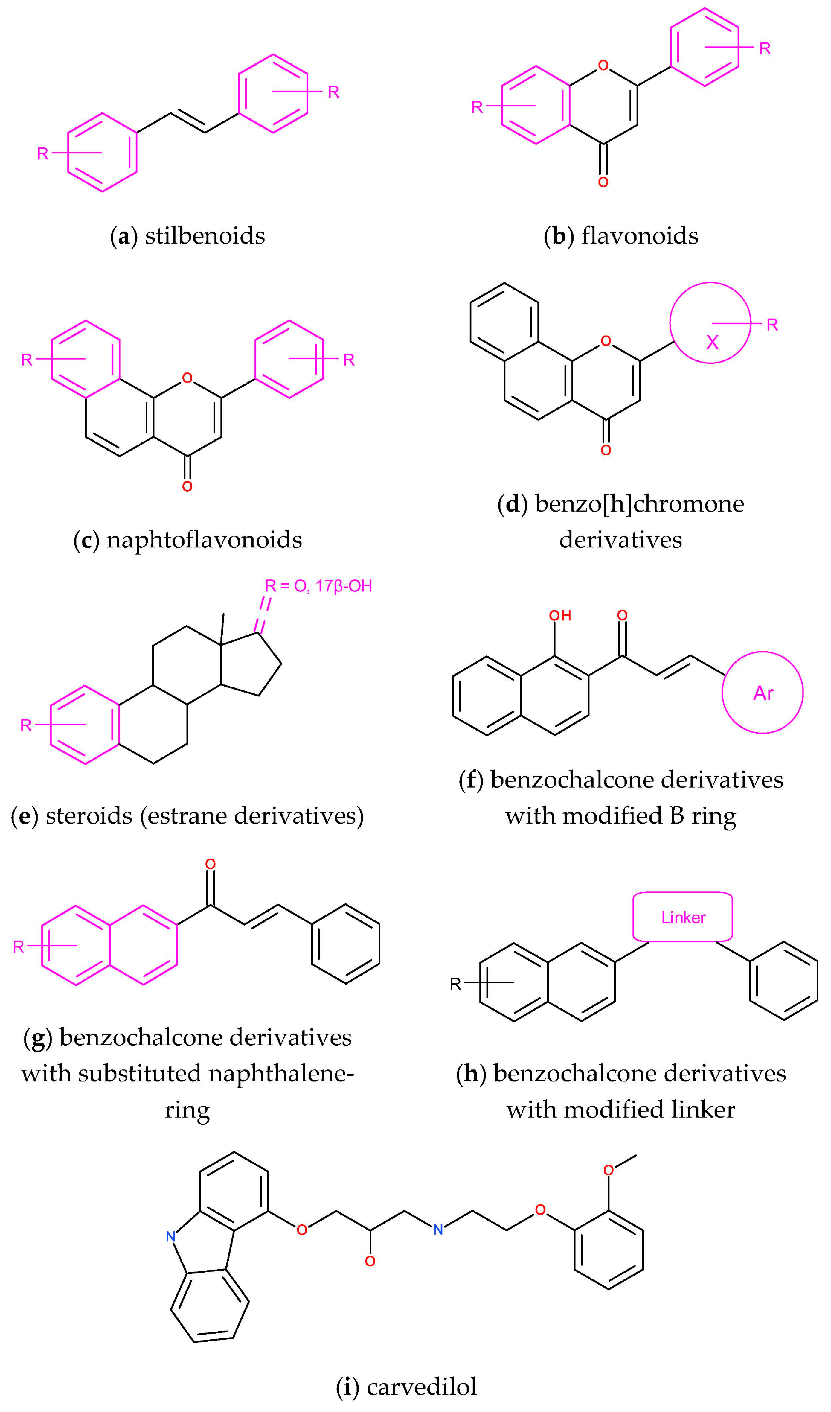
3.3. Design of Selective CYP1B1 Inhibitors

Inhibitors are chemical tools used in the studies of the molecular structure of enzymes. In turn, the knowledge of enzyme structures facilitates the studies on its function and allows the design of new molecules with stronger affinity to the binding site. In structure-based drug design (SBDD), many methods can be used to calculate the binding affinity of the molecules for their biological target prior to their synthesis [83]. Computational methods in the search for new efficient CYP1B1 inhibitors are widely used, highlighting the mechanism of enzyme inhibition and indicating possible structural modifications of known inhibitors and drug candidates, thereby improving inhibitory properties. A structure–activity relationship is a crucial issue in drug design development. Strategies in search of target-specific inhibitors were classified by Zhan and coworkers as target-guided approaches and derivatization approaches based on the known substrates and inhibitors [84]. Taking into account the design of effective inhibitors of individual enzyme isoforms, an additional feature to be met is the selectivity of their action [85]. The shape of the enzyme cavity, electrostatic properties of amino acids on its surface, the flexibility of the active site, and the location of water molecules bound in the active site must be taken into account in the process of the design of selective and potent enzyme inhibitors [86].
Stilbenoids and flavonoids (Figure 6) are two classes of compounds for which interactions with CYP1 isozymes were studied extensively for two decades. Stilbene and flavonoid derivatives were designed, synthesized, and evaluated as substrates and inhibitors of CYP1 isozymes with special regard to their molecular interactions in the enzyme binding sites. The high potency of trans-stilbene methoxy derivatives as selective inhibitors of CYP1B1 activity was analyzed with the use of computational methods, among others stilbene derivatives: 2,4,3′,5′-tetramethoxy-trans-stilbene (TMS) and 2,2′,4,6′-tetramethoxy-trans-stilbene [87,88,89]. In our studies, 2,3′,4′-trimethoxy-trans-stilbene was the most effective and selective CYP1B1 inhibitor among the studied series of poly-methoxystilbenes [63]. Molecular docking of 2,3′,4′-trimethoxy-trans-stilbene to the enzyme binding site with the use of the LigandFit procedure revealed that van der Waals forces were mostly responsible for the high affinity of this compound to the CYP1B1 binding site. In the energetically favorable orientation of 2,3′,4′-trimethoxy-trans-stilbene, 3′,4′-dimethoxy motif was directed to the heme, and π-π stacking of the phenyl ring with Phe231 was formed.
To explain the selectivity of 2,3′,4′-trimethoxy-trans-stilbene towards CYP1B1, its interactions at the CYP1B1 binding site were compared to 2,4′-dimethoxy-trans-stilbene, which inhibited CYP1B1 activity about 20 times less. Energetically favored poses of 2,4′-dimethoxy-trans-stilbene were directed to the heme with 4′-methoxyphenyl. The oxygen atom in position 2 of 2,4′-dimethoxy-trans-stilbene formed the hydrogen bond with the NH2 group of Gln332, whereas 2,3′,4′-trimethoxy-trans-stilbene interacted with Gln332 by means of π electrons of the stilbene phenyl ring. Additionally, π electrons of both compounds interacted with the peptide bond Gly329/Ala330 (π-amide stacking). For 2,4′-dimethoxy-trans-stilbene docked to the CYP1B1 binding site, the weak hydrogen bond between Asn228 and the polarized CH3 group (C-H---O=C) in position 2 was observed. For 2,3′,4′-trimethoxy-trans-stilbene oriented with 3,4-substituted phenyl to the heme in the CYP1B1 binding site, T-shaped π-π interaction occurred [90].
In a series of polymethoxy-trans-stilbenes, tetramethoxy and pentamethoxy derivatives displayed potent inhibitory activity toward CYP1A1 and CYP1B1 with IC50 < 1 μM, while the derivatives possessing methoxy substituents in positions 4 and 4′, which made the molecules longer, did not fit to the CYP1A2 binding site [63]. This confirms the findings of Liu et al. concerning the size and shape of CYP1cavities. CYP1A1 and CYP1B1 have narrow and long cavities compared to the CYP1A2 binding site, which can accommodate triangular molecules, showing a planar trigonal structure with side lengths 9.3 Å, 8.7 Å, and 7.2 Å, respectively [91].
More recently, the selectivity of stilbenoid inhibitors against CYP1 isoforms was studied with integrated fit docking and molecular dynamics simulation. The potency of CYP1 inhibitors was correlated with π-π stacking interactions with the phenylalanine residues in the F helix. Modification of 2,4,3′,5′ -tetramethoxy-trans-stilbene with thiophene and imidazole rings significantly improved the selectivity of the modified inhibitor against CYP1A2, while the potency against CYP1B1 did not change [85].
The effect of natural and synthetic flavonoids and naphthoflavonoids (Figure 6) on CYP1 activity was an objective of extended structure–activity studies [92]. Among a series of 33 flavonoids, 3,5,7-trihydroxyflavone (galangin) inhibited CYP1B1 activity with the IC50 value of 3 nM. Six flavone derivatives were docked to CYP1B1, indicating the occurrence of more than one binding site in the CYP1B1 cavity [93]. A and/or C rings were found near SRS4 and SRS5 and far from SRS1, SRS2, and SRS3. In these studies, the authors generated CYP1B1 structure by homology modeling based on the crystal structure of CYP1A2.
ANF is the best known strong inhibitor of CYP1s; however, this compound is weakly soluble in water, and it does not exert selectivity toward CYP1B1. Choosing ANF as a lead compound, its derivatives differing in the substituents and their positions were designed and synthesized. Cui and Dong with coworkers [66,94] set out some structural features determining the inhibitory potency of designed molecules. In their studies of a series of ANF derivatives, very potent CYP1B1 inhibitors were found, indicating the indispensable role of the keto group at the C-ring and the significant increase of inhibitory potency when a compound possessed a halogen atom at the 3′ position on the B-ring. ANF derivative, 3-hydroxy-3′-fluoro-6,7,10-trimethoxy-7,8-benzoflavone exhibited the highest potency against CYP1B1 ever reported with IC50 equal to 0.043 nM [65]. Equally high inhibitory potency with IC50 below 1 nM was found for B ring modified ANF derivatives [95]. The compounds possessing chloro- and trifluoromethyl groups substituted at position 3 of the B ring were 10-fold more potent than the parent compound. Molecular docking of 3-chloro-ANF derivative revealed the π-π stacking interaction with Phe231 and three hydrogen bonds to the amino and hydroxyl groups of Ser131 and to the carbonyl group of Asp326 with distances of 3.5, 2.1, and 2.8 Å, respectively. In the CYP1B1 active site, ANF derivatives adopt a different pose than the ANF binding pose. As in cases of other inhibitors, the affinity to CYP1B1 was increased by hydrophobic contacts with Ile399, Leu264, Phe268, Phe120, Ala330, and Ala121 [95]. Benzochalcones (Figure 6) was another series of synthesized compounds related to ANF, exhibiting a nanomolar inhibitory potency toward CYP1B1 and the cytotoxic activity against drug-resistant cancer cells [96].
In turn, two heterocycle-containing ANF derivatives are noteworthy as very potent CYP1B1 inhibitors: a fluorine-containing pyridin-2-yl substituted 6,7,10-trimethoxy-ANF (IC50 value of 0.07 nM) and a pyridin-3-yl substituted 6,7,10-trimethoxy-ANF with the 4-NH2 group at the ring B (IC50 value of 0.98 nM), whose solubility in water is much better than that of ANF [94].
The design of CYP1B1 inhibitors may be based on their substrates as lead compounds. Four series of A-ring substituted steroids (Figure 6) were synthesized, and their inhibitory activity against CYP1B1 was estimated. 2-(4-Fluorophenyl)-E2 was the best CYP1B1 inhibitor with IC50 = 0.24 μM. Docking results showed that this new estrane derivative fits better into the CYP1B1 binding site than that of CYP1A1 because its A-ring produces more π-interactions in the CYP1B1 cavity in comparison with CYP1A1. These included π−π T-shaped with Phe-231 and π−σ with Ala-330. Asn228 residue formed a hydrogen bond with the 17β-OH [97].
Another way to search for efficient and selective CYP1B1 inhibitors is the structure-based virtual screening of approved drugs registered in the FDA database, which Wang and his group applied. The screening results were correlated with experimental determination of inhibitory activity, indicating beta-blocker carvedilol (Figure 6) as a promising CYP1B1 inhibitor [98]. Carvedilol, a drug used in the treatment of high blood pressure and heart failure, was docked to the X-ray crystal structure of the enzyme (PDB code: 6IQ5). Its binding pose in the CYP1B1 cavity was stabilized by π-π stacking interaction formed by a 9H-carbazole group of carvedilol with Phe231. As in other cases of CYP1B1 inhibitors, hydrophobic interactions formed by the methoxy-substituted phenyl with heme and surrounding residues decided on the affinity of carvedilol to the CYP1B1 binding site [98].

3.4. Genetic Variability

CYP1B1 involvement in cancer therapy requires extensive knowledge of the CYP1B1 molecular structure, which determines its interactions with the ligands. Polymorphism of CYP1B1 is an objective of research concerning the increased risk of cancer [99,100]. Genetic variability of CYP1B1 was studied since 1997 when Stoilov and coworkers identified three truncated mutations of CYP1B1 as the cause of primary congenital glaucoma [9,101]. Pharmacological analyses of individual CYP1B1 variants indicated univocally on the association of CYP1B1 mutations with eye diseases [102,103,104,105].
The structures of CYP1B1 variants responsible for primary congenital glaucoma (PCG) and primary open angle glaucoma (POAG) were earlier generated using bacterial CYP102 or human CYP2C9 as a template [9,22,101]. More recently, structural determinants of the CYP1B1 variants associated with PCG and POAG in humans were investigated with the use of crystal structure generated with ANF as a ligand. In 2016, extensive normal mode analysis and MD simulations performed by Banerjee and coworkers [23] enabled the observations of possible effects of mutations on the CYP1B1 structure. Altered structural flexibility within the B-C and F-G loop region and altered tunnel properties were observed in a variant with changed metabolizing activity of 17-β-estradiol (E2) relative to the normal level [23].
Other authors indicate that the I helix plays an essential role in substrate binding, imposing its orientation in the binding cavity and consequently deciding on the site of metabolism. Valine at position 395 decides the stereochemistry of E2 hydroxylation. Wild type CYP1B1 predominantly metabolizes E2 to 4-OH-E2, while 2-OH-E2 is a product of CYP1A1 having leucine at position 395. Consistently, CYP1B1 mutant V395L, having valine replaced with leucine, also catalyzes hydroxylation at the 2-position due to the change in substrate orientation in relation to ferryl oxygen [106].
CYP1B1 variants are also investigated with regard to the metabolism of environmental pollutants [2]. Both allelic variants CYP1B1.1 and 1B1.3 preferentially oxidized 1-chloropyrene, a chlorinated polycyclic hydrocarbon, at the 6-position, while CYP1A1 catalyzed hydroxylation at the 6- and 8-positions more actively than that at the 3-position [107]. The orientation of 1-chloropyrene in the CYP1B1 binding site (3PM0) was determined with position 6 directed to the heme in the distance of 5.71 Å. However, the CYP1B1 variants did not differ in activity toward 1-chloropyrene. The structure of the CYP1B1 active site was compared with that of CYP2A13, displaying fargoing similarities, and both enzymes also showed overlapping substrate specificity [108].
The analysis of interactions between polymorphic forms of CYP1B1 and substrates/inhibitors is explored concerning the design of specifically targeted anti-cancer agents. The hypothesis that CYP1B1 variants coding impaired protein with reduced enzyme function is responsible for better response to anti-cancer treatment still needs to be confirmed. In the Chinese population, women with the homozygous variant genotype for CYP1B1 (single nucleotide polymorphism rs1056836) exhibited a significantly reduced risk of breast cancer; however, homozygous variant genes for GSTP1 and COMT were associated with an increased risk of developing breast cancer [109]. Several reports have shown that the CYP1B1.3 genetic variant (L432V) is associated with an increased risk of prostate, colorectal, and urinary system cancers [68,110,111,112,113].

3.5. Characteristics of in Silico Methods

The quality and reliability of the results obtained in molecular docking and molecular dynamics (MD) simulations largely depend on the models, approximations, and computational methods used in the experiments. Docking, used as a virtual screening tool to assess the affinity of ligands, uses fast, yet approximate methods, i.e., scoring functions (SFs). Commonly used SFs belong to four groups: force field-based, empirical, knowledge-based, and relatively new, machine-learning-based [114,115,116,117]. Better at distinguishing active from inactive molecules are scoring functions based on quantum mechanics, which give a much lower number of false positives, although at the cost of increased computation time by one to two orders of magnitude [118,119,120,121,122]. For ligand design, where individual molecules or a series of congeneric ligands are docked, the affinity assessment helps determine the most likely orientation (pose) of the ligand, which in turn determines the possibility of specific ligand-protein interactions [63,66,85,90,93,94,95,96,97].
The binding of a ligand to a protein usually changes the conformation of both the ligand and the protein. Thus, it is also important for flexible ligands to properly sample their conformational space during docking [116]. The more significant challenge is to account for the conformational changes in the protein during ligand binding [123,124,125,126,127,128]. One of the methods of studying this phenomenon is molecular dynamics. If the X-ray structure of the appropriate ligand-protein complex is not available, the poses selected in the docking can be used to set up MD simulations [85].
In drug design, molecular dynamics simulations allow the study of a wide variety of processes, including changes in protein conformation caused by ligand binding or unbinding and by the flexibility of different protein regions. MD simulations allow us to predict or explain the effects of the mutations on a protein’s structure and function [22,23,25]. Simulation-based methods also enable more accurate estimation of ligand binding affinity than docking [129,130,131]. Of course, molecular dynamics also have their limitations. They mainly result from approximations in force fields, the need for appropriate parameterization of ligands, the lack of taking into account electronic effects such as polarization, formation or breaking of covalent bonds, and the time scales of the studied phenomenon [129,131]. Advances in algorithms, improvements of computer hardware, especially the use of graphics processing units (GPUs) in MD simulations along with enhanced sampling techniques [132,133,134,135] allow simulations to study events taking place over a longer time scale [131,136].

4. Summary: Perspectives of CYP1B1 Studies in Drug Design

Considering the association of CYP1B1 with metabolic diseases, carcinogenesis, and eye diseases, the interaction of this enzyme with substrates/inhibitors is an important objective for studies with the use of the latest computational methods. Recent studies indicate the flexibility of ligand as a feature that helps in fitting to the enzyme cavity. In addition, the shape, electrostatic environment, and malleability of enzyme active site significantly determine ligand-enzyme interactions. Molecular docking and, in particular, molecular dynamics simulations, allow extending our knowledge of the enzyme’s molecular structure, which determines its function. Knowing the molecular mechanisms which drive the processes essential for organism functioning will enable us to cure the diseases associated with metabolic disorders and redox imbalance. The design of efficient and safe CYP1B1 inhibitors is still a developing area of research.

Author Contributions

R.M., Z.D. contributed equally to the manuscript development and preparation. Both authors have read and agreed to the published version of the manuscript.

Funding

This research received no external funding.

Institutional Review Board Statement

Not applicable.

Informed Consent Statement

Not applicable.

Data Availability Statement

Not applicable.

Acknowledgments

This study was supported by grants from Poznań University of Medical Sciences and Nicolaus Copernicus University in Toruń, Ludwik Rydygier Collegium Medicum, Statutory Research no. PDB WF411.

Conflicts of Interest

The authors declare no conflict of interest.

References

  1. Bièche, I.; Narjoz, C.; Asselah, T.; Vacher, S.; Marcellin, P.; Lidereau, R.; Beaune, P.; de Waziers, I. Reverse transcriptase-PCR quantification of mRNA levels from cytochrome (CYP)1, CYP2 and CYP3 families in 22 different human tissues. Pharm. Genom. 2007, 17, 731–742. [Google Scholar] [CrossRef] [PubMed]
  2. Shimada, T. Inhibition of carcinogen-activating cytochrome P450 Enzymes by xenobiotic chemicals in relation to antimutagenicity and anticarcinogenicity. Toxicol. Res. 2017, 33, 79–96. [Google Scholar] [CrossRef]
  3. Zanger, U.M.; Schwab, M. Cytochrome P450 enzymes in drug metabolism: Regulation of gene expression, enzyme activities, and impact of genetic variation. Pharmacol. Ther. 2013, 138, 103–141. [Google Scholar] [CrossRef]
  4. Go, R.-E.; Hwang, K.-A.; Choi, K.-C. Cytochrome P450 1 family and cancers. J. Steroid Biochem. Mol. Biol. 2015, 147, 24–30. [Google Scholar] [CrossRef]
  5. McFadyen, M.C.E.; McLeod, H.L.; Jackson, F.C.; Melvin, W.T.; Doehmer, J.; Murray, G.I. Cytochrome P450 CYP1B1 protein expression: A novel mechanism of anticancer drug resistance. Biochem. Pharmacol. 2001, 62, 207–212. [Google Scholar] [CrossRef]
  6. Rochat, B.; Morsman, J.M.; Figg, W.D.; Murray, G.I.; McLeod, H.L. Human CYP1B1 and anticancer agent metabolism: Mechanism for tumor-specific drug inactivation? J. Pharmacol. Exp. Ther. 2001, 296, 537–541. [Google Scholar]
  7. Zhu, Z.; Mu, Y.; Qi, C.; Wang, J.; Xi, G.; Guo, J.; Mi, R.; Zhao, F. CYP1B1 enhances the resistance of epithelial ovarian cancer cells to paclitaxel in vivo and in vitro. Int. J. Mol. Med. 2015, 35, 340–348. [Google Scholar] [CrossRef] [Green Version]
  8. Murray, G.I.; Taylor, M.C.; McFadyen, M.C.E.; McKay, J.A.; Greenlee, W.F.; Burke, M.D.; Melvin, W.T. Tumor-specific expression of cytochrome P450 CYP1B1. Cancer Res. 1997, 57, 3026–3031. [Google Scholar] [PubMed]
  9. Stoilov, I.; Akarsu, A.N.; Sarfarazi, M. Identification of three different truncating mutations in cytochrome P4501B1 (CYP1B1) as the principal cause of primary congenital glaucoma (Buphthalmos) in families linked to the GLC3A locus on chromosome 2p21. Hum. Mol. Genet. 1997, 6, 641–647. [Google Scholar] [CrossRef]
  10. Alsubait, A.; Aldossary, W.; Rashid, M.; Algamdi, A.; Alrfaei, B.M. CYP1B1 gene: Implications in glaucoma and cancer. J. Cancer 2020, 11, 4652–4661. [Google Scholar] [CrossRef] [PubMed]
  11. Carrera, A.N.; Grant, M.K.O.; Zordoky, B.N. CYP1B1 as a therapeutic target in cardio-oncology. Clin. Sci. 2020, 134, 2897–2927. [Google Scholar] [CrossRef]
  12. Nebert, D.W.; Dalton, T.P.; Okey, A.B.; Gonzalez, F.J. Role of Aryl Hydrocarbon Receptor-mediated Induction of the CYP1 Enzymes in Environmental Toxicity and Cancer. J. Biol. Chem. 2004, 279, 23847–23850. [Google Scholar] [CrossRef] [Green Version]
  13. Yang, X.; Solomon, S.; Fraser, L.R.; Trombino, A.F.; Liu, D.; Sonenshein, G.E.; Hestermann, E.V.; Sherr, D.H. Constitutive regulation ofCYP1B1 by the aryl hydrocarbon receptor (AhR) in pre-malignant and malignant mammary tissue. J. Cell. Biochem. 2008, 104, 402–417. [Google Scholar] [CrossRef]
  14. Kawajiri, K.; Fujii-Kuriyama, Y. Cytochrome P450 gene regulation and physiological functions mediated by the aryl hydrocarbon receptor. Arch. Biochem. Biophys. 2007, 464, 207–212. [Google Scholar] [CrossRef]
  15. Avilla, M.N.; Malecki, K.M.C.; Hahn, M.E.; Wilson, R.H.; Bradfield, C.A. The Ah receptor: Adaptive metabolism, ligand diversity, and the Xenokine model. Chem. Res. Toxicol. 2020, 33, 860–879. [Google Scholar] [CrossRef]
  16. Ingelman-Sundberg, M.; Zhong, X.B.; Hankinson, O.; Beedanagari, S.; Yu, A.M.; Peng, L.; Osawa, Y. Potential role of epigenetic mechanisms in the regulation of drug metabolism and transport. Drug Metab. Dispos. 2013, 41, 1725–1731. [Google Scholar] [CrossRef] [PubMed] [Green Version]
  17. Beedanagari, S.R.; Taylor, R.T.; Hankinson, O. Differential regulation of the dioxin-induced Cyp1a1 and Cyp1b1 genes in mouse hepatoma and fibroblast cell lines. Toxicol. Lett. 2010, 194, 26–33. [Google Scholar] [CrossRef] [Green Version]
  18. Beedanagari, S.R.; Taylor, R.T.; Bui, P.; Wang, F.; Nickerson, D.W.; Hankinson, O. Role of epigenetic mechanisms in differential regulation of the dioxin-inducible human CYP1A1 and CYP1B1 genes. Mol. Pharmacol. 2010, 78, 608–616. [Google Scholar] [CrossRef] [PubMed] [Green Version]
  19. Kang, G.H.; Lee, S.; Cho, N.Y.; Gandamihardja, T.; Long, T.I.; Weisenberger, D.J.; Campan, M.; Laird, P.W. DNA methylation profiles of gastric carcinoma characterized by quantitative DNA methylation analysis. Lab. Investig. 2008, 88, 161–170. [Google Scholar] [CrossRef] [PubMed] [Green Version]
  20. Habano, W.; Gamo, T.; Sugai, T.; Otsuka, K.; Wakabayashi, G.; Ozawa, S. CYP1B1, but not CYP1A1, is downregulated by promoter methylation in colorectal cancers. Int. J. Oncol. 2009, 34, 1085–1091. [Google Scholar] [CrossRef] [PubMed]
  21. Shah, B.R.; Xu, W.; Mraz, J. Cytochrome P450 1B1: Role in health and disease and effect of nutrition on its expression. RSC Adv. 2019, 9, 21050–21062. [Google Scholar] [CrossRef] [Green Version]
  22. Achary, M.S.; Reddy, A.B.M.; Chakrabarti, S.; Panicker, S.G.; Mandal, A.K.; Ahmed, N.; Balasubramanian, D.; Hasnain, S.E.; Nagarajaram, H.A. Disease-causing mutations in proteins: Structural analysis of the CYP1b1 mutations causing primary congenital glaucoma in humans. Biophys. J. 2006, 91, 4329–4339. [Google Scholar] [CrossRef] [Green Version]
  23. Banerjee, A.; Chakraborty, S.; Chakraborty, A.; Chakrabarti, S.; Ray, K. Functional and Structural Analyses of CYP1B1 Variants Linked to Congenital and Adult-Onset Glaucoma to Investigate the Molecular Basis of These Diseases. PLoS ONE 2016, 11, e0156252. [Google Scholar] [CrossRef] [PubMed] [Green Version]
  24. Rashid, M.; Yousaf, S.; Sheikh, S.A.; Sajid, Z.; Shabbir, A.S.; Kausar, T.; Tariq, N.; Usman, M.; Shaikh, R.S.; Ali, M.; et al. Identities and frequencies of variants in CYP1B1 causing primary congenital glaucoma in Pakistan. Mol. Vis. 2019, 25, 144–154. [Google Scholar] [PubMed]
  25. Ou, Z.; Liu, G.; Liu, W.; Deng, Y.; Zheng, L.; Zhang, S.; Feng, G. Bioinformatics analysis of CYP1B1 mutation hotspots in Chinese primary congenital glaucoma patients. Biosci. Rep. 2018, 38. [Google Scholar] [CrossRef] [Green Version]
  26. Falero-Perez, J.; Song, Y.-S.; Zhao, Y.; Teixeira, L.; Sorenson, C.M.; Sheibani, N. Cyp1b1 expression impacts the angiogenic and inflammatory properties of liver sinusoidal endothelial cells. PLoS ONE 2018, 13, e0206756. [Google Scholar] [CrossRef] [PubMed]
  27. Falero-Perez, J.; Song, Y.-S.; Sorenson, C.M.; Sheibani, N. CYP1B1: A key regulator of redox homeostasis. Trends Cell Mol. Biol. 2018, 13, 27–45. [Google Scholar] [PubMed]
  28. Maguire, M.; Larsen, M.C.; Foong, Y.H.; Tanumihardjo, S.; Jefcoate, C.R. Cyp1b1 deletion and retinol deficiency coordinately suppress mouse liver lipogenic genes and hepcidin expression during post-natal development. Mol. Cell. Endocrinol. 2017, 454, 50–68. [Google Scholar] [CrossRef] [PubMed]
  29. Andriopoulos, B.; Corradini, E.; Xia, Y.; Faasse, S.A.; Chen, S.; Grgurevic, L.; Knutson, M.D.; Pietrangelo, A.; Vukicevic, S.; Lin, H.Y.; et al. BMP6 is a key endogenous regulator of hepcidin expression and iron metabolism. Nat. Genet. 2009, 41, 482–487. [Google Scholar] [CrossRef] [PubMed] [Green Version]
  30. Meynard, D.; Kautz, L.; Darnaud, V.; Canonne-Hergaux, F.; Coppin, H.; Roth, M.P. Lack of the bone morphogenetic protein BMP6 induces massive iron overload. Nat. Genet. 2009, 41, 478–481. [Google Scholar] [CrossRef] [PubMed]
  31. Dinu, D.; Chu, C.; Veith, A.; Lingappan, K.; Couroucli, X.; Jefcoate, C.R.; Sheibani, N.; Moorthy, B. Mechanistic role of cytochrome P450 (CYP)1B1 in oxygen-mediated toxicity in pulmonary cells: A novel target for prevention of hyperoxic lung injury. Biochem. Biophys. Res. Commun. 2016, 476, 346–351. [Google Scholar] [CrossRef] [Green Version]
  32. Veith, A.C.; Aram, B.B.; Jiang, W.; Wang, L.; Zhou, G.; Jefcoate, C.R.; Couroucli, X.I.; Lingappan, K.; Moorthy, B. Mice Lacking the Cytochrome P450 1B1 Gene Are Less Susceptible to Hyperoxic Lung Injury Than Wild Type. Toxicol. Sci. 2018, 165, 462–474. [Google Scholar] [CrossRef]
  33. Kubli, S.P.; Bassi, C.; Roux, C.; Wakeham, A.; Göbl, C.; Zhou, W.; Jafari, S.M.; Snow, B.; Jones, L.; Palomero, L.; et al. AhR controls redox homeostasis and shapes the tumor microenvironment in BRCA1-associated breast cancer. Proc. Natl. Acad. Sci. USA 2019, 116, 3604–3613. [Google Scholar] [CrossRef] [PubMed] [Green Version]
  34. Li, F.; Zhu, W.; Gonzalez, F.J. Potential role of CYP1B1 in the development and treatment of metabolic diseases. Pharmacol. Ther. 2017, 178, 18–30. [Google Scholar] [CrossRef] [PubMed]
  35. Anderson, G.; Mazzoccoli, G. Left Ventricular Hypertrophy: Roles of Mitochondria CYP1B1 and Melatonergic Pathways in Co-Ordinating Wider Pathophysiology. Int. J. Mol. Sci. 2019, 20, 4068. [Google Scholar] [CrossRef] [Green Version]
  36. Elkhatali, S.; Maayah, Z.H.; El-Sherbeni, A.A.; Elshenawy, O.H.; Abdelhamid, G.; Shoieb, S.M.; El-Kadi, A.O.S. Inhibition of mid-chain HETEs protects against angiotensin II-induced cardiac hypertrophy. J. Cardiovasc. Pharmacol. 2017, 70, 16–24. [Google Scholar] [CrossRef]
  37. Maayah, Z.H.; Althurwi, H.N.; Abdelhamid, G.; Lesyk, G.; Jurasz, P.; El-Kadi, A.O.S. CYP1B1 inhibition attenuates doxorubicin-induced cardiotoxicity through a mid-chain HETEs-dependent mechanism. Pharmacol. Res. 2016, 105, 28–43. [Google Scholar] [CrossRef]
  38. Maayah, Z.H.; Althurwi, H.N.; El-Sherbeni, A.A.; Abdelhamid, G.; Siraki, A.G.; El-Kadi, A.O.S. The role of cytochrome P450 1B1 and its associated mid-chain hydroxyeicosatetraenoic acid metabolites in the development of cardiac hypertrophy induced by isoproterenol. Mol. Cell. Biochem. 2017, 429, 151–165. [Google Scholar] [CrossRef] [PubMed]
  39. Sahan-Firat, S.; Jennings, B.L.; Yaghini, F.A.; Song, C.Y.; Estes, A.M.; Fang, X.R.; Farjana, N.; Khan, A.I.; Malik, K.U. 2,3′,4,5′-Tetramethoxystilbene prevents deoxycorticosterone—Salt-induced hypertension: Contribution of cytochrome P-450 1B1. Am. J. Physiol. Hear. Circ. Physiol. 2010, 299. [Google Scholar] [CrossRef] [PubMed] [Green Version]
  40. Jennings, B.L.; Anderson, L.J.; Estes, A.M.; Yaghini, F.A.; Fang, X.R.; Porter, J.; Gonzalez, F.J.; Campbell, W.B.; Malik, K.U. Cytochrome P450 1B1 contributes to renal dysfunction and damage caused by angiotensin II in mice. Hypertension 2012, 59, 348–354. [Google Scholar] [CrossRef] [PubMed]
  41. Jennings, B.L.; Montanez, D.E.; May, M.E.; Estes, A.M.; Fang, X.R.; Yaghini, F.A.; Kanu, A.; Malik, K.U. Cytochrome P450 1B1 contributes to increased blood pressure and cardiovascular and renal dysfunction in spontaneously hypertensive rats. Cardiovasc. Drugs Ther. 2014, 28, 145–161. [Google Scholar] [CrossRef] [PubMed] [Green Version]
  42. Pingili, A.K.; Davidge, K.N.; Thirunavukkarasu, S.; Khan, N.S.; Katsurada, A.; Majid, D.S.A.; Gonzalez, F.J.; Navar, L.G.; Malik, K.U. 2-Methoxyestradiol reduces angiotensin II-induced hypertension and renal dysfunction in ovariectomized female and intact male mice. Hypertension 2017, 69, 1104–1112. [Google Scholar] [CrossRef]
  43. Malik, K.U.; Jennings, B.L.; Yaghini, F.A.; Sahan-Firat, S.; Song, C.Y.; Estes, A.M.; Fang, X.R. Contribution of cytochrome P450 1B1 to hypertension and associated pathophysiology: A novel target for antihypertensive agents. Prostaglandins Other Lipid Mediat. 2012, 98, 69–74. [Google Scholar] [CrossRef] [PubMed] [Green Version]
  44. Song, C.Y.; Ghafoor, K.; Ghafoor, H.U.; Khan, N.S.; Thirunavukkarasu, S.; Jennings, B.L.; Estes, A.M.; Zaidi, S.; Bridges, D.; Tso, P.; et al. Cytochrome P450 1B1 contributes to the development of atherosclerosis and hypertension in apolipoprotein E-deficient mice. Hypertension 2016, 67, 206–213. [Google Scholar] [CrossRef]
  45. Pingili, A.K.; Jennings, B.L.; Mukherjee, K.; Akroush, W.; Gonzalez, F.J.; Malik, K.U. 6β-Hydroxytestosterone, a metabolite of testosterone generated by CYP1B1, contributes to vascular changes in angiotensin II-induced hypertension in male mice. Biol. Sex Differ. 2020, 11, 4. [Google Scholar] [CrossRef]
  46. Liu, X.; Huang, T.; Li, L.; Tang, Y.; Tian, Y.; Wang, S.; Fan, C. CYP1B1 deficiency ameliorates obesity and glucose intolerance induced by high fat diet in adult C57BL/6J mice. Am. J. Transl. Res. 2015, 7, 761–771. [Google Scholar]
  47. Larsen, M.C.; Bushkofsky, J.R.; Gorman, T.; Adhami, V.; Mukhtar, H.; Wang, S.; Reeder, S.B.; Sheibani, N.; Jefcoate, C.R. Cytochrome P450 1B1: An unexpected modulator of liver fatty acid homeostasis. Arch. Biochem. Biophys. 2015, 571, 21–39. [Google Scholar] [CrossRef] [PubMed] [Green Version]
  48. Conway, D.E.; Sakurai, Y.; Weiss, D.; Vega, J.D.; Taylor, W.R.; Jo, H.; Eskin, S.G.; Marcus, C.B.; McIntire, L.V. Expression of CYP1A1 and CYP1B1 in human endothelial cells: Regulation by fluid shear stress. Cardiovasc. Res. 2009, 81, 669–677. [Google Scholar] [CrossRef]
  49. Yaghini, F.A.; Song, C.Y.; Lavrentyev, E.N.; Ghafoor, H.U.B.; Fang, X.R.; Estes, A.M.; Campbell, W.B.; Malik, K.U. Angiotensin II-induced vascular smooth muscle cell migration and growth are mediated by cytochrome p450 1b1-dependent superoxide generation. Hypertension 2010, 55, 1461–1467. [Google Scholar] [CrossRef] [Green Version]
  50. Thompson, P.A.; Khatami, M.; Baglole, C.J.; Sun, J.; Harris, S.A.; Moon, E.-Y.; Al-Mulla, F.; Al-Temaimi, R.; Brown, D.G.; Colacci, A.M.; et al. Environmental immune disruptors, inflammation and cancer risk. Carcinogenesis 2015, 36, S232–S253. [Google Scholar] [CrossRef] [Green Version]
  51. Wang, Z.; Dabrosin, C.; Yin, X.; Fuster, M.M.; Arreola, A.; Rathmell, W.K.; Generali, D.; Nagaraju, G.P.; El-Rayes, B.; Ribatti, D.; et al. Broad targeting of angiogenesis for cancer prevention and therapy. Semin. Cancer Biol. 2015, 35, S224–S243. [Google Scholar] [CrossRef] [PubMed]
  52. Ramjiawan, R.R.; Griffioen, A.W.; Duda, D.G. Anti-angiogenesis for cancer revisited: Is there a role for combinations with immunotherapy? Angiogenesis 2017, 20, 185–204. [Google Scholar] [CrossRef] [PubMed]
  53. Martínez-Poveda, B.; Torres-Vargas, J.A.; del Ocaña, M.C.; García-Caballero, M.; Medina, M.Á.; Quesada, A.R. The mediterranean diet, a rich source of angiopreventive compounds in cancer. Nutrients 2019, 11, 2036. [Google Scholar] [CrossRef] [Green Version]
  54. Kanda, Y.; Osaki, M.; Okada, F. Chemopreventive strategies for inflammation-related carcinogenesis: Current status and future direction. Int. J. Mol. Sci. 2017, 18, 867. [Google Scholar] [CrossRef]
  55. D’Uva, G.; Baci, D.; Albini, A.; Noonan, D.M. Cancer chemoprevention revisited: Cytochrome P450 family 1B1 as a target in the tumor and the microenvironment. Cancer Treat. Rev. 2018, 63, 1–18. [Google Scholar] [CrossRef]
  56. Dutour, R.; Poirier, D. Inhibitors of cytochrome P450 (CYP) 1B1. Eur. J. Med. Chem. 2017, 135, 296–306. [Google Scholar] [CrossRef]
  57. Cui, J.; Li, S. Inhibitors and prodrugs targeting CYP1: A novel approach in cancer prevention and therapy. Curr. Med. Chem. 2014, 21, 519–552. [Google Scholar] [CrossRef]
  58. Jiang, X.; Wang, J.; Deng, X.; Xiong, F.; Zhang, S.; Gong, Z.; Li, X.; Cao, K.; Deng, H.; He, Y.; et al. The role of microenvironment in tumor angiogenesis. J. Exp. Clin. Cancer Res. 2020, 39, 204. [Google Scholar] [CrossRef]
  59. Ware, W.R. Natural cancer therapy and prevention targeted on cancer cells and cancer stem cells based on the cytochrome P45O enzyme CYP1B1: A commentary. Altern Ther Heal. Med. 2017, 23, 50–58. [Google Scholar]
  60. Taylor, W.F.; Jabbarzadeh, E. The use of natural products to target cancer stem cells. Am. J. Cancer Res. 2017, 7, 1588–1605. [Google Scholar] [PubMed]
  61. Dutkiewicz, Z.; Mikstacka, R. Structure-based drug design for cytochrome P450 family 1 inhibitors. Bioinorg. Chem. Appl. 2018, 2018. [Google Scholar] [CrossRef]
  62. Sirerol, J.A.; Rodríguez, M.L.; Mena, S.; Asensi, M.A.; Estrela, J.M.; Ortega, A.L. Role of natural stilbenes in the prevention of cancer. Oxid. Med. Cell. Longev. 2016, 2016, 1–15. [Google Scholar] [CrossRef] [Green Version]
  63. Mikstacka, R.; Wierzchowski, M.; Dutkiewicz, Z.; Gielara-Korzańska, A.; Korzański, A.; Teubert, A.; Sobiak, S.; Baer-Dubowska, W. 3,4,2′-Trimethoxy-trans-stilbene—A potent CYP1B1 inhibitor. MedChemComm 2014, 5, 496–501. [Google Scholar] [CrossRef]
  64. Gao, X.; Xie, C.; Wang, Y.; Luo, Y.; Yagai, T.; Sun, D.; Qin, X.; Krausz, K.W.; Gonzalez, F.J. The antiandrogen flutamide is a novel aryl hydrocarbon receptor ligand that disrupts bile acid homeostasis in mice through induction of Abcc4. Biochem. Pharmacol. 2016, 119, 93–104. [Google Scholar] [CrossRef] [PubMed] [Green Version]
  65. Mitsui, Y.; Chang, I.; Fukuhara, S.; Hiraki, M.; Arichi, N.; Yasumoto, H.; Hirata, H.; Yamamura, S.; Shahryari, V.; Deng, G.; et al. CYP1B1 promotes tumorigenesis via altered expression of CDC20 and DAPK1 genes in renal cell carcinoma. BMC Cancer 2015, 15, 942. [Google Scholar] [CrossRef]
  66. Cui, J.; Meng, Q.; Zhang, X.; Cui, Q.; Zhou, W.; Li, S. Design and synthesis of new α-naphthoflavones as cytochrome P450 (CYP) 1B1 inhibitors to overcome docetaxel-resistance associated with CYP1B1 overexpression. J. Med. Chem. 2015, 58, 3534–3547. [Google Scholar] [CrossRef]
  67. Kwon, Y.-J.; Baek, H.-S.; Ye, D.-J.; Shin, S.; Kim, D.; Chun, Y.-J. CYP1B1 enhances cell proliferation and metastasis through induction of EMT and activation of Wnt/β-catenin signaling via Sp1 upregulation. PLoS ONE 2016, 11, e0151598. [Google Scholar] [CrossRef] [Green Version]
  68. Chang, I.; Mitsui, Y.; Kim, S.K.; Sun, J.S.; Jeon, H.S.; Kang, J.Y.; Kang, N.J.; Fukuhara, S.; Gill, A.; Shahryari, V.; et al. Cytochrome P450 1B1 inhibition suppresses tumorigenicity of prostate cancer via caspase-1 activation. Oncotarget 2017, 8, 39087–39100. [Google Scholar] [CrossRef] [PubMed]
  69. Wang, Z.; Chen, Y.; Drbohlav, L.M.; Wu, J.Q.; Wang, M.Z. development of an in vitro model to screen CYP1B1-targeted anticancer prodrugs. J. Biomol. Screen. 2016, 21, 1090–1099. [Google Scholar] [CrossRef] [PubMed] [Green Version]
  70. Cui, J.; Zhang, X.; Huang, G.; Zhang, Q.; Dong, J.; Sun, G.; Meng, Q.; Li, S. DMAKO-20 as a new multitarget anticancer prodrug activated by the tumor specific CYP1B1 enzyme. Mol. Pharm. 2019, 16, 409–421. [Google Scholar] [CrossRef] [PubMed]
  71. Meng, Q.; Wang, Z.; Cui, J.; Cui, Q.; Dong, J.; Zhang, Q.; Li, S. Design, synthesis, and biological evaluation of cytochrome P450 1B1 targeted molecular imaging probes for colorectal tumor detection. J. Med. Chem. 2018, 61, 10901–10909. [Google Scholar] [CrossRef] [PubMed]
  72. Itoh, T.; Takemura, H.; Shimoi, K.; Yamamoto, K. A 3D model of CYP1B1 explains the dominant 4-hydroxylation of estradiol. J. Chem. Inf. Model. 2010, 50, 1173–1178. [Google Scholar] [CrossRef]
  73. Wang, A.; Savas, U.; Stout, C.D.; Johnson, E.F. Structural characterization of the complex between α-naphthoflavone and human cytochrome P450 1B1. J. Biol. Chem. 2011, 286, 5736–5743. [Google Scholar] [CrossRef] [PubMed] [Green Version]
  74. Walsh, A.A.; Szklarz, G.D.; Scott, E.E. Human cytochrome P450 1A1 structure and utility in understanding drug and xenobiotic metabolism. J. Biol. Chem. 2013, 288, 12932–12943. [Google Scholar] [CrossRef] [PubMed] [Green Version]
  75. Bart, A.G.; Scott, E.E. Structures of human cytochrome P450 1A1 with bergamottin and erlotinib reveal active-site modifications for binding of diverse ligands. J. Biol. Chem. 2018, 293, 19201–19210. [Google Scholar] [CrossRef] [Green Version]
  76. Bart, A.G.; Takahashi, R.H.; Wang, X.; Scott, E.E. Human cytochrome P450 1A1 adapts active site for atypical nonplanar substrate. Drug Metab. Dispos. 2020, 48, 86–92. [Google Scholar] [CrossRef]
  77. Sansen, S.; Yano, J.K.; Reynald, R.L.; Schoch, G.A.; Griffin, K.J.; Stout, C.D.; Johnson, E.F. Adaptations for the oxidation of polycyclic aromatic hydrocarbons exhibited by the structure of human P450 1A2. J. Biol. Chem. 2007, 282, 14348–14355. [Google Scholar] [CrossRef] [Green Version]
  78. Kubo, M.; Yamamoto, K.; Itoh, T. Design and synthesis of selective CYP1B1 inhibitor via dearomatization of α-naphthoflavone. Bioorganic Med. Chem. 2019, 27, 285–304. [Google Scholar] [CrossRef] [PubMed]
  79. Bart, A.G.; Harris, K.L.; Gillam, E.M.J.; Scott, E.E. Structure of an ancestral mammalian family 1B1 cytochrome P450 with increased thermostability. J. Biol. Chem. 2020, 295, 5640–5653. [Google Scholar] [CrossRef] [Green Version]
  80. Liu, J.; Taylor, S.F.; Dupart, P.S.; Arnold, C.L.; Sridhar, J.; Jiang, Q.; Wang, Y.; Skripnikova, E.V.; Zhao, M.; Foroozesh, M. Pyranoflavones: A group of small-molecule probes for exploring the active site cavities of cytochrome P450 enzymes 1A1, 1A2, and 1B1. J. Med. Chem. 2013, 56, 4082–4092. [Google Scholar] [CrossRef] [PubMed] [Green Version]
  81. Sridhar, J.; Goyal, N.; Liu, J.; Foroozesh, M. Review of Ligand Specificity Factors for CYP1A Subfamily Enzymes from Molecular Modeling Studies Reported to-Date. Molecules 2017, 22, 1143. [Google Scholar] [CrossRef] [Green Version]
  82. Yu, X.; Cojocaru, V.; Wade, R.C. Conformational diversity and ligand tunnels of mammalian cytochrome P450s. Biotechnol. Appl. Biochem. 2013, 60, 134–145. [Google Scholar] [CrossRef]
  83. Dutkiewicz, Z. Computational methods for calculation of protein-ligand binding affinities in structure-based drug design. Phys. Sci. Rev. 2020. [Google Scholar] [CrossRef]
  84. Zhan, P.; Itoh, Y.; Suzuki, T.; Liu, X. Strategies for the discovery of target-specific or isoform-selective modulators. J. Med. Chem. 2015, 58, 7611–7633. [Google Scholar] [CrossRef]
  85. Lee, J.Y.; Cho, H.; Thangapandian, S.; Lim, C.; Chun, Y.J.; Lee, Y.; Choi, S.; Kim, S. Adaptable small ligand of CYP1 enzymes for use in understanding the structural features determining isoform selectivity. ACS Med. Chem. Lett. 2018, 9, 1247–1252. [Google Scholar] [CrossRef]
  86. Huggins, D.J.; Sherman, W.; Tidor, B. Rational approaches to improving selectivity in drug design. J. Med. Chem. 2012, 55, 1424–1444. [Google Scholar] [CrossRef] [PubMed]
  87. Chun, Y.-J.; Kim, S.; Kim, D.; Lee, S.-K.; Guengerich, F.P. A new selective and potent inhibitor of human cytochrome P450 1B1 and its application to antimutagenesis. Cancer Res. 2001, 61, 8164–8170. [Google Scholar] [PubMed]
  88. Chun, Y.J.; Oh, Y.K.; Kim, B.J.; Kim, D.; Kim, S.S.; Choi, H.K.; Kim, M.Y. Potent inhibition of human cytochrome P450 1B1 by tetramethoxystilbene. Toxicol. Lett. 2009, 189, 84–89. [Google Scholar] [CrossRef] [PubMed]
  89. Chun, Y.J.; Lim, C.; Ohk, S.O.; Lee, J.M.; Lee, J.H.; Choi, S.; Kim, S. trans-Stilbenoids: Potent and selective inhibitors for human cytochrome P450 1B1. MedChemComm 2011, 2, 402–405. [Google Scholar] [CrossRef]
  90. Wierzchowski, M.; Dutkiewicz, Z.; Gielara-Korzańska, A.; Korzański, A.; Teubert, A.; Teżyk, A.; Stefański, T.; Baer-Dubowska, W.; Mikstacka, R. Synthesis, biological evaluation and docking studies of trans-stilbene methylthio derivatives as cytochromes P450 family 1 inhibitors. Chem. Biol. Drug Des. 2017, 90, 1226–1236. [Google Scholar] [CrossRef] [PubMed]
  91. Liu, J.; Sridhar, J.; Foroozesh, M. Cytochrome P450 family 1 inhibitors and structure-activity relationships. Molecules 2013, 18, 14470–14495. [Google Scholar] [CrossRef] [PubMed] [Green Version]
  92. Dong, J.; Zhang, Q.; Cui, Q.; Huang, G.; Pan, X.; Li, S. Flavonoids and naphthoflavonoids: Wider roles in the modulation of cytochrome P450 family 1 enzymes. ChemMedChem 2016, 11, 2102–2118. [Google Scholar] [CrossRef] [PubMed]
  93. Shimada, T.; Tanaka, K.; Takenaka, S.; Murayama, N.; Martin, M.V.; Foroozesh, M.K.; Yamazaki, H.; Guengerich, F.P.; Komori, M. Structure–function relationships of inhibition of human cytochromes P450 1A1, 1A2, 1B1, 2C9, and 3A4 by 33 flavonoid derivatives. Chem. Res. Toxicol. 2010, 23, 1921–1935. [Google Scholar] [CrossRef] [PubMed] [Green Version]
  94. Dong, J.; Huang, G.; Cui, Q.; Meng, Q.; Li, S.; Cui, J. Discovery of heterocycle-containing α-naphthoflavone derivatives as water-soluble, highly potent and selective CYP1B1 inhibitors. Eur. J. Med. Chem. 2021, 209, 112895. [Google Scholar] [CrossRef]
  95. Dong, J.; Wang, Z.; Cui, J.; Meng, Q.; Li, S. Synthesis and structure-activity relationship studies of α-naphthoflavone derivatives as CYP1B1 inhibitors. Eur. J. Med. Chem. 2020, 187, 111938. [Google Scholar] [CrossRef]
  96. Dong, J.; Huang, G.; Zhang, Q.; Wang, Z.; Cui, J.; Wu, Y.; Meng, Q.; Li, S. Development of benzochalcone derivatives as selective CYP1B1 inhibitors and anticancer agents. MedChemComm 2019, 10, 1606–1614. [Google Scholar] [CrossRef]
  97. Dutour, R.; Roy, J.; Cortés-Benítez, F.; Maltais, R.; Poirier, D. Targeting cytochrome P450 (CYP) 1B1 enzyme with four series of a-ring substituted estrane derivatives: Design, synthesis, inhibitory activity, and selectivity. J. Med. Chem. 2018, 61, 9229–9245. [Google Scholar] [CrossRef]
  98. Wang, Y.; He, X.; Li, C.; Ma, Y.; Xue, W.; Hu, B.; Wang, J.; Zhang, T.; Zhang, F. Carvedilol serves as a novel CYP1B1 inhibitor, a systematic drug repurposing approach through structure-based virtual screening and experimental verification. Eur. J. Med. Chem. 2020, 193, 112235. [Google Scholar] [CrossRef] [PubMed]
  99. Gajjar, K.; Martin-Hirsch, P.L.; Martin, F.L. CYP1B1 and hormone-induced cancer. Cancer Lett. 2012, 324, 13–30. [Google Scholar] [CrossRef]
  100. Chun, Y.J.; Kim, D. Cancer activation and polymorphisms of human cytochrome P450 1B1. Toxicol. Res. 2016, 32, 89–93. [Google Scholar] [CrossRef] [Green Version]
  101. Stoilov, I.; Akarsu, A.N.; Alozie, I.; Child, A.; Barsoum-Homsy, M.; Turacli, M.E.; Or, M.; Lewis, R.A.; Ozdemir, N.; Brice, G.; et al. Sequence analysis and homology modeling suggest that primary congenital glaucoma on 2p21 results from mutations disrupting either the hinge region or the conserved core structures of cytochrome P4501B1. Am. J. Hum. Genet. 1998, 62, 573–584. [Google Scholar] [CrossRef] [Green Version]
  102. McLellan, R.A.; Oscarson, M.; Hidestrand, M.; Leidvik, B.; Jonsson, E.; Otter, C.; Ingelman-Sundberg, M. Characterization and functional analysis of two common human cytochrome P450 1B1 variants. Arch. Biochem. Biophys. 2000, 378, 175–181. [Google Scholar] [CrossRef]
  103. Aklillu, E.; Oscarson, M.; Hidestrand, M.; Leidvik, B.; Otter, C.; Ingelman-Sundberg, M. Functional analysis of six different polymorphic CYP1B1 enzyme variants found in an ethiopian population. Mol. Pharmacol. 2002, 61, 586–594. [Google Scholar] [CrossRef] [PubMed] [Green Version]
  104. Vasiliou, V.; Gonzalez, F.J. Role of CYP1B1 in Glaucoma. Annu. Rev. Pharmacol. Toxicol. 2008, 48, 333–358. [Google Scholar] [CrossRef]
  105. Prokudin, I.; Simons, C.; Grigg, J.R.; Storen, R.; Kumar, V.; Phua, Z.Y.; Smith, J.; Flaherty, M.; Davila, S.; Jamieson, R.V. Exome sequencing in developmental eye disease leads to identification of causal variants in GJA8, CRYGC, PAX6 and CYP1B1. Eur. J. Hum. Genet. 2014, 22, 907–915. [Google Scholar] [CrossRef] [Green Version]
  106. Nishida, C.R.; Everett, S.; de Montellano, P.R.O. Specificity determinants of CYP1B1 estradiol hydroxylation. Mol. Pharmacol. 2013, 84, 451–458. [Google Scholar] [CrossRef] [Green Version]
  107. Shimada, T.; Murayama, N.; Kakimoto, K.; Takenaka, S.; Lim, Y.R.; Yeom, S.; Kim, D.; Yamazaki, H.; Guengerich, F.P.; Komori, M. Oxidation of 1-chloropyrene by human CYP1 family and CYP2A subfamily cytochrome P450 enzymes: Catalytic roles of two CYP1B1 and five CYP2A13 allelic variants. Xenobiotica 2018, 48, 565–575. [Google Scholar] [CrossRef] [PubMed]
  108. Shimada, T.; Kim, D.; Murayama, N.; Tanaka, K.; Takenaka, S.; Nagy, L.D.; Folkman, L.M.; Foroozesh, M.K.; Komori, M.; Yamazaki, H.; et al. Binding of diverse environmental chemicals with human cytochromes P450 2A13, 2A6, and 1B1 and enzyme inhibition. Chem. Res. Toxicol. 2013, 26, 517–528. [Google Scholar] [CrossRef] [PubMed] [Green Version]
  109. Qiu, J.; Du, Z.; Liu, J.; Zhou, Y.; Liang, F.; Lü, Q. Association between polymorphisms in estrogen metabolism genes and breast cancer development in Chinese women A prospective case-control study. Medicine 2018, 97, e13337. [Google Scholar] [CrossRef]
  110. Yang, J.; Xu, D.L.; Lu, Q.; Han, Z.J.; Tao, J.; Lu, P.; Wang, C.; Di, X.K.; Gu, M. Prostate cancer risk and aggressiveness associated with the CYP1B1 4326C/G (Leu432Val) polymorphism: A meta-analysis of 2788 cases and 2968 controls. Asian J. Androl. 2012, 14, 560–565. [Google Scholar] [CrossRef] [PubMed] [Green Version]
  111. Cui, L.; Dillehay, K.; Chen, W.; Shen, D.; Dong, Z.; Li, W. Association of the CYP1B1 Leu432Val polymorphism with the risk of prostate cancer: A meta-analysis. Mol. Biol. Rep. 2012, 39, 7465–7471. [Google Scholar] [CrossRef]
  112. Xie, Y.; Liu, G.Q.; Miao, X.Y.; Liu, Y.; Zhou, W.; Zhong, D.W. CYP1B1 Leu432Val polymorphism and colorectal cancer risk among Caucasians: A meta-analysis. Tumor Biol. 2012, 33, 809–816. [Google Scholar] [CrossRef]
  113. Liu, Y.; Lin, C.S.; Zhang, A.M.; Song, H.; Fan, C.C. The CYP1B1 Leu432Val polymorphism and risk of urinary system cancers. Tumor Biol. 2014, 35, 4719–4725. [Google Scholar] [CrossRef]
  114. Jain, A.N. Scoring functions for protein-ligand docking. Curr. Protein Pept. Sci. 2006, 7, 407–420. [Google Scholar] [CrossRef] [PubMed]
  115. Moitessier, N.; Englebienne, P.; Lee, D.; Lawandi, J.; Corbeil, C.R. Towards the development of universal, fast and highly accurate docking/scoring methods: A long way to go. Br. J. Pharmacol. 2008, 153, S7–S26. [Google Scholar] [CrossRef] [Green Version]
  116. Spyrakis, F.; Cozzini, P.; Kellogg, G.E. Docking and Scoring in Drug Discovery. In Burger’s Medicinal Chemistry and Drug Discovery; Wiley: Hoboken, NJ, USA, 2010; pp. 601–684. [Google Scholar]
  117. Shen, C.; Ding, J.; Wang, Z.; Cao, D.; Ding, X.; Hou, T. From machine learning to deep learning: Advances in scoring functions for protein–ligand docking. WIREs Comput. Mol. Sci. 2020, 10, e1429. [Google Scholar] [CrossRef]
  118. Raha, K.; Merz, K.M. A quantum mechanics-based scoring function: Study of zinc ion-mediated ligand binding. J. Am. Chem. Soc. 2004, 126, 1020–1021. [Google Scholar] [CrossRef] [PubMed]
  119. Fanfrlík, J.; Bronowska, A.K.; Řezáč, J.; Přenosil, O.; Konvalinka, J.; Hobza, P. A reliable docking/scoring scheme based on the semiempirical quantum mechanical PM6-DH2 method accurately covering dispersion and H-bonding: HIV-1 protease with 22 ligands. J. Phys. Chem. B 2010, 114, 12666–12678. [Google Scholar] [CrossRef] [PubMed]
  120. Lepšík, M.; Řezáč, J.; Kolář, M.; Pecina, A.; Hobza, P.; Fanfrlík, J. The semiempirical quantum mechanical scoring function for in silico drug design. ChemPlusChem 2013, 78, 921–931. [Google Scholar] [CrossRef]
  121. Ajani, H.; Pecina, A.; Eyrilmez, S.M.; Fanfrlík, J.; Haldar, S.; Řezáč, J.; Hobza, P.; Lepšík, M. Superior performance of the SQM/COSMO scoring functions in native pose recognition of diverse protein-ligand complexes in cognate docking. ACS Omega 2017, 2, 4022–4029. [Google Scholar] [CrossRef] [Green Version]
  122. Cavasotto, C.N.; Aucar, M.G. High-throughput docking using quantum mechanical scoring. Front. Chem. 2020, 8, 246. [Google Scholar] [CrossRef] [PubMed]
  123. Alonso, H.; Bliznyuk, A.A.; Gready, J.E. Combining docking and molecular dynamic simulations in drug design. Med. Res. Rev. 2006, 26, 531–568. [Google Scholar] [CrossRef]
  124. Rao, B.C.; Subramanian, J.; Sharma, S.D. Managing protein flexibility in docking and its applications. Drug Discov. Today 2009, 14, 394–400. [Google Scholar] [CrossRef] [PubMed]
  125. Kokh, D.B.; Wade, R.C.; Wenzel, W. Receptor flexibility in small-molecule docking calculations. WIREs Comput. Mol. Sci. 2011, 1, 298–314. [Google Scholar] [CrossRef]
  126. Lexa, K.W.; Carlson, H.A. Protein flexibility in docking and surface mapping. Q. Rev. Biophys. 2012, 45, 301–343. [Google Scholar] [CrossRef] [PubMed] [Green Version]
  127. Feixas, F.; Lindert, S.; Sinko, W.; McCammon, J.A. Exploring the role of receptor flexibility in structure-based drug discovery. Biophys. Chem. 2014, 186, 31–45. [Google Scholar] [CrossRef] [Green Version]
  128. Antunes, D.A.; Devaurs, D.; Kavraki, L.E. Understanding the challenges of protein flexibility in drug design. Expert Opin. Drug Discov. 2015, 10, 1301–1313. [Google Scholar] [CrossRef] [Green Version]
  129. Durrant, J.D.; McCammon, J.A. Molecular dynamics simulations and drug discovery. BMC Biol. 2011, 9, 71. [Google Scholar] [CrossRef] [PubMed] [Green Version]
  130. Ganesan, A.; Coote, M.L.; Barakat, K. Molecular dynamics-driven drug discovery: Leaping forward with confidence. Drug Discov. Today 2017, 22, 249–269. [Google Scholar] [CrossRef]
  131. Hollingsworth, S.A.; Dror, R.O. Molecular dynamics simulation for all. Neuron 2018, 99, 1129–1143. [Google Scholar] [CrossRef] [Green Version]
  132. Bernardi, R.C.; Melo, M.C.R.; Schulten, K. Enhanced sampling techniques in molecular dynamics simulations of biological systems. Biochim. Biophys. Acta Gen. Subj. 2015, 1850, 872–877. [Google Scholar] [CrossRef] [Green Version]
  133. Spiwok, V.; Sucur, Z.; Hosek, P. Enhanced sampling techniques in biomolecular simulations. Biotechnol. Adv. 2015, 33, 1130–1140. [Google Scholar] [CrossRef]
  134. Yang, Y.I.; Shao, Q.; Zhang, J.; Yang, L.; Gao, Y.Q. Enhanced sampling in molecular dynamics. J. Chem. Phys. 2019, 151, 070902. [Google Scholar] [CrossRef] [PubMed] [Green Version]
  135. Lazim, R.; Suh, D.; Choi, S. Advances in molecular dynamics simulations and enhanced sampling methods for the study of protein systems. Int. J. Mol. Sci. 2020, 21, 6339. [Google Scholar] [CrossRef] [PubMed]
  136. Dror, R.O.; Dirks, R.M.; Grossman, J.P.; Xu, H.; Shaw, D.E. Biomolecular simulation: A computational microscope for molecular biology. Annu. Rev. Biophys. 2012, 41, 429–452. [Google Scholar] [CrossRef] [PubMed] [Green Version]
Figure 1. Cytochrome P450 1B1 functions and its association with pathogenesis of diseases.
Processes 09 00817 g001
Figure 2. The role of CYP1B1 in cancer chemoprevention and therapy.
Processes 09 00817 g002
Figure 3. Orientation of ANF in: (A) CYP1A2 (PDB id: 2hi4) and (B) CYP1B1 (PDB id: 3pm0). ANF (rendered with black carbon atoms), heme, and residues are presented as stick models in atom type.
Processes 09 00817 g003
Figure 4. The orientation of inhibitor with azide group (benzo[h]chromone derivative) in CYP1B1 binding site. Inhibitor molecule (black carbon atoms); heme, Phe134, Phe231, Gly329, and Ala330 residues are presented as stick models in atom type. Thr334 (magenta), Val395 (cyan) and Thr510 (orange) as stick models.
Processes 09 00817 g004
Figure 5. Four molecules of ANF bound to ancestral CYP1B1 (PDB id: 6OYU). ANF (black carbon atoms), heme (dark blue), and Phe231 (magenta) presented as stick models.
Processes 09 00817 g005
Figure 6. Core structures (scaffolds) used in the design of selective CYP1B1 inhibitors: (a) trans-stilbene derivatives; (b) flavone derivatives substituted in A and B rings, (c) α-naphtoflavone derivatives with B and D ring modifications; (d) benzo[h]chromone derivatives with various B-ring (X) structures; (e) pyridine-, 3-thio- and 2-aryl-estrane derivatives; (f) benzochalcone derivatives with modified B ring; (g) naphthalene ring-substituted benzochalcones; (h) linker modified benzochalcones; (i) carvedilol as a novel CYP1B1 inhibitor found in structure-based virtual screening.
Processes 09 00817 g006
Table 1. Crystal structures of CYP1 isozymes: CYP1A1, CYP1A2 and CYP1B1 deposited in Protein Data Base.
CYPPDB IdLigandReference
CYP1A14I8Vα-naphthoflavone[74]
6DWMbergamottin[75]
6DWNerlotinib[75]
6O5YPim kinase inhibitor GDC-0339[76]
6UDMDuocarmycin Prodrug (S) ICT-2726to be published
6UDLDuocarmycin Prodrug (S) ICT-2700to be published
CYP1A22HI4α-naphthoflavone[77]
CYP1B13PM0α-naphthoflavone[73]
6IQ5inhibitor having azide group[78]
Ancestral6OYUα-naphthoflavone[79]
CYP1B16OYVestradiol[79]
Publisher’s Note: MDPI stays neutral with regard to jurisdictional claims in published maps and institutional affiliations.

