Article

Iron-Stimulated Production and Antimicrobial Potential of a Novel Biosurfactant Produced by a Drilling Waste-Degrading Pseudomonas citronellolis Strain

1
Department of Civil and Environmental Engineering, University of Cyprus, 75 Kallipoleos, Nicosia 1678, Cyprus
2
Nireas International Water Research Centre, University of Cyprus, Nicosia 1678, Cyprus
3
Department of Chemistry, University of Cyprus, Nicosia 1678, Cyprus
*
Author to whom correspondence should be addressed.
Processes 2021, 9(4), 686; https://doi.org/10.3390/pr9040686
Submission received: 20 March 2021 / Revised: 10 April 2021 / Accepted: 12 April 2021 / Published: 14 April 2021
(This article belongs to the Special Issue Study of Biodegradation and Bioremediation)

Abstract

:
A Pseudomonas citronellolis strain was isolated from drilling waste (DW). This strain utilizes DW as the sole energy and carbon source to produce biosurfactants (BSs). The BS produced was thermally stable, amorphous and includes a peptide structure. FeSO4, FeCl3 and Fe(NO3)3 were supplemented at various concentration levels to assess possible enhancement of BS production and DW biodegradation. The limit concentration of Fe compounds between the increase in BS formation and microbial toxicity was 0.1 mM. FeCl3 enhanced DW biodegradation and more than doubled the BS formation yield, determining an optimization strategy for BS production. The BS was then partially purified and used against several Gram-negative and positive multi-drug resistant bacteria (such as Klebsiella pneumoniae, Pseudomonas aeruginosa, Escherichia coli spp, Acinetobacter baumaniii, Enterococcus faecalis spp, Streptococcus pneumoniae, Staphylococcus aureus, Salmonella enterica). The minimum inhibitory concentration was defined at a range of 0.25 to 10 mg/mL. The antimicrobial properties of the partially purified BS established its effectiveness and suggested a down-stream processing cost reduction, as no additional purification steps were necessary. The study could lead to a sustainable low-cost bioprocess towards a circular bioeconomy because waste, a non-expensive substrate, is used; while the BS holds great potential as a novel compound with antibiotic and disinfectant-like action, following toxicity testing with human cells.

1. Introduction

Surfactants, which are surface active agents, are classified into two main categories: synthetic surfactants and biosurfactants. Synthetic surfactants are produced by organic chemical reactions, whereas biosurfactants are produced by biological processes of microorganisms [1]. Biosurfactants (BS) are amphiphilic molecules possessing a hydrophilic and a hydrophobic moiety [2]. BSs production has been attracting increased attention due to their low toxicity, high biodegradability, better environmental compatibility, higher selectivity, high foaming capacity, stable activity at extreme pH, salinity and temperature, and compatibility with human skin [3,4], when compared to their synthetic counterparts. Given the different types of BS-producing microbial species and BS chemical structures, microbial surfactants are separated into four main categories: (1) glycolipids, (2) phospholipids, (3) polymeric surfactants, and (4) lipoproteins and lipopeptides [5].
Due to their exceptional properties, BSs are being increasingly used in various fields including: pharmaceutical and food industries as emulsifiers; surfactants in laundry products by detergent-producing industries; biological control agents in heavy oil spill mobilization for the control of oil pollution, for cleaning of oil sludge at storage facilities, as well as in the bioremediation of oil contaminated soil and (microbial) enhanced oil recovery ((M)EOR)) [6]. Interestingly, several BSs also exhibit antibacterial, antifungal, anticancer and antiviral activities, which render them appropriate candidate molecules for applications in combating many infections, viruses and diseases [7,8].
Even though interest in BS research and development is increasing, these compounds are not yet industrially and financially feasible, as opposed to synthetic surfactants. One of the main reasons for this is their high production cost, which is almost 50 times that of synthetic surfactants [9]. Nevertheless, the cost can be reduced by optimizing the medium composition through statistical methods, optimizing culture conditions, using high yield BS-producing strains [10], strain improvement, or by using alternative inexpensive substrates, such as agricultural and drilling waste and wastewater. The latter is important to the overall economy of the process, as it accounts for up to 50% of the final production cost and may also lead to a reduction in expenses as regards to waste treatment [11], thereby leading to circular economy and sustainability. Therefore, low-cost or underutilized substrates, such as industrial waste, can be utilized to address this issue [12].
The safe disposal of drilling waste (DW) poses important waste management and environmental problems to the oil and gas (O & G) industry, mainly due to the vast quantities generated and the high content of contaminants. DW is the second largest volume of waste, behind produced water, generated by the O & G industry [13]. It mainly consists of hydrocarbons, leading to contamination of the natural resources. Bioremediation by biodegradation is considered as one of the primary mechanisms for the elimination of hydrocarbon pollutants from the environment. The potential for DW bioremediation may efficiently contribute to effective waste management in the O & G industry. Furthermore, oil-related waste bioconversion to high added-value compounds, such as BSs, is an environmentally compatible and sustainable solution, enhancing the biodegradation process itself, while substantially reducing the production costs of BSs [14]. Well-known BSs used to enhance biodegradation of oil-contaminated sites and oil recovery are rhamnolipids from Pseudonomas spp. [15], sophorolipids from Candida spp. [16] and surfactins from Bacillus spp. [17].
Microorganisms are greatly affected by the growth conditions that prevail around them [18]. Different carbon and nitrogen sources, along with variations at pH and temperature, have resulted in increased yields of BSs [2]. In addition, metal supplementation was reported to be one of the critical factors required for the enhanced production of BSs [19]. Among the metal ions, Fe is the key microelement for BS production in several microorganisms, which also plays a key role in microbial metabolism [20].
Considering all the above, in the current study, a BS-producing microorganism, called Pseudomonas citronellolis, was isolated from DW. BS production by P. citronellolis will be exploited by degrading drill cutting fluid (DCF), a constituent of DW, and by supplementing different Fe compounds at different concentration levels. The BS will be characterized in principal and its antimicrobial potential will be tested against several Gram-negative and positive bacteria. To the best of author’s knowledge, this is the first collection of data on antimicrobial activity of BSs extracted from a P. citronellolis strain against a wide number of bacterial pathogens.

2. Materials and Methods

2.1. Microbial Enrichment Experiments

The microorganism was isolated using 0.5% (v/v) drill cutting fluid (DCF), provided by Innovating Environmental Solutions Center (IESC Ltd., Limassol, Cyprus), 5% (w/v) activated sludge coming from the municipal wastewater treatment plant of Limassol (Cyprus) as an inoculum, 10 times concentrated (10×) M9 minimal medium (i.e., 33.91 g Na2HPO4, 15 g KH2PO4, 15 g NaCl, 5 g NH4Cl in 0.5 L dH2O) [21] and 500 times concentrated (500×) MgSO4 (12.0372 g in 100 mL dH2O). Microbial enrichments were grown in a total volume of 50 mL in a 250 mL Erlenmeyer flask. In the flask, 10% of the volume was M9 minimal medium and 0.02% MgSO4. The total incubation period lasted 21 days, at a temperature of 30 °C. Within the 21 days, 10% (v/v) of the inoculum was transferred every 7 days to a freshly prepared media, which contained M9 minimal medium, MgSO4 and 0.5% (v/v) DCF. Following the total incubation period, 300 μL of the microbial enrichment was spread to a Petri dish containing agar with 10% M9 minimal medium, 0.02% MgSO4 and 0.5% (v/v) DCF. The Petri dish was incubated at 30 °C for 48 h. Following that, the culture of the Petri dish was spread to a new Petri dish, following the same procedure, until single colonies were observed. The process was repeated three times. Single colonies were then grown in liquid media containing 10% M9 minimal medium, 0.02% MgSO4 and 0.5% (v/v) DCF in a total volume of 50 mL in a 250 mL Erlenmeyer flask for 24 h in triplicates. Microbial culture aliquots were separated and stored in 25% (v/v) glycerol at −80 °C. Following that, the microbial cultures were centrifuged at 4000 rpm, the supernatant was discarded, and the biomass was proceeded for total DNA extraction.

2.2. Microorganism Identification and Homology

Total DNA was extracted from 100 mL microbial cultures using the DNeasy Kit (Qiagen, Düsseldorf, Germany), following the manufacturer’s instructions. For identification of the microbial culture, de novo sequencing was performed (Macrogen Ltd., Amsterdam, the Netherlands). A draft genome sequence was generated by performing the following steps: (i) DNA PCR-Free Library construction (Illumina TruSeq), (ii) 150 bp PE sequencing run-2Gb data (illumina NovaSeq), (iii) de novo assembly, (iv) genome annotation. The nearest relative strains were identified by Basic Local Alignment Search Tool (BLAST) analysis against the National Center for Biotechnology Information (NCBI).

2.3. Bacterial Culture Conditions

Subcultures of the isolated strain were pre-grown for 24 h at 30 °C and 100 rpm (Shaking Orbital Incubator SI50, Stuart) in M9 minimal medium [21] and MgSO4 supplemented with 1% (v/v) DCF (IESC company, Limassol, Cyprus). Each experiment was conducted in three independent cultures, which were prepared by diluting the 24 h culture in M9 minimal medium and MgSO4 to an initial optical density (OD) of 0.1 (0.1 L culture volume) at 600 nm (UV-Visible Spectrophotometer, JASCO V-530 PC, Nicosia, Cyprus). In each experiment, 10% of the volume was M9 minimal medium and 0.02% MgSO4. The exact recipe of the prepared M9 minimal medium and MgSO4 is described in Section 2.1. The M9 minimal medium and MgSO4 were supplemented with 1% (v/v) DCF, while varying additions of different Fe forms were further used, depending on the experiment performed (see below). Cultures were prepared using 500 mL conical Erlenmayer flasks, which were continuously stirred at the same conditions as pre-culture. The flasks were filled with medium up to one-fifth of their volume, to ensure sufficient oxygen availability to the microorganism.

2.4. Effect of Iron Forms and Concentration Level on the Isolated Strain

For every study, the pre-inoculum was prepared with 1% (v/v) DCF at M9 minimal media and MgSO4 and was left for 24 h at 30 °C and 100 rpm. For every experiment, the pre-inoculum was diluted to 0.1 OD. Certain concentrations of Fe of 3 different forms were added to the medium containing DCF. The different Fe compounds used in this study were: FeSO4 (Scharlau, Barcelona, Spain), Fe(NO3)3 (Carlo Erba, Barcelona, Spain), and FeCl3 (Carlo Erba, Barcelona, Spain). A control experiment without iron was also performed. To study the effect of iron concentration on biosurfactant production, bacterial growth and chemical oxygen demand (COD) removal, FeSO4 was added to the microbial cultures with DCF at concentrations of 0.1, 0.2, 0.4 and 0.6 mM. The compounds Fe(NO3)3 and FeCl3 were added at concentrations of 0.1 and 0.6 mM. In all experiments, the flasks were incubated in triplicate for 24 h at 30 °C and 100 rpm.

2.5. Biosurfactant Isolation, Extraction and Partial-Purification

For the isolation of the BS, a method similar to Suryanti et al. (2013) [22] was followed. In particular, following the completion of each experiment, the cultures were centrifuged for 25 min at 4500 rpm to precipitate and remove the biomass. The BS recovery process initiated from the cell-free supernatants of DCF containing media where the pH was adjusted to 2 using 1M H2SO4. The samples were incubated at 4 °C overnight. On the next day, the supernatant was centrifuged for 30 min at 4500 rpm, resulting in the precipitation of the BS, and the supernatant was discarded. A total of 5 mL of a mixture of chloroform: methanol (2:1 v/v) was added to the precipitated BS. The mixture was mixed vigorously, resulting in BS dissolution. The mixture was incubated at 30 °C for 15 min. Following that, the BS solution was centrifuged for 30 min at 4500 rpm, leading to the precipitation of the partially purified BS. The supernatant was discarded, the precipitate was left to dry, and was kept at −20 °C.

2.6. Analytical Methods: Chemical Oxygen Demand (COD)

Following the completion of each experiment, the cultures were centrifuged for 25 min at 4500 rpm to precipitate and remove the biomass. A total of 1 mL of the supernatant was collected and diluted in 4 mL of distilled H2O (×5 dilution). The method for the determination of COD was based on instructions by a kit (Supelco, Inc, Sigma-Aldrich, St. Louis, MI, USA) with COD range between 500 to 10,000 mg/L. The sample was diluted to fall within the kit COD range. The method agrees with the DIN ISO 15,705 and is similar to EPA410.4, APHA 5220D, and ASTM D1252-06 B. The kit provides the cell which contains sulfuric solution of potassium dichromate and silver sulfate as catalyst. Kit instructions for sample preparation, incubation and COD quantification were followed. Briefly, 1 mL of the diluted sample was added to the cell. The cell was vigorously mixed and then heated at 148 °C in a pre-heated thermoreactor (CR 3200, WTW, Xylem Analytics, Weilheim, Germany) for 120 min. Following complete cooling of the cell down to room temperature, the COD concentration was measured in a photometer (Spectroquant pharo 100, Merck Millipore, Darmstadt, Germany). The control for the COD measurement contained the medium and 1% (v/v) DCF.

2.7. Antimicrobial Activity

2.7.1. Bacterial Strains Tested

The antimicrobial activity of the studied biosurfactant was tested against Gram-negative and Gram-positive multi-drug resistant bacteria purchased from Leibniz Institute DSMZ-German Collection of Microorganisms and Cell Cultures GmbH (Germany). All the purchased microorganisms were grown in the relevant culture conditions recommended by the company. Furthermore, the antimicrobial activity was tested against an Escherichia coli strain and Enterococcus faecalis strain, isolated from the municipal wastewater treatment of Limassol (Cyprus). These two strains, in the present study, were called Escherichia coli resistant and Enterococcus faecalis resistant.
The Gram-negative bacteria purchased were: Klebsiella pneumoniae (DSM 681), Pseudomonas aeruginosa (DSM 19880), Escherichia coli (DSM 17076), Acinetobacter baumaniii (DSM 24110). The Gram-positive bacteria were: Enterococcus faecalis (DSM 20409), Streptococcus pneumoniae (DSM 11865), Staphylococcus aureus (DSM 799), Salmonella enterica (DSM 26655).

2.7.2. Preparation of Biosurfactant Solution

The BS was dissolved in 5% (v/v) methanol in distilled H2O. The pH of the solution was adjusted to 9. The concentration of the biosurfactant in the solution was adjusted to either 1 or 10 mg/mL and serial dilutions in distilled H2O were prepared to adjust to lower concentrations of the BS, as needed. In particular, 1 mg/mL was diluted to 0.25 and 0.5 mg/mL, whereas 10 mg/mL was diluted to 5 mg/mL to test the minimum inhibitory concentration of the BS, as described in Section 2.7.4 below. The control solution was 5% (v/v) methanol in distilled H2O with pH 9.

2.7.3. Agar Disc Susceptibility Test

The antimicrobial activity was determined using the standard clear zone inhibition halo method [23]. The susceptibility of the test strains against the BS was tested with the agar disc diffusion test. The method is similar to that used by Das et al. (2008), with a few modifications. For the test, all the bacterial test strains, including the isolated strains, were cultured overnight in sterile trypticase soy broth (TSB) at 37 °C. TSB contains 17 g tryptone (pancreatic digest of casein), 3 g soytone (peptic digest of soybean), 2.5 g glucose (= dextrose), 5 g sodium chloride, 2.5 g dipotassium phosphate per litter of dH2O at pH 7.3 ± 0.2. The OD of all broth cultures was adjusted to 0.1 equivalents to an inoculum of 1.5 × 10⁻⁸ CFU ml−1 (according to McFarland turbidity standards). Exactly 500 μL of each of the diluted in TSB cultures was used to inoculate agar plates by flooding their surface and spreading them uniformly on plates. The excess liquid broth was allowed to air dry for 15 min under the sterile environment of a biosafety laminar flow cabinet (Esco, Barnsley, UK). The agar disc diffusion was performed, following optimization experiments, on sterile TSB agar (Sigma-Aldrich ltd, St. Louis, MI, USA) (K. pneumoniae, P. aeruginosa, E. coli resistant, E. coli, A. baumaniii, S. pneumoniae), sterile TSB with yeast extract agar (Liofilchem, Roseto degli Abruzzi, Italy) (E. faecalis resistant, E. faecalis, S. aureus) and sterile nutrient agar (Scharlau, Barcelona, Spain) (S. enterica) plates. TSB with yeast extract contains 30 g TSB, 3 g yeast extract and 15 g agar per liter of dH2O. Nutrient agar contains 1 g meat extract, 2 g yeast extract, 5 g peptone, 5 g sodium chloride, 15 g agar per litter of dH2O. Sterile Whatman filter paper (No. 1) discs of 6 mm diameter, impregnated with 15 μL of the different stock solutions of the BS, were kept aseptically on the surface of these pre-inoculated plates. A disc soaked in the control solution (as described in Section 2.7.2 above) was kept as a negative control. The plates were incubated at 37 °C for 48 h and, after growth, the microbial growth inhibition zones around the discs were measured. All tests were performed in four replicates and the inhibition zone diameter values (mm) were expressed as the mean value ± standard deviation (SD).

2.7.4. Minimum Inhibitory Concentration (MIC) Agar Susceptibility Test

The agar disc diffusion test was carried out to determine the minimum inhibitory concentration (MIC) of the BS against the different tested microorganisms. The plates inoculated with the tested microorganisms were prepared as presented to Section 2.7.3. Sterile Whatman (No. 1) filter paper discs were impregnated with 15 μL of different dilutions of the solution of the partially purified BS. Those discs were then kept aseptically on the surface of the pre-inoculated plates. The plates were incubated at 37 °C for 48 h. The minimum concentration of the BS, which showed a clear zone of inhibition around the applied disc, was considered as their MIC against that particular microorganism. All tests were performed in four replicates and the inhibition zone diameter values (mm) were expressed as the mean value ± standard deviation (SD). A disc soaked in the control solution (as described in Section 2.7.2 above) was kept as a negative control.

2.8. Emulsification Index (E24)

The emulsification index (E24) was determined by adding 2 mL of DCF to the same amount of two different aqueous solutions (5% (w/v) and 1% (w/v)) of biosurfactant extracted as above. The DCF-biosurfactant solution was vortexed at maximum speed for 2 min and left to stand for 24 h. After that time period, the E24 was calculated as the percentage of height of emulsified layer (mm) divided by the total height of the liquid column (mm) [24] (Equation (1)). The emulsification percentage was determined based in triplicates.
E24 = (Height of emulsion layer/Total height of solution) × 100

2.9. Surface Tension

The BS surface tension was estimated based on the method of capillary rise [25,26,27]. The surface tension was determined based on the P. citronellolis isolated strain growth in M9 minimal media and MgSO4 at 30 °C and pH 7. The biomass was removed and the BS was extracted as described in Section 2.5. Before being stored at −20 °C, the BS was dissolved in water. The density of the BS was 1.78 g/mL. Surface tension was determined using Equation (2), where d is the liquid density, h the height of the capillary, r the capillary radius, γ the surface tension and θ the contact angle. Since the liquid completely wet the capillary walls, the contact angle θ was considered 0°, and cosθ was equal to 1.
γ   =   ( rhdg ) / ( 2 cos θ )

2.10. Fourier Transform Infrared Spectroscopy (FTIR)

The FTIR measurements were performed with a Bruker (Mannheim, Germany) Vertex 70 FTIR-spectrometer. The spectra were recorded in the attenuated total reflectance (ATR) mode by using a Pike Miracle ATR accessory configured with a single reflection ZnSe crystal. The biosurfactant was placed in the ZnSe crystal in powder form and a high-pressure clamp was used to improve contact between the sample and the ATR crystal. A background spectrum was recorded with a clean crystal before each measurement. ATR-FTIR spectra were recorded between 4000 and 600 cm−1 at a resolution of 4 cm−1.

2.11. Thermal Gravimetric Analysis (TGA) and Differential Thermal Analysis (DTA)

Simultaneous differential thermal analysis (DTA) and thermogravimetry (TG) on a representative sample of mass 13.61 mg of the extracted BS were undertaken using a Shimadzu (Kyoto, Japan) DTG-60H analyzer. These analyses were carried out from room temperature (ca. 25 °C) to 800 °C, at a heating rate of 5 °C/min, in a flowing dry nitrogen atmosphere (20 mL/min).

2.12. X-ray Diffraction (XRD) Analysis

The X-ray diffraction (XRD) analysis of the extracted BS (mass = 13.61 mg) was also performed using a Bruker (Mannheim, Germany) D8 Advance system with Cu Kα radiation (λ = 0.1541 nm) at 40 kV and 40 mA. The analysis was carried out with continual rotation of the sample and a step of 10°/min within the 10–80° 2θ angle range. The International Centre for Diffraction Data (ICDD) Powder Diffraction File (PDF) 4 database was used for the qualitative identification of the amino acids.

2.13. Statistical Analysis

For the antimicrobial activity test four technical replicates of each assay (i.e., agar disc susceptibility method and MIC) were performed on the same day with the same solution of the BS which was extracted from each experiment. Each experiment was performed in biological triplicates. The results are stated as mean ± SD.

3. Results and Discussion

3.1. Identification of the Microorganism, Production of BS and Determination of Surface Tension and Emulsification Index (E24)

DNA sequencing of three different single colonies derived from the isolation experiment showed that each colony was derived from the same microorganism. It is likely that the presence of biocides in the DCF prevented the isolation of more than one microorganism. According to BLAST results, following genome annotation of the draft genome sequence, the top ten (10) hits recognize that the microorganism is Pseudomonas citronellolis. The current isolated strain showed a 97–99% homology with P. citronellolis P3B5 or SJTE-3 (Table S1, Supplementary Material). Hence, current strain holds the potential to be a novel strain of P. citronellolis which, herein, was named P. citronellolis 620C. The genomes of both P3B5 and SJTE-3 strains were completely sequenced and both strains have been identified as hydrocarbon-degrading [28,29]. Furthermore, P3B5 has also been characterized as surfactant-producing using diesel as a substrate [30]. Nevertheless, analysis of the microorganism genome is beyond the scope of the present study.
BS formation is usually demonstrated from the late exponential phase until the end of the stationary phase [31], while the release of cell-bound BS in the medium leads to a decrease in surface tension, even after the stationary phase [32]. In the current study, it was observed that microbial growth, with 1% DCF as a substrate, was reduced at 26 h of culture and the BS was extracted at that time point. The surface tension of water and BS solution were 72.1 mN/m and 32.8 mN/m, respectively. Therefore, BS efficiently reduced the surface tension of water. The surface tension of the BS solution was similar to other studies in the literature, whereby BS was produced by Pseudomonas species. For instance, Singh et al. 2016 [31] reported a reduction in surface tension of a BS formed by P. otitidis P4 from 71.18 to 33.4 mN/m. Recently, a mixture of biosurfactants formed by P. syringae pv. tabaci reduced the surface tension of water down to 36.89 mN/m [33].
The formation of BS increases the bioavailability and biodegradation of hydrocarbons [34]. An indication of the increased bioavailability is the emulsification index (E24). The E24 using two solutions of BS with 1% (w/v) and 5% (w/v) BS concentration was 24.8% and 44.6%, respectively. The E24 results did not show high emulsification of the BS, despite the different concentration of BS used. This indicates that the current BS could be a low molecular weight molecule. Low molecular weight biosurfactants are the glycolipids or lipopeptides [35]. For instance, E24 of the glycolipid named rhamnolipid formed by P. fluorescens was up to 10% and 49% upon growth on hexadecane and olive oil, respectively [36]. Furthermore, several lipopeptides showed a E24 between 20–31% upon Pseudomonas spp growth on hexadecane biodegradation [37]. In another study, among several P. citronellolis isolates from hydrocarbon contaminated soil, the E24 ranged between 43.75–70.45% [38], showing low and high emulsification capability. Finally, upon E24 testing of a BS produced by P. citronellolis 222A, emulsification was not detected, indicating that the molecular weight of the BS produced by that strain was not high [39].

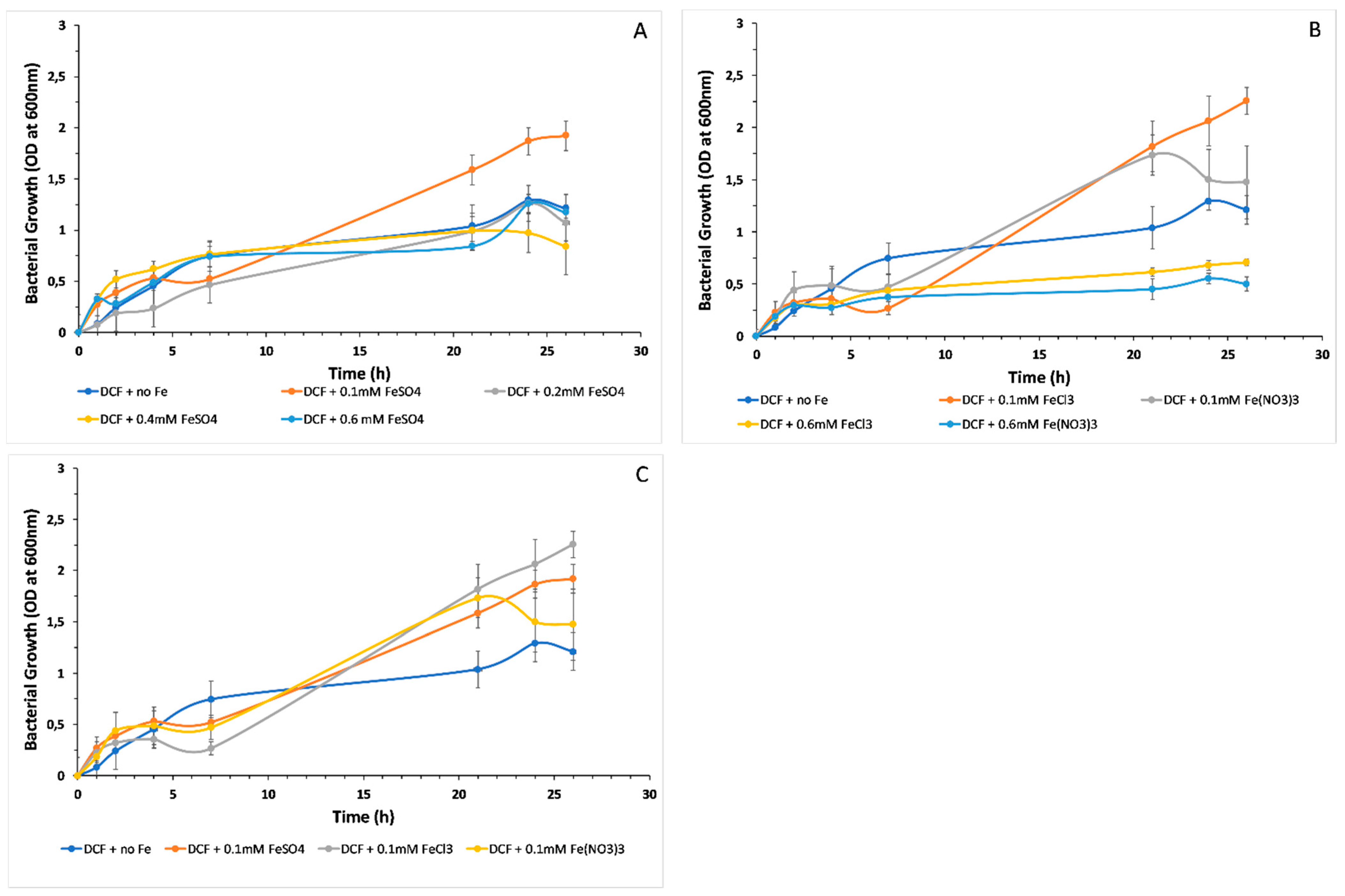
3.2. Microbial Growth and the Effect of Different Iron Forms

In this study, the effect of different iron forms, at different concentration levels, on the microbial growth of P. citronellolis isolate 620C was evaluated. In particular, FeSO4, FeCl3 and Fe(NO3)3 forms were studied based on the results reported by Santos et al. (2008) [40]. In that study, the effect of different iron forms on anthracene and other hydrocarbons degradation, and BS production was demonstrated using P. citronellolis and P. aeruginosa isolates. In the current study, to demonstrate the level of toxicity of iron forms on P. citronellolis 620C, FeSO4 supplementation was studied at concentration levels of 0.1, 0.2, 0.4 and 0.6 mM (Figure 1A). Santos et al. (2008) reported that the addition of iron forms at a concentration ≥0.5 mM was inhibitory for microbial growth. The results were compared to microbial growth without the addition of iron forms. It was observed that the addition of 0.1 mM of FeSO4 greatly enhanced microbial growth, similarly to Santos et al. 2008 [40] who chose that concentration level for further studies. Furthermore, unlike Santos et al. 2008 [40], the addition of higher than 0.1 mM concentrations of FeSO4 did not affect the growth of P. citronellolis isolate 620C.
Following that, the effect of adding FeCl3 and Fe(NO3)3 forms was studied. These iron forms were supplemented in the microbial culture at the lowest (i.e., 0.1 mM) and highest (i.e., 0.6 mM) concentration level of FeSO4. This was performed to check microbial growth at the concentration level of Fe compounds which promotes microbial growth, according to the previous experiment (i.e., 0.1 mM of FeSO4) and one above the limit of toxicity, according to Santos et al. (2008), which is 0.6 mM. Microbial growth kinetics were evaluated at regular time points (Figure 1B). The results were compared to microbial growth without the addition of iron forms. It was observed that 0.1 mM of both iron forms significantly enhanced microbial growth, while the addition of 0.6 mM resulted in the inhibition of P. citronellolis 620C growth. The latter can be attributed to the toxicity that may have been caused to the microorganism’s metabolism. These results are in accordance to Santos et al. 2008 [40]. Furthermore, the addition of 0.1 mM of the three different iron forms was compared to each other and to the microbial growth without the addition of any iron form (Figure 1C). Among the three iron forms, the addition of FeCl3 resulted in the highest microbial growth. Overall, when iron was supplemented in the microbial culture at the lowest concentration, microbial growth was significantly increased.
Several necessary functions of microbial metabolism are performed due to iron participation, such as the protection of the cells against superoxide radicals, whilst iron is also a key component of cytochromes and contributes to the Krebs cycle [41]. However, the limit between deficiency and toxicity is narrow. For instance, iron can interact with reactive oxygen species, such as hydrogen peroxide and superoxide, forming greatly damaging and reactive hydroxyl radicals. Furthermore, bacteria are highly sensitive in the presence of redox agents upon growth under iron-supplemented conditions [42]. Therefore, low amounts of iron are required.
The presence of oxygen leads to insolubility and low reactivity of Fe, and thus, low bioavailability and higher toxicity [40]. For instance, the predominant form of Fe, ferric iron, which exists in rhizosphere, is highly insoluble. Therefore, it is possible that appropriate concentrations of soluble iron forms can enhance the microbial growth, resulting in increased hydrocarbon biodegradation and, thus, higher microbial growth, as observed in the rhizosphere [43,44].
Consequently, in the present study, the effect of iron forms on microbial growth was more evident when more soluble iron forms were used. Specifically, microbial growth was further increased when FeCl3 and Fe(NO3)3 were added to the microbial cultures, compared to FeSO4 addition. The addition of more soluble forms of iron increases iron availability to the microorganism. FeSO4 is relatively insoluble in water, therefore, an increase in the concentration of FeSO4 leads to an increase in insolubility. Thus, the insignificant effect of ˃0.1 mM concentration of FeSO4 in the microbial culture may also be attributed to insolubility, which inevitably leads to a lower interaction of Fe with the microbial culture. Another cause of the lower effect of FeSO4 to the microbial growth could be that this compound may also cause microbial culture acidification [40].

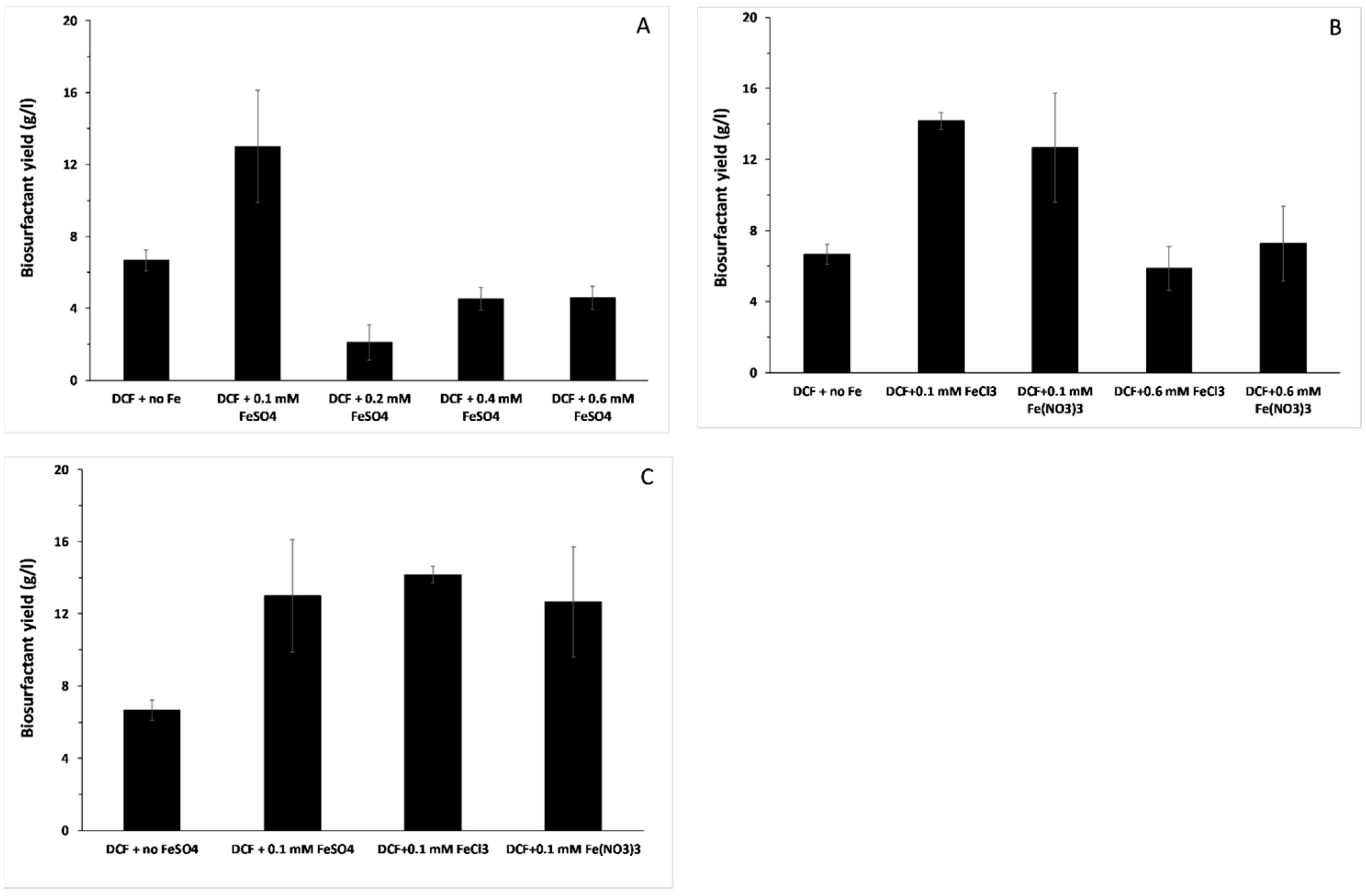
3.3. Biosurfactant Production and the Effect of Different Iron Forms

In the current study, BS production was evaluated upon supplementation of the microbial culture with FeSO4 at the concentration level of 0.1, 0.2, 0.4 and 0.6 mM (Figure 2A). Santos et al. (2008) reported that the addition of iron forms at concentrations ≥ 0.5 mM was inhibitory for microbial growth and, thus, BS production. The results were compared to BS production without the addition of iron forms. It was observed that the addition of 0.1 mM of FeSO4 greatly enhanced BS production, in line with the results of Santos et al. 2008 [40]. Specifically, the BS yield was increased by 95%, as shown in Figure 2A. Furthermore, addition of higher concentrations of FeSO4 led to lower levels of BS production than the control specimen, despite the fact that those concentration levels did not negatively affect the growth of P. citronellolis 620C. These results are consistent with those of Santos et al. 2008 [40], suggesting a strong correlation and dependence of iron and BS formation up to a limit where iron is not beneficial for BS formation. Following that, the addition of FeCl3 and Fe(NO3)3 were supplemented in the microbial culture at the lowest (i.e., 0.1 mM) and highest (i.e., 0.6 mM) concentration levels. It was observed that 0.1 mM of both iron forms greatly increased BS production. In particular, the addition of 0.1 mM of FeCl3 and Fe(NO3)3 resulted in a 112% and 90% increase, respectively, whereas the addition of 0.6 mM resulted in BS production that was lower than the control specimen (Figure 2B). The results were similar to the microbial growth results and they could be attributed to the toxicity that may have been caused to the microorganism’s metabolism, leading to low microbial growth and, thus, limited BS formation. These results are also in accordance to Santos et al. 2008 [40]. BS production was compared upon addition of 0.1 mM of the three different iron forms (Figure 2C). Among the three iron forms, the addition of FeCl3 resulted in the highest BS formation, which corresponds to the highest microbial growth. Overall, similarly to microbial growth, when iron was supplemented in the microbial culture at the lowest concentration, BS production was significantly increased. The addition of iron forms to the growth medium of Bacillus subtilis [45] and P. citronellolis strains [40] resulted in overproduction of the relevant BSs. Makkar and Cameotra (2002) also reported that metal ion addition greatly enhanced BS production [46]. Several other studies have also evaluated the positive effect of Fe nanoparticles on BS production [2,18].
Furthermore, in the present study, FeCl3 addition resulted in the highest BS yield, over-doubling the production. This is in line with Dinkla et al. (2001) [43], who reported the positive effect of FeCl3 on BS formation upon toluene biodegradation. In another study of the production of BS by marine Nocardiopsis lucentensis MSA04 in solid-state cultivation, FeCl3 was found to be the most suitable metal precursor [47]. Therefore, the addition of more soluble forms of iron at adequate concentrations could be a key strategy for the optimization of BS production.

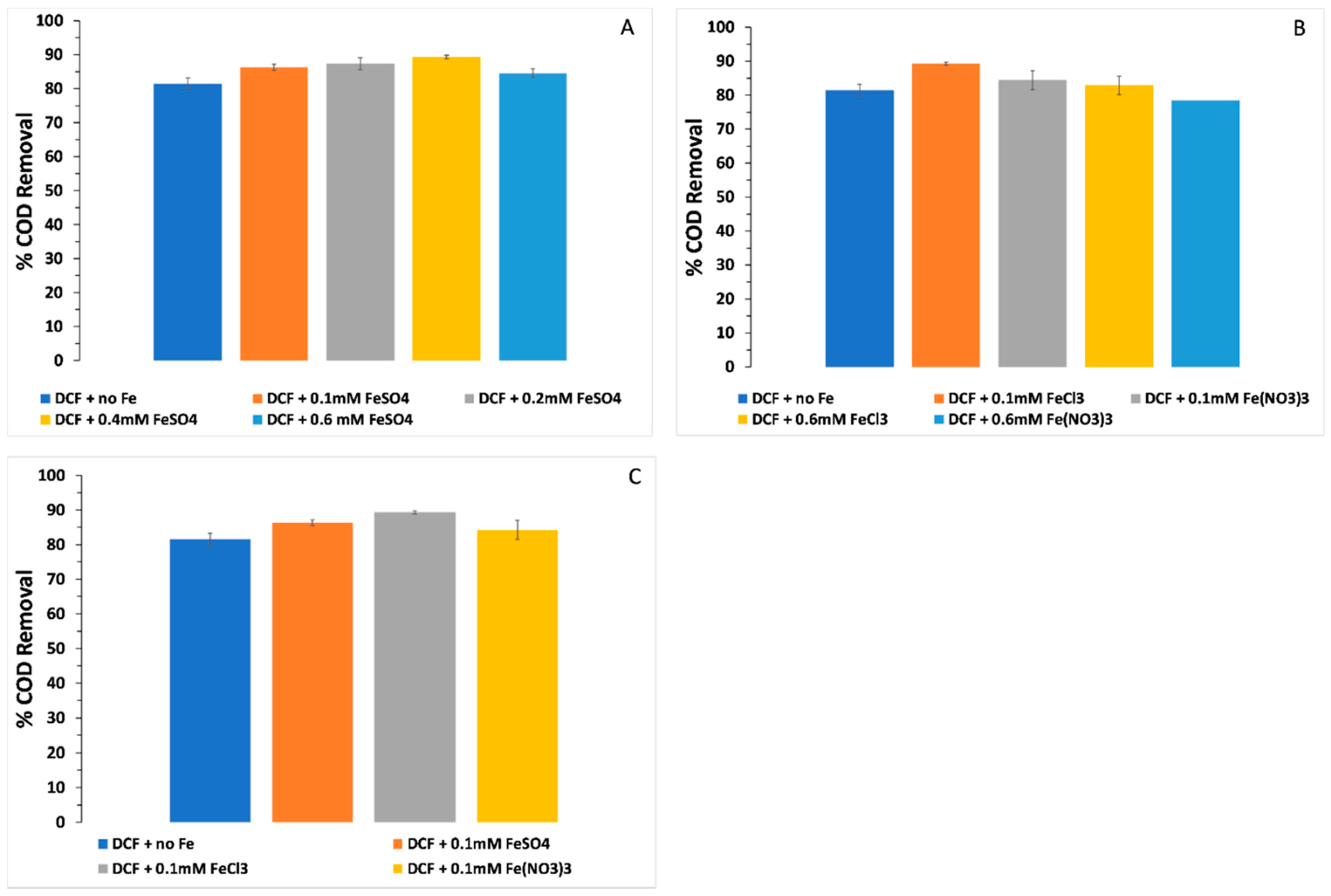
3.4. COD Removal and the Effect of Different Iron Forms

BSs enhance the bioremediation of oil waste [14]. Therefore, COD was studied to observe the effect of enhanced biodegradation due to BS production. The effect of the addition of the different iron forms was also evaluated through COD measurement. The experimental conditions were similar as those mentioned above (Section 3.2). The FeSO4 effect on % COD removal of DCF was studied upon the addition of 0.1, 0.2, 0.4 and 0.6 mM (Figure 3A), whereby a slight increase in the % COD removal was observed, compared to the control experiment (i.e., without iron addition). The results followed the microbial growth results, suggesting that FeSO4 addition does not greatly affect the metabolic pathways related to hydrocarbons and biomass formation activities. Following that, the effect of addition of 0.1 and 0.6 mM FeCl3 and Fe(NO3)3 was studied (Figure 3B), whereby at 0.1 mM, both forms increased the % COD removal, with the effect of FeCl3 being more pronounced, in agreement with the previous results of the present study. Additionally, at 0.1 mM addition, among the three iron forms, FeCl3, similarly to the microbial growth and BS production, further enhanced the % COD removal, demonstrating its positive effect in the bioprocess (Figure 3C).
It was noticed that, although the different iron forms resulted in increased % COD removal, compared to the COD removal without the supplementation of iron forms, the removal was not as high as that of the COD removal reported in other studies (e.g., Santos et al., 2008), whereby pure hydrocarbon forms were used, such as anthracene. It is suggested that the higher COD removal observed in the microbial cultures supplemented with iron was due to the higher availability of iron for induction, expression and activity of the enzymes catalyzing hydrocarbon biodegradation, as iron is a key element of oxygenases [48]. Furthermore, at low iron concentration, the direct dependence of the BS on the presence of iron, which resulted in BS over-production, probably increased hydrocarbon bioavailability to degradation, thus, leading to an increase in the % COD removal.
DCF is a highly toxic waste stream which, unless safely disposed, can lead to an adverse impact on human health and the environment due to its hazardous components and additives [49]. DCF biodegradation is an alternative eco-friendly approach for disposal management which is enhanced by BSs produced during the microbial bioprocess [50]. Therefore, the potential of P. citronellolis 620C to be used in a scaled-up bioprocess was examined. The enhancement of DCF biodegradation through the use of the BS-producing P. citronellolis 620C was also evaluated at higher than 1% (v/v) DCF concentrations (i.e., 5% and 10%) through monitoring of microbial growth kinetics and % COD removal. Despite the toxicity of the DCF (initial COD: 41.000 mg/L), the % COD removal was promising (i.e., 42% and 27.5%, respectively) (Supplementary Material Figure S1).

3.5. Biosurfactant Characterization
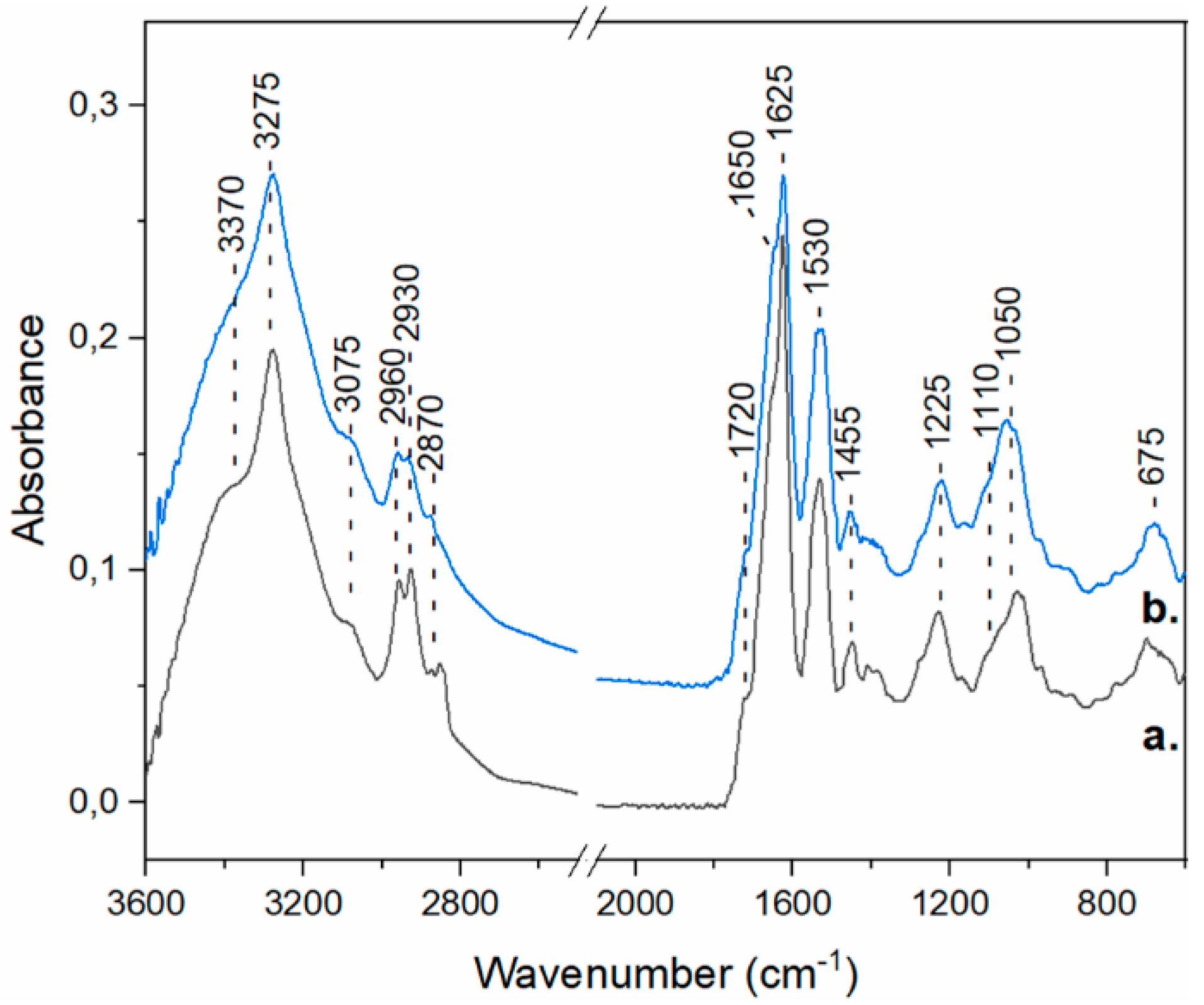
3.5.1. FTIR

FTIR spectroscopy is a valuable tool in the identification of the chemical structure of compounds, as FTIR spectra comprise bands that are characteristic for specific chemical bonds. In this work, the ATR-FTIR approach was employed, as it offers the advantage of working with solid or liquid samples with minimal sample preparation prior to spectral measurements. The ATR-FTIR spectra of the powder form of the BS are shown in Figure 4, with traces a and b corresponding to the spectra of the BS produced by the P. citronellolis 620C culture in the absence and presence of FeSO4, respectively. The vibrations detected in the ATR-FTIR spectra are characteristic of a peptide structure [51]. More specifically, the bands at 3275 and 3075 cm−1 were assigned to the amide A and amide B vibrations (N-H stretch) of the peptide, while contributions from O-H bonds in the 3000–3500 cm−1 region cannot be excluded. The peptide backbone, additionally, gave rise to the amide I vibrations (C=O stretch) observed at 1650 and 1625 cm−1, and to the amide II vibration (out-of-phase combination of the NH in plane bend and the CN stretching vibration) identified at 1530 cm−1 [51]. Furthermore, peaks originating from the C-H stretch of CH3 and CH2 groups were detected at 2960, 2930 and 2870 cm−1, demonstrating the presence of alkyl groups [51,52]. To identify additional functional groups in the BS, the 1300-1000 cm−1 region was examined. In this region, bands could be observed at 1225, 1110 and 1050 cm−1. These vibrations were attributed primarily to C-C and C-O(H) bonds and in conjunction with the contribution of O-H bonds in the ~3370 cm−1 region, they provide an indication of the presence of carbohydrate moieties in the peptide [52,53]. Finally, it should be noted that a shoulder was observed in the amide I band at 1720 cm−1. This band could have originated from the C=O stretch of Glu or Asp carboxylic acid side chains or from lipid ester group; therefore, a lipopeptide structure is also possible [51,54]. Overall, from the analysis of the ATR-FTIR spectra of the BS described herein and considering the similarity of these spectra to the FTIR spectra of biosurfactants produced by lactic acid [52] and marine [55] bacteria, a glycopeptide or lipopeptide structure is proposed. Furthermore, the FTIR spectra of the BS produced in the absence and presence of FeSO4 were similar, suggesting that Supplementation of the microbial culture with FeSO4 did not affect the structure of the BS.

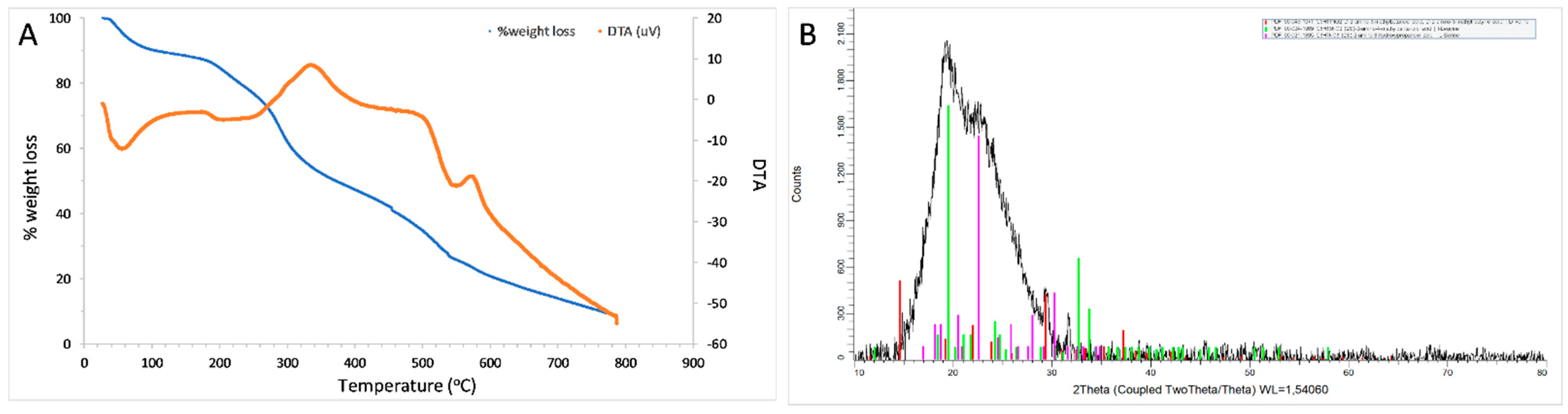
3.5.2. TGA, DTA and XRD

TGA/DTA, and XRD analyses were performed for the partially purified BS extracted by the microbial culture, where 1% DCF was used as the substrate. The thermal stability of BSs is a key property that is relevant to the applicability of these molecules in industrial and commercial processes [56]. In the first step of TGA (Figure 5A), during the temperature range 27–100 °C, the loss of BS weight due to the loss of water and solvent molecules was estimated at 9.73 %. Thermal degradation takes place upon mass loss of at least 5% [57]. In the current analysis, following the initial weight loss, 5% loss of BS weight was observed at 196. 4 °C. This step was followed by a rapid weight loss of 31.22% until 338.1 °C, which could be attributed to the degradation of thermolabile content, such as carbohydrates and peptides in the BS. The thermal decomposition at 338.1 °C was confirmed by the exothermic peak of the DTA analysis. These results are similar to those reported by Weiss et al. 2018 [58], where thermal decomposition of several amino acids (i.e., asparagine, glycine, glutamic acid, cysteine, aspartic acid, histidine, arginine and glutamine) was studied, demonstrating decomposition between 200–300 °C. The non-thermolabile content was gradually degraded, resulting in BS weight loss of 27.91 % over the temperature range of 338.1–546.9 °C. This could be attributed to fatty acids, which are commonly decomposed over a temperature of 300 °C [59]. At 546.9 °C, another exothermic peak on the DTA graph was noticed, further confirming the thermal decomposition. As determined from the TGA and DTA graphs, the degradation temperature of BS was found to be 546.9 °C, while complete weight loss took place after 800 °C. Therefore, the BS formed by P. citronellolis isolate 620C was determined to be thermostable. A similar TGA graph of a lipoprotein BS, produced by Bacillus subtilis, has been established [60].
The powder XRD pattern (Figure 5B) indicated a highly (57.2%) amorphous BS with crystallinity index (CIxrd) equal to 0.428. XRD was used to qualitatively assess the amino acids in the BS. Peptides are usually detected between 20–30° 2θ [61]. As confirmed by the FTIR analysis, the structure of the BS formed by the P. citronellolis 620C contains amino acids, the building blocks of peptides.

3.6. Antimicrobial Activity of the Biosurfactant

The antimicrobial activity was studied using the solution of the partially purified BS, extracted by the P. citronellolis 620C culture with 1% (v/v) DCF. The BS showed antimicrobial activity against all the multi-drug resistant Gram-positive and negative microorganisms tested (Table 1).
Firstly, the antimicrobial activity of the BS was tested at a concentration level of 1 mg/mL (Table 1). This concentration level is commonly used in the agar susceptibility method [62,63]. This BS concentration inhibited the growth of the Gram-negative bacteria K. pneumoniae, P. aeruginosa, E. coli resistant and E. coli. In contrast, this level of BS concentration did not inhibit the growth of the Gram-negative A. baumaniii and all the Gram-positive bacteria (i.e., E. faecalis resistant, E. faecalis, S. pneumoniae, S. aureus, S. enterica). These pathogens were inhibited at 10 mg/mL BS concentration (Table 1). These results indicate that some bacterial pathogens can be more resistant to antimicrobial agents compared to others. Commonly, the inhibition of BS on growth of bacterial pathogens is studied in a broad concentration range from few μg (e.g., 4.9 μg) [64] to 100 mg/mL [65]. Most probably, this is dependent on the chemical compartments which are contained in a BS, resulting in the antimicrobial activity, purification of the BS tested and the method used which can either be the agar susceptibility method or the micro-broth dilution technique. BSs, such as rhamnolipids at a concentration level of 1.12 mg/mL, caused an inhibition zone similar to that in the present study, against several Gram-negative and -positive bacterial pathogens [63]. The rhamnolipids were isolated and partially purified using a method similar to that of the present study.
Antimicrobial properties are more common for glycolipids or lipopeptides [62]. Most of the biosurfactants which possess antimicrobial properties have usually been produced from microorganisms isolated from contaminated and domestic sites [66]. Similarly, P. citronellolis 620C was isolated from a tank containing drilling waste.
The antimicrobial activity of BSs relies on the outer membrane of pathogens, which can be disrupted by the hydrophobic domain of the BSs, thus, breaking the structure of microorganisms and disabling pathogen activity. Therefore, the current BS has the potential to act as an alternative natural antibiotic, when tested against human cells. Furthermore, the current BS also has the potential to act as a disinfectant. Disinfectants are chemical means employed to inactivate pathogens, which are not inhibited anymore due to antimicrobial resistance [67]. Thus, further research on the toxicity of the BS to human skin and metabolic pathways is necessary to establish its antibiotic or disinfectant potential.
Although in recent years several studies on the antimicrobial activity of BSs have been performed, such studies are debated. This is because of the diversity of methodologies which have been employed to establish the activity and the different purity levels of the examined BSs. Upon use of the agar susceptibility method, Das et al., 2008a [62] provided a solution of the partially purified BS in methanol. The latter could be the main factor for bacterial pathogens lethality in that study. Furthermore, in other studies, the concentration of the BS used to determine antimicrobial activity was not mentioned, such as that of Ilori et al. (2008) [68]. Over recent years, materials and methods analysis of antimicrobial activity has become more detailed.
It has to be pointed out, that the majority of the studies found by the author exploit the antimicrobial activity of the purified BS acquired through the solvent extraction method described in Section 2.5. Therefore, biological activity can be attributed to the presence of several compounds, which may act synergistically, enabling antimicrobial action. Further purification of the BS may enhance antimicrobial properties. This is achieved through the exhaustive use of solvents and extraction steps [69] which, however, greatly increases the cost of the bioprocess, rendering down-stream processing unattractive. In the present study, the antimicrobial potential of the partially purified BS was examined, without further purification steps, leading to promising results and a promising, less-costly, down-stream processing method of the BS. However, further research on the purity, chemical structure and identification is necessary.

3.7. MIC of the Biosurfactant

The commonly used halo technique, using the agar disc diffusion method, was employed to determine the MIC of the BS (Table 2). The partially purified BS was extracted from the P. citronellolis 620C culture using 1% (v/v) DCF. The solution of the BS was able to inhibit bacterial growth in concentrations as low as 0.25 mg/mL. K. pneumoniae was affected at such a low BS concentration. P. aeruginosa, E. coli resistant and E. coli were observed to be inhibited at a relatively higher BS concentration (0.5 mg/mL). The BS inhibited the E. faecalis and S. enterica at concentrations as low as 5 mg/mL. However, in the agar plate of E. faecalis, only one out of four discs impregnated with BS solution showed an inhibition zone, resulting in a high SD (Table 2). Furthermore, A. baumaniii, S. pneumoniae and S. aureus were inhibited at relatively higher concentrations of 10 mg/mL. It was noticed that the Gram-positive pathogens were more resilient and insensitive to the BS solution, compared to the Gram-negative strains. Specifically, at least five and in most cases 10 times more BS solution was needed to inhibit these multi-drug resistant bacteria. This could be attributed to their structure, whereby the cell wall is relatively harder to break, compared to that of the Gram-negative bacteria. Interestingly, a higher dose of the BS solution was essential to observe inhibition of the Gram-negative A. baumaniii, demonstrating the high resistance of the pathogen [70], but also the importance of the antimicrobial activity of the current BS, which can efficiently inhibit the growth of this emerging pathogen. The differences observed in the sensitivity of the pathogens to the BS do not only depend on the composition of the BS. The antimicrobial activity, except of the purity of the BS, may also be influenced by the nutritional and environmental conditions, and adaptation of the bacterial pathogens [71].
MIC may be performed for highly pure BSs, such as rhamnolipids, reaching 99% purity [64]. Despite the purity level, the inhibition zone observed against E. coli, S. aureus and S. enterica is similar to that of the current study. The inhibition zone against E. coli strains was also similar to other studies such as Onbasli & Aslim (2008) [72].

4. Conclusions

In the current study, (i) a novel BS-producing microorganism was isolated from a waste site, (ii) BS formation was a result of DW biodegradation, (iii) an optimization strategy of BS production was explored through supplementation of iron forms, which led to a great production enhancement upon induction with 0.1 mM. Among FeSO4, FeCl3 and Fe(NO3)3, addition of FeCl3 resulted in the highest increase in both BS, microbial growth and DW biodegradation. Use of a waste stream as substrate may result in a significant reduction in the bioprocess cost. The partially purified BS has promising antimicrobial potential against several Gram-negative and -positive multi-drug resistant bacterial pathogens. Hence, as no extra purification steps are necessary, a reduction in down-stream processing costs is possible. Preliminary characterization of the aforementioned BS via a number of analytical methods established its thermostability, low crystallinity and the presence of a peptide structure. The BS hereby produced and studied appears to have great potential in biomedical and environmental biotechnological applications.

Supplementary Materials

The following are available online at https://www.mdpi.com/article/10.3390/pr9040686/s1, Figure S1: Bacterial growth and % COD removal upon biodegradation of 5 and 10% (v/v) DCF, Table S1: Top 10 hits of the BLAST results.

Author Contributions

A.T.: conceptualization, methodology, investigation, experimentation, validation, writing original manuscript, reviewing, editing. K.S.: methodology, investigation, experimentation for extraction of the BS, iron forms effect, antimicrobial properties of BS. M.P.: methodology, investigation, experimentation for extraction of the BS, iron forms effect. L.K.: methodology, TGA/DTA analysis. I.R.: methodology, XRD analysis, reviewing and editing of the manuscript. I.I.: methodology, TGA/DTA and XRD analysis, reviewing and editing of the manuscript. E.P. (Erato Papageorgiou): methodology, FTIR analysis. E.P. (Eftychia Pinakoulaki): methodology, FTIR analysis, writing of FTIR results, reviewing and editing of the manuscript. All authors have read and agreed to the published version of the manuscript.

Funding

This research was funded by University of Cyprus, Cyprus. The APC was funded by the Department of Civil and Environmental Engineering, University of Cyprus, Cyprus.

Institutional Review Board Statement

Not applicable.

Informed Consent Statement

Not applicable.

Data Availability Statement

Data available on request due to privacy restrictions. The data presented in this study are available on request from the corresponding author.

Acknowledgments

The corresponding author would like to thank IESC Ltd. (Limassol, Cyprus) for providing the drilling waste. Furthermore, the corresponding author would like to thank researcher Anna Korelidou for providing the multi-drug resistant strains isolated from the municipal wastewater treatment of Limassol and the University of Cyprus for financial support.

Conflicts of Interest

The authors declare no conflict of interest.

References

  1. Hemlata, B.; Selvin, J.; Tukaram, K. Optimization of iron chelating biosurfactant production by Stenotrophomonas maltophilia NBS-11. Biocatal. Agric. Biotechnol. 2015, 4, 135–143. [Google Scholar] [CrossRef]
  2. Liu, J.; Vipulanandan, C.; Cooper, T.F.; Vipulanandan, G. Effects of Fe nanoparticles on bacterial growth and biosurfactant production. J. Nanoparticle Res. 2013, 15, 1–13. [Google Scholar] [CrossRef]
  3. Banat, I.M.; Makkar, R.S.; Cameotra, S.S. Potential commercial applications of microbial surfactants. Appl. Microbiol. Biotechnol. 2000, 53, 495–508. [Google Scholar] [CrossRef]
  4. Naughton, P.; Marchant, R.; Naughton, V.; Banat, I. Microbial biosurfactants: Current trends and applications in agricultural and biomedical industries. J. Appl. Microbiol. 2019, 127, 12–28. [Google Scholar] [CrossRef] [PubMed] [Green Version]
  5. Dhiman, R.; Meena, K.R.; Sharma, A.; Kanwar, S.S. Biosurfactants and their Screening Methods Biosurfactants and their Screening Methods. Res. J. Recent Sci. 2016, 5, 1–6. [Google Scholar]
  6. Shekhar, S.; Sundaramanickam, A.; Balasubramanian, T. Biosurfactant Producing Microbes and their Potential Applications: A Review. Crit. Rev. Environ. Sci. Technol. 2015, 45, 1522–1554. [Google Scholar] [CrossRef]
  7. Markande, A.R.; Patel, D.; Varjani, S. A review on biosurfactants: Properties, applications and current developments. Bioresour. Technol. 2021, 330, 124963. [Google Scholar] [CrossRef] [PubMed]
  8. Smith, M.L.; Gandolfi, S.; Coshall, P.M.; Rahman, P.K.S.M. Biosurfactants: A Covid-19 Perspective. Front. Microbiol. 2020, 11, 1341. [Google Scholar] [CrossRef] [PubMed]
  9. Franzetti, A.; Gandolfi, I.; Bestetti, G.; Smyth, T.J.P.; Banat, I.M. Production and applications of trehalose lipid biosurfactants. Eur. J. Lipid Sci. Technol. 2010, 112, 617–627. [Google Scholar] [CrossRef]
  10. Kaskatepe, B.; Yildiz, S. Rhamnolipid Biosurfactants Produced by Pseudomonas Species. Braz. Arch. Biol. Technol. 2016, 59, 1–16. [Google Scholar] [CrossRef] [Green Version]
  11. Jimoh, A.A.; Lin, J. Biosurfactant: A new frontier for greener technology and environmental sustainability. Ecotoxicol. Environ. Saf. 2019, 184, 109607. [Google Scholar] [CrossRef] [PubMed]
  12. Santos, D.K.; Rufino, R.D.; Luna, J.M.; Santos, V.A.; Salgueiro, A.A.; Sarubbo, L.A. Synthesis and evaluation of biosurfactant produced by Candida lipolytica using animal fat and corn steep liquor. J. Pet. Sci. Eng. 2013, 105, 43–50. [Google Scholar] [CrossRef]
  13. Onwukwe, S.I.; Nwakaudu, M.S. Drilling Wastes Generation and Management Approach. Int. J. Environ. Sci. Dev. 2012, 3, 252–257. [Google Scholar] [CrossRef]
  14. Karlapudi, A.P.; Venkateswarulu, T.; Tammineedi, J.; Kanumuri, L.; Ravuru, B.K.; Dirisala, V.R.; Kodali, V.P. Role of biosurfactants in bioremediation of oil pollution-a review. Petroleum 2018, 4, 241–249. [Google Scholar] [CrossRef]
  15. Yan, P.; Lu, M.; Yang, Q.; Zhang, H.-L.; Zhang, Z.-Z.; Chen, R. Oil recovery from refinery oily sludge using a rhamnolipid biosurfactant-producing Pseudomonas. Bioresour. Technol. 2012, 116, 24–28. [Google Scholar] [CrossRef]
  16. Elshafie, A.E.; Joshi, S.J.; Al-Wahaibi, Y.M.; Al-Bemani, A.S.; Al-Bahry, S.N.; Al-Maqbali, D.; Banat, I.M. Sophorolipids Production by Candida bombicola ATCC 22214 and its Potential Application in Microbial Enhanced Oil Recovery. Front. Microbiol. 2015, 6, 1324. [Google Scholar] [CrossRef] [Green Version]
  17. Datta, P.; Tiwari, P.; Pandey, L.M. Isolation and characterization of biosurfactant producing and oil degrading Bacillus subtilis MG495086 from formation water of Assam oil reservoir and its suitability for enhanced oil recovery. Bioresour. Technol. 2018, 270, 439–448. [Google Scholar] [CrossRef]
  18. Kiran, G.S.; Nishanth, L.A.; Priyadharshini, S.; Anitha, K.; Selvin, J. Effect of Fe nanoparticle on growth and glycolipid biosurfactant production under solid state culture by marine Nocardiopsissp. MSA13A. BMC Biotechnol. 2014, 14, 48. [Google Scholar] [CrossRef] [PubMed] [Green Version]
  19. Jimoh, A.A.; Lin, J. Enhancement of Paenibacillus sp. D9 Lipopeptide Biosurfactant Production Through the Optimization of Medium Composition and Its Application for Biodegradation of Hydrophobic Pollutants. Appl. Biochem. Biotechnol. 2019, 187, 724–743. [Google Scholar] [CrossRef]
  20. Haferburg, G.; Kothe, E. Microbes and metals: Interactions in the environment. J. Basic Microbiol. 2007, 47, 453–467. [Google Scholar] [CrossRef] [PubMed]
  21. Sambrook, J.; Fritsch, E.F.; Maniatis, E. Molecular Cloning: A Laboratory Manual; Cold Spring Harbour Press: Cold Spring Harbor, NY, USA, 1989. [Google Scholar]
  22. Suryanti, V.; Marliyana, S.D.; Handayani, D.S.; Ratnaningrum, D. Production and characterization of biosurfactant by pseudomonas fluorescens using cassava flour wastewater as media. Indones. J. Chem. 2013, 13, 229–235. [Google Scholar] [CrossRef] [Green Version]
  23. De Rienzo, M.A.D.; Stevenson, P.; Marchant, R.; Banat, I.M. Antibacterial properties of biosurfactants against selected Gram-positive and -negative bacteria. FEMS Microbiol. Lett. 2016, 363, fnv224. [Google Scholar] [CrossRef] [PubMed] [Green Version]
  24. Desai, J.D.; Banat, I.M. Microbial production of surfactants and their commercial potential. Microbiol. Mol. Biol. Rev. 1997, 61, 47–64. [Google Scholar] [CrossRef]
  25. Huck-Iriart, C.; De-Candia, A.; Rodriguez, J.; Rinaldi, C. Determination of Surface Tension of Surfactant Solutions through Capillary Rise Measurements: An Image-Processing Undergraduate Laboratory Experiment. J. Chem. Educ. 2016, 93, 1647–1651. [Google Scholar] [CrossRef]
  26. Eldin, A.M.; Kamel, Z.; Hossam, N. Isolation and genetic identification of yeast producing biosurfactants, evaluated by different screening methods. Microchem. J. 2019, 146, 309–314. [Google Scholar] [CrossRef]
  27. Wang, Q.; Fang, X.; Bai, B.; Liang, X.; Shuler, P.J.; Iii, W.A.G.; Tang, Y. Engineering bacteria for production of rhamnolipid as an agent for enhanced oil recovery. Biotechnol. Bioeng. 2007, 98, 842–853. [Google Scholar] [CrossRef] [PubMed]
  28. Remus-Emsermann, M.N.; Schmid, M.; Gekenidis, M.-T.; Pelludat, C.; Frey, J.E.; Ahrens, C.H.; Drissner, D. Complete genome sequence of Pseudomonas citronellolis P3B5, a candidate for microbial phyllo-remediation of hydrocarbon-contaminated sites. Stand. Genom. Sci. 2016, 11, 1–12. [Google Scholar] [CrossRef] [PubMed] [Green Version]
  29. Zheng, D.; Wang, X.; Wang, P.; Peng, W.; Ji, N.; Liang, R. Genome Sequence of Pseudomonas citronellolis SJTE-3, an Estrogen- and Polycyclic Aromatic Hydrocarbon-Degrading Bacterium. Genome Announc. 2016, 4, 16–17. [Google Scholar] [CrossRef] [PubMed] [Green Version]
  30. Oso, S.; Walters, M.; Schlechter, R.O.; Remus-Emsermann, M.N.P. Utilisation of hydrocarbons and production of surfactants by bacteria isolated from plant leaf surfaces. FEMS Microbiol. Lett. 2019, 366, 1–10. [Google Scholar] [CrossRef]
  31. Singh, P.; Tiwary, B.N. Isolation and characterization of glycolipid biosurfactant produced by a Pseudomonas otitidis strain isolated from Chirimiri coal mines, India. Bioresour. Bioprocess. 2016, 3, 42. [Google Scholar] [CrossRef] [Green Version]
  32. Ron, E.Z.; Rosenberg, E. Biosurfactants and oil bioremediation. Curr. Opin. Biotechnol. 2002, 13, 249–252. [Google Scholar] [CrossRef]
  33. Haidar, C.N.; Pereira, M.M.; Lima, Á.S.; Nerli, B.B.; Malpiedi, L.P. Biosurfactants produced by Pseudomonas syringae pv tabaci: A versatile mixture with interesting emulsifying properties. Process. Biochem. 2020, 97, 121–129. [Google Scholar] [CrossRef]
  34. Meliani, A. Enhancement of Hydrocarbons Degradation by Use of Pseudomonas Biosurfactants and Biofilms. J. Pet. Environ. Biotechnol. 2014, 05, 1–7. [Google Scholar] [CrossRef] [Green Version]
  35. Rosenberg, E.; Ron, E.Z. Biosurfactants. In The Prokaryotes: Applied Bacteriology and Biotechnology; Rosenberg, E.F., DeLong, S., Lory, E., Stackebrandt, F., Thompson, E., Eds.; Springer: Berlin/Heidelberg, Germany, 2013; pp. 281–294. [Google Scholar] [CrossRef]
  36. Abouseoud, M.; Maachi, R.; Amrane, A.; Boudergua, S.; Nabi, A. Evaluation of different carbon and nitrogen sources in production of biosurfactant by Pseudomonas fluorescens. Desalination 2008, 223, 143–151. [Google Scholar] [CrossRef]
  37. Bak, F.; Bonnichsen, L.; Jørgensen, N.O.G.; Nicolaisen, M.H.; Nybroe, O. The biosurfactant viscosin transiently stimulates n-hexadecane mineralization by a bacterial consortium. Appl. Microbiol. Biotechnol. 2015, 99, 1475–1483. [Google Scholar] [CrossRef] [Green Version]
  38. Ismail Isa, T.; Ngoshe, I.Y.; Gajere, H.M. Biosurfactant Production by Bacteria Isolated from Hydrocarbon-impacted soil. Bioremed. Sci. Technol. Res. 2019, 6, 4–8. [Google Scholar]
  39. Jacques, R.J.S.; Santos, E.C.; Haddad, R.; Catharino, R.R.; Eberlin, M.N.; Bento, F.M.; Camargo, F.A.D.O. Mass spectrometry analysis of surface tension reducing substances produced by a pah-degrading Pseudomonas citronellolis strain. Braz. J. Microbiol. 2008, 39, 353–356. [Google Scholar] [CrossRef] [Green Version]
  40. Santos, E.C.; Jacques, R.J.; Bento, F.M.; Peralba, M.D.C.R.; Selbach, P.A.; Sá, E.L.; Camargo, F.A. Anthracene biodegradation and surface activity by an iron-stimulated Pseudomonas sp. Bioresour. Technol. 2008, 99, 2644–2649. [Google Scholar] [CrossRef]
  41. Wei, Y.-H.; Wang, L.-F.; Changy, J.-S.; Kung, S.-S. Identification of induced acidification in iron-enriched cultures of Bacillus subtilis during biosurfactant fermentation. J. Biosci. Bioeng. 2003, 96, 174–178. [Google Scholar] [CrossRef]
  42. Andrews, S.C.; Robinson, A.K.; Rodrı́guez-Quiñones, F. Bacterial iron homeostasis. FEMS Microbiol. Rev. 2003, 27, 215–237. [Google Scholar] [CrossRef]
  43. Dinkla, I.J.T.; Gabor, E.M.; Janssen, D.B. Effects of Iron Limitation on the Degradation of Toluene by Pseudomonas Strains Carrying the TOL (pWWO) Plasmid. Appl. Environ. Microbiol. 2001, 67, 3406–3412. [Google Scholar] [CrossRef] [Green Version]
  44. Dinkla, I.; Janssen, D. Simultaneous Growth on Citrate Reduces the Effects of Iron Limitation during Toluene Degradation in Pseudomonas. Microb. Ecol. 2003, 45, 97–107. [Google Scholar] [CrossRef] [Green Version]
  45. Wei, Y.-H.; Chu, I.-M. Enhancement of surfactin production in iron-enriched media by bacillus subtilis ATCC 21332. Enzym. Microb. Technol. 1998, 22, 724–728. [Google Scholar] [CrossRef]
  46. Makkar, S.C.R. An update on the use of unconventional substrates for biosurfactant production and their new applications. Appl. Microbiol. Biotechnol. 2002, 58, 428–434. [Google Scholar] [CrossRef]
  47. Kiran, G.S.; Thomas, T.A.; Selvin, J. Production of a new glycolipid biosurfactant from marine Nocardiopsis lucentensis MSA04 in solid-state cultivation. Colloids Surf. B Biointerfaces 2010, 78, 8–16. [Google Scholar] [CrossRef]
  48. Dunham, N.P.; Arnold, F.H. Nature’s Machinery, Repurposed: Expanding the Repertoire of Iron-Dependent Oxygenases. ACS Catal. 2020, 10, 12239–12255. [Google Scholar] [CrossRef]
  49. Ismail, A.R.; Alias, A.H.; Sulaiman, W.R.W.; Jaafar, M.Z.; Ismail, I. Drilling fluid waste management in drilling for oil and gas wells. Chem. Eng. Trans. 2017, 56, 1351–1356. [Google Scholar] [CrossRef]
  50. De Almeida, D.G.; Silva, R.D.C.F.S.D.; Luna, J.M.; Rufino, R.D.; Santos, V.A.; Banat, I.M.; Sarubbo, L.A. Biosurfactants: Promising Molecules for Petroleum Biotechnology Advances. Front. Microbiol. 2016, 7, 1718. [Google Scholar] [CrossRef] [PubMed] [Green Version]
  51. Barth, A. Infrared spectroscopy of proteins. Biochim. Biophys. Acta (BBA) Bioenerg. 2007, 1767, 1073–1101. [Google Scholar] [CrossRef] [PubMed] [Green Version]
  52. Moldes, A.B.; Paradelo, R.; Vecino, X.; Cruz, J.M.; Gudiña, E.; Rodrigues, L.; Teixeira, J.A.; Domínguez, J.M.; Barral, M.T. Partial Characterization of Biosurfactant fromLactobacillus pentosusand Comparison with Sodium Dodecyl Sulphate for the Bioremediation of Hydrocarbon Contaminated Soil. BioMed Res. Int. 2013, 2013, 1–6. [Google Scholar] [CrossRef] [PubMed] [Green Version]
  53. Derenne, A.; Derfoufi, K.-M.; Cowper, B.; Delporte, C.; Goormaghtigh, E. FTIR spectroscopy as an analytical tool to compare glycosylation in therapeutic monoclonal antibodies. Anal. Chim. Acta 2020, 1112, 62–71. [Google Scholar] [CrossRef] [PubMed]
  54. Krilov, D.; Balarin, M.; Kosovic, M.; Gamulin, O.; Brnjas-Kraljević, J. FT-IR spectroscopy of lipoproteins—A comparative study. Spectrochim. Acta Part A: Mol. Biomol. Spectrosc. 2009, 73, 701–706. [Google Scholar] [CrossRef] [PubMed] [Green Version]
  55. Kiran, G.S.; Priyadharsini, S.; Sajayan, A.; Priyadharsini, G.B.; Poulose, N.; Selvin, J. Production of Lipopeptide Biosurfactant by a Marine Nesterenkonia sp. and Its Application in Food Industry. Front. Microbiol. 2017, 8, 1138. [Google Scholar] [CrossRef] [PubMed]
  56. Chandankere, R.; Yao, J.; Cai, M.; Masakorala, K.; Jain, A.; Choi, M.M. Properties and characterization of biosurfactant in crude oil biodegradation by bacterium Bacillus methylotrophicus USTBa. Fuel 2014, 122, 140–148. [Google Scholar] [CrossRef]
  57. Kourmentza, C.; Costa, J.; Azevedo, Z.; Servin, C.; Grandfils, C.; De Freitas, V.; Reis, M. Burkholderia thailandensis as a microbial cell factory for the bioconversion of used cooking oil to polyhydroxyalkanoates and rhamnolipids. Bioresour. Technol. 2018, 247, 829–837. [Google Scholar] [CrossRef]
  58. Weiss, I.M.; Muth, C.; Drumm, R.; Kirchner, H.O.K. Thermal decomposition of the amino acids glycine, cysteine, aspartic acid, asparagine, glutamic acid, glutamine, arginine and histidine. BMC Biophys. 2018, 11, 1–15. [Google Scholar] [CrossRef] [Green Version]
  59. Reeves, C.J.; Menezes, P.L.; Jen, T.-C.; Lovell, M.R. The influence of fatty acids on tribological and thermal properties of natural oils as sustainable biolubricants. Tribol. Int. 2015, 90, 123–134. [Google Scholar] [CrossRef]
  60. Saranya, P.; Swarnalatha, S.; Sekaran, G. Lipoprotein biosurfactant production from an extreme acidophile using fish oil and its immobilization in nanoporous activated carbon for the removal of Ca2+and Cr3+in aqueous solution. RSC Adv. 2014, 4, 34144–34155. [Google Scholar] [CrossRef]
  61. Hutchinson, J.A.; Hamley, I.W.; Torras, J.; Alemán, C.; Seitsonen, J.; Ruokolainen, J. Self-Assembly of Lipopeptides Containing Short Peptide Fragments Derived from the Gastrointestinal Hormone PYY3–36: From Micelles to Amyloid Fibrils. J. Phys. Chem. B 2019, 123, 614–621. [Google Scholar] [CrossRef] [PubMed]
  62. Das, P.; Mukherjee, S.; Sen, R. Antimicrobial potential of a lipopeptide biosurfactant derived from a marine Bacillus circulans. J. Appl. Microbiol. 2008, 104, 1675–1684. [Google Scholar] [CrossRef]
  63. Ndlovu, T.; Rautenbach, M.; Vosloo, J.A.; Khan, S.; Khan, W. Characterisation and antimicrobial activity of biosurfactant extracts produced by Bacillus amyloliquefaciens and Pseudomonas aeruginosa isolated from a wastewater treatment plant. AMB Express 2017, 7, 1–19. [Google Scholar] [CrossRef] [PubMed] [Green Version]
  64. Ferreira, J.D.F.; Vieira, E.A.; Nitschke, M. The antibacterial activity of rhamnolipid biosurfactant is pH dependent. Food Res. Int. 2019, 116, 737–744. [Google Scholar] [CrossRef]
  65. Karlapudi, A.P.; T.C., V.; Srirama, K.; Kota, R.K.; Mikkili, I.; Kodali, V.P. Evaluation of anti-cancer, anti-microbial and anti-biofilm potential of biosurfactant extracted from an Acinetobacter M6 strain. J. King Saud Univ. Sci. 2020, 32, 223–227. [Google Scholar] [CrossRef]
  66. Das, P.; Mukherjee, S.; Sen, R. Genetic Regulations of the Biosynthesis of Microbial Surfactants: An Overview. Biotechnol. Genet. Eng. Rev. 2008, 25, 165–186. [Google Scholar] [CrossRef] [PubMed]
  67. Singh, A.K.; Sharma, P. Disinfectant-like activity of lipopeptide biosurfactant produced by Bacillus tequilensis strain SDS21. Colloids Surfaces B: Biointerfaces 2020, 185, 110514. [Google Scholar] [CrossRef]
  68. Ilori, M.O.; Adebusoye, S.A.; Ojo, A.C. Isolation and characterization of hydrocarbon-degrading and biosurfactant-producing yeast strains obtained from a polluted lagoon water. World J. Microbiol. Biotechnol. 2008, 24, 2539–2545. [Google Scholar] [CrossRef]
  69. Tang, Y.; Ma, Q.; Du, Y.; Ren, L.; Van Zyl, L.J.; Long, X. Efficient purification of sophorolipids via chemical modifications coupled with extractions and their potential applications as antibacterial agents. Sep. Purif. Technol. 2020, 245, 116897. [Google Scholar] [CrossRef]
  70. Howard, A.; O’Donoghue, M.; Feeney, A.; Sleator, R.D. Acinetobacter baumannii. Virulence 2012, 3, 243–250. [Google Scholar] [CrossRef]
  71. Otzen, D.E. Biosurfactants and surfactants interacting with membranes and proteins: Same but different? Biochim. Biophys. Acta (BBA) Biomembr. 2017, 1859, 639–649. [Google Scholar] [CrossRef]
  72. Onbasli, D.; Aslim, B. Determination of antimicrobial activity and production of some metabolites by Pseudomonas aeruginosa B1 and B2 in sugar beet molasses. Afr. J. Biotechnol. 2008, 7, 4614–4619. [Google Scholar]
Figure 1. Microbial net growth of P. citronellolis 620C with 1% (v/v) drill cutting fluid (DCF) as substrate: (A) with and without the supplementation of different concentration levels of FeSO4, (B) with and without the addition of high and low concentration level of FeCl3 and Fe(NO3)3, (C) comparison when different 0.1 mM iron forms are supplemented.
Processes 09 00686 g001
Figure 2. Biosurfactant production by P. citronellolis 620C with 1% (v/v) DCF as substrate, based on the product yield of the biosurfactant (BS) over drilling waste consumption: (A) with and without supplementation of different concentration levels of FeSO4, (B) with and without the addition of high and low concentration levels of FeCl3 and Fe(NO3)3, (C) comparison when different 0.1mM iron forms were supplemented.
Processes 09 00686 g002
Figure 3. % chemical oxygen demand (COD) removal of DCF in P. citronellolis 620C culture with 1% (v/v) DCF as substrate: (A) with and without the supplementation of different concentration levels of FeSO4, (B) with and without the addition of high and low concentration levels of FeCl3 and Fe(NO3)3, (C) comparison when different 0.1 mM iron forms were supplemented.
Processes 09 00686 g003
Figure 4. FT-IR analysis of the novel BS extracted and partially purified from P. citronellolis isolate 620C culture with 1% DCF as substrate: (a) without and (b) with the supplementation of FeSO4.
Processes 09 00686 g004
Figure 5. (A) Thermal gravimetric analysis (TGA) and differential thermal analysis (DTA) curves, and (B) XRD analysis of the novel BS extracted and partially purified by P. citronellolis isolate 620C culture with 1% DCF as substrate.
Processes 09 00686 g005
Table 1. In Vitro antimicrobial activity of the partially purified biosurfactant against multidrug-resistant pathogenic bacterial strains, using the agar susceptibility method.
Biosurfactant (mg/mL)Antimicrobial Zone Diameter (mm)
Klebsiella pneumoniae19.0 ± 0.7
Pseudomonas aeruginosa18.8 ± 0.8
Escherichia coli resistant 110.5 ± 1.5
Escherichia coli18.8 ± 0.4
Enterococcus faecalis resistant 109.3 ± 2.2
Enterococcus faecalis108.3 ± 1.9
Acinetobacter baumaniii107.3 ± 5.3
Streptococcus pneumoniae109.8 ± 1.0
Staphylococcus aureus109.0 ± 0.8
Salmonella enterica109.3 ± 0.5
Table 2. Minimum inhibitory concentration (MIC) of the partially purified BS against multidrug-resistant pathogenic bacterial strains, using the agar susceptibility method.
Antimicrobial Zone Diameter (mm)
Biosurfactant (mg/mL):10 510.50.250.1
Klebsiella pneumoniae--9.0 ± 0.78.0 ± 0.07.0 ± 0.00.0 ± 0.0
Pseudomonas aeruginosa--8.8 ± 0.88.5 ± 0.60.0 ± 0.00.0 ± 0.0
Escherichia coli resistant --10.5 ± 1.57.3 ± 0.50.0 ± 0.00.0 ± 0.0
Escherichia coli--8.8 ± 0.40.0 ± 0.00.0 ± 0.00.0 ± 0.0
Enterococcus faecalis resistant 9.3 ± 2.20.0 ± 0.00.0 ± 0.0---
Enterococcus faecalis8.3 ± 1.91.8 ± 3.50.0 ± 0.0---
Acinetobacter baumaniii7.3 ± 5.30.0 ± 0.00.0 ± 0.0---
Streptococcus pneumoniae9.8 ± 1.00.0 ± 0.00.0 ± 0.0---
Staphylococcus aureus9.0 ± 0.80.0 ± 0.00.0 ± 0.0---
Salmonella enterica9.3 ± 0.58.3 ± 1.90.0 ± 0.0---
Publisher’s Note: MDPI stays neutral with regard to jurisdictional claims in published maps and institutional affiliations.

