Article

Differentially Expressed Genes Correlated with Fibrosis in a Rat Model of Chronic Partial Bladder Outlet Obstruction

1
Department of Medical Science Industries, College of Health Sciences, Chang Jung Christian University, Tainan 711, Taiwan
2
Institute of Clinical Pharmacy and Pharmaceutical Sciences, College of Medicine, National Cheng Kung University, Tainan 701, Taiwan
3
School of Pharmacy, College of Medicine, National Cheng Kung University, Tainan 701, Taiwan
4
Department of Pharmacy, National Cheng Kung University Hospital, College of Medicine, National Cheng Kung University, Tainan 701, Taiwan
5
Department of Pharmacy, National Cheng Kung University Hospital, Dou-Liou Branch, Douliu 640, Taiwan
6
Graduate Institute of Medical Sciences, College of Health Sciences, Chang Jung Christian University, Tainan 711, Taiwan
7
Division of Urology, Department of Surgery, Chang Gung Memorial Hospital, Chia-Yi 613, Taiwan
8
Graduate Institute of Clinical Medical Sciences, Chang Gung University, Taoyuan 333, Taiwan
9
Chang Gung Institute of Technology, Chia-Yi 613, Taiwan
*
Author to whom correspondence should be addressed.
Processes 2021, 9(12), 2219; https://doi.org/10.3390/pr9122219
Submission received: 27 October 2021 / Revised: 27 November 2021 / Accepted: 2 December 2021 / Published: 9 December 2021
(This article belongs to the Special Issue Pharmaceutical Development and Bioavailability Analysis)

Abstract

:
Chronic partial bladder outlet obstruction (PBOO) is a prevalent clinical problem that may result from multiple etiologies. PBOO may be a secondary condition to various anatomical and functional abnormalities. Bladder fibrosis is the worst outcome of PBOO. However, gene alterations and the mechanism of fibrosis development after PBOO onset are not clear. Therefore, we aimed to investigate gene expression alterations during chronic PBOO. A rat model of PBOO was established and validated by a significant increase in rat bladder weight. The bladder samples were further analyzed by microarray, and differentially expressed genes (DEGs) that are more related to PBOO compared with the control genes were selected. The data showed that 16 significantly upregulated mRNAs and 3 significantly downregulated mRNAs are involved in fibrosis. Moreover, 13 significantly upregulated mRNAs and 12 significantly downregulated mRNAs are related to TGFB signaling. Twenty-two significantly upregulated mRNAs and nine significantly downregulated mRNAs are related to the extracellular matrix. The genes with differential expressions greater than four-fold included Grem1, Thbs1, Col8a1, Itga5, Tnc, Lox, Timp1, Col4a1, Col4a2, Bhlhe40, Itga1, Tgfb3, and Gadd45b. The gene with a differential expression less than a quarter-fold was Thbs2. These findings show the potential roles of these genes in the physiology of PBOO.

1. Introduction

Chronic partial bladder outlet obstruction (PBOO) is a prevalent clinical problem that leads to multiple etiologies. Obstruction can further induce numerous anatomical and functional abnormalities, including urethral stricture and benign prostatic hyperplasia. Benign prostatic hyperplasia occurs in 50% to 80% of men over 50 years old, which leads to more than 8 million clinic visits per year. Despite a relief of obstruction, bladder dysfunction can persist, which includes decreased voided volume, increased postvoid residual urine, urine retention, or even an increase in recurrent urinary tract infections. Earlier studies revealed that bladder dysfunction and erectile dysfunction are closely related, independent of age and comorbidities [1,2]. Establishing a relationship between these two disorders and identifying the molecular mechanisms linking them are important.
The bladder reacts to outlet obstruction-induced pressure overload by undergoing “bladder remodeling”, which is not a clearly defined hyperplastic or hypertrophic process [3,4]. At the beginning of bladder remodeling, the bladder tends to expel more urine against the obstruction. With time, the size and volume of the bladder increase and affect its contract efficiency. Ultimately, with continued obstruction, fibrosis of the bladder occurs, and the bladder enters a decompensated stage in which detrusor compliance decreases, leaving the bladder atonic. Although the production of numerous factors has been found during PBOO, the broad-spectrum of cellular signals critical for mediating changes in bladder muscle and collagen after obstruction remain to be elucidated.
Recent studies have shown some factors involved in PBOO-induced fibrosis. Using transforming growth factor-β (TGFB) knockout mice, Anumanthan et al. demonstrated that stromal loss of TGFB signaling decreases collagen deposition after PBOO [5]. In contrast to collagen production by recruited macrophages, stromal TGFB signaling appears to be the primary source of fibrosis after PBOO. In another study, radioligand binding was used, and the results showed a decrease in the density of functional angiotensin-II receptors (AGTRs) in obstructed bladders [6]. However, the role of angiotensin in PBOO is unclear. Palmer et al. demonstrated that, in rats, neither the angiotensin-converting enzyme inhibitor captopril nor the AGTR antagonist losartan significantly ameliorated specific histological changes seen in PBOO [7]. In an identical experimental model, Palmer et al. failed to show any differences in bladder weight or cystometric profiles between untreated obstructed bladders and those treated with losartan [4]. Additionally, Myers et al. found that cell cycle proteins and elements of the mitogen-activated protein kinase pathway (CCND3, HSP70, and JUN) are upregulated in mice with PBOO [8]. On the other hand, Tanaka et al. demonstrated that cells derived from bone marrow were recruited to the bladder by bladder outlet obstruction and were present in the urothelial and stromal layers [9]. Stromal bone marrow-derived cells may be important for hypertrophy and fibrosis. Taken together, these results still lack evidence of complete gene expression alterations in bladder tissue after PBOO.
There are rare microarray and NGS studies on PBOO-induced gene expression. Yang et al. tried to investigate the gene expressions of ECM proteins, receptors, and metabolism regulators in a rat PBOO model [10]. However, only few differentially expressed genes (DEGs) were significantly decreased in the PBOO group compared with the sham group. Another study showed that the levels of brain-derived neurotrophic factor (BDNF) and fibroblast growth factor 2 (FGF2) were increased in a rat PBOO model compared with the control group [11]. Therefore, to investigate the alterations in the expression of all genes related to fibrosis, TGFB signaling, and extracellular matrix after PBOO, a PBOO rat model was established in our study. The bladders of rats with PBOO were collected and analyzed by microarray. Our data showed that 16 upregulated mRNAs and 3 downregulated mRNAs were related to fibrosis. Moreover, 13 upregulated mRNAs and 12 downregulated mRNAs were related to TGFB signaling. Twenty-two upregulated mRNAs and nine downregulated mRNAs were related to the extracellular matrix. These findings pinpoint the potential roles of genes involved in the physiology of PBOO.

2. Materials and Methods

2.1. PBOO Animal Model

The PBOO animal model was established on the basis of a previous study [12]. The procedures for animal housing and treatment followed the guidelines of the Handbook of the Laboratory Animal Breeding and Research Centre of the Ministry of Science and Technology of Taiwan and were approved by the Animal Ethics Committee of the Chang Gung Medical Foundation (IACUC number 2012121205). Briefly, rats were housed under a constant temperature (23 ± 2 °C) and humidity (40–70%), exposed to a 12 h light/dark cycle, and provided with access to standard pelleted feed and water ad libitum. Six 8-week-old male Sprague–Dawley rats were obtained from the Laboratory Animal Centre in Chiayi, Taiwan. Before surgery, the rats were weighed and assigned equally into a control (C; N = 3) and a 4-week PBOO (O4; N = 3) group.
For surgery-induced PBOO, each rat was anesthetized with 2–3% aerosolized isoflurane. The control group received a sham treatment. In the experimental group, rats were induced by bladder catheterization with a PE-50 tube via the urethra, followed by the placement of a 2-zero silk ligature loosely around the catheterized proximal urethra in each rat and subsequent catheter removal for four weeks (O4). Four weeks after surgery, the rats were weighed and sacrificed, and their bladder samples were collected for further analysis.

2.2. Bladder Tissue Preparation and RNA Preparation

Each bladder was surgically removed from the rats. The bladders were harvested and then washed with 0.9% normal saline solution. The bladders were dissected into small pieces and immediately placed in an organ bath with liquid nitrogen and then stored in a refrigerator at −80 °C. Further experiments for RNA extraction and OneArray Plus microarrays were performed by the Phalanx Biotech Group, Taiwan. Briefly, the RNA was isolated from the bladder using the RNeasy Mini Kit (Zymed, MA, USA) according to the manufacturer’s instructions. RNA quantity and purity was determined by NanoDrop ND-1000. Pass criteria for absorbance ratios were established at A260/A280 ≥ 1.8 and A260/A230 ≥ 1.5, indicating acceptable RNA purity. RNA integrity number (RIN) values were ascertained using Agilent RNA 6000 Nano assay to determine RNA integrity. Pass criteria for RIN value were established at ≥6 indicating acceptable RNA integrity. gDNA contamination was evaluated by gel electrophoresis.

2.3. Rat OneArray Plus Microarrays

Target preparation was performed using an Eberwine-based amplification method with the Amino Allyl MessageAmp II aRNA Amplification Kit (Ambion, AM1753) to generate amino-allyl antisense RNA (aa-aRNA). Labeled aRNA coupled with NHS-CyDye was prepared and purified prior to hybridization. Purified coupled aRNA was quantified using NanoDrop ND-1000; the pass criteria for CyDye incorporation efficiency were at >10 dye molecular/1000 nt. Then, rat OneArray Plus microarrays (Phalanx Biotech Group, Taiwan) were performed to generate the expression profiling data. The details of the microarray quality control is listed in Table A1.

2.4. Data Analysis

The Rosetta Resolver® System (Rosetta Biosoftware) was used to process the data analysis. Pearson’s correlation coefficient was used to calculate technical replicates to assess reproducibility. Standard selection criteria were used to identify differentially expressed genes established at log2 ∣Fold change∣ ≧ 1 and p < 0.05. Gene clustering by the average linkage algorithm was performed on selected differentially expressed gene lists after data transformation and mean centering. Differentially expressed genes (DEGs) related to PBOO when compared with the control were selected. DEGs with fold change values higher than 2 or lower than −2 and with p-values less than 0.05 were selected.

3. Results

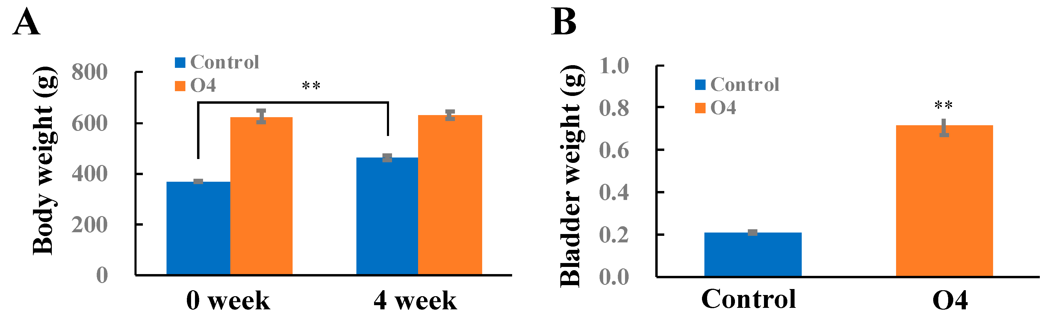
PBOO Increased Bladder Weight and Induced DEG Alterations Involved in Fibrosis, Downstream TGFB, and Extracellular Matrix

PBOO rat models were established following the description in the Materials and Methods section. While PBOO occurred, the bladder weight increased. Therefore, we measured the bladder weight to confirm the PBOO model. After surgery for four weeks, body weight significantly increased in the control group but not in the PBOO group (Figure 1A). The bladder weight in the PBOO group was significantly increased compared with that in the control group (Figure 1B). These bladder weight increases showed the outcome of PBOO.
Then, bladder tissues from the PBOO rats and control rats were collected. To further investigate the gene expression profile in rats with PBOO, mRNA of the bladder tissues was extracted, and the expression levels were then analyzed by Rat OneArray Plus microarrays, including “fibrosis”, “Tgfb signal pathway”, “extracellular matrix” (Table 1), and “Il6 and Stat3 signal-related genes” (Table A2). Genome probe 20,715 was used to screen the differentially expressed genes (DEGs) between the PBOO group and the control group. In this study, we focused on DEGs in fibrosis, TGFB downstream, and extracellular matrix. Table 1 shows the fold change of DEGs greater than 2 in fibrosis compared with the control group, including Grem1 (82.6), Thbs1 (25.8), Lox (6.3), Serpine1 (6.2), Timp1 (6.2), Itga1 (4.5), Tgfb3 (4.5), Ldha (3.9), Ilk (2.8), Timp3 (2.8), Ccl12 (2.6), Mmp14 (2.4), Ltbp1 (2.4), Stat6 (2.2), Itgb1 (2.2), and Nfkb1 (2.0). The fold change of DEGs less than −2 in fibrosis included Thbs2 (0.24), Stat1 (0.47), and Bmp7 (0.48).
In addition to fibrosis marker alteration in the bladder tissue of rats with PBOO, the microarray data also showed DEGs downstream of TGFB and extracellular matrix. Table 1 shows the DEGs with a fold change greater than 2 downstream from TGFB: Thbs1 (25.8), Serpine1 (6.2), Bhlhe40 (5.0), Gadd45b (4.5), Ldha (3.9), Hmox1 (3.3), Ptgs2 (3.2), Fn1 (2.7), Ptk2 (2.7), Cryab (2.6), Ctnnb1 (2.2), Fos (2.1), and Sox4 (2.0). The DEGs with a fold change than −2 in downstream TGFB included Srebf2 (0.33), Rybp (0.35), Bcl2l1 (0.36), Notch1 (0.37), Id2 (0.39), Ptk2b (0.40), Gtf2i (0.41), Msx2 (0.44), Herpud1 (0.45), Acvrl1 (0.47), Crebbp (0.49), and Cdkn1b (0.49).
Table 1 also shows the DEGs with a fold change greater than 2 in the extracellular matrix: Thbs1 (25.8), Col8a1 (15.2), Itga5 (9.3), Tnc (6.6), Timp1 (6.2), Col4a1 (6.1), Col4a2 (5.7), Ldha (3.9), Emilin1 (3.5), Col5a1 (3.3), Ncam1 (3.2), Lamc1 (3.0), Cd44 (2.8), Vcan (2.8), Timp3 (2.8), Fn1 (2.7), Adamts1 (2.6), Mmp14 (2.4), Ctnnb1 (2.2), Spock1 (2.2), Itgb1 (2.2), and Lama1 (2.0). DEGs with a fold change less than −2 in the extracellular matrix included Thbs2 (0.24), Tgfbi (0.26), Vcam1 (0.28), Cdh1 (0.30), Mmp15 (0.35), Mmp11 (0.38), Mmp16 (0.42), Pecam1 (0.45), and Cdh3 (0.45).

4. Discussion

Fibrosis is a common consequence after PBOO damage and leads to tissue or organ dysfunction. Fibrosis formation has been well studied in the heart [13] because most cardiovascular diseases eventually lead to heart fibrosis and loss of its function. Similar to “bladder remodeling”, structural remodeling of the heart, including myocardial hypertrophy and fibrosis, is the key determinant for the clinical outcome of heart failure [14]. A variety of growth factors, including angiotensin II, EDN1, TGFB, connective tissue growth factor, and periostin, promote fibrotic responses to the heart [15]. TGFB has been demonstrated to promote myofibroblast formation and collagen production. Another study showed that cAMP could inhibit the fibroblast-to-myofibroblast transition via an RHOA-dependent pathway [15,16]. RHO kinase upregulates proinflammatory cytokines and mediators such as IL6, monocyte chemoattractant protein-1, and TGFB and enhances inflammation and tissue fibrosis. Therefore, RHO kinase activation plays a role in the diastolic heart failure [17]. RHO kinase inhibition by fasudil attenuates angiotensin II-induced cardiac hypertrophy and fibrosis [18]. Angiotensin II is another key factor because it could sensitize fibroblasts to this transition by directly inducing TGFB signaling, SMAD3 expression, and phospho-SMAD3 translocation into the nucleus [19]. Additionally, the level of RHO-kinase increases in bladder tissue after PBOO. The expression of RHO-kinase increased significantly to approximately the same extent in the 1–4-week obstructed groups and further increased in the 8-week obstructed group at both the mRNA and protein levels [20].
Our data showed that Mmp14, Timp1, and Timp3 were increased but that Mmp11, Mmp15, and Mmp16 were reduced in the PBOO group compared with the control group. Although MMP14, MMP15, and MMP16 are expressed in most tissues, MMP14 is highly expressed in bladder. Interestingly, MMP11 is reported to be enriched in endometrium and placenta. Taylor et al. demonstrated that MMP14 promotes fibrous tissue expansion in the tendons [21]. Another study showed that the levels of TIMP1 and MMP2 decreased, but that the levels of MMP7, MMP11, MMP13, and MMP16 increased in significant fibrotic myocardium [22]. In a renal fibrosis model of streptozotocin (STZ)-induced diabetic rats, the proteins expression of TGFB, ERK1/2, TIMP1, TIMP2, MMP2, MMP7, MMP8, MMP11, and MMP14 increased [23]. Taken together, MMP14 plays a critical role in PBOO, STZ-induced renal fibrosis, and fibrotic tendons. Moreover, other DEGs, such as Timp1, Mmp11, and Mmp16, may have different roles in PBOO and other types of fibrosis.
ROS activate a broad variety of hypertrophy signaling kinases and transcription factors, such as tyrosine kinase, SRC, GTPBP, RAS, PKC, and MAPKs, including extracellular response kinase and extracellular signal–regulated kinase, and JUN [24]. ROS also exert potent effects on the extracellular matrix, stimulating cardiac fibroblast proliferation and activating MMPs, which are central to fibrosis and matrix remodeling [25,26]. MMPs are generally secreted in an inactive form and are activated post-translationally by ROS as a result of targeted interactions with critical cysteine residues in the pro-peptide autoinhibitory domain [27]. ROS also stimulate the transcription factors NFKB, ETS, and AP-1 to stimulate MMP expression [28].
Several studies have tried to treat PBOO with different strategies. Mesenchymal stem cells (MSCs) have been used to treat rats with PBOO. Al-Saikan et al. demonstrated that MSC treatment resulted in a significant decrease in bladder capacity, accompanied by the mRNA downregulation of Tgfb, Hif1a, Rhoa, and Hspa5 in PBOO rats [29]. Wiafe et al. found that inflammatory markers, such as Tgfb, Hif1a, Hif3a, Mtor, Col1, and Col3, were upregulated in the bladder after PBOO [30]. After MSC treatment of PBOO rats, the bladder had physiological benefits, and Tgfb, Hif1a, Hif3a, Mtor, Col1, and Col3 were downregulated. Yang et al. demonstrated that tolterodine, a drug used to delay PBOO progression, can reduce collagen volume in the bladder wall of PBOO [10]. Moreover, tolterodine also induced MMP7, ITGA4, ITGB2, TIMP1, and FN1, which may be a mechanism of tolterodine for PBOO treatment. 17-DMAG, an HSP90 inhibitor, has been demonstrated to inhibit HIF1A [31]. Treatment with 17-DMAG increased the bladder pressure at micturition and nonvoid contractions, leading to urodynamic parameters in PBOO mice [32]. These results demonstrate that treatment with 17-DMAG, a HIF inhibitor, significantly alleviated PBOO-induced bladder pathology in vivo. Sodium tanshinone IIA sulfonate (STS) has been demonstrated to ameliorate PBOO-induced Tgfb, phospho-Smad2, and phospho-Smad3 upregulation in rats [33]. In addition, STS abolished PBOO-induced mRNA upregulation of Acta2, Col1, and Col3. Taken together, these approaches pinpoint the importance of dissecting PBOO-induced gene expression in fibrosis, TGFB signaling, and extracellular matrix.
There were some limitations of the present study. First, real-time PCR analysis and histological analysis on DEGs were not performed in our PBOO rat model. Second, a further investigation on the role of these DEGs in physiology of PBOO will dissect the mechanism of its pathogenesis. Third, a further validation of our findings on PBOO patients will strength the importance.

5. Conclusions

In conclusion, PBOO initiates a pathophysiologic cascade that leads to structural and functional changes in the bladder. In this study, we discovered DEGs in fibrosis, TGFB signaling, and extracellular matrix after PBOO compared with in the control. The expressions of 19 mRNAs in fibrosis, 25 mRNAs in TGFB signaling, and 31 mRNAs in the extracellular matrix were significantly altered. Some DEGs with a fold change greater than 4 included Grem1, Thbs1, Col8a1, Itga5, Tnc, Lox, Timp1, Col4a1, Col4a2, Bhlhe40, Itga1, Tgfb3, and Gadd45b. One DEG was expressed with a fold change less than −4: Thbs2. These findings show the genes involved in the pathophysiology during PBOO. Further investigations of these DEGs may help clarify the mechanism of PBOO and provide potential therapeutic targets to reverse PBOO formation.

Author Contributions

Conceptualization, S.-Y.K. and W.-Y.L.; data curation and formal analysis, Y.-P.L.; funding acquisition, W.-Y.L.; supervision, W.-Y.L.; writing—original draft, Y.-S.H. and H.-H.C.; writing—review and editing, Y.-S.H., H.-H.C. and W.-Y.L. All investigators participated in the interpretation of the data and approved the final manuscript. All authors have read and agreed to the published version of the manuscript.

Funding

This research was funded by the Chang Gung Medical Foundation, grant numbers CMRPG6H0091 and CMRPG6J0101-2, and by the Taiwan and Ministry of Science and Technology, grant numbers MOST 110-2314-B-309-001, MOST 109-2314-B-309-002, and MOST 104-2321-B-400-019-MY3 in Executive Yuan, Taiwan.

Institutional Review Board Statement

The procedures for animal housing and treatment followed the guidelines of the Handbook of the Laboratory Animal Breeding and Research Centre of the Ministry of Science and Technology of Taiwan and were approved by the Animal Ethics Committee of the Chang Gung Medical Foundation (IACUC number 2012121205).

Informed Consent Statement

Not applicable.

Data Availability Statement

The data that support the findings of this study are available from Wei-Yu Li. Restrictions apply to the availability of these data, which were used under the license for this study. The data presented in this study are available from Wei-Yu Li upon request.

Acknowledgments

We thank Chih-Ying Lin (Institute of Clinical Pharmacy and Pharmaceutical Sciences, College of Medicine, National Cheng Kung University National Cheng Kung University) for administrative support.

Conflicts of Interest

The authors declare no conflict of interest.

Appendix A

Table A1. Microarray quality control.
Microarray Quality Control
Check ItemsDescriptionSpecification
Background arerage intensityCy5:B635 MedianCy5 < 100
Intrinsic Hybridization Controls (IHCs)IHC Probes designed for monitoring overall hybridization efficiencyIHC < 15,000
IHC CV < 15%
Extrinsic Target Quality Control (ETQC) with SpikiinsETQC probes and labeled spike-ins designed for monitoring the specificity of hybridization at low, medium, and high concentrationsHigh, Median, Low
Noise48 negative probes used in Rosetta Resolver for monitoring non-specific hybridizationNumber of negative intensity > 200 should be less than 10
Sample integrity in processingIntrinsic Target Quality Control (ITQC) 3, 6, 7 probes designed for monitoring the sample integrityNumber of ITQC rations within spec (S1/S2 * < 3.5) >= 2
Technical reproducibilityPearson’s correlation coefficient between technical replicatesR value >= 0.975
* Two probes per gene were selected from seven consistently expressed housekeeping genes. For each gene, one probe is designed of 300–600 bp (S1), 900–1200 bp (S2), respectively, from each 3’’ end of the transcript.
Table A2. DEGs related to Il6 and Stat3 signals in the PBOO group.
Il6 and Stat3 Signal Related Genes
GeneLdhaSocs3Ccl12OsmrNfkb1Il11Ccl5Cxcl12Cdc25a
Fold3.92.92.62.42.02.00.280.300.39
RNA of bladder tissues from the PBOO and control groups was extracted and then analyzed by Rat OneArray Plus microarrays. The expression of genes in fibrotic bladder tissues in the PBOO rats were compared to that in the control rats. DEGs with fold change values higher than 2 (A) or lower than −2 (B) and with p-values less than 0.05 were selected.

References

  1. Rosen, R.; Altwein, J.; Boyle, P.; Kirby, R.S.; Lukacs, B.; Meuleman, E.; O’Leary, M.P.; Puppo, P.; Robertson, C.; Giuliano, F. Lower urinary tract symptoms and male sexual dysfunction: The multinational survey of the aging male (MSAM-7). Eur. Urol. 2003, 44, 637–649. [Google Scholar] [CrossRef] [PubMed]
  2. Vallancien, G.; Emberton, M.; Harving, N.; van Moorselaar, R.J.; Alf-One Study, G. Sexual dysfunction in 1274 European men suffering from lower urinary tract symptoms. J. Urol. 2003, 169, 2257–2261. [Google Scholar] [CrossRef] [PubMed]
  3. Landau, E.H.; Jayanthi, V.R.; Churchill, B.M.; Shapiro, E.; Gilmour, R.F.; Khoury, A.E.; Macarak, E.J.; McLorie, G.A.; Steckler, R.E.; Kogan, B.A. Loss of elasticity in dysfunctional bladders: Urodynamic and histochemical correlation. J. Urol. 1994, 152 Pt 2, 702–705. [Google Scholar] [CrossRef]
  4. Palmer, L.S.; Lee, C.; Decker, R.S.; Lang, S.; Kaplan, W.E.; Firlit, C.F.; Cheng, E.Y. The effect of angiotensin converting enzyme inhibition and angiotensin II receptor antagonism on obstructed rat bladder. J. Urol. 1997, 158 Pt 2, 1100–1104. [Google Scholar] [CrossRef]
  5. Anumanthan, G.; Tanaka, S.T.; Adams, C.M.; Thomas, J.C.; Wills, M.L.; Adams, M.C.; Hayward, S.W.; Matusik, R.J.; Bhowmick, N.A.; Brock, J.W., 3rd; et al. Bladder stromal loss of transforming growth factor receptor II decreases fibrosis after bladder obstruction. J. Urol. 2009, 182 (Suppl. 4), 1775–1780. [Google Scholar] [CrossRef] [Green Version]
  6. Yamada, S.; Takeuchi, C.; Oyunzul, L.; Ito, Y. Bladder angiotensin-II receptors: Characterization and alteration in bladder outlet obstruction. Eur. Urol. 2009, 55, 482–489. [Google Scholar] [CrossRef]
  7. Persson, K.; Pandita, R.K.; Waldeck, K.; Andersson, K.E. Angiotensin II and bladder obstruction in the rat: Influence on hypertrophic growth and contractility. Am. J. Physiol. 1996, 271 Pt 2, R1186–R1192. [Google Scholar] [CrossRef]
  8. Myers, J.B.; Dall’era, J.E.; Koul, S.; Kumar, B.; Khandrika, L.; Flynn, B.J.; Koul, H.K. Biochemical alterations in partial bladder outlet obstruction in mice: Up-regulation of the mitogen activated protein kinase pathway. J. Urol. 2009, 181, 1926–1931. [Google Scholar] [CrossRef]
  9. Tanaka, S.T.; Martinez-Ferrer, M.; Makari, J.H.; Wills, M.L.; Thomas, J.C.; Adams, M.C.; Brock, J.W., 3rd; Pope, J.C.t.; Bhowmick, N.A. Recruitment of bone marrow derived cells to the bladder after bladder outlet obstruction. J. Urol. 2009, 182 (Suppl. 4), 1769–1774. [Google Scholar] [CrossRef]
  10. Yang, T.X.; Luo, D.Y.; Lin, Y.F.; Liu, Q.; Cai, X.; Ai, J.Z.; Li, H.; Shen, H.; Wang, K.J. The Antimuscarinic Agent Tolterodine Regulates Bladder Extracellular Matrix in Partial Bladder Outlet Obstruction in Rats. Cell Physiol. Biochem. 2018, 46, 36–45. [Google Scholar] [CrossRef]
  11. Gao, Y.; Liu, P.; He, F.; Yang, X.; Wu, R.; Chen, W.; Li, L.; Yang, Z. Fibroblast Growth Factor 2 Promotes Bladder Hypertrophy Caused by Partial Bladder Outlet Obstruction. Front. Cell Dev. Biol. 2021, 9, 630228. [Google Scholar] [CrossRef]
  12. Lin, W.Y.; Lin, Y.P.; Levin, R.M.; Chen, M.L. The relevance of immune responses to partial bladder outlet obstruction and reversal. Neurourol. Urodyn. 2017, 36, 1306–1312. [Google Scholar] [CrossRef]
  13. Frangogiannis, N.G.; Dewald, O.; Xia, Y.; Ren, G.; Haudek, S.; Leucker, T.; Kraemer, D.; Taffet, G.; Rollins, B.J.; Entman, M.L. Critical role of monocyte chemoattractant protein-1/CC chemokine ligand 2 in the pathogenesis of ischemic cardiomyopathy. Circulation 2007, 115, 584–592. [Google Scholar] [CrossRef]
  14. Cohn, J.N.; Ferrari, R.; Sharpe, N. Cardiac remodeling–concepts and clinical implications: A consensus paper from an international forum on cardiac remodeling. Behalf of an International Forum on Cardiac Remodeling. J. Am. Coll Cardiol. 2000, 35, 569–582. [Google Scholar] [CrossRef] [Green Version]
  15. Berk, B.C.; Fujiwara, K.; Lehoux, S. ECM remodeling in hypertensive heart disease. J. Clin. Investig. 2007, 117, 568–575. [Google Scholar] [CrossRef]
  16. Swaney, J.S.; Roth, D.M.; Olson, E.R.; Naugle, J.E.; Meszaros, J.G.; Insel, P.A. Inhibition of cardiac myofibroblast formation and collagen synthesis by activation and overexpression of adenylyl cyclase. Proc. Natl. Acad. Sci. USA 2005, 102, 437–442. [Google Scholar] [CrossRef] [Green Version]
  17. Shimokawa, H.; Takeshita, A. Rho-kinase is an important therapeutic target in cardiovascular medicine. Arterioscler. Thromb. Vasc. Biol. 2005, 25, 1767–1775. [Google Scholar] [CrossRef]
  18. Wang, Y.X.; da Cunha, V.; Martin-McNulty, B.; Vincelette, J.; Li, W.; Choy, D.F.; Halks-Miller, M.; Mahmoudi, M.; Schroeder, M.; Johns, A.; et al. Inhibition of Rho-kinase by fasudil attenuated angiotensin II-induced cardiac hypertrophy in apolipoprotein E deficient mice. Eur. J. Pharmacol. 2005, 512, 215–222. [Google Scholar] [CrossRef]
  19. Sorescu, D. Smad3 mediates angiotensin II- and TGF-beta1-induced vascular fibrosis: Smad3 thickens the plot. Circ. Res. 2006, 98, 988–989. [Google Scholar] [CrossRef] [Green Version]
  20. Guven, A.; Onal, B.; Kalorin, C.; Whitbeck, C.; Chichester, P.; Kogan, B.; Levin, R.; Mannikarottu, A. Long term partial bladder outlet obstruction induced contractile dysfunction in male rabbits: A role for Rho-kinase. Neurourol. Urodyn. 2007, 26, 1043–1049. [Google Scholar] [CrossRef]
  21. Taylor, S.H.; Yeung, C.Y.; Kalson, N.S.; Lu, Y.; Zigrino, P.; Starborg, T.; Warwood, S.; Holmes, D.F.; Canty-Laird, E.G.; Mauch, C.; et al. Matrix metalloproteinase 14 is required for fibrous tissue expansion. Elife 2015, 4, e09345. [Google Scholar] [CrossRef]
  22. Xiao, T.; Zeng, O.; Luo, J.; Wu, Z.; Li, F.; Yang, J. Effects of hydrogen sulfide on myocardial fibrosis in diabetic rats: Changes in matrix metalloproteinases parameters. Biomed. Mater. Eng. 2015, 26 (Suppl. 1), S2033–S2039. [Google Scholar] [CrossRef] [Green Version]
  23. Li, Y.; Li, L.; Zeng, O.; Liu, J.M.; Yang, J. H2S improves renal fibrosis in STZ-induced diabetic rats by ameliorating TGF-beta1 expression. Ren. Fail. 2017, 39, 265–272. [Google Scholar] [CrossRef] [PubMed] [Green Version]
  24. Brieger, K.; Schiavone, S.; Miller, F.J., Jr.; Krause, K.H. Reactive oxygen species: From health to disease. Swiss Med. Wkly. 2012, 142, w13659. [Google Scholar] [CrossRef]
  25. Spinale, F.G. Bioactive peptide signaling within the myocardial interstitium and the matrix metalloproteinases. Circ. Res. 2002, 91, 1082–1084. [Google Scholar] [CrossRef] [Green Version]
  26. Siwik, D.A.; Pagano, P.J.; Colucci, W.S. Oxidative stress regulates collagen synthesis and matrix metalloproteinase activity in cardiac fibroblasts. Am. J. Physiol. Cell Physiol. 2001, 280, C53–C60. [Google Scholar] [CrossRef]
  27. Lin, W.Y.; Guven, A.; Juan, Y.S.; Neuman, P.; Whitbeck, C.; Chichester, P.; Kogan, B.; Levin, R.M.; Mannikarottu, A. Free radical damage as a biomarker of bladder dysfunction after partial outlet obstruction and reversal. BJU Int. 2008, 101, 621–626. [Google Scholar] [CrossRef]
  28. Takimoto, E.; Kass, D.A. Role of oxidative stress in cardiac hypertrophy and remodeling. Hypertension 2007, 49, 241–248. [Google Scholar] [CrossRef]
  29. Al-Saikan, B.; Ding, J.; Tredget, E.; Metcalfe, P. Benefits of mesenchymal stem cells after partial bladder outlet obstruction. Can Urol. Assoc. J. 2016, 10, E1–E6. [Google Scholar] [CrossRef] [Green Version]
  30. Wiafe, B.; Kadam, R.; Metcalfe, P.D. Intraperitoneal administration of mesenchymal stem cells is effective at mitigating detrusor deterioration after pBOO. Am. J. Physiol. Renal. Physiol. 2020, 318, F549–F556. [Google Scholar] [CrossRef]
  31. Semenza, G.L. Targeting HIF-1 for cancer therapy. Nat. Rev. Cancer 2003, 3, 721–732. [Google Scholar] [CrossRef] [PubMed]
  32. Iguchi, N.; Donmez, M.I.; Malykhina, A.P.; Carrasco, A., Jr.; Wilcox, D.T. Preventative effects of a HIF inhibitor, 17-DMAG, on partial bladder outlet obstruction-induced bladder dysfunction. Am. J. Physiol. Renal. Physiol. 2017, 313, F1149–F1160. [Google Scholar] [CrossRef] [PubMed] [Green Version]
  33. Jiang, X.; Chen, Y.; Zhu, H.; Wang, B.; Qu, P.; Chen, R.; Sun, X. Sodium Tanshinone IIA Sulfonate Ameliorates Bladder Fibrosis in a Rat Model of Partial Bladder Outlet Obstruction by Inhibiting the TGF-beta/Smad Pathway Activation. PLoS ONE 2015, 10, e0129655. [Google Scholar] [CrossRef] [PubMed] [Green Version]
Figure 1. Body weight and bladder weight of rats in the PBOO group and control group. Rat models of PBOO were established as described in the Materials and Methods section. Each rat was weighed before surgery and after surgery for four weeks (A). Four weeks after surgery, the rats were sacrificed, and the bladder was collected and weighed (B). C: control; O4: four-week PBOO. ** p < 0.01.
Processes 09 02219 g001
Table 1. DEGs related to fibrosis, Tgfb signaling, and extracellular matrix in the PBOO group.
Fibrosis-Related Genes
GeneGrem1Thbs1LoxSerpine1Timp1Itga1Tgfb3LdhaIlkTimp3Timp3
Fold82.625.86.36.26.24.54.53.92.82.82.8
GeneCcl12Mmp14Ltbp1Stat6ItgbNfkbThbs2Stat1Bmp7
Fold2.62.42.42.22.22.00.240.470.48
TgfbSignaling-Related Genes
GeneThbs1Serpine1Bhlhe40Gadd45bLdhaHmox1Ptgs2Fn1Ptk2CryabCtnnb1
Fold25.86.25.04.53.93.33.22.72.72.62.2
GeneFosSox4Srebf2RybpBcl2l1Notch1Id2Ptk2bGtf2iMsx2Herpud1
Fold2.12.00.330.350.360.370.390.400.410.440.45
GeneAcvrl1CrebbpCdkn1b
Fold0.470.490.49
Extracellular Matrix-Related Genes
GeneThbs1Col8a1Itga5TncTimp1Col4a1Col4a2LdhaEmilin1Col5a1Ncam1
Fold25.815.29.36.66.26.15.73.93.53.33.2
GeneLamc1Cd44VcanTimp3Fn1Adamts1Mmp14Ctnnb1Spock1Itgb1Lama1
Fold3.02.82.82.82.72.62.42.22.22.22.0
GeneThbs2TgfbiVcam1Cdh1Mmp15Mmp11Mmp16Pecam1Cdh3
Fold0.240.260.280.300.350.380.420.450.45
RNA of bladder tissues from the PBOO and control groups was extracted and then analyzed by Rat OneArray Plus microarrays. The expression of genes in fibrotic bladder tissues in the PBOO rats were compared with that in the control rats. DEGs with fold change values higher than 2 (A) or lower than −2 (B) and with p-values less than 0.05 were selected.
Publisher’s Note: MDPI stays neutral with regard to jurisdictional claims in published maps and institutional affiliations.

