Article

Expression of the Thermobifida fusca β-1,3-Glucanase in Yarrowia lipolytica and Its Application in Hydrolysis of β-1,3-Glucan from Four Kinds of Polyporaceae

1
Department of Cosmetic Science, Providence University, Taichung 43301, Taiwan
2
Cosmetic Industry Research & Development Center, Providence University, Taichung 43301, Taiwan
3
Xtremes Pure Company, Taipei 10652, Taiwan
*
Author to whom correspondence should be addressed.
Processes 2021, 9(1), 56; https://doi.org/10.3390/pr9010056
Submission received: 5 December 2020 / Revised: 26 December 2020 / Accepted: 27 December 2020 / Published: 29 December 2020
(This article belongs to the Special Issue Biological Activity Evaluation Process of Natural Antioxidants)

Abstract

:
The gene encoding a thermostable β-1,3-glucanase was cloned from Thermobifida fusca and expressed constitutively by Yarrowia lipolytica using plasmid pYLSC1. The expression level of the recombinant β-1,3-glucanase reached up to 270 U/mL in the culture medium. After a treatment with endo-β-N-acetyl-glucosaminidase H, the recombinant protein appeared as a single protein band, with a molecular size of approximately 66 kDa on the SDS-polyacrylamide gel. The molecular weight was consistent with the size predicted from the nucleotide sequence. The optimum temperature and pH of the transformant β-1,3-glucanase were 60 °C and pH 8.0, respectively. This β-1,3-glucanase was tolerant to 10% methanol, ethanol, and DMSO, retaining 70% activity. The enzyme markedly hydrolyzed Wolfiporia cocos and Pycnoporus sanguineus glucans. The DPPH and ABTS scavenging potential, reducing power and total phenolic contents of these two Polyporaceae hydrolysates, were significantly increased after 18 h of the enzymatic reaction. The present results indicate that T. fusca β-1,3-glucanase from Y. lipolytica transformant (pYLSC1-13g) hydrolyzes W. cocos and P. sanguineus glucans and improves the antioxidant potential of the hydrolysates.

1. Introduction

Edible medicinal fungi have been used in China for over two thousand years, and relevant information is available in historical relics. Numerous Polyporaceae are often used in Chinese medicine and dietary conditioning, such as Wolfiporia cocos (Fu-Ling), Pycnoporus sanguineus (Red fungus), Polyporus umbellatus (Chu-Ling), Laccocephalum mylittae (Lei-Wan), since they are rich in β-1,3 glucan.
Studies on traditional medicine have reported that β-glucan has anti-inflammatory, anti-allergic, antitumor, immunoregulatory, and antioxidant activity; thus, preventing bacterial infections and regulating other physiological phenomena [1]. β-glucan is one of the principal components of fungal cell walls. These medicinal fungi are rich in β-glucan, but have different chemical compositions. Polysaccharides with β (1 → 3) and β (1 → 6) or β (1 → 4) and β (1 → 6) glycosidic bonds, rather than those with pure β (1 → 4) glycosidic bonds, usually have stronger pharmacological activity [2,3]. The previous reports show that the low molecular weight β-glucans of oat and yeast reveals anticancer activity, antitumor, and immunological properties, respectively [4,5]. Bioactivity of polysaccharides is highly related to their chain structure, chemical composition, molecular weight, backbone, and degree of branching [6,7]. β-glucans from various fungi vary greatly in length and the degree of branching. Although the flavonoids, saponins, tannins, and terpenes are antioxidant compounds in the mushroom [8]. Therefore, the antioxidant properties of fungi are primarily attributed to β-glucan [9]. β-1,3 glucanase has been used to destroy cell walls of some fungi and crack the fragments the fragments having immunoregulatory activity [10]. It is able to reduce molecular weight polysaccharide for β-1,3-glucan to produce low molecular weight β-1,3-glucan with pharmacological activity properties [11]. It has been reported that, Ganoderma lucidum β-1,3 glucan with low molecular weight and low molecular weight β-glucan of yeast had better immunological and antioxidant activities, respectively [4,12].
W. cocos are used in approximately 10% of traditional Chinese medicine preparations. W. cocos contains abundant β-1,3-glucan and is the principal component. Insolubility of β-1,3-glucan has low biological activity and can be chemically modified to increase its hydrophilicity. W. cocos β-1,3-glucan has various bioactivities [13], and its low molecular weight facilitates free radical scavenging activity and helps to prevent DNA damage [14]. P. sanguineus β-1,3-glucan reportedly scavenges DPPH free radicals [15] and has antimicrobial and antitumor effects [16], and its ethanol extract has different biological activities. P. umbellatus is used to treat various symptoms including edema, oliguria, and jaundice [17]. Typical of other β-glucans, P. umbellatus polysaccharide (β-1,3-glucan) reportedly has anticancer, hepatoprotective, immunoregulatory, and antitumor activity, and prevents kidney damage [18]. P. umbellatus polysaccharide has received increasing attention owing to its prominent physiological and biological functions [19]. L. mylittae is a traditional Chinese medicinal fungus. It has been used as a vermifuge against many kinds of parasites, including roundworms, cestodes, and ancylostoma [20].
Interestingly, it has been reported that the thermophilic actinomycetes, Thermobifida fusca, can produce extracellular thermostable enzymes [21,22]. The β-1,3-glucanase gene from T. fusca YX was also cloned and expressed in Escherichia coli BL-21 Codon Plus (DE3)-RIPL [23].
In order to reduce energy costs in the production process, a mesophilic host is often used as the host to express the enzymes originally isolated from thermophilic organisms. Some available mesophilic expression systems have been reported. E coli expression system is usually the first choice because of its simple genetic manipulation, rapid growth, and high transformation efficiency. The inclusion body formation and intracellular accumulation limit its use [24]. The Pichia pastoris expression system was another option for mesophilic expression. However, P. pastoris showed low transformation efficiency and high false positives in transformant. Yarrowia lipolytica, generally regarded as safe (GRAS) yeast, also serves as a mesophilic host for heterologous protein expression [25]. There are many optional gene engineering tools for use in this host-vector system [26].
Therefore, this study aimed to constitutively express heterologous the β-1,3-glucanase gene in a Y. lipolytica mesophilic expression system and hydrolyze polysaccharides from edible-medicinal Polyporaceae, and to evaluate the antioxidant properties of their partial hydrolysates.

2. Materials and Methods

2.1. Materials and Microorganisms

Whole, dry sporocarps of P. sanguineus, W. cocos, P. umbellatus, and L. mylittae were purchased from a local Chinese pharmacy in Taichung, Taiwan, in February 2020. They were identified and stored by the Department of Cosmetic Science, Providence University, Taiwan. The actinomycetes, Thermobifida fusca BCRC 19214, isolated from a compost sample collected in Taiwan and stocked in Bioresource Collection and Research Center (BCRC) in Hsinchu, Taiwan was used herein [22]. Yarrowia lipolytica expression system (strain P01g and pYLSC1) was purchased from Yeastern Biotech Co., Ltd. (Taipei, Taiwan) [24]. The VioTag DNA polymerase, PCR buffer, and dNTP were purchased from Viogene (Sunnyvale, CA, USA). The YPD (Yeast Extract Peptone Dextrose) medium (Y1500), yeast nitrogen base without amino acids (Y0626), agar, restriction endo endodeoxyribonucleases, T4 DNA ligation kit, and all other chemicals were purchased from Sigma-Aldrich (St. Louis, MO, USA).

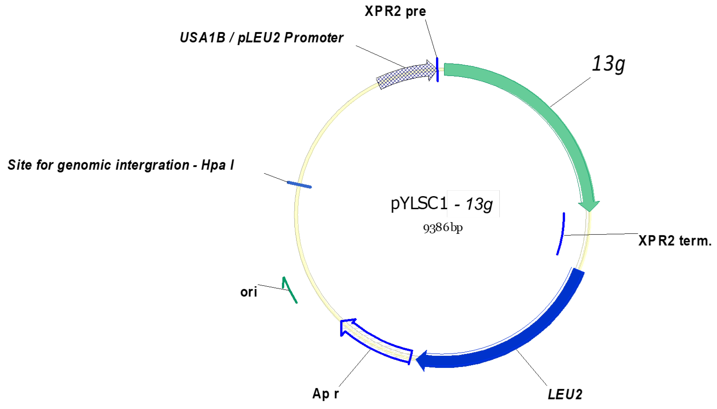
2.2. Construction of the β-1,3-Glucanase Expression Plasmid

The β-1,3-glucanase gene was amplified from the chromosomal DNA of T. fusca BCRC 19214 as template by PCR using the primers 5′ -AAA GGC CGT TCT GGC CGT CCG ACT AGG CTC CGG C- 3′ (SfiI site is underlined) and 5′-AAA GGT ACC TCA ATG ATG ATG ATG ATG ATG CCC GGT CGC CAA CTG C-3′ (KpnI site is underlined) based on the gene sequence (NCBI accession number WP_011292553.1) of T. fusca YX. The amplified PCR product was further digested with SfiI and KpnI, and then ligated with SfiI-KpnI-treated pYLSC1. The resulting plasmid pYLSC1-13g (Figure 1) was used for recombinant enzyme production in Y. lipolytica P01g. DNA manipulation was carried out as previously reported [27].

2.3. Transformation and Screening of Y. lipolytica Transformant

The NotI-linearized pYLSC1-13g was introduced into Y. lipolytica P01g [25]. The transformants cells were spread on agar plates, which contained 6.7 g/L yeast nitrogen base without amino acids (Y0626), 20.0 g/L glucose, and 15.0 g agar/L, and cultivated at 28 °C for 4 days. The selected transformants were grown in 50 mL YPD medium (Y1500) in 250-mL Hinton flask, 28 °C, 200 rpm, and those with apparent β-1,3-glucanase activity were chosen for further study. Integration of the target gene into chromosome of Y. lipolytica P01g was checked by genomic PCR.

2.4. Enzyme Expression and Purification

Y. lipolytica transformant (pYLSC1-13g) with high-β-1,3-glucanase-activity was incubated in 200 mL YPD medium in 500-mL Hinton flasks on a reciprocal shaker (200 rpm) at 28 °C for 120 h. After cultivation, the culture supernatant was collected by centrifugation at 10,000× g under 4 °C for 30 min and served as a crude enzyme solution.
The crude enzyme solution was applied to a 10 mL Ni2+-NTA column (Merck KGaA, Darmstadt, Germany). After washed with 100 mL wash buffer (50 mM phosphate (pH 7.5), and 10 mM imidazole), the protein bound to the column was eluted with elution buffer (20 mM Tris-HCl (pH 7.5), 500 mM NaCl and 500 mM imidazole). The eluted fractions containing the purified enzyme were collected.

2.5. Determination of β-1,3-Glucanase Activity

The 1 mL reaction solution was prepared by mixing 0.1 mL of the enzyme solution, 0.8 mL of Tris-HCl buffer (20 mM, pH 8.0) and 0.1 mL of laminarin (10 mg/mL), followed by incubation at 60 °C. The reducing sugars resulting from laminarin hydrolysis were assayed by the DNS assay [28]. One unit of β-1,3-glucanase activity was defined as the amount of enzyme that release 1 μg glucose per min at 60 °C. To determine the metal effect on the enzymatic activity, 1 mM metal ion, including Na+, K+, Ca2+, Mg2+, Zn2+, Cu2+, Mn2+, Co2+, and Hg2+ was added into the reaction solution before incubation at 60 °C. Solvents including methanol, ethanol, isopropanol, DMSO, acetonitrile, and acetone was individually included in the reaction solution at 10% or 20% to determine the solvent stability of the enzyme.

2.6. Hydrolysis of Polyporaceae Glucans

Various Polyporaceae were placed in 100-mL serum bottles, and β-1,3-glucanase and 20 mM Tris-HCl buffer (pH 8.0) were added to adjust the total volume to 80 mL, followed by hydrolysis for 24 h at 60 °C. The hydrolysis degree was computed based on the following equation:
Hydrolysis degree (%) = (The total sugar of the supernatant (μg)/substrate (g)) × 100%.

2.7. Quantification of Reducing Sugars and Total Sugar

Levels of reducing sugars were directly determined via the 2,4-dinitrosalicylic acid (DNS) assay. The total sugar contents of the samples were measured through the phenol–sulfuric acid method.

2.8. Assessment of Antioxidant Properties

Free radical scavenging activity of 1,1-diphenyl-2-picrylhydrazyl (DPPH) and 2 2’-azino-bis- (3-ethylbenzothiazoline-6- sulfonic acid) (ABTS), total phenolic contents, and reducing power of the hydrolysate were evaluated using previously reported methods [29].

2.9. Statistical Analysis

All data were analyzed in triplicate. The mean values were compared to the appropriate control using Student’s t-test. p-values less than 0.05 indicated statistically significant differences.

3. Results and Discussion

3.1. Amplification and Construction of the β-1,3-Glucanase Gene in a Y. lipolytica Expression System

The β-1,3-glucanase gene 13g was cloned in frame into a pYLSC1 vector by SfiI and XbaI restriction sites to form expression plasmid pYLSC1-13g that was then transformed into Y. lipolytica P01g. The Y. lipolytica transformant (pYLSC1-13g) that displayed the most stable and highest β-1,3-glucanase activity was then elected for further experiments. The coding sequence of the β-1,3-glucanase from T. fusca BCRC 19214 was 99.86% identical to that of WP_011292553.1 (Tfu-2130) from the translation protein of Tfu-2130 gene (Lam81A) [23]. On comparing the sequences of the β-1,3-glucanase from T. fusca BCRC 19214 with the Tfu-2130 sequence from T. fusca YX, three mismatched nucleotides were identified: positions 459 (C, T), 1647 (A, G), and 2097 (G, A).

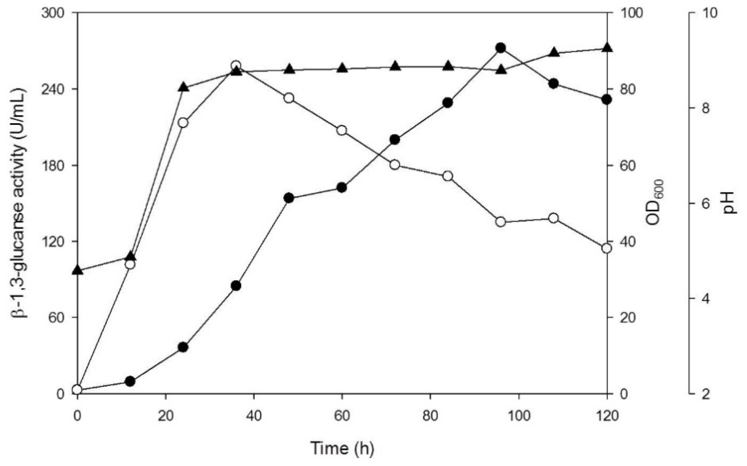
3.2. Production of β-1,3-Glucanase from Y. lipolytica Transformant (pYLSC1-13g)

Figure 2 showed that the transformant (pYLSC1-13g) rapidly grew from 0 to 36 h. The concentration of yeast (OD600) was 86 after 36 h of incubation. β-1,3-glucanase activity in the culture supernatant rapidly accumulated along with the incubation time, with a peak (270 U/mL) at 96 h, No β-1,3-glucanase activity was observed in medium of the control strain, Y. lipolytica (pYLSC1), under the same culture conditions.

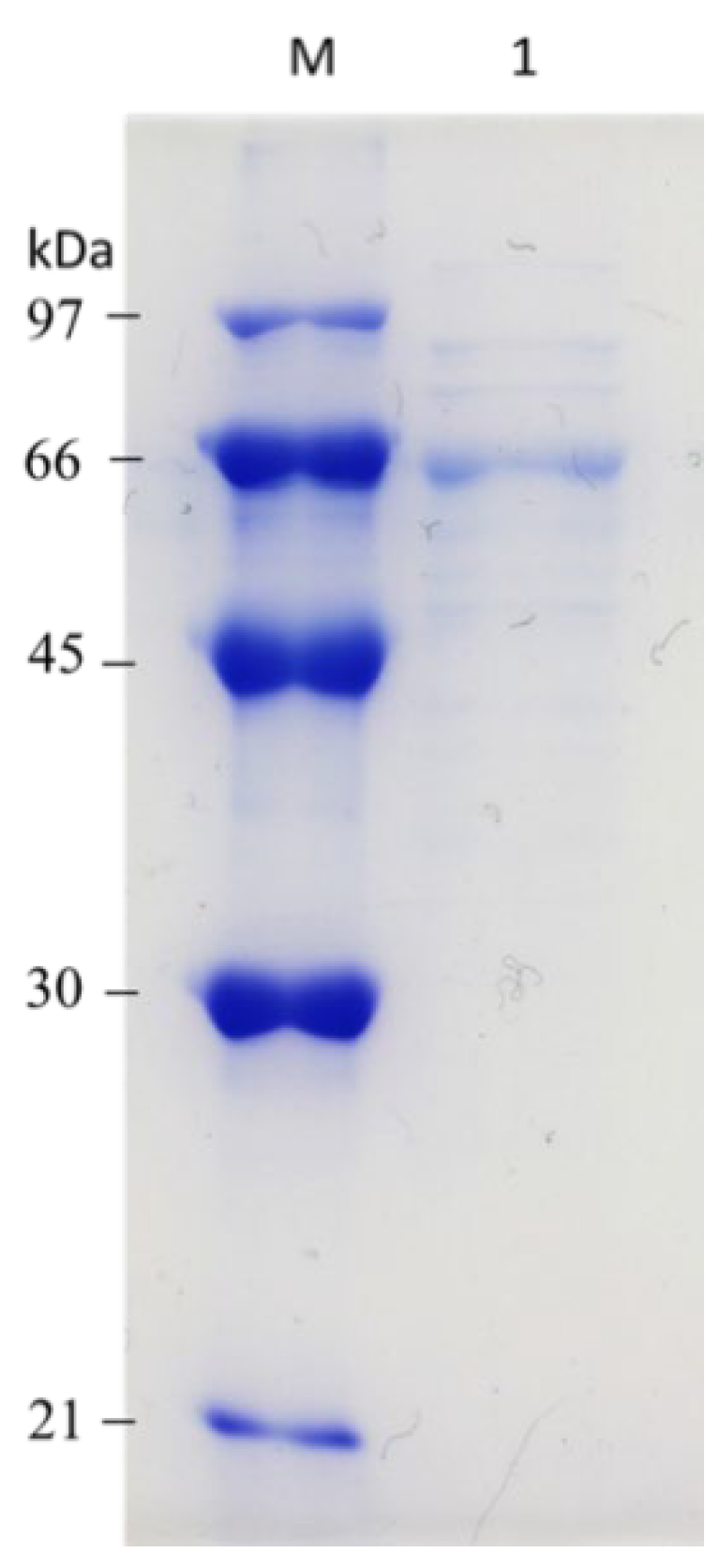
3.3. Purification of β-1,3-Glucanase from Y. lipolytica Transformant (pYLSC1-13g)

β-1,3-glucanase in the culture supernatant was purified using a Ni2+-NTA column as described in the Materials and Methods Section (Section 2). The yield and purification fold of the process was 29.27% and 3.93-fold, respectively (Table 1). The miscellaneous proteins in the culture supernatant of the Y. lipolytica transformant was fewer than that of the E. coli expression system. This will accelerate the application of the β-1,3-glucanase in the industrial process conveniently without numerous purification procedures.

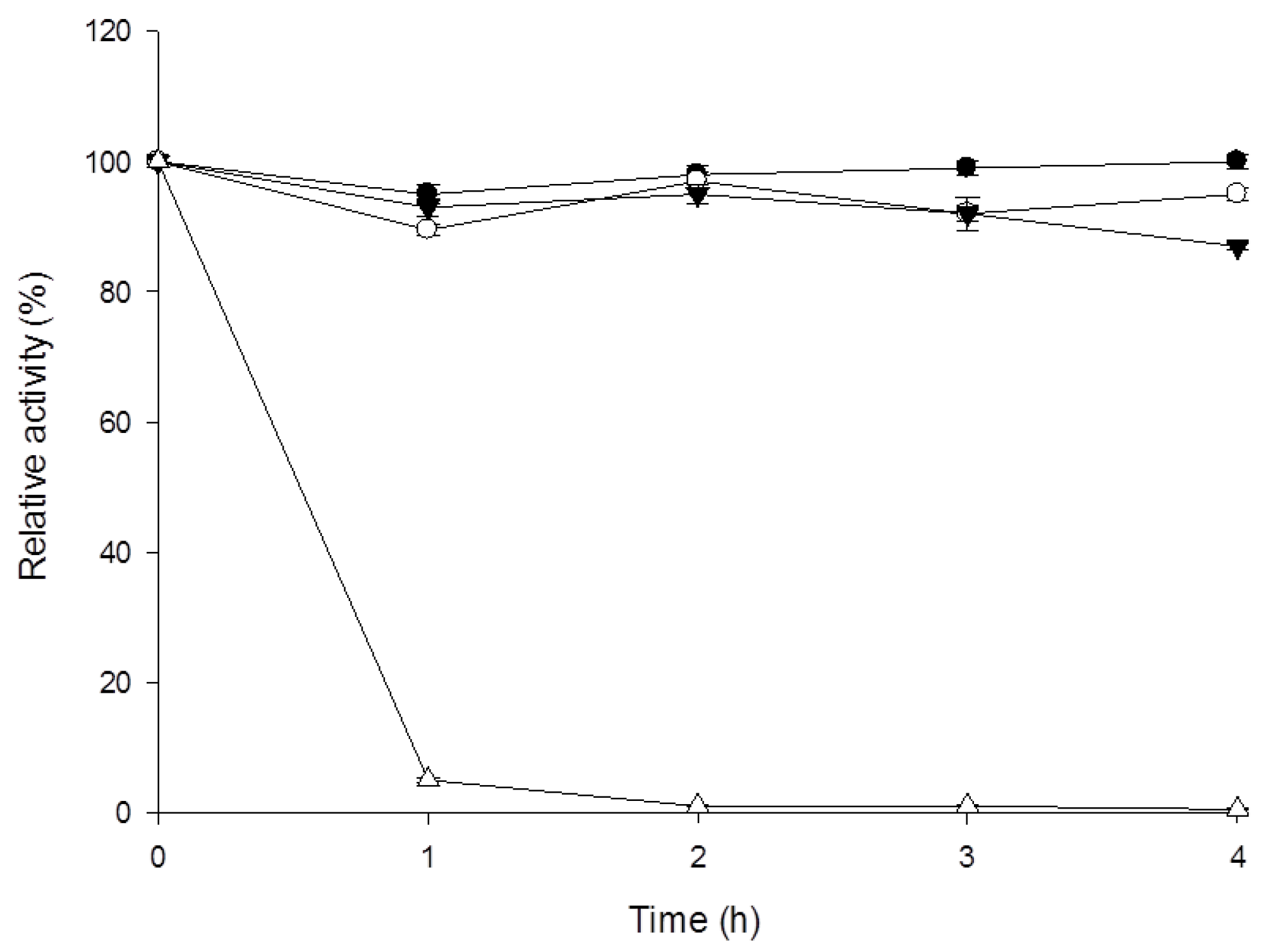
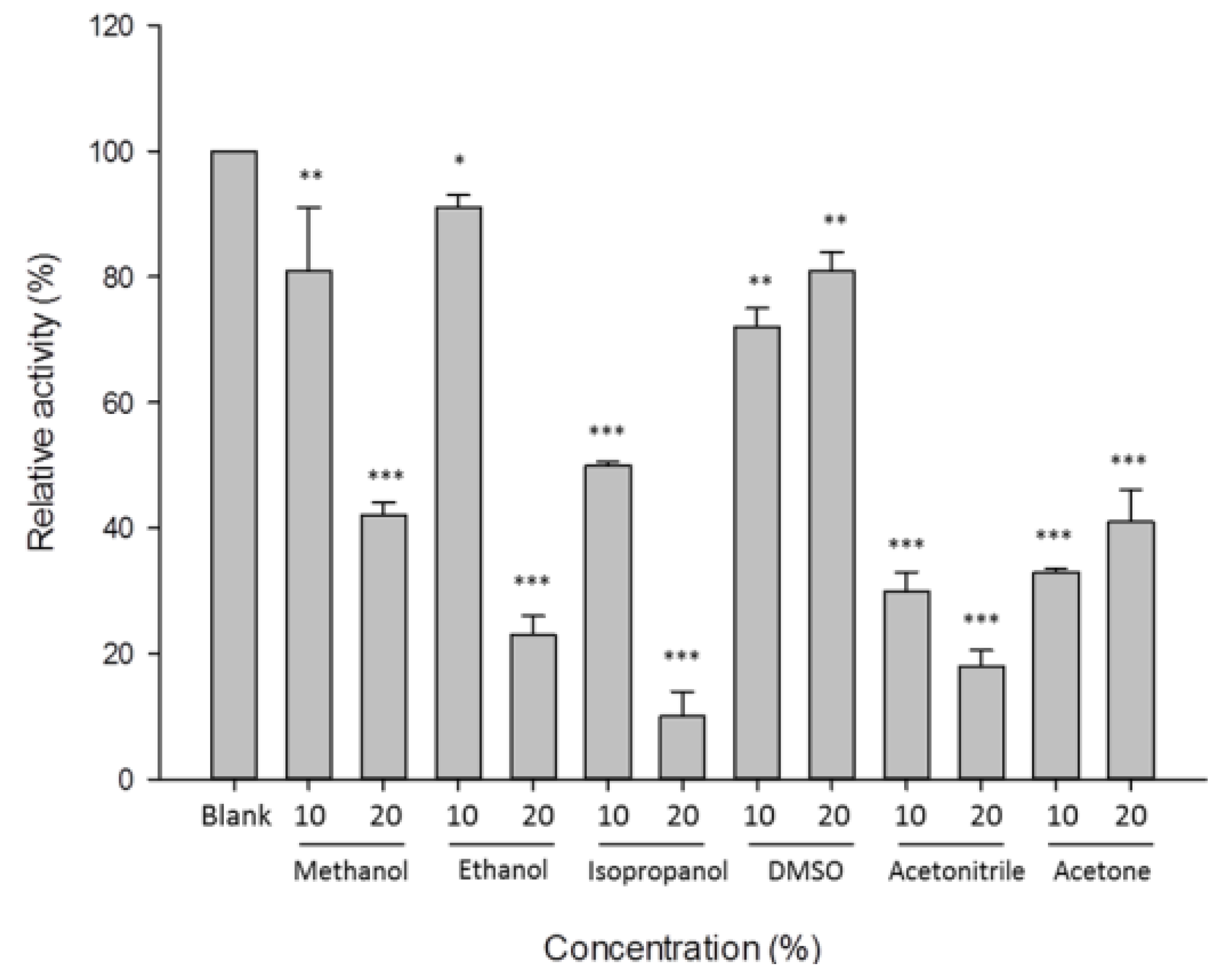
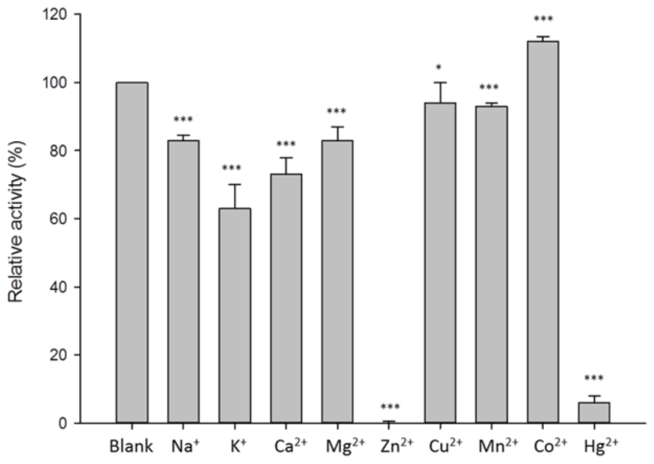
3.4. Characterization of β-1,3-Glucanase from Y. lipolytica Transformant (pYLSC1-13g)

As shown in Figure 3, the purified β-1,3-glucanase from Y. lipolytica transformant (pYLSC1-13g) was obtained as a single protein band on SDS–PAGE, with an estimated molecular weight of approximately 66 kDa. The optimal pH and temperature of the purified β-1,3-glucanase were pH 8.0 and 60 °C, respectively, with approximately >85% activity retained at 40~60 °C for 4 h. Enzyme activity decreased rapidly at 70 °C, with only 10% activity retained after 1 h of incubation (Figure 4).
The optimal temperature of β-1,3-glucanase produced by Y. lipolytica transformant (pYLSC1-13g) was 65 °C, similar to that of Trichoderma harzianum and Wickerhamomyces anomalus β-1,3-glucanase [30,31]. The optimal pH of β-1,3-glucanases from Pseudomonas cepacia, Phaseolus vulgaris L, and T. harzianum were pH. 5.0, 5.0, and 4.4, respectively, concurrent with previous reports [32,33,34]. In contrast, the optimal pH of β-1,3-glucanase from T. fusca BCRC 19214 was pH 8.0, displaying high pH stability at pH 7.0–9.0.
Furthermore, approximately 80% β-1,3-glucanase activity from Y. lipolytica transformant (pYLSC1-13g) was retained in 10% methanol and ethanol and decreased to 42% and 24% with methanol and ethanol, respectively. Isopropanol more markedly inhibits β-1,3-glucanase activity, and this inhibitory effect becomes more obvious with an increase in the concentration. β-1,3-Glucanase displayed high tolerance towards DMSO, with >70% activity retained, while acetonitrile and acetone significantly decreased enzyme activity up to 50% (Figure 5). These results are concurrent with those of previous studies reporting that methanol, ethanol, and DMSO (all 10%) decreased the relative activity of Thermotoga maritima β-1,3-glucanase by 52%, 34% and 16%, respectively [35].
In addition, enzyme activity was retained at >80% upon treatment with Na+, Mg2+, Cu2+, Mn2+, and Co2+ (Figure 6). K+ and Ca2+ inhibited enzyme activity by 30–40%, while 1 mM Zn2+ and Hg2+ further inhibited enzyme activity.
Compared with other β-1,3-glucanases, the activity of β-1,3-glucanases produced from T. harzianum, T. koningii, and Flavobacterium dormitator were inhibited by Hg2+ [34,36,37]. Hg2+ significantly inhibited the activity of β-1,3-glucanase, presumably because of the presence of sulfur-containing amino acids or indole-containing amino acids.

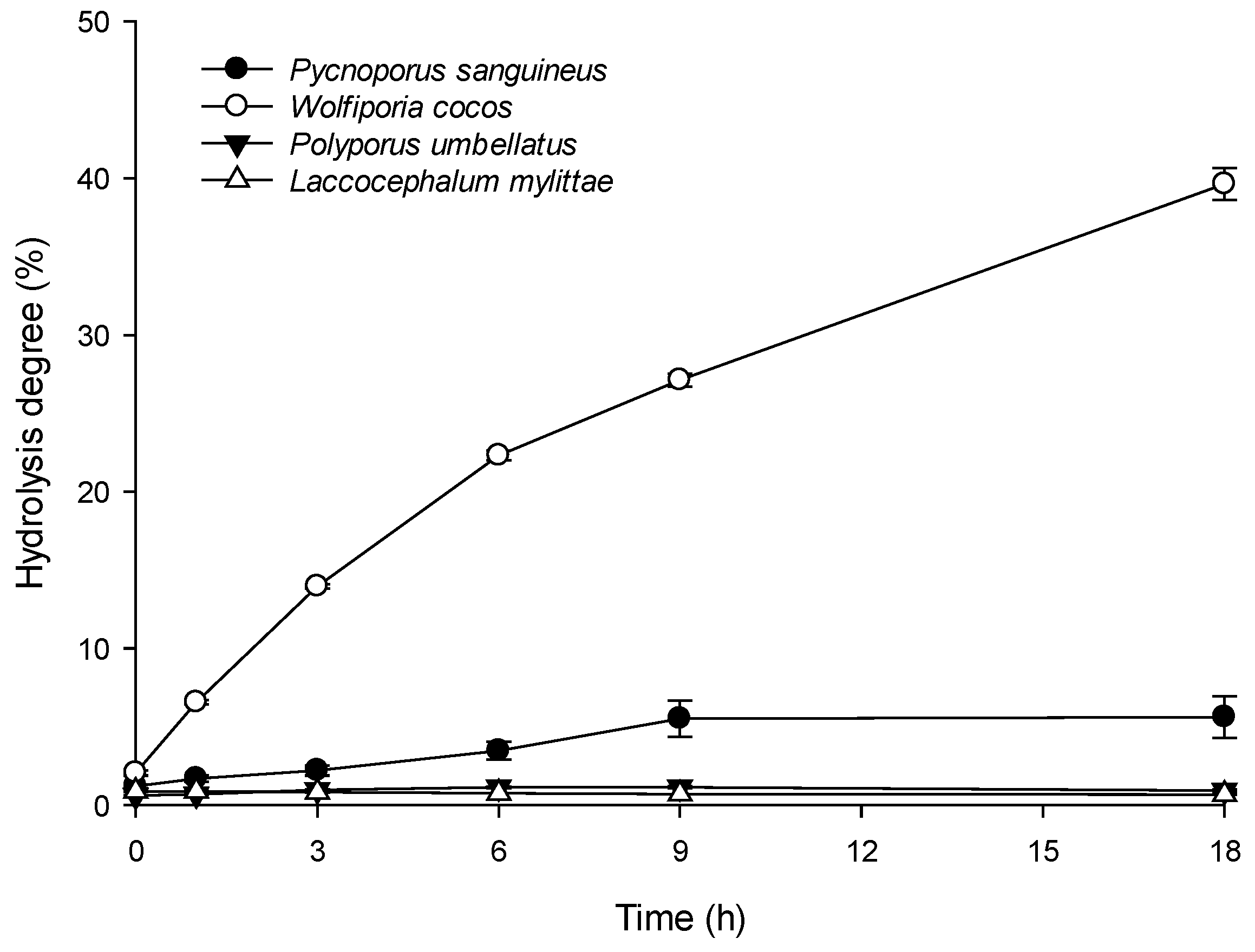
3.5. Enzymatic Hydrolysis of Polyporaceae Glucans

Four types of the Polyporaceae (P. sanguineus, W. cocos, P. umbellatus, and L. mylittae) glucans were hydrolyzed by β-1,3-glucanase from Y. lipolytica transformant (pYLSC1-13g), and the enzymatic hydrolysis degree were determined. As shown in Figure 7, the hydrolysis degree of W. cocos and P. sanguineus increased significantly during hydrolysis. After 18 h of hydrolysis, the hydrolysis degree for W. cocos and P. sanguineus were 40% and 6%, respectively, while those for P. umbellatus and L. mylittae were <1%.

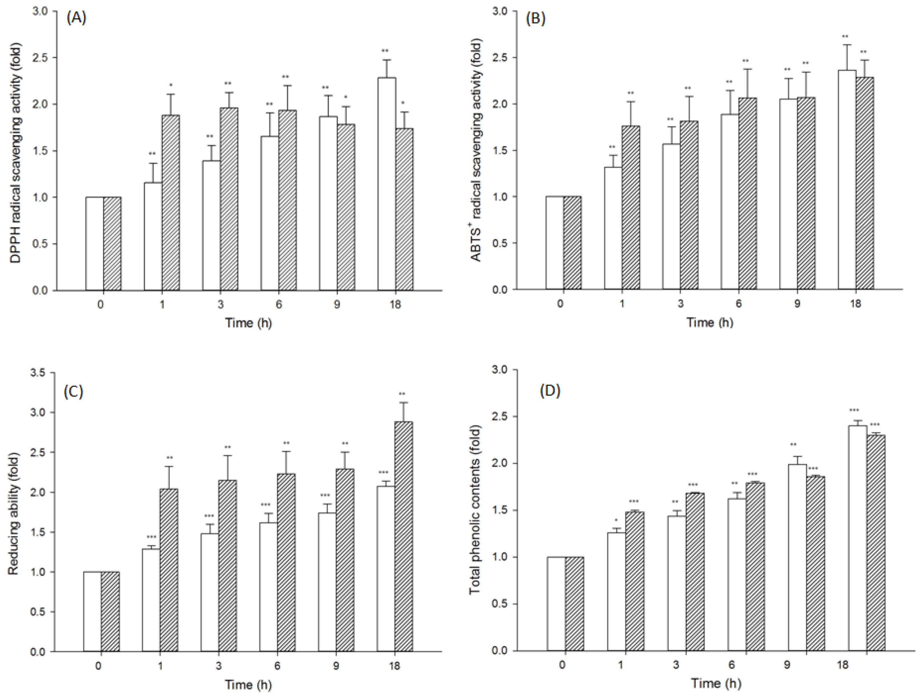
3.6. Evaluation of the Antioxidant Activity of Polyporaceae Hydrolysates

After glucans from the aforementioned the W. cocos and P. sanguineus were hydrolyzed by β-1,3-glucanase for 18 h, the antioxidant activities of enzyme hydrolysates were determined. The free radical scavenging assay revealed a significant increase in the DPPH scavenging potential of the two hydrolysates (Figure 8A).
The DPPH scavenging potential of P. sanguineus hydrolysates was consistent with an increase in its hydrolysis time. The maximum DPPH scavenging potential approached 2.3-fold after 18 h of hydrolysis. On hydrolyzing W. cocos glucans with β-1,3-glucanase for 1 h, the DPPH scavenging potential peaked and plateaued with an increase in the hydrolysis time.
The ABTS+ free radical scavenging activity of the enzyme hydrolysates of the W. cocos and P. sanguineus increased significantly on increasing the enzymatic hydrolysis time (Figure 8B).
The reducing power of the W. cocos and P. sanguineus increased significantly with an increase in the hydrolysis time (Figure 8C). The maximum reducing power approached 2.1-fold and 2.9-fold, respectively, after 18 h of hydrolysis.
As shown in Figure 8D, the total phenolic contents of the hydrolysates of the W. cocos and P. sanguineus used herein increased significantly with an increase in the enzymatic hydrolysis time. The P. sanguineus hydrolysate had the higher total phenolic contents after 18 h of hydrolysis.
Low-molecular-weight polysaccharides reportedly display higher antioxidant activity than high-molecular-weight polysaccharides [38,39,40]. Peasura et al. reported that low-molecular-weight polysaccharides have more reducing ends and can be made to react with free radicals; thus, displaying increased antioxidant activity [41]. The antioxidant activity of fungal polysaccharides is associated with the presence of phenolics [42]. In this study, the total phenolic contents increased as a result of Polyporaceae hydrolysis. The “total phenolic contents” have gained much attention, due to their free radical scavenging abilities and antioxidant activities, which potentially have beneficial implications in human health [43,44]. The “total phenolic contents” is usually considered as one of the important factors for evaluating antioxidant activity. It may suggest the deglycosylation of polyphenols from β-1,3-glucan. This could be confirmed by further experiments. As shown in Figure 7, W. cocos and P. sanguineus had a better hydrolysis rate than L. mylittae and P. umbellatus, with no significant hydrolysis in the latter two Polyporaceae. Although the main polysaccharides in four kinds of Polyporaceae are β-1,3-glucans. However, the content of β-1,3-glucan in various mushrooms is different. The structures of polysaccharides is complex. There are several types of linear or branched glucans in various mushroom species [45]. These differences in content and structure make the degree of hydrolysis different. After 18 h of incubation, enzymatic hydrolysates of the W. cocos and P. sanguineus presented higher total phenolic contents and displayed significantly improved DPPH and ABTS+ scavenging activity and reducing power.

4. Conclusions

Incubation of Y. lipolytica transformant (pYLSC1-13g) with YPD medium for 96 h increased the β-1,3-glucanase activity to 270 U/mL in the culture broth. The transformant β-1,3-glucanase had high thermostability and was tolerant to 10% methanol, ethanol, and DMSO. The transformant β-1,3-glucanase significantly hydrolyzed W. cocos and P. sanguineus. The DPPH and ABTS scavenging abilities, reducing power, and total phenolic contents of the aforementioned two Polyporaceae hydrolysates were significantly increased after 18 h of hydrolysis. This study shows that W. cocos and P. sanguineus glucans hydrolysates produced using the Thermobifida fusca β-1,3-glucanase in a Y. lipolytica transformant (pYLSC1-13g) have high antioxidant activities.

Author Contributions

C.-H.Y. and W.-L.C. designed the study. W.-L.C. and C.-H.Y. wrote the paper. W.-L.C., J.-C.H., C.-L.L., and C.-Y.C. performed the enzymatic assay, gene sequencing, and antioxidant assays. C.-H.Y. and W.-L.C. supervised the study. All authors have read and agreed to the published version of the manuscript.

Funding

This research was funded by the Ministry of Science and Technology of the Republic of China and grant number MOST 109-2313-B-126-001-MY2.

Institutional Review Board Statement

Not applicable.

Informed Consent Statement

Not applicable.

Data Availability Statement

The data presented in this study are available on request from the corresponding author.

Conflicts of Interest

The authors declare no conflict of interest.

References

  1. Choi, E.Y.; Lee, S.S.; Hyeon, J.Y.; Choe, S.H.; Keum, B.R.; Lim, J.M.; Park, D.C.; Chio, I.S.; Cho, K.K. Effects of β-glucan on the release of nitric oxide by macrophages stimulated with lipopolysaccharide. Asian-Australas J. Anim. Sci. 2016, 29, 1664–1674. [Google Scholar] [CrossRef] [PubMed]
  2. Kitamura, S.; Hori, T.; Kurita, K.; Takeo, K.; Hara, C.; Itoh, W.; Tabata, K.; Elgsaeter, A.; Stokke, B.T. An antitumor, branched (1→ 3)-β-D-glucan from a water extract of fruiting bodies of Cryptoporus volvatus. Carbohydr. Res. 1994, 263, 111–121. [Google Scholar] [CrossRef]
  3. Huiqin, F.; Qingyao, Y.; Xiaotong, Y.; Ke, M.; Lingyun, Z. Analysis of polysaccharide isolated from the fruit bodies and mycelia of Grifola frondosa. Huadong Shifan Daxue Xuebao J. East China Norm. Univ. 2001, 3, 9–96. [Google Scholar]
  4. Lei, N.; Wang, M.; Zhang, L.; Xiao, S.; Fei, C.; Wang, X.; Zhang, K.; Zheng, W.; Wang, C.; Yang, R. Effects of low molecular weight yeast β-glucan on antioxidant and immunological activities in mice. Int. J. Mol. Sci. 2015, 16, 21575–21590. [Google Scholar] [CrossRef] [Green Version]
  5. Choromanska, A.; Kulbacka, J.; Rembialkowska, N.; Pilat, J.; Oledzki, R.; Harasym, J.; Saczko, J. Anticancer properties of low molecular weight oat beta-glucan–An in vitro study. Int. J. Biol. Macromol. 2015, 80, 23–28. [Google Scholar] [CrossRef] [PubMed]
  6. Tada, R.; Tanioka, A.; Iwasawa, H.; Hatashima, K.; Shoji, Y.; Ishibashi, K. Structural characterisation and biological activities of a unique type β-D-glucan obtained from Aureobasidium pullulans. Glycoconj J. 2008, 25, 851–861. [Google Scholar] [CrossRef]
  7. Vetvicka, V.; Vetvickova, J. Physiological effects of different types of β-glucan. Biomed. Pap. Med. Fac. Univ. Palacky Olomouc Czech Repub. 2007, 151, 225–231. [Google Scholar] [CrossRef] [Green Version]
  8. Tuong, T.D.; Chu, D.X.; Dieu, B.T.M. Antioxidant activity of fruiting body extracts from Pycnoporus sanguineus (L.: Fr.) Murrill mushroom. Vietnam J. Sci. Technol. 2020, 58, 143–151. [Google Scholar]
  9. Kozarski, M.; Klaus, A.; Jakovljevic, D.; Todorovic, N.; Vunduk, J.; Petrović, P. Antioxidants of edible mushrooms. Molecules 2015, 20, 19489–19525. [Google Scholar] [CrossRef] [Green Version]
  10. Dzhavakhiya, V.G.; Ozeretskovskaya, O.L.; Zinovyeva, S.V. Chapter 10-Immune response. In Comprehensive and Molecular Phytopathology; Dyakov, Y.T., Dzhavakhiya, V.G., Korpela, T., Eds.; Elsevier: Amsterdam, The Netherlands, 2007; pp. 265–314. [Google Scholar]
  11. Wu, Q.; Dou, X.; Wang, Q.; Guan, Z.; Cai, Y.; Liao, X. Isolation of β-1,3-Glucanase-Producing Microorganisms from Poria cocos Cultivation Soil via Molecular Biology. Molecules 2018, 23, 1555. [Google Scholar] [CrossRef] [Green Version]
  12. Kao, P.F.; Wang, S.H.; Hung, W.T.; Liao, Y.H.; Lin, C.M.; Yang, W.B. Structural characterization and antioxidative activity of low-molecular-weights beta-1, 3-glucan from the residue of extracted Ganoderma lucidum fruiting bodies. J. Biomed. Biotechnol. 2012, 1, 673764. [Google Scholar]
  13. Wang, H.; Mukerabigwi, J.F.; Zhang, Y.; Han, L.; Jiayinaguli, T.; Wang, Q.; Liu, L.; Cao, Y.; Sun, R.; Huang, X. In vivo immunological activity of carboxymethylated-sulfated (1→ 3)-β-d-glucan from sclerotium of Poria cocos. Int. J. Biol. Macromol. 2015, 79, 511–517. [Google Scholar] [CrossRef] [PubMed]
  14. Tang, J.; Nie, J.; Li, D.; Zhu, W.; Zhang, S.; Ma, F.; Sun, Q.; Song, J.; Zheng, Y.; Chen, P. Characterization and antioxidant activities of degraded polysaccharides from Poria cocos sclerotium. Carbohydr. Polym. 2014, 105, 121–126. [Google Scholar] [CrossRef] [PubMed]
  15. He, P.F.; Zhang, A.Q.; Wang, X.L.; Qu, L.; Li, G.L.; Li, Y.P.; Sun, P.L. Structure elucidation and antioxidant activity of a novel polysaccharide from Polyporus umbellatus sclerotia. Int. J. Biol. Macromol. 2016, 82, 411–417. [Google Scholar] [CrossRef]
  16. Wu, F.; Zhou, L.W.; Yang, Z.L.; Bau, T.; Li, T.H.; Dai, Y.C. Resource diversity of Chinese macrofungi: Edible, medicinal and poisonous species. Fungal Divers. 2019, 98, 1–76. [Google Scholar] [CrossRef]
  17. Yang, L.J.; Wang, R.T.; Liu, J.S.; Tong, H.; Deng, Y.Q.; Li, Q.H. The effect of Polyporus umbellatus polysaccharide on the immunosuppression property of culture supernatant of S180 cells. Cell Mol. Immunol. 2004, 20, 234–237. [Google Scholar]
  18. Vannucci, L.; Krizan, J.; Sima, P.; Stakheev, D.; Caja, F.; Rajsiglova, L.; Horak, V.; Saieh, M. Immunostimulatory properties and antitumor activities of glucans. Int. J. Oncol. 2013, 43, 357–364. [Google Scholar] [CrossRef] [Green Version]
  19. Cao, J.; Zhang, H.J.; Xu, C.P. Culture characterization of exopolysaccharides with antioxidant activity produced by Pycnoporus sanguineus in stirred-tank and airlift reactors. J. Taiwan Inst. Chem. Eng. 2014, 45, 2075–2080. [Google Scholar] [CrossRef]
  20. Zhou, L.H.; Zhang, Y.Q.; Wang, R.J.; Shen, X.L.; Li, Y.Q.; Guan, W.J. Optimization of mycelial biomass and protease production by Laccocephalum mylittae in submerged fermentation. Afr. J. Biotechnol. 2009, 8, 1591–1601. [Google Scholar]
  21. Tuncer, M.; Ball, A.S.; Rob, A.; Wilson, M.T. Optimization of extracellular lignocellulolytic enzyme production by a thermophilic actinomycetes Thermomonospora fusca BD25. Enzyme Microb. Technol. 1999, 25, 38–47. [Google Scholar] [CrossRef]
  22. Yang, C.H.; Liu, W.H. Purification and properties of an acetylxylan esterase from Thermobifida fusca. Enzyme Microb. Technol. 2008, 42, 181–186. [Google Scholar] [CrossRef] [PubMed]
  23. McGrath, C.E.; Wilson, D.B. Characterization of a Thermobifida fusca β-1,3-glucanase (Lam81A) with a potential role in plant biomass degradation. Biochemistry 2006, 45, 14094–14100. [Google Scholar] [CrossRef] [PubMed]
  24. Bordes, F.; Fudalej, F.; Dossat, V.; Nicaud, J.M.; Marty, A. A new recombinant protein expression system for high-throughput screening in the yeast Yarrowia lipolytica. J. Microbiol. Methods 2007, 70, 393–502. [Google Scholar] [CrossRef] [PubMed]
  25. Madzak, C.; Treton, B.; Blanchin-Roland, S. Strong hybrid promoters and integrative expression/secretion vectors for quasi-constitutive expression of heterologous proteins in the yeast Yarrowia lipolytica. J. Mol. Microbiol. Biotechnol. 2000, 2, 207–216. [Google Scholar]
  26. Madzak, C.; Gaillardin, C.; Beckerich, J.M. Heterologous protein expression and secretion in the non-conventional yeast Yarrowia lipolytica. J. Biotechnol. 2004, 109, 63–81. [Google Scholar] [CrossRef]
  27. Sambrook, J.; Russell, D.W. Molecular Cloning: A Laboratory Manual, 3rd ed.; Cold Spring Harbor Laboratory Press: New York, NY, USA, 2001. [Google Scholar]
  28. Miller, G.L. Use of dinitrosalicylic acid reagent for determination of reducing sugar. Anal. Chem. 1959, 31, 426–428. [Google Scholar] [CrossRef]
  29. Huang, Y.C.; Chen, C.Y.; Chen, W.-L.; Ciou, Y.-P.; Yang, T.Y.; Yang, C.H. Production and antioxidant properties of the ferulic acid-rich destarched wheat bran hydrolysate by feruloyl esterases from thermophilic actinomycetes. BioResources 2013, 8, 4981–4991. [Google Scholar] [CrossRef] [Green Version]
  30. El-Katatny, M.; Gudelj, M.; Robra, K.H.; Elnaghy, M.; Gübitz, G. Characterization of a chitinase and an endo-β-1,3-glucanase from Trichoderma harzianum Rifai T24 involved in control of the phytopathogen Sclerotium rolfsii. Appl. Microbiol. Biotechnol. 2001, 56, 137–143. [Google Scholar] [CrossRef]
  31. Schwentke, J.; Sabel, A.; Petri, A.; König, H.; Claus, H. The yeast Wickerhamomyces anomalus AS1 secretes a multifunctional exo-β-1,3-glucanase with implications for winemaking. Yeast 2014, 31, 349–359. [Google Scholar] [CrossRef] [Green Version]
  32. Fridlender, M.; Inbar, J.; Chet, I. Biological control of soilborne plant pathogens by a β-1,3-glucanase-producing Pseudomonas cepacia. Soil Biol. Biochem. 1993, 25, 1211–1221. [Google Scholar] [CrossRef]
  33. Abeles, F.B.; Forrence, L.E. Temporal and hormonal control of β-1,3-glucanase in Phaseolus vulgaris L. Plant Physiol. 1970, 45, 395–400. [Google Scholar] [CrossRef] [PubMed] [Green Version]
  34. Noronha, E.F.; Ulhoa, C. Characterization of a 29-kDa β-1,3-glucanase from Trichoderma harzianum. FEMS Microbiol. Lett. 2000, 183, 119–123. [Google Scholar] [CrossRef]
  35. Woo, C.B.; Kang, H.N.; Lee, S.B. Molecular cloning and anti-fungal effect of endo-β-1,3-glucanase from Thermotoga maritima. Food Sci. Biotechnol. 2014, 23, 1243–1246. [Google Scholar] [CrossRef] [Green Version]
  36. Monteiro, V.N.; Ulhoa, C.J. Biochemical characterization of a β-1,3-glucanase from Trichoderma koningii induced by cell wall of Rhizoctonia solani. Curr. Microbiol. 2006, 52, 92–96. [Google Scholar] [CrossRef] [PubMed]
  37. Nagasaki, S.; Mori, H.; Yamamoto, S. Characterization of Endo β-1,3-Glucanase IV from Flavobacterium dormitator var. glucanolyticae FA-5. Agric. Biol. Chem. 1981, 45, 2689–2694. [Google Scholar] [CrossRef]
  38. Li, B.; Liu, S.; Xing, R.; Li, K.C.; Li, R.F.; Qin, Y.K.; Wang, X.Q.; Wei, Z.H.; Li, P.C. Degradation of sulfated polysaccharides from Enteromorpha prolifera and their antioxidant activities. Carbohydr. Polym. 2013, 92, 1991–1996. [Google Scholar] [CrossRef]
  39. Guo, X.; Ye, X.Q.; Sun, Y.J.; Wu, D.; Wu, N.A.; Hu, Y.Q.; Chen, S.G. Ultrasound effects on the degradation kinetics, structure, and antioxidant activity of sea cucumber fucoidan. J. Agric. Food Chem. 2014, 62, 1088–1095. [Google Scholar] [CrossRef]
  40. Zha, S.H.; Zhao, Q.S.; Zhao, B.; Ouyang, J.; Mo, J.L.; Chen, J.J.; Cao, L.L.; Zhang, H. Molecular weight controllable degradation of Laminaria japonica polysaccharides and its antioxidant properties. J. Ocean Univ. China 2016, 15, 637–642. [Google Scholar] [CrossRef]
  41. Peasura, N.; Laohakunjit, N.; Kerdchoechuen, O.; Wanlapa, S. Characteristics and antioxidant of Ulva intestinalis sulphated polysaccharides extracted with different solvents. Int. J. Biol. Macromol. 2015, 81, 912–919. [Google Scholar] [CrossRef]
  42. Dinh, T.V.; Saravana, P.S.; Woo, H.C.; Chun, B.S. Ionic liquid-assisted subcritical water enhances the extraction of phenolics from brown seaweed and its antioxidant activity. Sep. Sci. Technol. 2018, 196, 287–299. [Google Scholar]
  43. Imeh, U.; Khokhar, S. Distribution of conjugated and free phenols in fruits: Antioxidant activity and cultivar variations. J. Agric. Food Chem. 2002, 50, 6301–6306. [Google Scholar] [CrossRef] [PubMed]
  44. Ross, J.A.; Kasum, C.M. Dietary flavonoids: Bioavailability, metabolic effects, and safety. Ann. Rev. Nutr. 2002, 22, 19–34. [Google Scholar] [CrossRef] [PubMed]
  45. Semedo, M.C.; Karmali, A.; Fonseca, L. A high throughput colorimetric assay of β-1,3-d-glucans by Congo red dye. J. Microbiol. Methods 2015, 109, 140–148. [Google Scholar] [CrossRef] [PubMed]
Figure 1. Plasmid map of pYLSC1-13g.
Processes 09 00056 g001
Figure 2. Time course of β-1,3-glucanase activity in a Y. lipolytica transformant (pYLSC1-13g). (-●-) extracellular β-1,3-glucanase activity; (-○-), Optical density (OD) 600 nm; (-▲-), pH. The culture conditions were: temperature 25 °C; shaking speed 250 rpm.
Processes 09 00056 g002
Figure 3. SDS-PAGE of the purified β-1,3-glucanase from Y. lipolytica transformant (pYLSC1-13g). Lane M: molecular weight marker; Lane 1: purified β-1,3-glucanase.
Processes 09 00056 g003
Figure 4. Thermal stability of β-1,3-glucanase from Y. lipolytica transformant (pYLSC1-13g). -●-: 40 °C; -○-: 50 °C; -▼-: 60℃; -△-: 70 °C.
Processes 09 00056 g004
Figure 5. Effect of organic solvents on β-1,3-glucanase from Y. lipolytica transformant (pYLSC1-13g). Data: mean ± SE from triplicate experiments (Student’s t-test P values: * p < 0.05; ** p < 0.01; *** p < 0.001).
Processes 09 00056 g005
Figure 6. Effect of metal salts on β-1,3-glucanase from Y. lipolytica transformant (pYLSC1-13g). Data: mean ± SE from triplicate experiments (Student’s t-test p values: * p < 0.05; *** p < 0.001).
Processes 09 00056 g006
Figure 7. Hydrolysis degree of Polyporaceae hydrolysate by β-1,3-glucanase from Y. lipolytica transformant (pYLSC1-13g).
Processes 09 00056 g007
Figure 8. Antioxidant activity of edible medicinal Polyporaceae enzyme hydrolysate prepared using β-1,3-glucanase from Y. lipolytica transformant (pYLSC1-13g). (A) DPPH radical scavenging assay; (B) ABTS+ radical scavenging activity; (C) reducing power assay; (D) total phenolic content assay. Processes 09 00056 i001 Pycnoporus sanguineus; Processes 09 00056 i002 Wolfiporia cocos. Data: mean ± SE from triplicate experiments (Student’s t-test p values: * p < 0.05; ** p < 0.01; *** p < 0.001).
Processes 09 00056 g008
Table 1. Purification of β-1,3-glucanase from Y. lipolytica transformant (pYLSC1-13g).
Total Activity
(U)
Total Protein
(mg)
Specific Activity
(U/mg)
Purification
(Fold)
Yield
(%)
Culture supernatant101,341.3812.368199.141100
Ni2+-NTA column29,661.150.9232,240.383.9329.27
Publisher’s Note: MDPI stays neutral with regard to jurisdictional claims in published maps and institutional affiliations.

