Article

Compound Identification and In Vitro Cytotoxicity of the Supercritical Carbon Dioxide Extract of Papaya Freeze-Dried Leaf Juice

by
Kooi-Yeong Khaw
1,2,*,
Paul Nicholas Shaw
1,*,
Marie-Odile Parat
1,
Saurabh Pandey
1 and
James Robert Falconer
1,*
1
School of Pharmacy, The University of Queensland, Brisbane, QLD 4072, Australia
2
Biofunctional Molecule Exploratory Research Group (BMEX) School of Pharmacy, Monash University Malaysia, Jalan Lagoon Selatan, Bandar Sunway 47500, Malaysia
*
Authors to whom correspondence should be addressed.
Processes 2020, 8(5), 610; https://doi.org/10.3390/pr8050610
Submission received: 10 May 2020 / Revised: 15 May 2020 / Accepted: 19 May 2020 / Published: 20 May 2020
(This article belongs to the Special Issue Innovation in Chemical Plant Design)

Abstract

:
Carica papaya leaves are used as a remedy for the management of cancer. Freeze-dried C. papaya leaf juice was extracted using a supercritical fluid extraction system. Compound identification was carried out using analytical techniques including liquid chromatography coupled to high-resolution quadrupole time-of-flight mass spectrometry (LC–QToF-MS) and gas chromatography–mass spectrometry (GC–MS). The cytotoxic activities of the scCO2 extract and its chemical constituents were determined using the 3-(4,5-dimethylthiazol-2-yl)-2,5-diphenyltetrazolium bromide (MTT) assay on squamous cell carcinoma (SCC25) and human keratinocyte (HaCaT) cell lines. The chemical constituents were quantified by QToF-MS. The supercritical carbon dioxide (scCO2) extract of papaya freeze-dried leaf juice showed cytotoxic activity against SCC25. Three phytosterols, namely, β-sitosterol, campesterol, and stigmasterol, together with α-tocopherol, were confirmed to be present in the scCO2 extract. Quantitative analysis showed that β-sitosterol was the major phytosterol present followed by α-tocopherol, campesterol, and stigmasterol. β-Sitosterol and campesterol were active against SCC25 (half maximal inhibitory concentration (IC50) ≈ 1 µM), while stigmasterol was less active (~33 µM) but was biologically more selective against SCC25. Interestingly, an equimolar mixture of phytosterols was not more effective (no synergistic effect was observed) but was more selective than the individual compounds. The compounds identified are likely accountable for at least part of the cytotoxicity and selectivity effects of C. papaya.

1. Introduction

Carica papaya is a herbaceous plant indigenous to tropical Mexico, Central America, and northern South America [1]. It is widely cultivated in the tropical and subtropical countries for its nutritional edible fruit [2]. Apart from the fruit of C. papaya, the leaf is used as a food and medicine. Anecdotal evidence indicates that the leaf is used in Australia in the form of a decoction to treat cancer [3]. In other parts of the world, the decoction of the leaf is used as a tea to treat high blood pressure, diabetes, digestion disorder, and jaundice, as well as dengue fever, rheumatic complaints, and elephantoid growths [4,5,6,7].
Several chemical constituents from C. papaya were identified [7]. The leaf of papaya is reported to contain alkaloids, tocopherol, flavonoids, tannins, phytosterols, saponin, phenolic compounds, and chlorogenic acid [8,9]. Epidemiology studies showed that phytochemicals from plants are beneficial in reducing the risk of dementia, stroke, diabetes, cardiovascular disease, and cancers [10]. Some studies demonstrated that leaf extracts from C. papaya were selectively cytotoxic to skin cancer in vitro [11,12]. Nguyen and colleagues reported that phenoside A from papaya leaf juice was potently cytotoxic to the cancerous SCC25 cell line. However, it was also cytotoxic against the non-cancerous HaCaT cell line [12]. Active and selective anti-cancer compounds from leaf juice remain to be fully elucidated.
This research gap prompted our interest to discover the bioactive chemical constituents of C. papaya leaves with selective activity against skin cancer. Supercritical fluid extraction (SFE) offers an alternative method, whereby different chemical constituents are selectively extracted from those components yielded by conventional methods. This approach may, therefore, reduce the analytical workload in identifying chemical constituents of interest. Several analytical methods were used to identify and quantify chemical constituents from papaya leaves including LC–MS, GC–MS, and nuclear magnetic resonance (NMR) spectroscopy [11,13,14]. For example, linoleic and linolenic acids were identified from the ethyl acetate fraction of C. papaya leaves by GC–MS [15].
Over the course of our continuing efforts to characterize anti-cancer compounds from C. papaya, we aimed to identify bioactive chemical constituents from the scCO2 extract of C. papaya. The tentatively identified compounds were obtained commercially and compared with those present in the scCO2 extract (for their liquid chromatography retention times, accurate mass, and MS/MS fragmentations). The identified compounds were analyzed and quantified. Furthermore, the cytotoxicity of the identified active compounds alone and in combination were evaluated.

2. Materials and Methods

2.1. Materials

DL-α-Tocopherol (purity >96%), stigmasterol (purity >95%), and 3-(4,5-dimethylthiazol-2-yl)-2,5-diphenyltetrazolium bromide were purchased from Sigma-Aldrich (Castle Hill, NSW, Australia). β-Sitosterol (purity >95%) was from Chromadex (USA) and campesterol (purity >95%) was from Novachem (Heidelberg West, VIC, Australia). Ethanol, HPLC-grade methanol, and HPLC-grade acetone were purchased from Merck (Darmstadt, Germany). Dulbecco’s modified Eagle’s medium (DMEM), DMEM/F12, trypsin, penicillin/streptomycin, and fetal bovine serum (FBS) were obtained from Invitrogen (Life Technologies, Mulgrave, VIC, Australia). Freeze-dried leaf juice was prepared from Australian C. papaya leaves (grown organically) gifted by Tropical Fruit World Pty Ltd., (Duranbah, New South Wales, Australia) according to the protocol described previously [16].

2.2. Supercritical Fluid Extraction (SFE)

SFE of C. papaya leaf juice was performed using a laboratory-scale extraction system. Earlier experiments determined that freeze-dried leaf juice was a better starting material in comparison with fresh leaves or freeze-dried leaves when considering both extraction yield and selective toxicity to cancer cells. The SFE system comprised a liquid carbon dioxide (CO2) reservoir, high-pressure syringe pump (Teledyne Isco 260D), a vertical 60-mL stainless-steel (SS-316) extraction vessel (University of Nottingham), backpressure regulator, heating jacket (WatLow, USA), overhead stirrer (200 rpm) and fixed straight blade paddle, and SS precipitation chamber; details are described in previous work [16]. Glass wool (Merck, Darmstadt, Germany) was placed inside the extraction vessel and covered by a stainless-steel mesh to prevent any entrainment of the sample. We previously optimized the conditions of SFE that provided the extract exhibiting most cytotoxicity toward cancer cells [16]. The extraction was operated with the following conditions: pressure 250 bar; temperature 35 °C; freeze-dried leaf juice 5 g; extraction time 3 h. Following completion of the extraction, the supercritical fluid (scCO2) extract was separated to the precipitation chamber via a capillary nozzle (0.0625 inch; 1.5875 mm) and the CO2 gas was discharged to the atmosphere. The extract was weighed and stored at −20 °C for further analysis. The experiments were repeated three times.

2.3. Gas Chromatography–Mass Spectrometry

Analysis of the scCO2 extract was carried out using a Shimadzu GCMS-TQ8040. Separation was obtained on an Rtx-5ms column (30 m × 0.25 mm, 0.25 µm film thickness; Restek, USA) with helium as the carrier gas at a constant linear velocity of 46.6 cm/s. The injection volume was 1 µL with a split ratio of 10. The initial column temperature was held at 160 °C for 1 min and then increased to 300 °C at a rate of 10 °C/min. The final column temperature was maintained at 300 °C for another 10 min. The temperatures of the injector and the detector were 240 °C and 200 °C, respectively. The interface temperature was set to 300 °C. Mass acquisition was performed in the range of 42–500 m/z using electron impact ionization at 70 eV. The components detected in the sample were identified by performing spectral database matching against the National Institute of Standards and Technology (NIST) library (v14).

2.4. Sample Preparation for UHPLC–QToF-MS Analysis

The standard stock solutions of dl-α-tocopherol, stigmasterol, β-sitosterol, and campesterol were prepared by dissolving accurately weighed standards in HPLC-grade methanol to give a concentration of 1 mg/mL. Each stock solution was filtered through a 0.22-μm polyvinylidene fluoride (PVDF) sterile filter (Merck Millipore, Germany) and stored in a freezer at −20 °C prior to analysis.

2.5. UHPLC–QToF-MS Analysis

The chromatography analysis of dl-α-tocopherol and phytosterols was performed on an Agilent 1290 UHPLC system (Agilent Technologies, Santa Clara, CA, USA) equipped with an Agilent 6520 high-resolution accurate mass quadruple time of flight (QToF) mass spectrometer. Chromatographic separation was conducted by a 2.0 × 150 mm, 100 Å, 2.6 µm C18 analytical column (Phenomenex, USA). Ultra-purified MilliQ water was mobile phase A, while HPLC-grade methanol was mobile phase B. The gradient elution conditions were as follows: 50% B for the first 5 min; 50% B increasing to 90% B from 5–40 min; 90% B increasing to 100% B from 40–60 min; 100% B decreasing to 50% B from 60–75 min. The sample injection volume was 5 µL with the flow rate of 0.2 mL/min. Mass spectral acquisition was monitored by MassHunter software (version B.02.01 SP3 –Agilent). The operating conditions for the mass spectrometer included a scan rate of 0.8 cycles/per second with the following conditions: nebuliser pressure 30 psi, m/z scan 100–1700, drying gas flow 5.0 L/min, gas temperature 300 °C, fragmenting voltage 175 V, and skimmer voltage 65.0 V.

2.5.1. Calibration Curves, Linearity Ranges, Limits of Detection (LOD), and Limits of Quantification (LOQ)

In this study, four biomarker analytes were evaluated at concentrations ranging from 1.56 to 50 µg/mL for the determination of the linear dynamic ranges. Each individual analyte at a fixed concentration was injected three times and the resultant peak heights obtained. Calibration curves were constructed by plotting peak heights against the analyte concentrations prepared, and the linearity of response to the four compounds was evaluated by linear regression analysis.

2.5.2. Quantification of dl-α-Tocopherol and Phytosterols by LC–QToF-MS

A 5-µL volume of the sample was injected into the LC–MS system. The concentration and profile of dl-α-tocopherol and phytosterols in the extracts were obtained using optimized LC–QToF-MS parameters. Peak identifications were performed by matching the retention times and accurate masses with the standard analytes. The sample was quantified using the external standard method.

2.6. Sample Preparation for the Cell Viability Assay

dl-α-Tocopherol, campesterol, stigmasterol, and β-sitosterol were dissolved in ethanol at concentrations of 10 mg/mL or 25 mM for the combined phytosterol assay, while the scCO2 extract was solubilized in ethanol at a concentration of 50 mg/mL. All samples were sterile-filtered by a 0.22-µm polyvinylidene fluoride (PVDF) filter (Merck Millipore, Germany), resulting in stock solutions that were diluted with serum-free medium to the indicated final concentrations prior to performing the experiments.

2.7. Cell Culture Conditions

Human oral squamous cell carcinoma (SCC25) cells were obtained from ATCC® CRL-1628™, Manassas, VA, USA. The cells were maintained in DMEM/F12 medium added with 10% v/v heat-inactivated FBS, penicillin (100 units/mL), streptomycin (100 µg/mL), and hydrocortisone (0.4 μg/mL). Non-cancerous human keratinocyte (HaCaT) cells were a generous gift from Professor Fusenig. The cells were grown in DMEM containing 10% FBS, penicillin (100 units/mL), and streptomycin (100 µg/mL). All cell lines were cultured in a humidified atmosphere with 5% CO2 at 37 °C. The cultures were passaged every third day, at which point they were approximately 70%–90% confluent.

Cell Viability Assay

The 3-(4,5-dimethylthiazol-2-yl)-2,5-diphenyltetrazolium bromide (MTT) assay was performed according to the method previously optimized for anticancer bioactive discovery [11,12]. In brief, 6000 cells (SCC25) per well or 3000 cells (HaCaT) were seeded in each well of a 96-well microtiterTM microplate. The cells were allowed to attach for 24 h prior to addition of samples. The culture medium was replaced with samples to be tested diluted in serum-free medium. The samples were incubated for 48 h at 37 °C, and the serum-free medium was replaced with a 0.5 mg/mL MTT solution. After a 2-h incubation, the medium was replaced with 100 μL of dimethyl sulfoxide (DMSO) on an orbital shaker for 20 min. The absorbance values were measured at 595 nm using an Lmark plate reader (BioRad, Hercules, California, USA). Wells containing no cells was being used as blanks whose absorbance was subtracted. Results are expressed as the percentage of viable cells with control, with untreated cells taken as 100%. The half maximal inhibitory concentration (IC50) values of the compounds were estimated using non-linear regression analysis implemented in Prism 7 (GraphPad software Inc., San Diego, CA, USA).

2.8. Statistical Analysis

All statistical analysis was performed using Prism 7 (GraphPad software Inc., San Diego, CA, USA). All data are presented as means ± standard error of mean (SEM). Two-way ANOVA with a Sidak post hoc test was employed to compare the differences between the two cell lines.

3. Results

3.1. Supercritical Carbon Dioxide Extraction

Freeze-dried leaf juice of C. papaya was extracted using a scCO2 extraction system. The extraction yield of scCO2 freeze-dried leaf juice was 3.2% (n = 3).

3.2. Chemical Analysis of scCO2 Extract with GC–MS

GC–MS results showed that at least 11 volatile compounds were present in the scCO2 extract of freeze-dried leaf juice of papaya (Figure A1).
The mass spectral database of these compounds was matched (>95%) with the NIST library. Lipophilic compounds including fatty acids, vitamin E, and the phytosterols were tentatively assigned, and they are listed in Table 1. The results showed that dl-α-tocopherol (25.15%) and β-sitosterol (40.96%) were the major compounds detected, followed by campesterol (9.87%) and stigmasterol (8.86%).

3.3. Compound Identification by Comparison with Authentic Standards

Based on the results from the database searching and the tentative identification of compounds from GC–MS analysis, direct comparison of the four compounds with four commercially available standards (dl-α-tocopherol, β-sitosterol, stigmasterol, and campesterol) with the scCO2 extract of freeze-dried leaf juice was performed using LC–MS.
Table 2 shows the comparison of the masses and retention times of the respective standards and features derived from the scCO2 extract of freeze-dried leaf juice detected in positive ion mode. All protonated phytosterols likely lost a molecule of water and, hence, the ions detected by the mass spectrometer are of the form [M + H − H2O]+. All compounds were matched to the retention times, accurate masses, and MS/MS fragmentations of the reference compounds (Figure 1a–d).

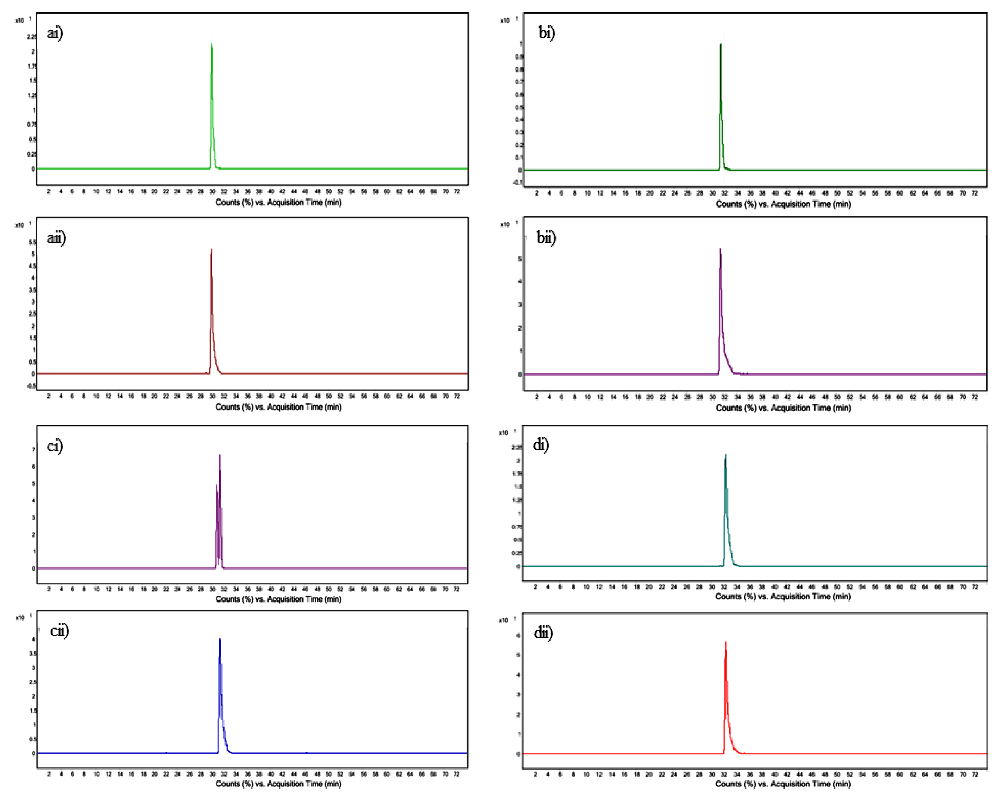
3.4. Quantification of Tocopherol and Phytosterols by UPLC–QToF-MS

The responses of all compounds possessed good linearity where dl-α-tocopherol, campesterol, and β-sitosterol were linear over the concentration range of 1.56–50 µg/mL (r2 = 0.9992, 0.9987, and 0.9991, respectively), and stigmasterol was linear over the range 3.12–50 µg/mL. The LODs were calculated using a signal-to-noise ratio of 3. The LOD was 0.39 µg/mL for stigmasterol campesterol, and β-sitosterol, and 0.19 µg/mL for dl-α-tocopherol. The LOQs were calculated at a signal-to-noise ratio of 10. The LOQ was 1.56 µg/mL for stigmasterol, campesterol, and β-sitosterol, and 0.78 µg/mL for dl-α-tocopherol (Table 3).
The concentrations of tocopherol and phytosterols from C. papaya were evaluated. The content of each compound was calculated as μg/100 mg of original leaf. The concentrations of tocopherol and phytosterols were in the range of 0.33 to 1.91 μg/100 mg of original leaf material. β-Sitosterol was present at the highest concentration, followed by dl-α-tocopherol, campesterol, and stigmasterol (Table 4).

3.5. Cytotoxicity of scCO2 Extract and Identified Compounds

The cytotoxic effects of scCO2 extract, dl-α-tocopherol, stigmasterol, campesterol, and β-sitosterol were evaluated against cancerous SCC25 and non-cancerous HaCaT cell lines using the MTT assay. The concentrations used ranged from 1 to 100 µg/mL for the individual compounds and 30 to 500 µg/mL for scCO2 extract. Figure 2 and Figure 3 show SCC25 HaCaT cell viabilities upon exposure to extracts or pure compounds. The scCO2 extract and phytosterols, but not dl-α-tocopherol, showed statistically significant cytotoxic effects against SCC25 and HaCaT cell lines in a dose-dependent manner. Approximately 60% of SCC25 cells survived when exposed to a concentration of 1 µg/mL campesterol or β-sitosterol, while 1 µg/mL stigmasterol only reduced cell viability to 77.1%. Approximately 60% of SCC25 cells survived after treatment with 250 µg/mL of the scCO2 leaf juice extract. Interestingly, when comparing the selectivity of the phytosterols at 1 µg/mL, stigmasterol resulted in the most significant difference in viabilities between the two cell lines (13.4% difference), whereas campesterol and β-sitosterol demonstrated statistically significant selectivity, but with only 3.4% and 5.6% differences, respectively, at the same concentrations. On the other hand, the scCO2 extract showed significant selectivity between the two cell lines (p < 0.001, two-way ANOVA) with the post hoc test comparison showing the significance at each concentration tested.
Table 5 summarizes the IC50 values of the compounds and scCO2 extract toward each cell line. Campesterol was very active but not selective (had the same IC50 against SCC25 and HaCaT cells). Stigmasterol and β-sitosterol exhibited some selectivity toward SCC25, and stigmasterol was less toxic than the other two sterols tested. The scCO2 extract was cytotoxic against SCC25 and HaCaT cell lines at concentrations of 88.07 to 120.60 µg/mL, respectively, confirming its selectivity.

3.6. Effect of Combined Phytosterols against SCC25 and HaCaT Cell Lines

To determine if there was a synergistic interaction between the compounds of interest, the cytotoxic effects of an equimolar mixture of stigmasterol, campesterol, and β-sitosterol on SCC25 and HaCaT cell lines were evaluated at a range of concentrations from 2.5 to 250 µM (equivalent to 1–100 μg/mL). In this experiment, 1 μM of the mixture represents a 0.333 μM concentration of each individual sterol. Figure 4 reveals the effects of the mixture against SCC25 and HaCaT cell lines. The mixture of phytosterols showed cytotoxicity with an IC50 of 5.98 and 15.60 µM toward SCC25 and HaCaT cells, respectively. The selectivity of the mixture was the highest at a concentration of 5 µM with 89% viable HaCaT and 68% viable SCC25 cells (** p < 0.01, two-way ANOVA with the Sidak post hoc test). Two-way ANOVA analysis indicated that, overall, the treatment effect was different between the two cell lines (p < 0.0001).
The IC50 of the mixture of phytosterols indicates that the mixture of compounds is not more potent than campesterol or β-sitosterol on either cell line (Table 5), thereby excluding significant synergistic effects. It is interesting to note, however, that, with an IC50 ~3 times lower on cancer cells than non-cancer cells, the mixture showed better selectivity than any of the individual sterols or the extract.

4. Discussion

Several groups reported the potential anticancer properties of C. papaya leaves prepared by different extraction methods [12,17,18]. However, the phytochemicals responsible for cytotoxicity and selectivity are yet to be identified. An orthogonally different extraction method such as supercritical CO2 extraction method is necessary to ease the identification of compounds of interest from extracts of simplified composition [19]. There are no scientific studies in the literature which characterize the scCO2 extracts from freeze-dried leaf juice of C. papaya and, therefore, the present study provides the first insights into the chemotherapeutic potential of identified compounds from scCO2 extract from freeze-dried leaf juice of C. papaya.
Among the phytosterols confirmed to be present in the scCO2 extract in our study (stigmasterol, campesterol, and β-sitosterol), β-sitosterol was found to be the most abundant compound, followed by campesterol and stigmasterol. This quantitative result is consistent with the phytosterol biosynthesis pathway, which suggests that the most abundant end-product of plant sterol synthesis is β-sitosterol, followed by campesterol and stigmasterol [20].
The search for potential chemotherapeutic drugs involves screening for compounds selectively cytotoxic toward cancerous cells, sparing non-cancerous cells. In the present study, although campesterol and β-sitosterol were potently cytotoxic to SCC25 (with ~1 μM IC50), only stigmasterol demonstrated statistically and biologically significant selectivity over a range of concentrations. For campesterol, the selective cytotoxicity is statistically significant but of an amplitude unlikely to be of biological relevance. By comparing the structural features of all three phytosterols, a double bond in the side chain (C-22) of stigmasterol might be hypothesized to selectively affect cancer cells; however, more data are needed to confirm this claim. Another study showed that stigmasterol demonstrates chemo-preventive properties against dimethylbenz[a]anthracene-induced carcinoma in a mouse model at concentrations of 200 mg/kg and 400 mg/kg body weight [21]. This supports the further evaluation of stigmasterol as a therapeutic agent, as well as the possibility that the traditional use of papaya leaves may be derived from such activity.
While a previous study showed that phenoside A was more cytotoxic toward non-cancerous HaCaT cells, this study for the first time proves that the cytotoxicity and selectivity of the C. papaya leaf extract was due to phytosterol derivatives. However, more studies are needed to discover potential compounds that possess better selectivity toward SCC25. Some strategies including dereplication and single-compound purification from the crude extract may lead to fruitful outcomes. The scaffold of stigmasterol is for medicinal chemists to investigate its structural activity relationship and selectivity. Some of the successful cases for semi-synthetic compounds including dihydroartemisinin (arteminisin analogue) are used together with holotranferrin to treat breast cancer [22]. Interestingly, the combination of sterols demonstrated greater selectivity against SCC25 than individual sterols. This study paves the way for further in vivo study for future chemotherapy purposes and important clinical implications.
A synergistic effect is the result of the interaction of multiple agents exerting a greater effect than the total of their individual effects [23]. For example, a mixture of berberine and evodiamine demonstrated greater inhibition of the human hepatocellular carcinoma cells SMMC-7221 than single treatment by berberine or evodiamine [24]. We evaluated the possibility that the three most abundant sterols could act synergistically in selectively impairing cancer cell viability. The results suggest that no benefit results from combining equimolar concentrations of β-sitosterol, campesterol, and stigmasterol in terms of half maximal effective concentration (EC50). However, it is interesting to note that the mixture showed better selectivity than each compound applied individually. A study by Csupor-Löffler and co-workers reported that the mixture of β-sitosterol plus stigmasterol (at an unknown ratio) from the roots of Conyza canadensis [22] was five times more cytotoxic against skin carcinoma (A431) cells than non-cancerous human fetal fibroblasts (MRC-5). The IC50 values of each individual compound against A431 and MRC-5 were not determined.

5. Conclusions

In this study, four compounds from scCO2 freeze-dried leaf juice extract were identified by UPLC–QToF-MS-based chemometric and GC–MS analysis. Phytosterols were reported for the first time from papaya freeze-dried leaf juice for their cytotoxic activities against skin cancer. Stigmasterol was selective toward cancerous SCC25 cells in comparison to non-cancerous HaCaT cells. β-Sitosterol was found to be the most abundant, followed by campesterol and stigmasterol. The present study provides evidence for further studies on the mechanism of action and in vivo efficacy of papaya leaf extracts in skin cancer.

Author Contributions

J.R.F., M.-O.P., P.N.S., and K.-Y.K. conceptualized the study; K.-Y.K. performed the experiments, analyzed the data, and wrote the manuscript; J.R.F., M.-O.P., S.P., and P.N.S. critically reviewed the manuscript. All authors have read and agreed to the published version of the manuscript.

Funding

This research was not funded.

Acknowledgments

We wish to thank Tropical Fruit World Pty Ltd., 29 Duranbah Road, Duranbah, NSW 2487 for providing fresh papaya leaves (website: tropicalfruitworld.com.au). We would like to thank Elvis Chua for his research contribution toward the GC–MS analysis.

Conflicts of Interest

The authors declare no conflicts of interest.

Appendix A

Figure A1. GC–MS analysis of scCO2 extract.
Processes 08 00610 g0a1

References

  1. Antunes Carvalho, F.; Renner, S.S. A dated phylogeny of the papaya family (Caricaceae) reveals the crop’s closest relatives and the family’s biogeographic history. Mol. Phylogenet. Evol. 2012, 65, 46–53. [Google Scholar] [CrossRef] [PubMed]
  2. O’Hare, T.J.; Williams, D.J. Papaya as a Medicinal Plant. In Genetics and Genomics of Papaya; Ming, R., Moore, P.H., Eds.; Springer: New York, NY, USA, 2014; pp. 391–407. [Google Scholar] [CrossRef]
  3. Nguyen, T.T.T.; Shaw, P.N.; Parat, M.-O.; Hewavitharana, A.K. Anticancer activity of Carica papaya: A review. Mol. Nutr. Food Res. 2013, 57, 153–164. [Google Scholar] [CrossRef] [PubMed]
  4. Ahmad, N.; Fazal, H.; Ayaz, M.; Abbasi, B.H.; Mohammad, I.; Fazal, L. Dengue fever treatment with Carica papaya leaves extracts. Asian Pac. J. Trop. Biomed. 2011, 1, 330–333. [Google Scholar] [CrossRef] [Green Version]
  5. Gunjan, M.; Karna, L.; Dayalan, K.; Sasigaran, P. A review and search of phytomedicine used by traditional people in Malaysia (Ipoh, Perak). Int. J. Phytoterapy Res. 2012, 2, 26–41. [Google Scholar]
  6. Kapoor, S.; Saraf, S. Topical herbal therapies an alternative and complementary choice to combat acne. Res. J. Med. Plant 2011, 5, 650–669. [Google Scholar] [CrossRef]
  7. Lim, T.K. Carica papaya, Edible Medicinal and Non-Medicinal Plants: Volume 1, Fruits; Springer: Dordrecht, The Netherlands, 2012; pp. 693–717. [Google Scholar] [CrossRef]
  8. Pandey, S.; Cabot, P.J.; Shaw, P.N.; Hewavitharana, A.K. Anti-inflammatory and immunomodulatory properties of Carica papaya. J. Immunotoxicol. 2016, 13, 590–602. [Google Scholar] [CrossRef] [Green Version]
  9. Vij, T.; Prashar, Y. A review on medicinal properties of Carica papaya Linn. Asian Pac. J. Trop. Dis. 2015, 5, 1–6. [Google Scholar] [CrossRef]
  10. Boeing, H.; Bechthold, A.; Bub, A.; Ellinger, S.; Haller, D.; Kroke, A.; Leschik-Bonnet, E.; Müller, M.J.; Oberritter, H.; Schulze, M.; et al. Critical review: Vegetables and fruit in the prevention of chronic diseases. Eur. J. Nutr. 2012, 51, 637–663. [Google Scholar] [CrossRef] [Green Version]
  11. Nguyen, T.; Parat, M.-O.; Hodson, M.; Pan, J.; Shaw, P.; Hewavitharana, A. Chemical Characterization and in vitro Cytotoxicity on Squamous Cell Carcinoma Cells of Carica Papaya Leaf Extracts. Toxins 2016, 8, 7. [Google Scholar] [CrossRef]
  12. Nguyen, T.T.; Parat, M.-O.; Shaw, P.N.; Hewavitharana, A.K.; Hodson, M.P. Traditional Aboriginal Preparation Alters the Chemical Profile of Carica papaya Leaves and Impacts on Cytotoxicity towards Human Squamous Cell Carcinoma. PLoS ONE 2016, 11, e0147956. [Google Scholar] [CrossRef] [Green Version]
  13. Gogna, N.; Hamid, N.; Dorai, K. Metabolomic profiling of the phytomedicinal constituents of Carica papaya L. leaves and seeds by 1H NMR spectroscopy and multivariate statistical analysis. J. Pharm. Biomed. Anal. 2015, 115, 74–85. [Google Scholar] [CrossRef] [PubMed]
  14. Lieb, V.M.; Esquivel, P.; Cubero Castillo, E.; Carle, R.; Steingass, C.B. GC–MS profiling, descriptive sensory analysis, and consumer acceptance of Costa Rican papaya (Carica papaya L.) fruit purees. Food Chem. 2018, 248, 238–246. [Google Scholar] [CrossRef] [PubMed]
  15. Melariri, P.; Campbell, W.; Etusim, P.; Smith, P. Antiplasmodial Properties and Bioassay-Guided Fractionation of Ethyl Acetate Extracts from Carica papaya Leaves. J. Parasitol. Res. 2011, 2011, 104954. [Google Scholar] [CrossRef] [PubMed] [Green Version]
  16. Khaw, K.-Y.; Parat, M.-O.; Shaw, P.N.; Nguyen, T.T.T.; Pandey, S.; Thurecht, K.J.; Falconer, J.R. Factorial design-assisted supercritical carbon-dioxide extraction of cytotoxic active principles from Carica papaya leaf juice. Sci. Rep. 2019, 9, 1716. [Google Scholar] [CrossRef] [PubMed]
  17. Pandey, S.; Walpole, C.; Cabot, P.J.; Shaw, P.N.; Batra, J.; Hewavitharana, A.K. Selective anti-proliferative activities of Carica papaya leaf juice extracts against prostate cancer. Biomed. Pharmacother. 2017, 89, 515–523. [Google Scholar] [CrossRef]
  18. Otsuki, N.; Dang, N.H.; Kumagai, E.; Kondo, A.; Iwata, S.; Morimoto, C. Aqueous extract of Carica papaya leaves exhibits anti-tumor activity and immunomodulatory effects. J. Ethnopharmacol. 2010, 127, 760–767. [Google Scholar] [CrossRef]
  19. Khaw, K.-Y.; Parat, M.-O.; Shaw, P.N.; Falconer, J.R. Solvent Supercritical Fluid Technologies to Extract Bioactive Compounds from Natural Sources: A Review. Molecules 2017, 22, 1186. [Google Scholar] [CrossRef]
  20. Hartmann, M.-A. 5 Sterol metabolism and functions in higher plants. In Lipid Metabolism and Membrane Biogenesis; Daum, G., Ed.; Springer: Berlin/Heidelberg, Germany, 2004; pp. 183–211. [Google Scholar] [CrossRef]
  21. Ali, H.; Dixit, S.; Ali, D.; Alqahtani, S.M.; Alkahtani, S.; Alarifi, S. Isolation and evaluation of anticancer efficacy of stigmasterol in a mouse model of DMBA-induced skin carcinoma. Drug Des. Dev. Ther. 2015, 9, 2793–2800. [Google Scholar] [CrossRef] [Green Version]
  22. Csupor-Löffler, B.; Hajdú, Z.; Zupkó, I.; Molnár, J.; Forgo, P.; Vasas, A.; Kele, Z.; Hohmann, J. Antiproliferative Constituents of the Roots of Conyza canadensis. Planta Med. 2011, 77, 1183–1188. [Google Scholar] [CrossRef] [Green Version]
  23. van Vuuren, S.; Viljoen, A. Plant-Based Antimicrobial Studies—Methods and Approaches to Study the Interaction between Natural Products. Planta Med. 2011, 77, 1168–1182. [Google Scholar] [CrossRef] [Green Version]
  24. Wang, X.N.; Han, X.; Xu, L.N.; Yin, L.H.; Xu, Y.W.; Qi, Y.; Peng, J.Y. Enhancement of apoptosis of human hepatocellular carcinoma SMMC-7721 cells through synergy of berberine and evodiamine. Phytomedicine 2008, 15, 1062–1068. [Google Scholar] [CrossRef] [PubMed]
Figure 1. (ai) Extracted ion chromatogram (EIC) of m/z 431.3884 for the scCO2 extract. (aii) EIC of m/z 431.3823 for the dl-α-tocopherol standard; (bi) EIC of m/z 383.3662 for the scCO2 extract; (bii) EIC of m/z 383.3683 for the campesterol standard; (ci) EIC of m/z 395.3668 for the scCO2 extract; (cii) EIC of m/z 395.3669 for the stigmasterol standard; (di) EIC of m/z 397.1361for the scCO2 extract; (dii) EIC of m/z 397.1347 for the β-sitosterol standard.
Processes 08 00610 g001
Figure 2. Effect of scCO2 extract on the viability of SCC25 and HaCaT cell lines. Results are expressed as means ± standard error of the mean (SEM) (n = 3 independent experiments). Statistical significance was determined by two-way ANOVA with the Sidak post hoc test, comparing the survival of HaCaT vs, scc25 (* p < 0.05, ** p < 0.01, *** p < 0.001).
Processes 08 00610 g002
Figure 3. Effect of stigmasterol (a), dl-α-tocopherol (b), campesterol (c), and β-sitosterol (d) on the cell viabilities of SCC25 and HaCaT cell lines. Results are expressed as means ± SEM (n = 3 independent experiments). Statistical significance was determined by two-way ANOVA with the Sidak post hoc test, comparing the survival of HaCaT vs. SCC25 (* p < 0.05, ** p < 0.01, *** p < 0.001).
Processes 08 00610 g003
Figure 4. Effect of an equimolar mix of the three phytosterols on SCC25 and HaCaT cell lines. Statistical significance was determined by two-way ANOVA with the Sidak post hoc test, comparing the survival of HaCaT vs. SCC25 (* p < 0.05, ** p < 0.01).
Processes 08 00610 g004
Table 1. Volatile compounds identified by GC–MS.
PeakRetention Time
(Min)
Peak
Area
a Peak
Area
(%)
Tentative Compound Identification
18.673863,4271.71Linolenic acid
213.820542,7841.07Squalene
315.4172,380,8124.71γ-Tocopherol
415.884624,0021.23β-Sitosterol acetate
516.03412,690,75025.15dl-α-Tocopherol
617.0004,980,2499.87Campesterol
717.2934,474,0038.86Stigmasterol
817.8920,663,84140.96β-Sitosterol
918.081,819,7713.60α-Amyrin
1018.741,403,9962.78Brassicasterin
1119.511,289,1652.533-Oxocholest-4-en-27-yl acetate
a Percentage of peak area relative to the total peak area.
Table 2. Phytochemicals determined by LC–electrospray ionization (ESI)-ToF MS in the scCO2 extract of freeze-dried leaf juice (positive mode).
StandardMass of Standard
[M + H − H2O]+ m/z
Retention Time of Standard
(min)
Product ion Standard
m/z
Mass of Feature
[M + H − H2O]+
m/z
Retention Time of Feature (min)Product ion
of Feature
m/z
Molecular Formula
DL-α-Tocopherol431.382330.049165.120431.388430.043165.139C29H50O2
Campesterol383.368331.429161.180383.366231.464161.187C28H48O
Stigmasterol395.366931.457255.187395.366831.497255.220C29H48O
β-Sitosterol397.134732.407161.185397.136132.409161.188C29H50O
Table 3. Linearity, limit of detection (LOD), and limit of quantification (LOQ) of tocopherol and phytosterols.
CompoundCalibration CurveR2Linear Range
(µg/mL)
LODs
(µg/mL)
LOQs
(µg/mL)
DL-α-Tocopheroly = 2668.2x + 4301.40.999250–1.560.190.78
Stigmasteroly = 956.46x + 2623.60.997950–3.120.391.56
Campesteroly = 1008.1x + 1213.60.998750–1.560.391.56
β-Sitosteroly = 1718.1x + 1316.60.999150–1.560.391.56
Table 4. Concentrations of tocopherol and phytosterols in Carica papaya leaves.
SampleContent (µg/100 mg ± SEM of Original Leaf) n = 3 Independent Extractions
DL-α-TocopherolStigmasterolCampesterolβ-Sitosterol
scCO2 extract1.07 ± 0.360.33 ± 0.100.55 ± 0.081.91 ± 0.45
Table 5. Half maximal inhibitory (IC50) values of tested compounds and extract for SCC25 and HaCaT cells.
Cell LinesIC50 (95% Confidence Interval)
DL-α-TocopherolStigmasterolCampesterolβ-SitosterolEquimolar Sterol * Mixture scCO2 Extract
µg/mLµMµg/mLµMµg/mLµMµM
SCC25ND14.35
(11.8–18.2)
34.80.63
(0.51–0.74)
1.570.41
(0.2–0.56)
0.985.98
(3.9–8.1)
88.07
(67.9–111)
HaCaTND23.41
(19.1–28.7)
57.70.63
(0.53–0.73)
1.570.78
(0.65–0.91)
1.8815.60
(11.2–21.6)
120.60
(95.7–152.3)
* Equal concentrations of stigmasterol, campesterol, and β-sitosterol. ND means not detected. Parentheses show the 95% confidence interval for each compound.

