Article

Catalytic Oxidation of Methylene Blue by Use of Natural Zeolite-Based Silver and Magnetite Nanocomposites

by
Aldiyar Kuntubek
1,2,
Nurassyl Kinayat
1,2,
Kulyash Meiramkulova
3,
Stavros G. Poulopoulos
1,2,
Joseph C. Bear
4,* and
Vassilis J. Inglezakis
1,2,*
1
Environmental Science & Technology Group (ESTg), Department of Chemical & Materials Engineering, School of Engineering & Digital Sciences, Nazarbayev University, Nur-Sultan 020000, Kazakhstan
2
The Environment & Resource Efficiency Cluster (EREC), Nazarbayev University, Nur-Sultan 010000, Kazakhstan
3
Department of Environmental Engineering & Management, L.N.Gumilyov Eurasian National University, Nur-Sultan 02000, Kazakhstan
4
School of Life Science, Pharmacy & Chemistry, Kingston University, Penrhyn Road, London KT1 2EE, UK
*
Authors to whom correspondence should be addressed.
Processes 2020, 8(4), 471; https://doi.org/10.3390/pr8040471
Submission received: 21 January 2020 / Revised: 25 February 2020 / Accepted: 27 February 2020 / Published: 16 April 2020
(This article belongs to the Special Issue Sustainable Remediation Processes Based on Zeolites)

Abstract

:
This work reports the synthesis of natural zeolite-based silver and magnetite nanocomposites and their application for the catalytic oxidation of methylene blue in water. The zeolite was impregnated with 5.5 wt.% Fe in the form of magnetite nanoparticles with size of 32 nm, and with 6.4 wt.% Ag in the form of silver oxide and metallic silver nanoparticles with sizes of 42 and 20 nm, respectively. The results showed that physical adsorption contributed to the removal of methylene blue by 25–36% and that Fe3O4@NZU is superior to Ag2O@NZU and Ag0@NZU, leading to 55% removal without oxidant and 97% in the presence of H2O2. However, there is no evidence of significant mineralization of methylene blue. The application of reaction rate models showed that the reaction order changes from zero to first and second order depending on the H2O2 concentration.

Graphical Abstract

1. Introduction

Pollution of water with organic dyes is a major concern due to their harmful effects to the environment and human health [1,2,3,4]. Textile, leather, paper and other industries use dyes to color their products and consequently waste significant amounts of water. This results in polluted and colored wastewater that requires treatment [5]. An estimation shows that there are more than 100,000 commercial dyes available on the market with over 7 × 10⁵ tons dye produced per year [6,7,8]. Unusual color of the water is the first sign of contaminated water with dyes; even 1 ppm of some dyes can be easily recognized without sophisticated equipment [9,10].
Methylene blue (MB) or methylthioninium chloride is an organic thiazine-class cationic dye commonly used in the cotton, pharmaceuticals, wood and textile industry. The structure of MB is shown in Figure 1. As far as toxicity to humans is concerned, acute levels of intravenous exposure to MB can lead to hypertension, sweating, chest pain, confusion, nausea, vomiting, dizziness, and cyanosis, as well as to urine and stool discoloration [11]. Other health effects after exposure to MB can be carcinoma of the bladder [12], tissue necrosis [13], spinal cord necrosis [14], paraplegia [11], infancy problems [15,16,17,18,19], membrane rupture [20], and reproductive effects [21]. Finally, MB is a severe eye irritant [11,22] and high concentrations might lead to corneal and conjunctival injury and other ocular disorders [23].
Physical adsorption and catalytic oxidation are the most common processes for MB removal from water. Hydrogen peroxide in combination with catalysts, UV or visible light is frequently used as an oxidant in degradation of many toxic and harmful organic compounds, including dyes in wastewater, due to its high efficiency and relative low toxicity as it decomposes into water and oxygen gas [24]. Recently, natural and synthetic zeolites modified with metal ions have attracted attention as effective catalysts in oxidation reactions of dyes such as MB by hydrogen peroxide. Zeolites are promising catalysts due to their low cost, stability, high surface area and ion exchange capacity [25,26]. Zeolites are a group of aluminosilicates, which contain open tetrahedral framework, allowing ion exchange and reversible dehydration [27]. The spaces between adjacent polyhedral cavities are of suitable size to let neutral molecules and/or cations to travel through the crystal from one cavity into another [28]. The aforementioned spaces can form channels allowing large guest species to be exchanged. The tetrahedra in the framework are connected by twofold coordinated oxygen atoms creating strong bonds that form the framework itself [29]. Also, the affinity of zeolites towards guest ions and molecules depend on the Si/Al ratio and differences in this ratio result in variations in several properties such as the distribution of negative charge density in the zeolitic framework, pores of different diameters and amount and location of extra framework cations. The affinity of zeolites towards guest molecules is also strongly dependent on the Si/Al ratio. Consequently, useful properties, such as uniform pores, exchange and enclosure of active metal species, make zeolites convenient and effective in catalytic processes [30]. Furthermore, zeolites can be synthesized on an industrial scale along with aluminosilicate hydrogels, which can be prepared from various precipitated silicas, aluminum salts, blast furnace slag and many other compounds, or clay minerals are used as a raw material [31]. The properties of zeolites are especially useful for Fenton-like reactions, which constitute a common method for MB degradation. For instance, 4Å zeolite-supported zero valence iron nanoparticles (nZVI/4A) was studied by Wu et al. [25]. The authors highlighted the effect of pH on the oxidative activity in Fenton-like reactions, showing that the catalyst promoted oxidation by OH. radicals at low pH. The efficiency for MB removal ranged from 70% to 100%, favored by low pH at 2–3 and depends on the medium and the dosage of the catalyst. Lopes et al. studied different zeolites as catalysts for MB decolorization and used numerous transitional metals to modify them. For instance, NaY and Naβ zeolites were modified into Y-Fe and Y-Cu, β-Fe and β-Cu, respectively [32]. The main finding was that despite the fact that the surface area was higher for Y-Fe and Y-Cu than for β-Fe and β-Cu, the cavity size being microporous for the former prevented the diffusion of MB molecules within the pores, which lowered the efficiency of the oxidation, whereas the cavity size for the latter, being mesoporous, enhanced the catalytic oxidation. Consequently, the Y-Cu and Y-Fe did not achieve significant oxidation of MB, showcasing adsorption rather than oxidation as the main mechanism for the dye removal. The efficiency for MB oxidation by the β-Cu and the β-Fe was 80% and 90%, respectively.
Table 1 summarizes the published papers on MB oxidation by use of metal-doped zeolites. The zeolites used in the oxidation of MB are of both synthetic and natural and the most used oxidant is H2O2. As is evident, there are several gaps in the literature that can be summarized as follows:
  • The studies on zeolitic nanocomposites are few
  • Magnetite zeolitic nanocomposites have not been studied
  • A direct comparison of different nanocomposites in a single study is missing
  • In most of studies the mineralization of MB via total carbon measurements is not studied
  • The effect of the H2O2 concentration on the reaction order is rarely presented
The present work comes to address these gaps by presenting the synthesis of three forms of clinoptilolite-based nanocomposite catalysts, namely Ag0, Ag2O and Fe3O4 and their catalytic activity towards discoloration of MB solutions. The study includes total carbon measurements and kinetics modeling for studying the effect of H2O2 concentration on the reaction order.

2. Materials and Methods

2.1. Materials

The natural zeolite was purchased from Transcarpathian Zeolitic Factory (Khust District, Ukraine) and after washing and sieving the 800–1400 µm fraction was used. AgNO3, NaCl, NaBH4, FeSO4·7H2O and FeCl3 anhydrous (all ACS reagent grade) purchased from Sigma Aldrich (St. Louis, MO, USA) were used for zeolite modification. For the preparation of all solutions deionized water (DI) was used obtained from Millipore (Burlington, MA, USA) equipment equipped with a reverse osmosis module.

2.2. Characterization Equipment

X-ray Fluorescence (XRF) analysis was performed on XRF Axios Max (PANalytical, Malvern, UK) equipped with standard analysis software (Omnian, Malvern, UK). X-ray Diffraction was performed on an XRD system (Shimadzu, Kyoto, Japan) equipped with a CuKα radiation source (0.1549 nm) over the 2θ range from 5° to 90°. X-ray photoelectron spectroscopy (XPS) spectra were recorded using a K-alpha instrument (Thermo Scientific Ltd., Waltham, MA, USA). All spectra were collected using an Al-Kα radiation (1486.6 eV), monochromatized by a twin crystal monochromator, yielding focused an elliptical X-ray spot with a major axis length of 400 µm at 3 mA × 12 kV. SEM/EDS measurements were carried out by scanning electron microscopy (SEM) using a Crossbeam 540 device (ZEISS, Oberkochen, Germany) operating in LV mode at 15 kV equipped with a backscattered electron detector was used for morphological characterization. Transmission Electron Microscopy (TEM) images were captured by a JEM 1400 Plus instrument (JEOL, Tokyo, Japan). The porous structure was studied using low temperature nitrogen adsorption measured on AutoSorb iQ (Quantachrome Instruments, Boynton Beach, FL, USA).

2.3. Modification of Zeolites

2.3.1. Modification with Sodium Chloride (NZU-Na)

Natural zeolite (NZU, 100 g) was mixed with 2 L of 1 M NaCl solution under stirring and heating at 60 °C. The solution was replaced every day for 7 days. Following modification, the samples were washed with DI water until chloride ions were not detected after adding AgNO3 to the solution. Samples were then dried in the oven at 60 °C for 8 h and placed in a desiccator.

2.3.2. Modification with Silver Oxide (Ag2O@NZU)

NZU-Na (10 g) was mixed with 600 mL of 0.1 M AgNO3 solution in dark conditions for 48 h. Samples were then washed with DI water 3–5 times and dried in an oven for 8 h at 60 °C and placed in a desiccator.

2.3.3. Modification with Silver Nanoparticles (Ag0@NZU)

NZU-Na (13.5 g) was mixed with 135 mL of 0.01 M AgNO3 solution in dark conditions for 24 h. The procedure was repeated with fresh AgNO3 solution and then the samples were washed with DI water and dried in an oven at 100°C for 3 h. The samples were placed in a beaker kept in a tank with ice and mixed with 0.05 M NaBH4 under dark conditions for 1 h. Following the reduction step the samples were washed with DI water several times until pH 7 dried at 100°C in an oven overnight and stored in a desiccator.

2.3.4. Modification with Magnetite (Fe3O4@NZU)

A 400 mL solution of 0.02 M FeSO4·7H2O and 0.04 M of FeCl3 was prepared with DI water under continuous intensive stirring on a heating plate at 80 °C. After 30 min of 1 M NaOH was added dropwise at a rate of 1 drop/sec until pH 12. Then the solution was left for an hour on a hot plate, after which the precipitate was removed by centrifugation at 3500 rpm. The sample was washed several times, dried in an oven at 105 °C for 8 h and stored in a desiccator.

2.4. Kinetic Measurements of Dye Discoloration

MB solution (500 mL, 50 ppm) was prepared with DI water. 0.5 g of catalyst were added to a 100 mL solution containing 25 ppm MB and 42.4 mM or 212 mM H2O2. The H2O2 added corresponds to 10 and 50 times the stoichiometric concentration for complete oxidation of MB to carbon dioxide. The pH of the solutions was 6.3 and remained the same after 24h of contact with the solids. Two control experiments were used, namely (a) NZU in the absence of H2O2 and (b) all reagents without solids. The MB concentration in the solution was measured by a PhotoLab 6600 UV-Vis spectrophotometer (WTW, White Plains, NY, USA) at 660–668 nm and total carbon by a Multi N/Cb 3100 (Analytik Jena, Jena, Germany).

3. Results & Discussion
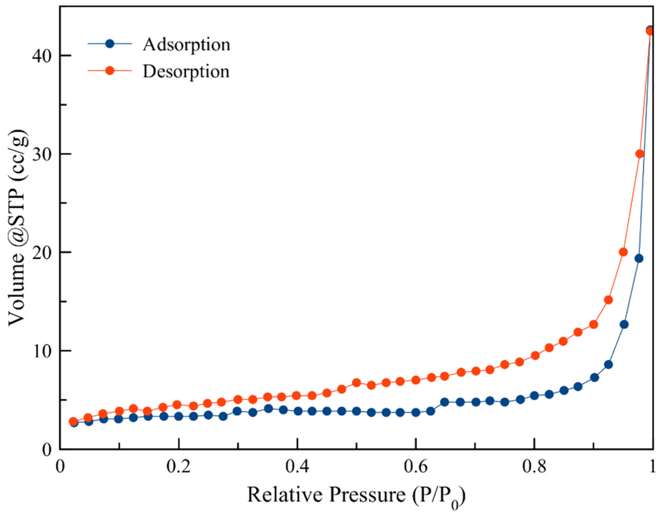
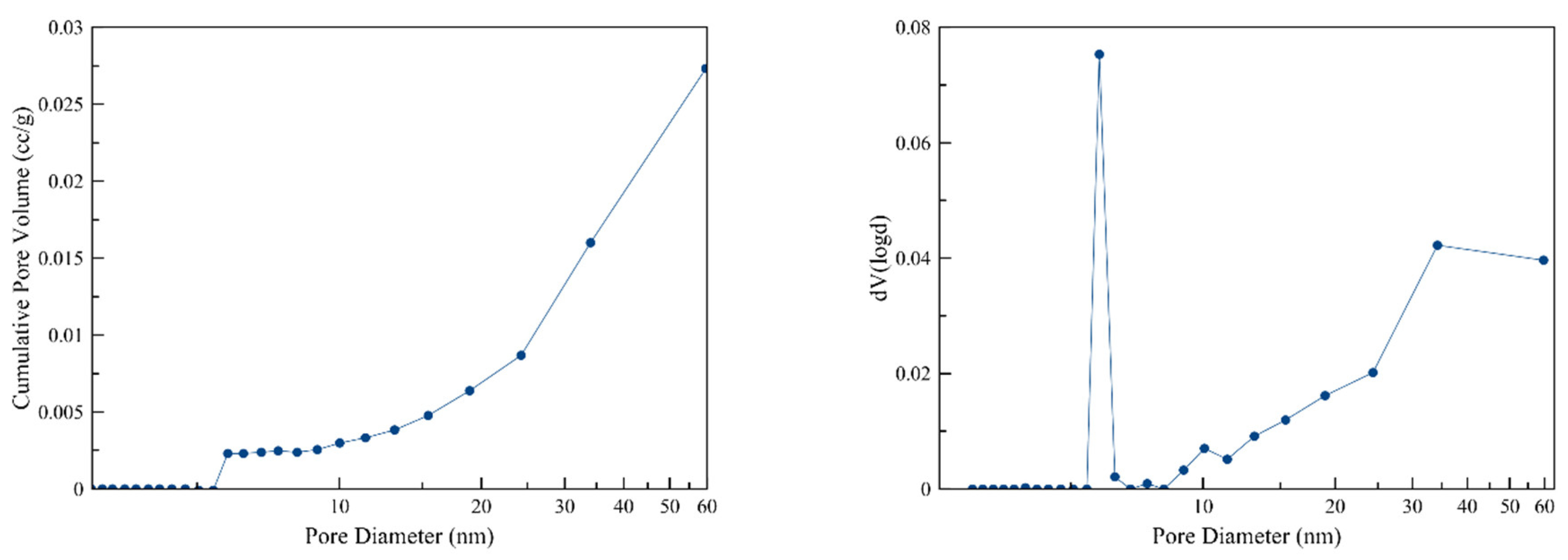
3.1. Porosimetry

Porosimetry results are shown in Figure 2 and Figure 3. The surface area of NZU is 12.6 m2/g, the pore volume 0.03 cm3/g and the average pore diameter 5.8 nm. The adsorption-desorption isotherms with the hysteresis branch are characteristic of microporous-mesoporous zeolites such as clinoptilolite [44]. Also, the modification of samples had no significant impact on the surface area, pore volume and pore diameter.

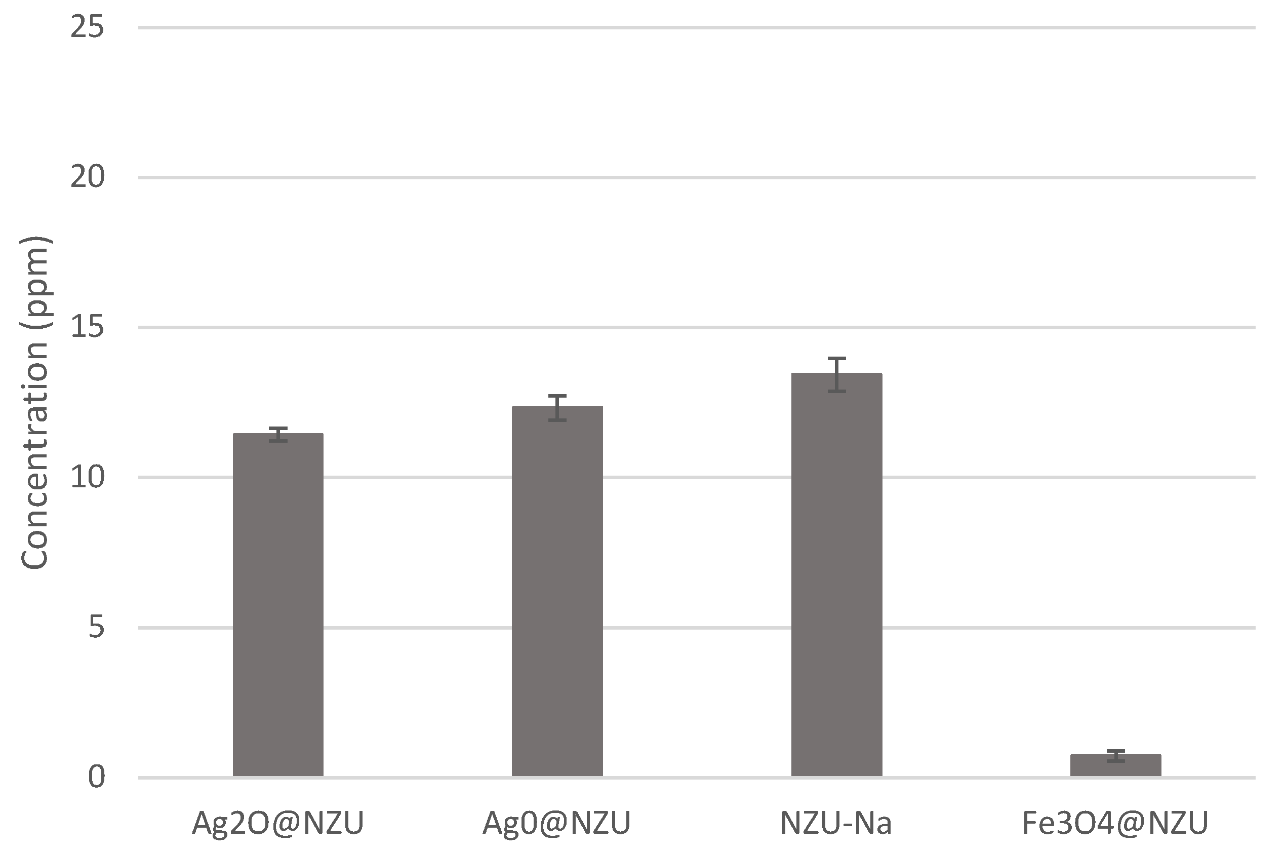
3.2. X-ray Fluorescence (XRF)

The XRF chemical analysis results are shown in Table 2. Ag and Fe content is similar in the synthesized samples with Ag samples retaining a significant amount of Fe which comes from NZU and may act as catalyst.

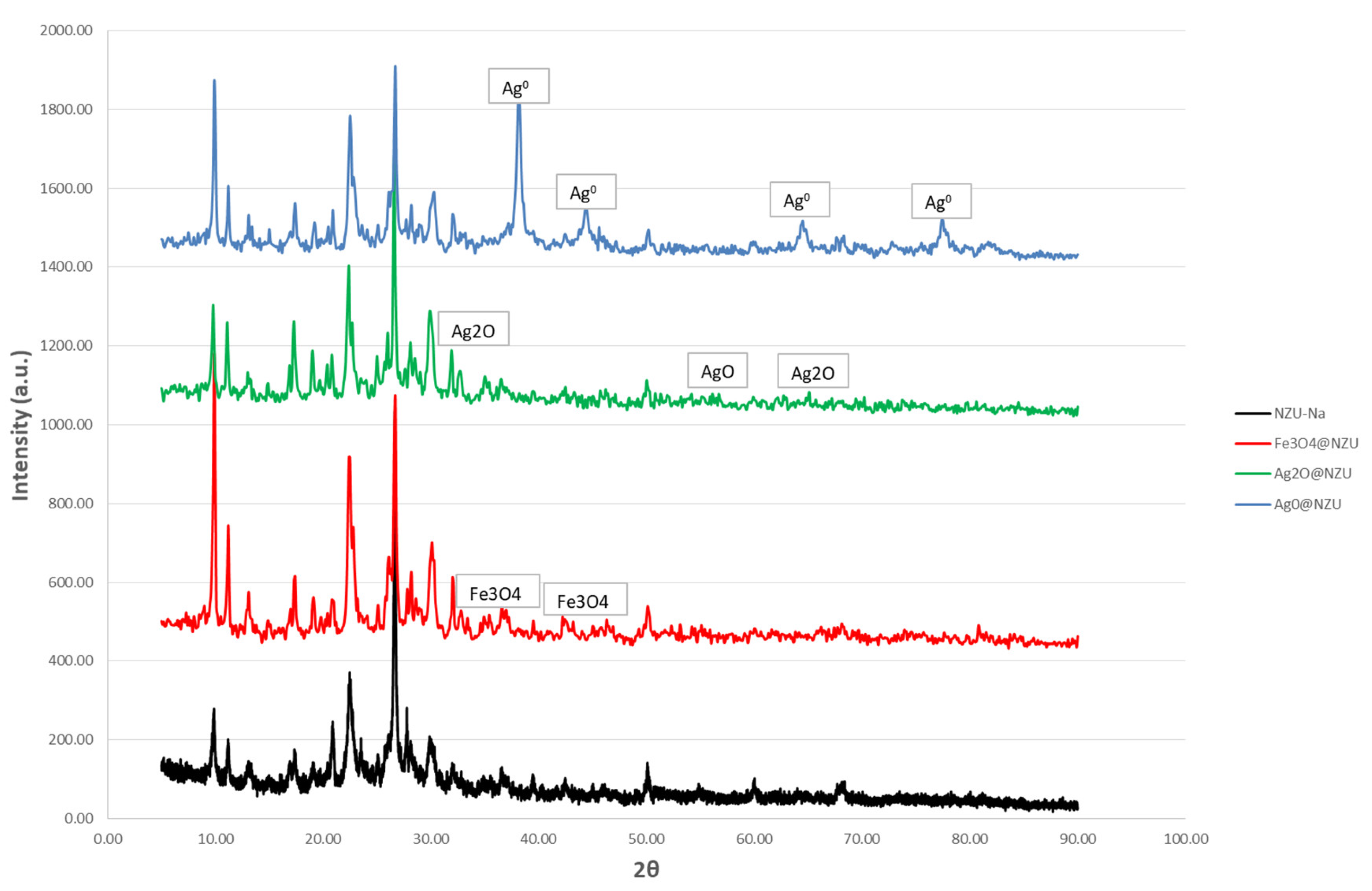
3.3. X-ray Diffraction (XRD)

From the XRD patterns in Figure 4, it can be seen that the zeolite is clinoptilolite with peaks at 2θ of 9.93, 17.23, 22.43, 29.87, 50.04.
Based on the material certificate provided by the manufacturer the purity is 80%. The peaks at 2θ of 35.48° and 43.25° in the Fe3O4@NZU sample are attributed to magnetite, at 2θ of 56.70° and 32.68°, 65.05° in the Ag2O@NZU sample to AgO and Ag2O, respectively and at 2θ of 38.16°, 44.30°, 64.47° and 77.40° in the Ag0@NZU sample to Ag0. The approximate NPs size calculated based on the Schrerrer’s formula is 32 nm for Fe3O4, 42 nm for Ag2O and 20 nm for Ag0.

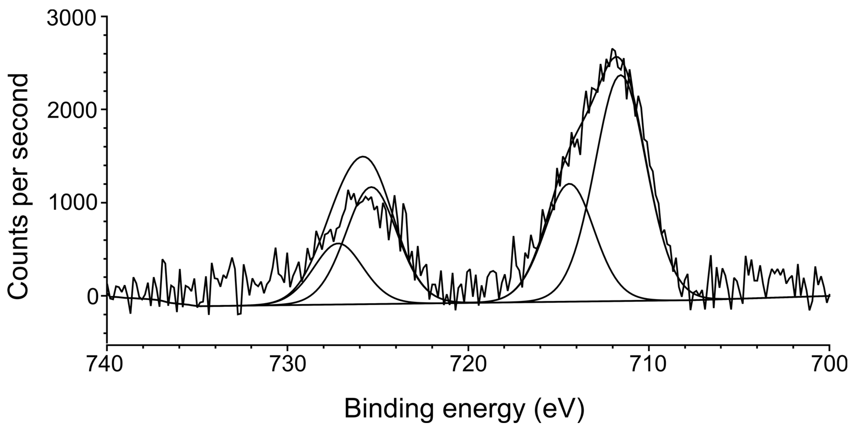
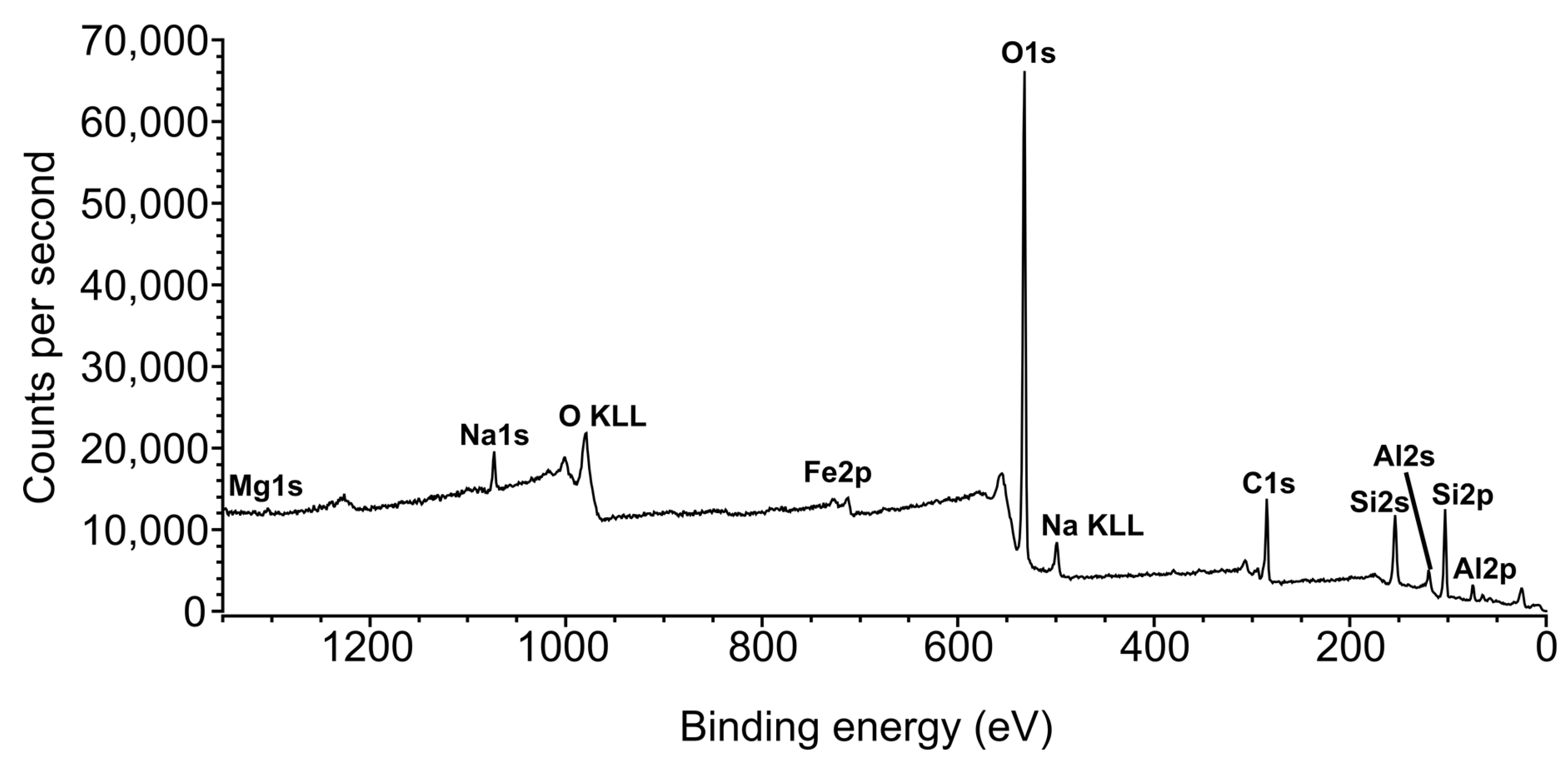
3.4. X-ray Photoelectron Spectroscopy (XPS)

XPS was done only for the Na-NZU in order to obtain the oxidation forms of the Fe contained in the parent zeolite as it can affect the redox reactions. The results showed that Na-NZU contains both Fe2+ and Fe3+(III) species which are catalytic active with approximate content of 68.3% and 31.7%, respectively. The pale green color of the Na-NZU is in agreement with the predominance of Fe2+ species. Figure 5 shows a high resolution scan in the Fe2p region, with two iron environments which agrees with oxidized iron as given in the NIST XPS database. Fe2p Scan A is recorded at 711.58 eV and Scan B at 714.38 eV, commensurate with Fe2+ in magnetite [45] and an Fe3+ species, most likely the Fe2O3 component of an Fe3O4/Fe2O3 mixture respectively. Finally, the survey scan shown in the ESI supports the elemental composition as analysed by EDS spectroscopy, showing the presence of oxygen, carbon, magnesium, iron, sodium, aluminium and silicon (Figure 6).

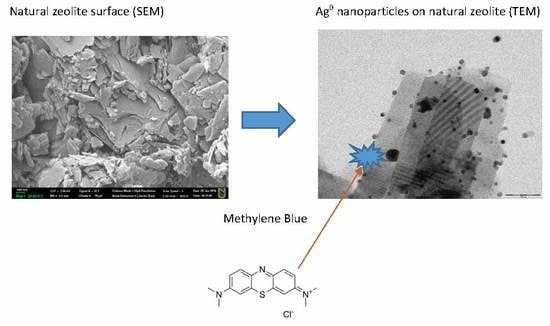
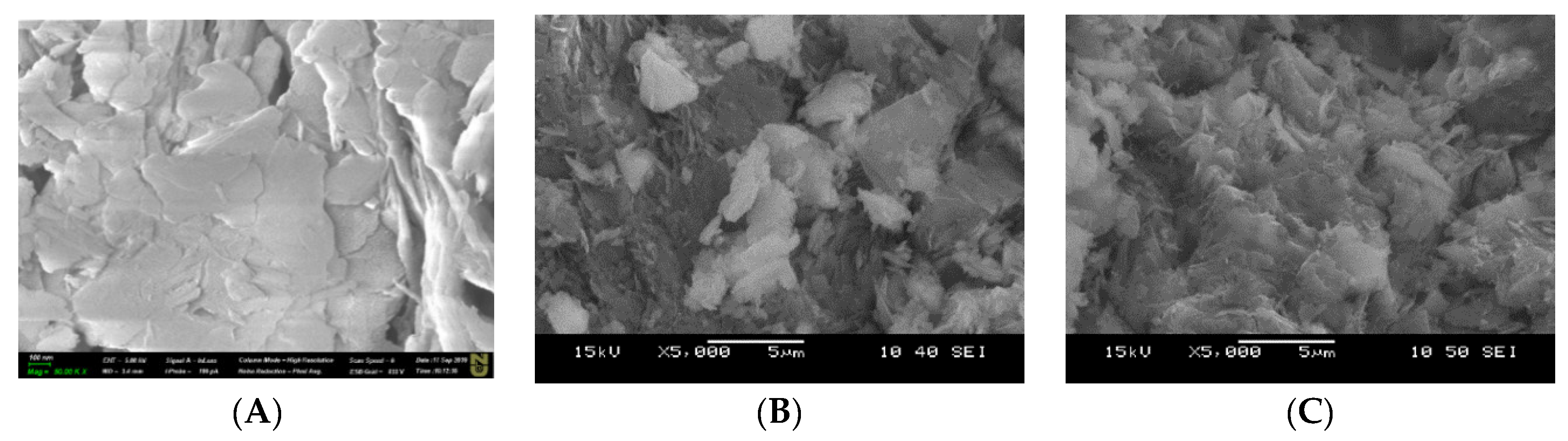
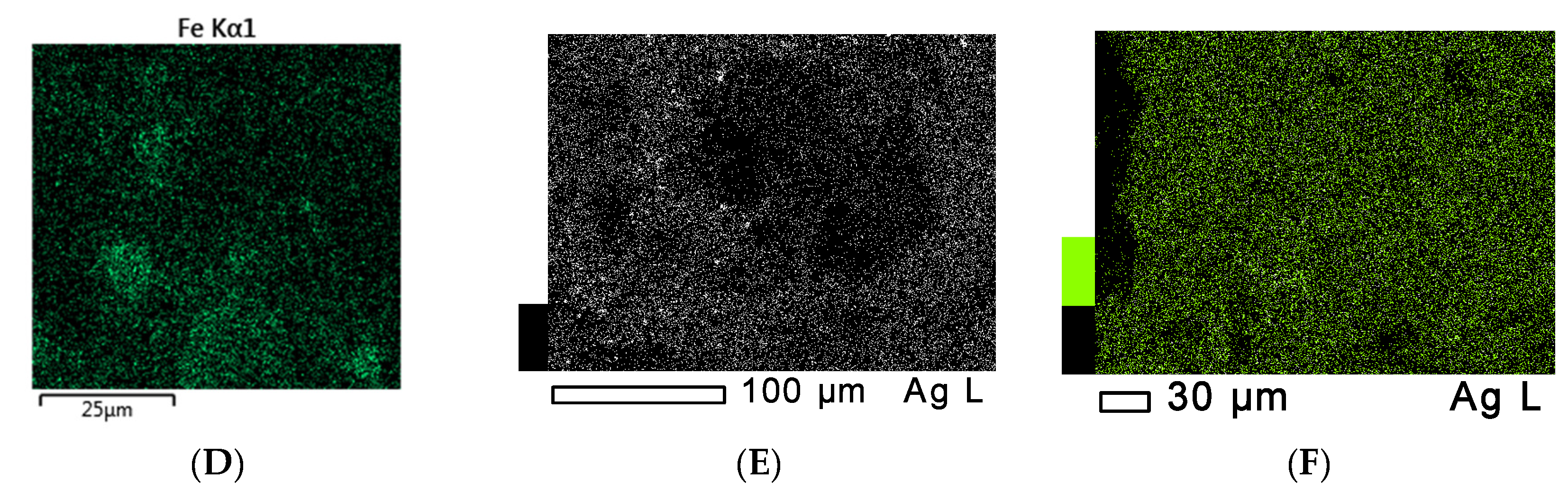
3.5. Scanning Electron Microscopy (SEM) and Energy Dispersive X-ray (EDS) Spectroscopy Measurements

Figure 7 shows the SEM images of the samples. SEM images demonstrate that there are no changes on the morphology of the zeolite surface. The EDS show metals uniformly distributed on the zeolite surface. There is no change after the adsorption and reaction with MB.

3.6. Transmission Electron Microscopy (TEM)

Figure 8 shows the TEM images of the samples on which the presence of nanoparticles is clear. Fe3O4 NPs size ranged in 5–50 nm, while the Ag2O NPs size was below 5 nm and the Ag0 NPs size was between 15–20 nm. With the exception of Ag2O these values are in general agreement with the NPs size calculations based on XRD data.

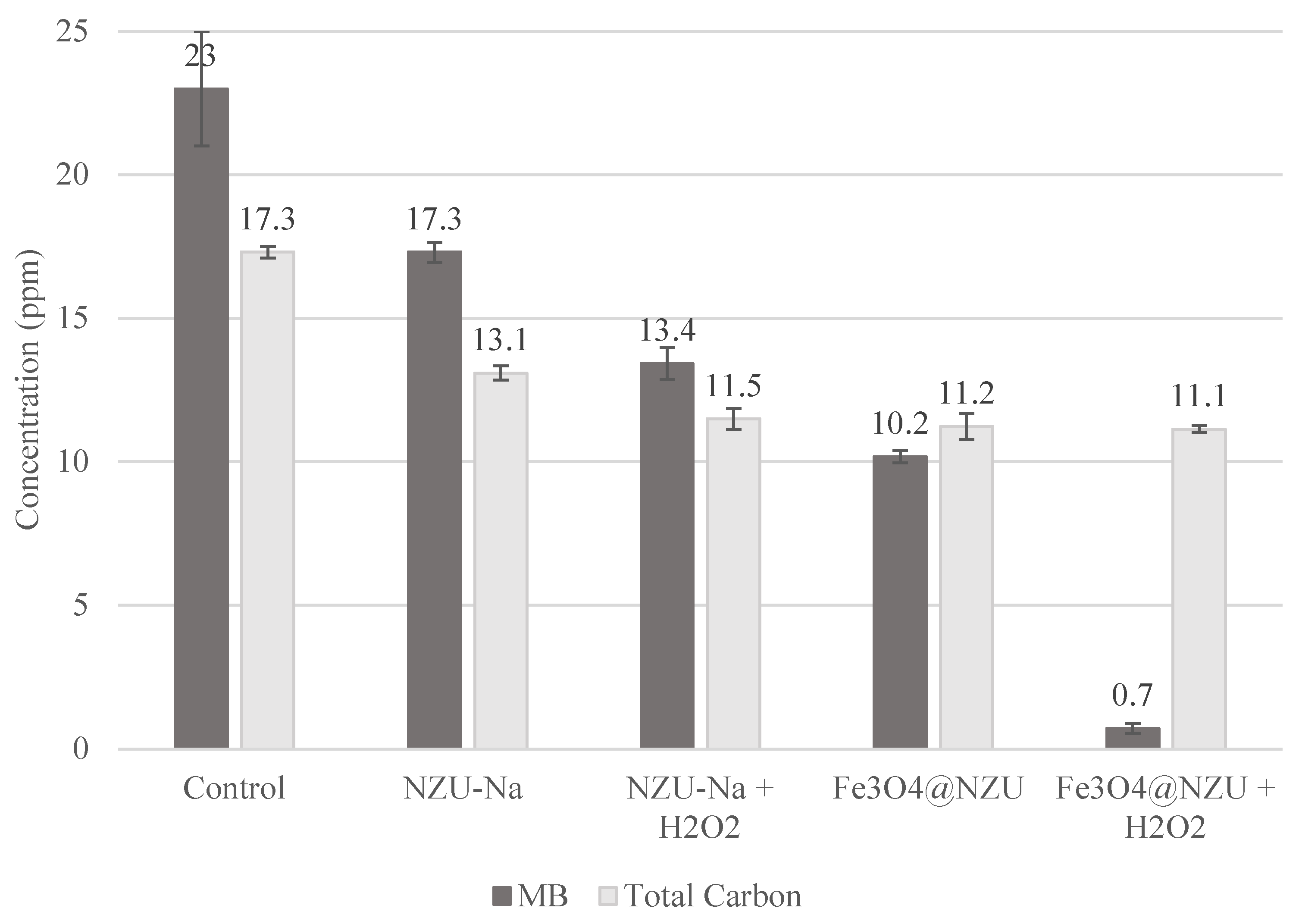
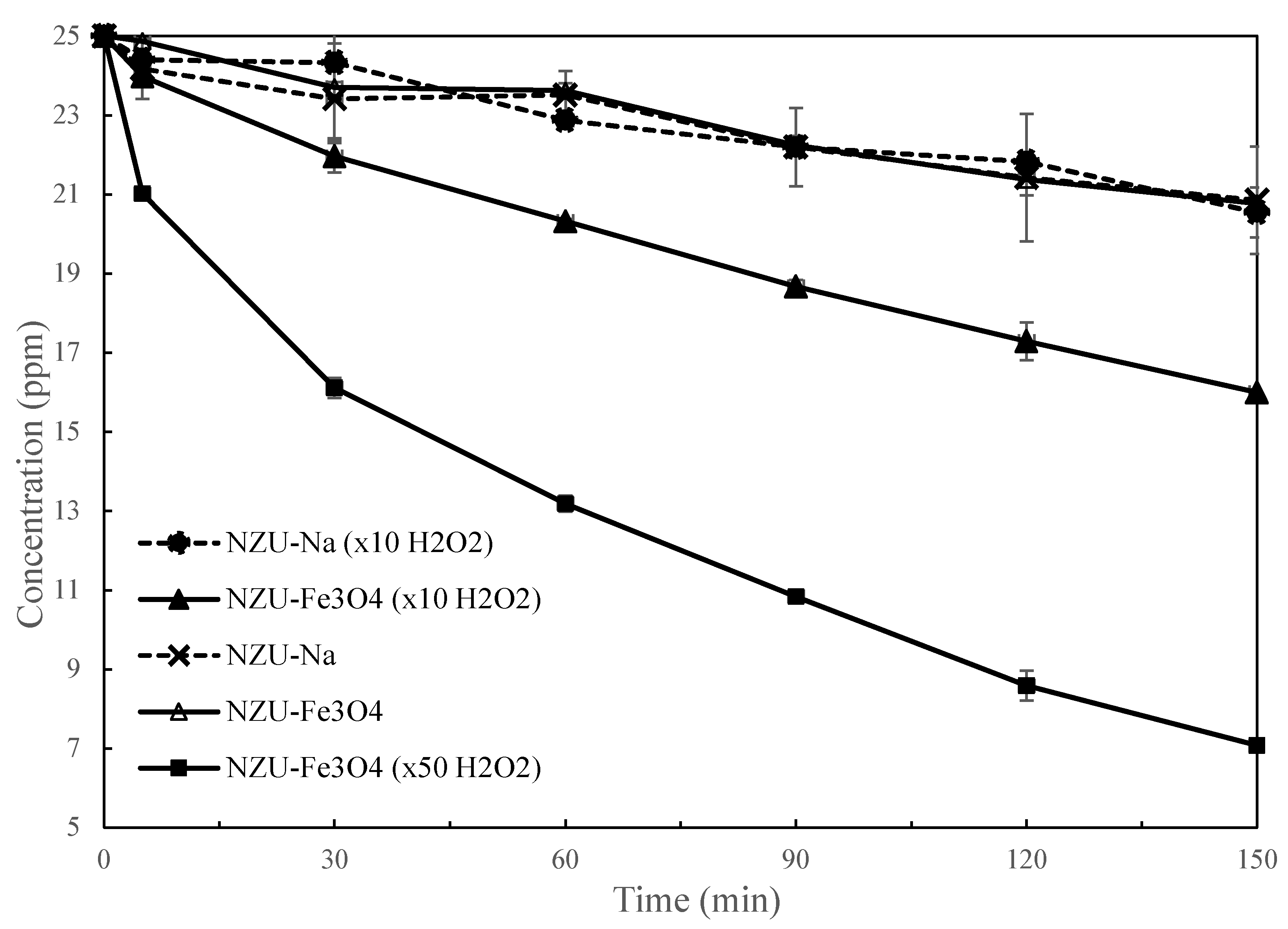
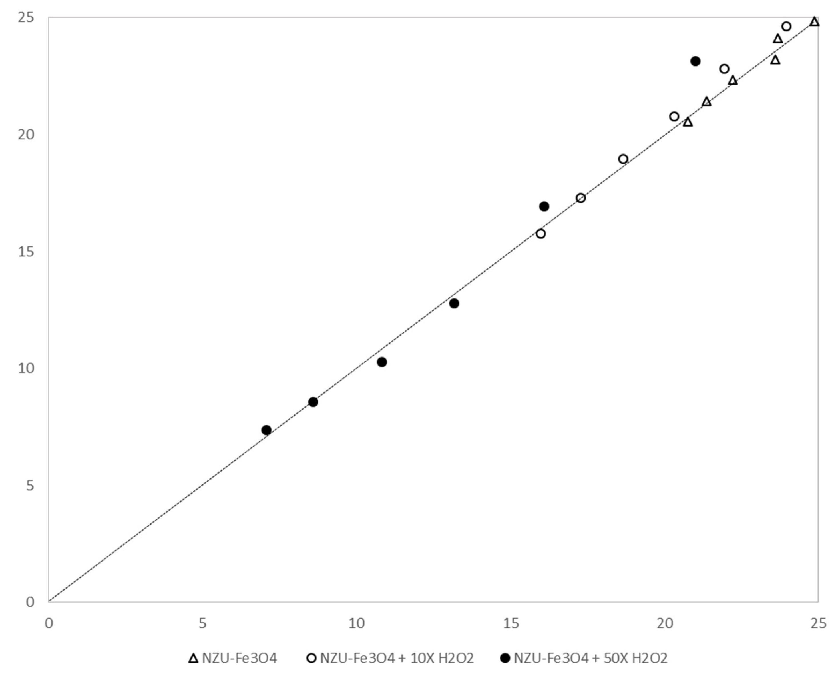
3.7. Catalytic Discoloration of MB

Figure 9 shows the discoloration of MB with H2O2 over the surfaces of the samples after 24 h. It is obvious that the Fe3O4@NZU exhibited the best performance in comparison to other samples. Also, NZU-Na removes almost 42% of MB owing to its adsorption properties and Fe content allowing Fenton or Fenton-like reactions to occur (see XPS results). Given this level of removal by NZU-Na, it is obvious that Ag2O@NZU and Ag0@NZU showed very low activity. The Ag0 sample performance is considerably lower that this found by Colar et al. who used Ag0 supported on clinoptilolite reaching 84% removal [36]. However, this can be explained as the main difference is that in the paper mentioned photocatalysis was employed and also different experimental conditions e.g., different solid mass to liquid volume ratio and pH.
Further experiments were carried out under the same experimental conditions by using only NZU-Na and Fe3O4@NZU samples with the addition of total carbon (TC) measurements (Figure 10). Although TC measurements do not provide information on the formed degradation intermediates, it is a simple and efficient way to study the mineralization of MB [33]. The complete oxidation of MB is represented by the following reaction [25]:
C16H18N3SCl + 52 H2O2 → 16 CO2 + 0.5 O2 + 58 H2O + 3 HNO3 + H2SO4 + HCl
The results obtained show that without the addition of H2O2 the NZU-Na removed 25% of MB and the Fe3O4@NZU sample 55%, which upon the addition of H2O2 was increased to 42% and 97%, respectively. The TC removal was between 25–36% for all samples, with or without H2O2, which is mainly attributed to adsorption, and thus the mineralization of MB to CO2 is either absent or not significant. A proposed degradation and mineralization MB mechanism has been provided elsewhere [35]. During the oxidation Cl− is first ionized and then several bonds in the MB molecule brake under the effect of radicals; first N–CH3 oxidized into HCHO or HCOOH, followed by the C–S and C–N which produce molecules like phenyl thiophene, 4-aminobenzenesulfonic acid and p-dihydroxybenzene. The final products are inorganic ions, such as CO2, H2O, Cl, SO42− and NO3”. The maximum MB removal achieved is comparable to other zeolite-based catalysts presented in Table 1.
These results show that MB adsorption is the main mechanism for the TC removal. The ability of natural zeolites to adsorb MB is well documented [36,46]. Briefly, MB upon dissolution releases cations into the solution, which are attracted by the permanent negative lattice charge of the zeolites. Also, the temporary surface charge of natural zeolites is governed by the solution pH and becomes more negative as pH is increasing contributing to the adsorption of cations [47]. However, we postulate that the pore size could hinder the adsorption by excluding large molecules from the zeolite structure.
The iron of the parent zeolite contributed to the oxidation of MB. Specifically, iron is well known to catalyze the decomposition of hydrogen peroxide into hydroxyl radicals that are powerful oxidants for organic compounds. This reaction is known as the Fenton reaction. The Fenton reaction is among the most widely applied advanced oxidation processes, in which the Fenton’s reagent, a mixture of a ferrous salt (catalyst) and hydrogen peroxide (oxidant), is used to destroy refractory organic compounds [48,49]. The Fenton reaction depends on the production of hydroxyl radicals and it is considered as a convenient and economical process [50]. The mechanism is described by the following reactions when no organic compounds are present in the solution [51]:
Fe 2 + +   H 2 O 2 Fe 3 + + OH + OH
Fe 3 + +   H 2 O 2 Fe 2 + + HO 2 + H +
OH + H 2 O 2   HO 2 +   H 2 O
OH + Fe 2 + Fe 3 + + OH
Fe 3 + +   HO 2 Fe 2 + + O 2 H +
Fe 2 + + HO 2 + H + Fe 3 + + H 2 O 2
2 HO 2   H 2 O 2 + O 2
Equation (2) occurs in acidic conditions. The chemical Equation (3) between ferric ions (Fe3+) and H2O2 is known as a Fenton-like reaction. When Fe2+ is initially present all reactions above take place, while for Fe(III) Reaction (3) is the first step. Hydroxyl radicals attack organic molecules according to the following reaction:
RH + OH H 2 O + R further   oxidation
As is clear from the results, the magnetite greatly enhanced the oxidation of MB and oxidizes part of MB even in the absence of H2O2. This is a result of higher content of Fe3+ in the zeolite (see Table 2).
In Figure 11 the kinetics of discoloration for the first 150 min are presented. It is clear that in the presence of H2O2 Fe3O4@NZU sample resulted in fast discoloration of MB, while the higher H2O2 concentration greatly enhances the kinetics. Although the differences between Na-NZU with and without H2O2 and the Fe3O4@NZU without H2O2 after 24h, they show the same removal up to 150 min supporting the hypothesis of adsorption as the predominant mechanism of removal at the initial stages followed immediately by oxidation.
Reaction rate models were applied on Fe3O4@NZU experimental data and the results are shown in Figure 12 and Table 3. The models used are the following:
Zero order:
C t = C o k · t
First order:
C t = C o · exp ( k · t )
Second order:
1 C t = 1 C o + k · t
where Co and Ct the initial and at time (t) concentration and (k) the reaction rate constant. The average absolute error shows that the application of the model is satisfactory. The overall reaction is of zero order in the absence of H2O2, first order for low concentration of H2O2 and second order for high H2O2 concentration, an indication that the reaction path depends on the oxidant [H2O2] concentration. It has been reported that the products distribution is strongly related to the initial molar ratio of H2O2/organic compound [52,53]. As a result, different initial concentrations of oxidant, namely of H2O2, can lead to different reaction pathways and products. The opposite trend was observed by Jakab et al. [33] who used a Cu2+ exchanged natural zeolite, however UV was combined with H2O2 and thus the oxidation conditions were different. Zhang et al. [40] observed no influence of the H2O2 on the reaction order but they used an organic form of Fe2+ and thus the reaction system is different. Finally, literature review shows that the H2O2 oxidation reaction is of first order in most studies (Table 1).

4. Conclusions

Natural zeolite-based silver and magnetite nanocomposites were successfully synthesized and tested in the catalytic oxidation of methylene blue in water. The zeolite was impregnated with approximately 5.5 wt% of Fe in the form of magnetite nanoparticles of size of 32 nm and 6.4 wt% Ag in the form of silver oxide and metallic silver nanoparticles of sizes of 42 and 20 nm, respectively. The results show that MB adsorption contributes significantly to the total removal, the iron of the parent zeolite contributes to the oxidation of MB, the magnetite oxidizes part of MB even in the absence of H2O2 and the addition of magnetite greatly enhances the oxidation of MB. The oxidation of MB is partial without evidence of mineralization. The application of reaction rate models on the experimental data where H2O2 was used was satisfactory with an average absolute error between 0.8–2.8%. In the absence of H2O2 the reaction for Na-NZU and Fe3O4@NZU is of zeroth order confirming that adsorption is the main mechanism at the initial stages followed by oxidation in later stages. The reaction order for Fe3O4@NZU swifts from zero, to first and second order depending on the H2O2 concentration, an indication that the reaction path changes.

Author Contributions

Conceptualization, V.J.I.; Methodology, V.J.I.; Software, J.C.B.; Validation, V.J.I., S.P. and J.C.B.; Formal Analysis, V.J.I. and S.P.; Investigation, A.K., N.K. and K.M.; Resources, V.J.I.; Data Curation, V.J.I.; Writing—Original Draft Preparation, V.J.I.; Writing—Review & Editing, V.J.I. and S.P.; Visualization, V.J.I.; Supervision, V.J.I.; Project Administration, V.J.I.; Funding Acquisition, V.J.I. and J.C.B. All authors have read and agreed to the published version of the manuscript.

Funding

This research was funded Nazarbayev University, Grant Number 110119FD4536 and Kingston University c/o GCRF Grant Number P2034-106. The APC was funded by Nazarbayev University.

Acknowledgments

The authors would like to thank the University of Alicante for the XPS analysis.

Conflicts of Interest

The authors declare no conflict of interest.

References

  1. Métivier-Pignon, H.; Faur-Brasquet, C.; le Cloirec, P. Adsorption of dyes onto activated carbon cloths: Approach of adsorption mechanisms and coupling of ACC with ultrafiltration to treat coloured wastewaters. Sep. Purif. Technol. 2003, 31, 3–11. [Google Scholar] [CrossRef]
  2. Rafatullah, M.; Sulaiman, O.; Hashim, R.; Ahmad, A. Adsorption of methylene blue on low-cost adsorbents: A review. J. Hazard. Mater. 2010, 177, 70–80. [Google Scholar] [CrossRef] [PubMed]
  3. Sheng, J.; Xie, Y.; Zhou, Y. Adsorption of methylene blue from aqueous solution on pyrophyllite. Appl. Clay Sci. 2009, 46, 422–424. [Google Scholar] [CrossRef]
  4. Li, Y.; Du, Q.; Liu, T.; Peng, X.; Wang, J.; Sun, J.; Wang, Y.; Wu, S.; Wang, Z.; Xia, Y.; et al. Comparative study of methylene blue dye adsorption onto activated carbon, graphene oxide, and carbon nanotubes. Chem. Eng. Res. Des. 2013, 91, 361–368. [Google Scholar] [CrossRef]
  5. Ravikumar, K.; Deebika, B.; Balu, K. Decolourization of aqueous dye solutions by a novel adsorbent: Application of statistical designs and surface plots for the optimization and regression analysis. J. Hazard. Mater. 2005, 122, 75–83. [Google Scholar] [CrossRef]
  6. McMullan, G.; Meehan, C.; Conneely, A.; Kirby, N.; Robinson, T.; Nigam, P.; Banat, I.M.; Marchant, R.; Smyth, W.F. Microbial decolourisation and degradation of textile dyes. Appl. Microbiol. Biotechnol. 2001, 56, 81–87. [Google Scholar] [CrossRef]
  7. Pearce, C.I.; Lloyd, J.R.; Guthrie, J.T. The removal of colour from textile wastewater using whole bacterial cells: A review. Dye. Pigment. 2003, 58, 179–196. [Google Scholar] [CrossRef]
  8. Lee, J.W.; Choi, S.P.; Thiruvenkatachari, R.; Shim, W.G.; Moon, H. Evaluation of the performance of adsorption and coagulation processes for the maximum removal of reactive dyes. Dye. Pigment. 2006, 69, 196–203. [Google Scholar] [CrossRef]
  9. Robinson, T.; McMullan, G.; Marchant, R.; Nigam, P. Remediation of dyes in textile effluent: A critical review on current treatment technologies with a proposed alternative. Bioresour. Technol. 2001, 77, 247–255. [Google Scholar] [CrossRef]
  10. Banat, I.M.; Nigam, P.; Singh, D.; Marchant, R. Microbial decolorization of textile-dye-containing effluents: A review. Bioresour. Technol. 1996, 58, 217–227. [Google Scholar] [CrossRef]
  11. IARC. Chemical and physical data. Pharm. Drugs 2000, 1981, 100–104. [Google Scholar]
  12. Perry anc Meinhard, P.M.E. Necrotic subcutaneous abscesses following injections of methylene blue. Br. J. Clin. Pract. 1974, 28, 289–291. [Google Scholar]
  13. Ruhlen, J.L. Tissue necrosis. Cutaneous and subcutaneous damage following extravasation of methylene blue. J. Kans. Med. Soc. 1982, 83, 236–260. [Google Scholar] [PubMed]
  14. Sharr, M.M.; Weller, R.O.; Brice, J.G. Spinal cord necrosis after intrathecal injection of methylene blue. J. Neurol. Neurosurg. Psychiatry 1978, 41, 384–386. [Google Scholar] [CrossRef] [Green Version]
  15. Troche, B.I. The Methylene Blue Baby. N. Engl. J. Med. 1989, 320, 1756–1757. [Google Scholar] [CrossRef]
  16. Spahr, R.C.; Salsburey, D.J.; Krissberg, A.; Prin, W. Intraamniotic injection of methylene blue leading to methemoglobinemia in one of twins. Int. J. Gynecol. Obstet. 1980, 17, 477–478. [Google Scholar] [CrossRef]
  17. Vincer, M.J.; Allen, A.C.; Evans, J.R.; Nwaesei, C.; Stinson, D.A. Methylene-blue-induced hemolytic anemia in a neonate. Can. Med. Assoc. J. 1987, 136, 503–504. [Google Scholar]
  18. Serota, F.; Bernbaum, J.; Schwartz, E. The methylene-blue baby. Lancet 1979, 314, 1142–1143. [Google Scholar] [CrossRef]
  19. Cowett, R.M.; Hakanson, D.O.; Kocon, R.W.; Oh, W. Untoward neonatal effect of intraamniotic administration of methylene blue. Obstet. Gynecol. 1976, 48, 74S–75S. [Google Scholar]
  20. McEnerney, J.K.; McEnerney, L.N. Unfavorable neonatal outcome after intraamniotic injection of methylene blue. Obstet. Gynecol. 1983, 61, 35S–37S. [Google Scholar]
  21. Coddington, C.C.; Anderson, T.L.; Accetta, C.R.; Swanson, J.; Kruger, T.; Hodgen, G.D. Adverse effects of methylene blue on human sperm motility, components of human reproductive tract fluids, and mouse embryo cleavage. Fertil. Steril. 1989, 51, 480–485. [Google Scholar] [CrossRef]
  22. Turner, J. Book review. J. Transp. Geogr. 2013, 106. [Google Scholar] [CrossRef]
  23. Brownstein, S.; Liszauer, A.D.; Jackson, W.B. Ocular complications of a topical methylene blue-vasoconstrictor-anesthetic preparation. Can. J. Ophthalmol. 1989, 24, 317–324. [Google Scholar] [PubMed]
  24. Salem, I.A.; El-Maazawi, M.S. Kinetics and mechanism of color removal of methylene blue with hydrogen peroxide catalyzed by some supported alumina surfaces. Chemosphere 2000, 41, 1173–1180. [Google Scholar] [CrossRef]
  25. Wu, Y.; Yang, M.; Hu, S.; Wang, L.; Yao, H. Characteristics and mechanisms of 4A zeolite supported nanoparticulate zero-valent iron as Fenton-like catalyst to degrade methylene blue. Toxicol. Environ. Chem. 2014, 96, 227–242. [Google Scholar] [CrossRef]
  26. Simoncic, P.; Armbruster, T. Cationic methylene blue incorporated into zeolite mordenite-Na: A single crystal X-ray study. Microporous Mesoporous Mater. 2005, 81, 87–95. [Google Scholar] [CrossRef]
  27. Smith, J.V. Definition of a zeolite. Zeolites 1984, 4, 309–310. [Google Scholar] [CrossRef]
  28. Liebau, F. Zeolites and clathrasils-Two distinct classes of framework silicates. Zeolites 1983, 3, 191–193. [Google Scholar] [CrossRef]
  29. Sartbaeva, A.; Wells, S.A.; Treacy, M.M.J.; Thorpe, M.F. The flexibility window in zeolites. Nat. Mater. 2006, 5, 962–965. [Google Scholar] [CrossRef]
  30. Groen, J.C.; Peffer, L.A.A.; Moulijn, J.A.; Pérez-Ramírez, J. On the introduction of intracrystalline mesoporosity in zeolites upon desilication in alkaline medium. Microporous Mesoporous Mater. 2004, 69, 29–34. [Google Scholar] [CrossRef]
  31. Roland, E. Hindustrial Production of Zeolites. Stud. Surf. Sci. Catal. 1989, 46, 645–659. [Google Scholar] [CrossRef]
  32. Lopes, J.H.; Nogueira, F.G.; Gonçalves, M.; Oliveira, L.C. Modified Zeolite with Transition Metals Cu and Fe for Removal of Methylene Blue from Aqueous Medium: Mass Spectrometry Study. Bull. Chem. React. Eng. Catal. 2015, 10, 237–248. [Google Scholar] [CrossRef]
  33. Jakab, Á.; Colar, L.A.; Pode, R.; Cocheci, L.; Manea, F. Catalytic photodegradation and mineralization of cationic dye methylene blue from aqueous solution onto copper doped zeolite. Rev. Chim. 2012, 63, 1016–1022. [Google Scholar]
  34. Wardhani, S.; Rahman, M.F.F.; Purwonugroho, D.; Tjahjanto, R.T.; Damayanti, C.A.; Wulandari, I.O. Photocatalytic Degradation of Methylene Blue Using TiO2-Natural Zeolite as A Photocatalyst. J. Pure Appl. Chem. Res. 2016, 5, 19–27. [Google Scholar] [CrossRef] [Green Version]
  35. Karimi-Shamsabadi, M.; Nezamzadeh-Ejhieh, A. Comparative study on the increased photoactivity of coupled and supported manganese-silver oxides onto a natural zeolite nano-particles. J. Mol. Catal. A Chem. 2016, 418–419, 103–114. [Google Scholar] [CrossRef]
  36. Colar, L.A.; Jakab, A.; Manea, F.; Pode, R.; Orha, C. Photocatalytic performance of Ag-modified natural zeolite catalyst for photocatalysis degradation of Methylene Blue (MB) under VIS irradiation. WIT Trans. Ecol. Environ. 2012, 164, 335–344. [Google Scholar] [CrossRef] [Green Version]
  37. SRajabi, K.; Sohrabnezhad, S. Synthesis and characterization of magnetic core with two shells: Mordenite zeolite and CuO to form Fe3O4@MOR@CuO core-shell: As a visible light driven photocatalyst. Microporous Mesoporous Mater. 2017, 242, 136–143. [Google Scholar] [CrossRef]
  38. Tedla, H.; Díaz, I.; Kebede, T.; Taddesse, A.M. Synthesis, characterization and photocatalytic activity of zeolite supported ZnO/Fe2O3/MnO2 nanocomposites. J. Environ. Chem. Eng. 2015, 3, 1586–1591. [Google Scholar] [CrossRef]
  39. Zhang, Y.; Shang, J.; Song, Y.; Rong, C.; Wang, Y.; Huang, W.; Yu, K. Selective Fenton-like oxidation of methylene blue on modified Fe-zeolites prepared via molecular imprinting technique. Water Sci. Technol. 2017, 75, 659–669. [Google Scholar] [CrossRef] [Green Version]
  40. Zhang, J.; Hu, F.T.; Liu, Q.Q.; Zhao, X.; Liu, S.Q. Application of heterogenous catalyst of tris(1,10)-phenanthroline iron(II) loaded on zeolite for the photo-Fenton degradation of methylene blue. React. Kinet. Mech. Catal. 2011, 103, 299–310. [Google Scholar] [CrossRef]
  41. Subramanian, E.; Subbulekshmi, N.L. Enhanced heterogeneous wet hydrogen peroxide catalytic oxidation performance of fly ash-derived zeolite by CuO incorporation. Sci. Iran. 2017, 24, 1189–1202. [Google Scholar] [CrossRef] [Green Version]
  42. Nassar, M.Y.; Abdelrahman, E.A. Hydrothermal tuning of the morphology and crystallite size of zeolite nanostructures for simultaneous adsorption and photocatalytic degradation of methylene blue dye. J. Mol. Liq. 2017, 242, 364–374. [Google Scholar] [CrossRef]
  43. Wojtaszek-Gurdak, A.; Zielinska, M.; Ziolek, M. MWW layered zeolites modified with niobium species—Surface and catalytic properties. Catal. Today 2019, 325, 89–97. [Google Scholar] [CrossRef]
  44. Stylianou, M.; Inglezakis, V.; Agapiou, A.; Itskos, G.; Jetybayeva, A.; Loizidou, M. A comparative study on phyllosilicate and tectosillicate mineral structural properties. Desalin. Water Treat. 2018, 112, 119–146. [Google Scholar] [CrossRef] [Green Version]
  45. Allen, G.C.; Curtis, M.T.; Hooper, A.J.; Tucker, P.M. X-ray photoelectron spectroscopy of iron–oxygen systems. J. Chem. Soc. Dalt. Trans. Dalt. Trans. 1974, 1525–1530. [Google Scholar] [CrossRef]
  46. Wang, S.; Zhu, Z.H. Characterisation and environmental application of an Australian natural zeolite for basic dye removal from aqueous solution. J. Hazard. Mater. 2006, 136, 946–952. [Google Scholar] [CrossRef]
  47. Wang, X.; Nguyen, A.V. Characterisation of electrokinetic properties of clinoptilolite before and after activation by sulphuric acid for treating CSG water. Microporous Mesoporous Mater. 2016, 220, 175–182. [Google Scholar] [CrossRef] [Green Version]
  48. Andreozzi, R.; Caprio, V.; Insola, A.; Marotta, R. Advanced oxidation processes (AOP) for water purification and recovery. Catal. Today. 1999, 53, 51–59. [Google Scholar] [CrossRef]
  49. Tarr, M.A. Chemical Degradation Methods for Wastes and Pollutants: Environmental and Industrial Applications; CRC Press: Boca Raton, FL, USA, 2003. [Google Scholar]
  50. Pignatello, J.J.; Oliveros, E.; MacKay, A. Advanced oxidation processes for organic contaminant destruction based on the fenton reaction and related chemistry. Crit. Rev. Environ. Sci. Technol. 2006, 36, 1–84. [Google Scholar] [CrossRef]
  51. Jain, B.; Singh, A.K.; Kim, H.; Lichtfouse, E.; Sharma, V.K. Treatment of organic pollutants by homogeneous and heterogeneous Fenton reaction processes. Environ. Chem. Lett. 2018, 16, 947–967. [Google Scholar] [CrossRef] [Green Version]
  52. Lipczynska-Kochany, E.; Bolton, J.R. Flash photolysis/HPLC applications. 2. Direct photolysis vs. hydrogen peroxide mediated photodegradation of 4-chlorophenol as studied by a flash photolysis/HPLC technique. Environ. Sci. Technol. 1992, 26, 259–262. [Google Scholar] [CrossRef]
  53. Poulopoulos, S.G.; Arvanitakis, F.; Philippopoulos, C.J. Photochemical treatment of phenol aqueous solutions using ultraviolet radiation and hydrogen peroxide. J. Hazard. Mater. 2006, 129, 64–68. [Google Scholar] [CrossRef] [PubMed]
Figure 1. The molecular structure of MB.
Processes 08 00471 g001
Figure 2. Adsorption-desorption isotherms of NZU.
Processes 08 00471 g002
Figure 3. Cumulative pore volume (left) and pore size distribution of NZU (right).
Processes 08 00471 g003
Figure 4. XRD spectra of NZU, Fe3O4@NZU, Ag2O@NZU and Ag0@NZU samples.
Processes 08 00471 g004
Figure 5. X-ray photoelectron spectroscopy of Na-NZU. Doublet separation was 13.50 eV.
Processes 08 00471 g005
Figure 6. XPS survey scan of the Na-NZU zeolite over the range 0–1350 eV.
Processes 08 00471 g006
Figure 7. Scanning electron microscopy images of (A) Fe3O4@NZU, (B) Ag2O@NZU, (C) Ag0@NZU and energy dispersive X-ray spectroscopy of (D) iron on Fe3O4@NZU and silver on (E) Ag2O@NZU and (F) Ag0@NZU.
Processes 08 00471 g007aProcesses 08 00471 g007b
Figure 8. Transmission electron microscopy images of (A) Fe3O4@NZU, (B) Ag2O@NZU and (C) Ag0@NZU.
Processes 08 00471 g008
Figure 9. Discoloration of MB solutions by the modified zeolites (H2O2 concentration: 42.4 mM).
Processes 08 00471 g009
Figure 10. Discoloration of MB solutions and and TC removal by the modified zeolites (H2O2 concentration: 42.4 mM).
Processes 08 00471 g010
Figure 11. Kinetics of MB solutions discoloration by the modified zeolites.
Processes 08 00471 g011
Figure 12. Models results.
Processes 08 00471 g012
Table 1. Metals-doped zeolites used for the oxidation of the MB.
Oxidants ZeoliteMetal 1 Catalyst Concentration (% w/w)Initial MB Concentration (ppm)Zeolite Mass to Solution Volume (g/L)Removal (Max. %)Reaction Order 2Ref.
H2O2 (10–50 mM) and UVNatural zeoliteCu2+ -50 1871.3–2 *[33]
H2O2 and UVClinoptiloliteTiO2
(anatase)
8020 2981[34]
H2O2 and UVClinoptiloliteMnO (NPs)
Ag2O (NPs)
10
2.1
4 0.25851[35]
VisClinoptiloliteAg0 (NPs)-20–1000.25–1.584-[36]
H2O2 (0.2 mL, 3 wt%) and UVMordeniteCuO (NPs)
Fe3O4
3.7
68.1
3.2–100.05–297-[37]
H2O2 (10 mM)4A Fe0 (NPs)12.730 0.2–51001[25]
VisZeolite YZnO (NPs)
Fe2O3
MnO2
3.7–13.6 (Zn)
1.9–6.9 (Fe)
0.7–0.8 (Mn)
10–1000.05–0.1580-[38]
H2O2 (2 mL, 30% v/v)NaY
Naß
Fe3O4
Cu2O
-100 1 1000[39]
H2O2 (6% v/v)4A Fe2O3
Fe2Al4Si5O18
17.1 15 0.3 881[39]
H2O2 (2.2–11 mM) and UVNaY Tris (1, 10)
phenanthroline
Fe2+
0.0111.5 40 771 *[40]
H2O2 (1 mL, 30% v/v)Zeolite XCuO
Fe2O3
29.6100 0.25100-[41]
H2O2 (2 mL, 0.5M) and UVAnalcimeC9H21O3Al-10 21001[42]
H2O2 (5 mL, 30% v/v)Synthetic zeoliteNb5+1.9–2.280 1100-[43]
1 The notation (NPs) in this table is used only for studies where the formation of nanoparticles is confirmed by XRD and/or TEM. 2 The notation (*) is used for cases where the effect of the H2O2 concentration on the reaction order was studied.
Table 2. X-ray Fluorescence (XRF) results in wt%.
SampleAl2O3SiO2Fe2O3Ag2O
NZU12.977.31.60
NZU-Na12.778.51.50
Fe3O4@NZU9.575.67.90
Ag2O@NZU11.772.81.56.9
Ag0@NZU11.669.91.46.8
Table 3. Models results.
H2O2 (Mm)k (min−1)Reaction OrderAverage Absolute Error (%)
00.029300.8
42.4 0.0030711.7
2120.0006422.8

