Article

Modification of the Bacterial Cell Wall—Is the Bioavailability Important in Creosote Biodegradation?

Institute of Chemical Technology and Engineering, Poznan University of Technology, Berdychowo 4, 60-695 Poznan, Poland
*
Authors to whom correspondence should be addressed.
Processes 2020, 8(2), 147; https://doi.org/10.3390/pr8020147
Submission received: 31 December 2019 / Revised: 17 January 2020 / Accepted: 21 January 2020 / Published: 23 January 2020
(This article belongs to the Special Issue Study of Biodegradation and Bioremediation)

Abstract

:
Creosote oil, widely used as a wood preservative, is a complex mixture of different polycyclic aromatic compounds. The soil contamination result in the presence of a specific microcosm. The presented study focuses on the most active strains involved in bioremediation of long-term creosote-contaminated soil. In three soil samples from different boreholes, two Sphingomonas maltophilia (S. maltophilia) and one Paenibacillus ulginis (P. ulginis) strain were isolated. The conducted experiments showed the differences and similarities between the bacteria strains capable of degrading creosote from the same contaminated area. Both S. maltophilia strains exhibit higher biodegradation efficiency (over 50% after 28 days) and greater increase in glutathione S-transferase activity than P. ulginis ODW 5.9. However, S. maltophilia ODW 3.7 and P. ulginis ODW 5.9 were different from the third of the tested strains. The growth of the former two on creosote resulted in an increase in cell adhesion to Congo red and in the total membrane permeability. Nevertheless, all three strains have shown a decrease in the permeability of the inner cell membrane. That suggests the complex relationship between the cell surface modifications and bioavailability of the creosote to microorganisms. The conducted research allowed us to broaden the current knowledge about the creosote bioremediation and the properties of microorganisms involved in the process.

1. Introduction

Creosote is a complex mixture of carbonaceous substances obtained from the distillation of tar. The two main types of creosote are wood-tar creosote and coal-tar creosote. The latter has been widely used for over 150 years as a wood preservative and water-proofing agent, in railroad and utilities industries, construction (roofing), bridge and pier decking, fencing or equipment for children’ playgrounds [1,2,3]. Creosote oil is characterized by a yellowish to dark brown/black color and a characteristic strong odor. It is slightly soluble in water and well soluble in organic solvents. The chemical compositions of the coal-tar creosotes are usually inconsistent and depend on the origin of the coal used and the nature of the distilling process. Despite the fact that average creosote oil contains several hundreds of chemicals, only less than 20% are present in the amounts greater than 1% [4]. However, the dominant classes of the compounds found in creosote oil may be distinguished as follows:
  • Aromatic hydrocarbons, including polycyclic aromatic hydrocarbons (PAHs), alkylated PAHs (up to 90%) and benzene-toluene-ethylbenzene-xylene (BTEX) pollutants group,
  • Phenolic compounds, such as cresols, phenols and xylenols,
  • Heterocycles containing nitrogen, sulfur or oxygen (e.g., pyridines, quinolones, benzotiophenes, dibenzofurans and their derivatives),
  • Aromatic amines, such as aniline, aminonaphtalens or diphenylamines [1,4,5].
Due to the wide applications of creosote oil, the substances contained in it can easily enter all environmental compartments. Among them, sediments, soil and groundwater are believed to be the most contaminated. However, the transport and distribution of creosote within the environment depend on the physicochemical properties of its constituents [6,7,8]. In light of these facts, the chemicals in creosote can be transferred into animals and plants tissues posing a serious threat for humans [9,10,11,12] and implementation of new, environmentally friendly strategies of its removal is extremely important.
When the effectiveness of the biodegradation of hydrophobic impurities is considered, the role of bioavailability in this process is increasingly pointed out [13]. The low solubility of PAHs in water as well as their high sorption to soil particles affect the bioaccumulation of these compounds in the environment. Moreover, the bacterial cells that show increased affinity to aromatic hydrocarbons can easier use these compounds as a source of carbon and energy [14,15]. The surface properties of cells are determined by the hydrophobicity of their outer layers. Van der Waals interactions and electrostatic forces largely depend on the chemical nature of the functional groups of compounds that build the outside of the cell wall [16]. On the one hand, the cell wall modification may lead to an increase of cell hydrophobicity and, in consequence, enhances the bioavailability of hydrophobic compounds. On the other hand the cells can modify their surface properties to decrease their affinity to pollutant, which allows us to minimize the contaminant’s toxic impact on cells [13]. However, it should be emphasized that the hydrophobicity of cell surfaces is not the only key factor in the efficiency of biodegradation processes. It has been reported that the transport across the membrane of a biodegradable compound also determines the rate of its assimilation [17,18]. However, increased cell membrane permeability means not only increased carbon source transport, but also increased cell exposure to toxic xenobiotics. This negative impact may eventually lead to the death of biomass [19]. Nevertheless, the changes in cell membrane permeability are the way that cells can use to regulate the interaction with pollutants. The compromise between a more efficient transport and lower exposure to toxic contaminant is one of the key factors regulating the biodegradation efficiency. Ultimately, bioavailability is the result of many mechanisms and only a simultaneous measurement of several parameters describing them can provide a broader view of its role.
Hence, the aim of the study was to determine the role of surface modification of environmental strains cells in their biodegradation by creosote. For this purpose, environmental strains were isolated from soil samples contaminated with PAHs, and the biodegradation efficiency was established and bound with the measured parameters describing the cell surface properties. Finally, the results obtained became the basis for verification of the research hypothesis that bioavailability is a factor of significant importance in biodegradation of the hydrocarbons present in creosote oil.

2. Materials and Methods

2.1. Chemicals

All chemicals used in the research were of analytical grade. The mineral medium for microorganisms cultivation as well as other aqueous solutions were prepared using ultra-pure MilliQ water (18.2 MΩ cm). The creosote (type B) was purchased from Centrala Obrotu Towarami Masowymi DAW-BYTOM Sp. z o.o. (Bytom, Poland).

2.2. Bacteria Strains Isolation

Samples of soil were collected from the area of a railway sleeper treatment plant in Koźmin Wielkopolski (Central Poland; 51°50’09.1"N, 17°26’20.1"E) in order to isolate bacterial strains. This area has been subjected to permanent contamination with creosote oil for more than 40 years. The samples were collected in triplicates from three different boreholes (from a depth of 0.5–4.0 m) with the use of a mechanical drill.
The most active strains that displayed the ability to degrade creosote hydrocarbons were isolated in accordance with the following procedure. Approximately 10 g of each soil sample was suspended in a sterile culture medium [20] and 2 mL of a 20% sodium succinate solution was added to each suspension. The systems were incubated at 22 °C for 24 h, then 10 mL of the suspension was introduced into 90 mL of a fresh sterile culture medium, which was supplemented with 1.5 mL of the 20% sodium succinate solution as well as 50 μL of creosote. This step was repeated every 7 days, however the subsequent cultures included only 50 μL of creosote as the sole carbon source. After 28 days, approximately 0.1 mL of the final culture was used to inoculate agar plates (bioMerieux, Warsaw, Poland). The plates were incubated (24 h, 22 °C) and then used to isolate colonies of individual bacterial strains. The isolates were subjected to identification based on their 16S rRNA gene sequence 8F 5′AGTTTGATCATCGCTCAG 3′ and 1492R 5′GGTTACCTTGTTACGACTT3′. Furthermore, Vitek 2 Compact (bioMerieux, Warsaw, Poland) kits were used to determine the biochemical profiles of the isolates [21]. Finally, a hemolysis test was carried out for each isolated strain in accordance with the procedure described by Hassanshahian [22].

2.3. Creosote Biodegradation Test

The following procedure was employed in order to determine the biodegradation efficiency of selected bacterial isolates. Liquid cultures were prepared 100-mL glass bottles, which contained 20 mL of the culture medium and 1 mL of a 20% sodium succinate solution. The bottles were inoculated with a full loop of cells collected from an agar plate used to store the corresponding bacterial strain. After incubation (24 h, 22 °C) the biomass was separated by centrifugation (4500× g, 5 min), rinsed and re-suspended in the mineral medium. Afterwards, 18 mL of the medium that contained the cells was introduced into sterile 100-mL glass bottles, then 2 mL of the described cell suspension (OD600 = 1.0) from the inoculum was added, followed by 50 μL of creosote. After incubation for 28 days (22 °C in the dark) the residual hydrocarbons were extracted using 8 mL of hexane and subjected to a quantitative analysis using a Pegasus 4D GCxGC-TOFMS (LECO, St. Joseph, MI, USA) equipped with a BPX-5 column (28 m, 250 μm, 0.25 μm). Helium was used as a carrier gas (1 mL min−1) and the following temperature program was employed: 40 °C for the first 2 min and increased to 300 °C at 15 °C min−1 (the final temperature was kept for 15 min). The quantity of the residual hydrocarbons was established using a calibration curve and the final content was corrected based on the values determined for control and abiotic samples.

2.4. Cell Wall Properties

The changes of cell wall properties were evaluated using bacterial cells collected from 7-day cultures, which were prepared in accordance with the procedure used for creosote biodegradation tests. The cells were rinsed twice and then re-suspended in sterile mineral medium in order to obtain an OD600 value equal to 1.0. Two different series of cultures were prepared for all studied strains: the first included creosote, while the second included 20% sodium succinate solution.
The hydrophobic–hydrophilic properties of the cells were analyzed based on the adsorption of Congo red dye on the surface of microbial cells (CR assay), as previously described by Ambalam et al. [23]. Additionally, inner membrane permeability was analyzed based on the o-nitrophenyl-β-D-galactoside assay (ONPG), which is based on the measurement of the concentration of β-galactosidase, which is released into the solution after hydrolysis of ONPG [24]. The total membrane permeability was analyzed based on crystal violet uptake (CV assay) by microbial cells using colorimetric measurements [25].

2.5. Glutathione S-Transferase Activity

In order to investigate the impact of creosote on microbial activity of glutathione S-transferases (GST), the bacterial cultures of Stenotrophomonas maltophilia ODW 2.4.2, Stenotrophomonas maltophilia ODW 3.7 and Paenibacillus ulginis ODW 5.9 were established in the same manner as those prepared for cell wall properties measurements. The 7-day, washed bacterial cells were lysed using the CelLytic™ B Plus Kit purchased from Sigma-Aldrich, according to the manufacturer’s protocol. The bacterial lysates were used to measure total GST using a Glutathione S-Transferase Assay Kit (Sigma Aldrich, MO, USA), which utilizes 1-Chloro-2,4-dinitrobenzene (CDNB). Upon conjugation of the thiol group of glutathione to the CDNB substrate, there was an increase in the absorbance at 340 nm, which was measured using a Multiskan Sky Microplate Spectrophotometer (Thermo Fisher Scientific, Agawam, MA, USA). Later on, the content of proteins was established using a Pierce™ BCA Protein Assay (Thermo Fisher Scientific, Agawam, MA, USA). The activity of GST was calculated and expressed as the activity of an enzyme per milligram of total protein (Sigma-Aldrich, St. Louis, MO, USA).

2.6. Statistical Analysis

The results presented in the study were calculated as an average value from at least three independent experiments. One-way analysis of variance (ANOVA) with Tukey’s range test applied as a post-hoc analysis was applied in order to determine the statistical significance of differences between the average values. The differences were considered as statistically significant at p < 0.05. The statistical analysis was carried out using Statistica v13 (StatSoft, Cracow, Poland).

3. Results and discussion

3.1. Bacterial strains

As a result of selective cultures, one of the most active bacterial strains, which showed the ability to degrade creosote oil as the only source of carbon and energy, was isolated from each soil sample. Table 1 contains the strains names and the most important information characterizing them.
In the soil samples taken from the creosote-contaminated area, the most active bacterial strains capable of degrading PAHs represented two species of microorganisms. In soil samples O10 and O6 Gram-negative Stenotrophomonas maltophilia strains were found, and Gram-positive Paenibacillus ulginis in sample O8, respectively. What is more, the S. maltophilia strains were characterized by alpha hemolysis. In contrast, the P. ulginis cell showed beta hemolysis. Several previous studies have shown that the most common PAH-degrading strains are those belonging to the genera Pseudomonas, Sphingomonas and Sphingobium, slightly less often Rhodococcus, Ochrobactrum or Acinetobacter [26]. However, bacteria of the genus Paenibacillus were also observed among them, which is confirmed by studies carried out by Daane et al. [27]. Furthermore, Mesbaiah et al. [28], by studying PAH-degrading strains, isolated strain of the genus Paenibacillus, as well, and found its ability to produce extracellular biosurfactant. This corresponds to the observations made for the strain Paenibacillus ulginis ODW 5.9, which show a beta hemolysis characteristic of microorganisms producing surfactant compounds [29]. The literature also contains reports on the ability of strains belonging to the species Stenotrophomonas maltophilia to degrade pyrene [30] and anthracene, phenanthrene, naphthalene or fluorene [31].

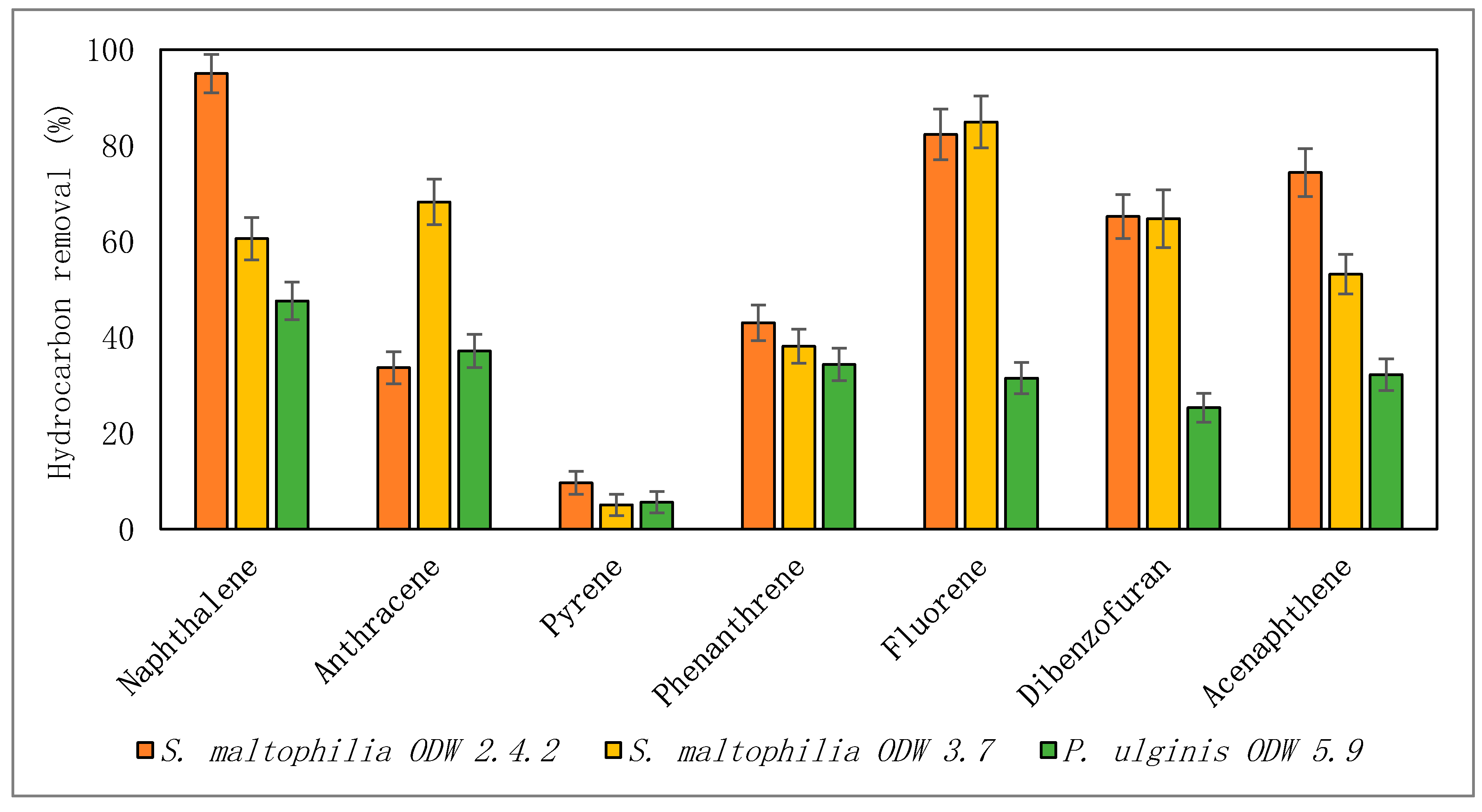
3.2. Creosote Biodegradation

Long-term contact of microorganisms with highly toxic PAHs present in the soil affects the biodiversity of bacteria found there. Three microbial consortia were isolated from the contaminated soil and then in selective cultures, the bacteria with high biodegradability potential in relation to hydrocarbons present in creosote oil were isolated. The highest creosote oil biodegradation was observed for microorganisms isolated from sample O6 (51% ± 9%). This microbial consortia effectively biodegraded dibenzofuran (81%), and anthracene and acenaphtene in approximately 70%. The most active strain isolated from this sample was S. maltophilia ODW 2.4.2, for which a similar level of creosote oil biodegradation (50% ± 4%) after seven days of the process was obtained (Figure 1). Moreover, high biodegradation of naphthalene (95%), fluorene (82%) and acenaphthene (74%) was noticed (Figure 1). What is more, from sample O6 also a strain belonging to the species S. maltophilia ODW 3.7 was isolated, for which a similar level of creosote oil biodegradation was observed (51%). However, this strain was characterized by different biodegradable activity in relation to different creosote oil components. Unlike sample O10, microbial community of O8 sample removed only 35% of creosote. The most active strain was identified as P. ulginis ODW 5.9. Nevertheless, the biodegradation efficiency of creosote and creosote components by this strain was the lowest from among the tested bacteria strains. The effectiveness of biodegradation depends on the chemical structure of creosote components, their concentration, as well as on the age of the contaminant [2]. Low molecular weight PAHs (consisting of two and three rings) are faster removed than the compounds of high molecular weight [32,33,34]. Naphthalene, a two-ringed PAH, is degraded relatively easily [35]. It has been observed that with increasing size of a PAH molecule, the hydrophobicity and electrochemical stability increase [36] and slow down the rates of biodegradation. This high hydrophobicity of the compounds present in creosote and related low water solubility influence their low bioavailability for microorganisms and therefore biodegradation efficiency [37]. Different species of bacteria show great biodegradation potential of polycyclic aromatic hydrocarbons [38]. Muangchinda et al. [39] have demonstrated high potential of Actinobacteria in PAH degradation in river sediments. Subashchandrabose et al. [40] have observed high biodegradation of phenanthrene by Rhodococcus wratislaviensis strain 9. This strain degraded also pyrene and benzo[a]pyrene. Our results indicated high biodegradation potential of Stenotrophomonas maltophilia, which could be used in the degradation of even aged PAHs.

3.3. Enzymatic Activity in the Presence of Creosote

The glutathione S-transferase (GST) activity was tested in three bacterial strains. Each strain was cultivated for seven days with the addition of creosote oil (marked as CREO) and without the addition of this component (control samples, marked as CTRL). Each culture was established in three biological repetitions and three technical repetitions of each culture were subjected to analysis. The final results presented in the graph (Figure 2) stand for the average values of these measurements. Regarding control samples of all strains tested, the highest activity of GST was measured in the strain S. maltophilia ODW 3.7 and the lowest value was noted for P. ulginis ODW 5.9. Similar directions of changes were observed for bacterial cells exposed to creosote oil. What can be clearly seen in the figure was a significant increase in GST activity in bacterial cells of the strains S. maltophilia ODW 2.4.2 and S. maltophilia ODW 3.7 exposed to creosote in comparison to control samples. In the cell extract of P. ulginis ODW 5.9 bacterial strain, the activity of GST measured in CREO sample was slightly higher (when compared to P. ulginis ODW 5.9 CTRL), however, the modifications were not statistically significant. In general, GST is an enzyme involved, among others, in detoxification of a wide range of xenobiotics. It works via nucleophilic conjugation of glutathione with electrophilic substrates. Although, the latter is described as a major detoxification mechanism in mammals [41,42], the activity of GST in bacterial strains was postulated as well. GST is believed to play an important role in biological degradation of xenobiotic compounds by bacteria, what was described by Allocati et al. (2009) [43] and Zablotowicz et al. (1995) [44]. The results of our research confirm the results of these researchers since the higher activity was measured in samples containing toxic, hazardous compounds for bacteria and the cells (especially those of S. maltophilia ODW 2.4.2 and S. maltophilia ODW 3.7 strains) might have initiated the reaction of biological degradation through GST enzymes.

3.4. Cell Wall Properties

The next stage of the research has been devoted to the analysis of cell adaptation processes to degradation of hydrophobic hydrocarbons, which consisted of modification of cell wall properties. Such adaptations are a very important parameter, which determines the bioavailability of compounds with low water solubility such as PAH to a high extent [13]. Table 2 includes the results of measurements of Congo red absorption on the cell surface as well as membrane permeability analyzes. Congo red adsorption studies indicate that all strains were characterized by slight dye adhesion. In addition, the ambiguous response of cells to contact with creosote oil should be emphasized. Strain S. maltophilia ODW 2.4.2 in the culture with an easily digestible sodium succinate exhibited an adsorption of 7%, whereas in the case of creosote culture this value was lower than 2%. The S. maltophilia ODW 3.7 strain, which belongs to the same species, behaved differently. In this case, a slight increase in dye adsorption was observed after comparing the cells from the culture with creosote and succinate. An analogous but much more pronounced increase (from 3.4% to 13.6%) was observed for P. ulginis ODW 5.9 cells. This is coherent with the previous studies, such as these reported by Ghosh and Mukherji [45] or Obuekwe [46], which established that the biodegradation of PAHs is associated with the increase of cell hydrophobicity. Moreover, Bezza and Chirwa [47] associated the increase of the hydrophobicity of cells during the biodegradation of pyrene with the interaction of biosurfactant produced by the bacteria.
The differences between the two tested strains of S. maltophilia were also evident during the analysis of total membrane permeability measured by the degree of penetration of crystal violet. S. maltophilia ODW 2.4.2 exhibited a clear decrease (from 3.7% to 24%) of membrane permeability of cells cultured using creosote, while S. maltophilia ODW 3.7 cells increased their membrane permeability after the comparison of samples from creosote-supplemented cultures with those from cultures with sodium succinate. Additionally, in the case of strain P. ulginis ODW 5.9, the presence of aromatic hydrocarbons resulted in an increased membrane permeability. The influence of the contact of the tested strains with creosote on the permeability of the inner cell membrane was relatively unambiguous and analogous. In all cases, the rate of o-nitrophenol formation resulting from the reaction catalyzed with intracellular galactosidase decreased in cultures with a mixture of PAHs. In the case of P. ulginis ODW 5.9, there was a significant ten-fold decrease of the measured parameter (from 1.24 to 0.13 µM min−1). However, it should be emphasized that the measured value could be affected by both the permeability of the internal membrane and the amount of enzyme produced by the cell. Jiang et al. [48] underlined that the xenobiotic can be a factor that enhances the biodegradation by altering the permeability of bacterial cell membrane. Additionally, the study of Kuyukina et al. [49] suggests that organic solvents primarily affect bacterial membranes. This results in the loss of membrane integrity, but the cells may remain viable. However, the decrease in membrane permeability may be a cellular defense response to the presence of a toxic compound [50]. These two mentioned mechanisms can affect the measured membrane permeability to a different degree, which explains the observed differences in the membrane permeability of the strains studied in the framework of this study. Moreover, it can be assumed that the very low inner membrane permeability and relatively higher Congo red adsorption ratio is associated with lower bioavailability of creosote to bacterial cells, which may explain the low biodegradation of the mixture of these PAHs by P. ulginis ODW 5.9 in comparison with S. maltophilia strains.

4. Conclusions

The conducted experiments showed the differences and similarities between strains capable of degrading creosote from the same contaminated area. The S. maltophilia strains exhibited higher biodegradation efficiency and the more significant increase in glutathione S-transferase than P. ulginis. It can suggest the higher adaptation potential of the strains from this genus than the P. ulginis strain. High level of the S-transferase activity indicates activation of the metabolic pathways involved in biotransformation of the creosote hydrocarbons. Moreover, it can be presumed that the P. ulginis strain uses other enzymes to degrade the pollutant. However, studies of cell surface properties show differences between S. maltophilia strains. S. maltophilia ODW 3.7 and P. ulginis ODW 5.9 distinguished from the third of the tested strains in that the growth on creosote resulted in an increase in both cases in cell adhesion to Congo red, as well as the total membrane permeability. However, all three strains shown decreased in inner membrane permeability. To summarize, the obtained results brought new valuable information on the variety of characteristics and properties of microorganisms involved in the bioremediation processes of creosote oil in contaminated soil.

Author Contributions

Conceptualization, E.K. and W.S.; methodology, A.P. and W.S.; validation, A.P.; writing—original draft preparation, W.S, A.P. and E.K. All authors have read and agreed to the published version of the manuscript.

Funding

This research was financed as a part of the project “Modern technology for bioremediation soil contaminated with creosote oil on the premises of Sleeper Treating Plant Spółka Akcyjna in Koźmin Wielkopolski”, no. POIR.04.01.02-00-0057/17-00 implemented under sub-measure 4.1.2 “Regional Science and Research Agendas”, co-financed by the European Regional Development Fund.

Conflicts of Interest

The authors declare no conflict of interest.

References

  1. Hicknell, B.N.; Mumford, K.G.; Kueper, B.H. Laboratory study of creosote removal from sand at elevated temperatures. J. Contam. Hydrol. 2018, 219, 40–49. [Google Scholar] [CrossRef]
  2. Madrid, F.; Rubio-Bellido, M.; Villaverde, J.; Peña, A.; Morillo, E. Natural and assisted dissipation of polycyclic aromatic hydrocarbons in a long-term co-contaminated soil with creosote and potentially toxic elements. Sci. Total Environ. 2019, 660, 705–714. [Google Scholar] [CrossRef]
  3. Webb, D.A. Creosote, its biodegradation and environmental effects. Amer. Wood-Preserv. Assoc. 1980, 76, 65–69. [Google Scholar]
  4. Melber, C.; Kielhorn, J.; Mangelsdorf, I. Concise International Chamical Assessment Document 62. Coal Car Creosote; World Health Organization: Geneva, Switzerland, 2004. [Google Scholar]
  5. Ye, Y.-F.; Ma, F.-Y.; Wu, M.; Wei, X.-Y.; Liu, J.-M. Increase of acenaphthene content in creosote oil by hydrodynamic cavitation. Chem. Eng. Process. 2016, 104, 66–74. [Google Scholar] [CrossRef]
  6. Blum, P.; Sagner, A.; Tiehm, A.; Martus, P.; Wendel, T.; Grathwohl, P. Importance of heterocylic aromatic compounds in monitored natural attenuation for coal tar contaminated aquifers: A review. J. Contam. Hydrol. 2011, 126, 181–194. [Google Scholar] [CrossRef] [PubMed]
  7. Mueller, J.G.; Chapman, P.J.; Pritchard, P.H. Creosote-contaminated sites. Their potential for bioremediation. Environ. Sci. Technol. 1989, 23, 1197–1201. [Google Scholar] [CrossRef]
  8. Vähäoja, P.; Piltonen, P.; Hyvönen, A.; Niinimäki, J.; Jalonen, J.; Kuokkanen, T. Biodegradability Studies of Certain Wood Preservatives in Groundwater as Determined by the Respirometric Bod Oxitop Method. Water Air Soil Pollut. 2005, 165, 313–324. [Google Scholar] [CrossRef]
  9. Carlsten, C.; Hunt, S.C.; Kaufman, J.D. Squamous Cell Carcinoma of the Skin and Coal Tar Creosote Exposure in a Railroad Worker. Environ. Health Perspect. 2005, 113, 96–97. [Google Scholar] [CrossRef] [Green Version]
  10. Clapp, R.W.; Jacobs, M.M.; Loechler, E.L. Environmental and Occupational Causes of Cancer: New Evidence 2005-2007. Rev. Environ. Health 2008, 23, 1–38. [Google Scholar] [CrossRef]
  11. Hu, J.; Mao, Y.; White, K. Renal cell carcinoma and occupational exposure to chemicals in Canada. Occup. Med. 2002, 52, 157–164. [Google Scholar] [CrossRef] [Green Version]
  12. Krzyśko-Łupicka, T.; Cybulska, K.; Kołosowski, P.; Telesiński, A.; Sudoł, A. Influence of environmental pollution with creosote oil or its vapors on biomass and selected physiological groups of microorganisms. E3S Web Conf. 2017, 22, 00092. [Google Scholar] [CrossRef] [Green Version]
  13. Kaczorek, E.; Pacholak, A.; Zdarta, A.; Smułek, W. The Impact of Biosurfactants on Microbial Cell Properties Leading to Hydrocarbon Bioavailability Increase. Colloids Interfaces 2018, 2, 35. [Google Scholar] [CrossRef] [Green Version]
  14. Ren, X.; Zeng, G.; Tang, L.; Wang, J.; Wan, J.; Liu, Y.; Yu, J.; Yi, H.; Ye, S.; Deng, R. Sorption, transport and biodegradation—An insight into bioavailability of persistent organic pollutants in soil. Sci. Total Environ. 2018, 610, 1154–1163. [Google Scholar] [CrossRef] [PubMed]
  15. Zeng, Z.; Liu, Y.; Zhong, H.; Xiao, R.; Zeng, G.; Liu, Z.; Cheng, M.; Lai, C.; Zhang, C.; Liu, G.; et al. Mechanisms for rhamnolipids-mediated biodegradation of hydrophobic organic compounds. Sci. Total Environ. 2018, 634, 1–11. [Google Scholar] [CrossRef]
  16. Xing, S.-F.; Sun, X.-F.; Taylor, A.A.; Walker, S.L.; Wang, Y.-F.; Wang, S.-G. D-Amino acids inhibit initial bacterial Adhesion: Thermodynamic evidence. Biotechnol. Bioeng. 2015, 112, 696–704. [Google Scholar] [CrossRef] [PubMed] [Green Version]
  17. Hearn, E.M.; Patel, D.R.; van den Berg, B. Outer-membrane transport of aromatic hydrocarbons as a first step in biodegradation. Proc. Natl. Acad. Sci. USA 2008, 105, 8601–8606. [Google Scholar] [CrossRef] [Green Version]
  18. Hua, F.; Wang, H.Q. Uptake and trans-membrane transport of petroleum hydrocarbons by microorganisms. Biotechnol. Biotechnol. Equip. 2014, 28, 165–175. [Google Scholar] [CrossRef] [Green Version]
  19. Sikkema, J.; de Bont, J.A.; Poolman, B. Mechanisms of membrane toxicity of hydrocarbons. Microbiol. Rev. 1995, 59(2), 201–222. [Google Scholar] [CrossRef]
  20. Kaczorek, E.; Urbanowicz, M.; Olszanowski, A. The influence of surfactants on cell surface properties of Aeromonas hydrophila during diesel oil biodegradation. Colloids Surf. B 2010, 81, 363–368. [Google Scholar] [CrossRef]
  21. Kaczorek, E.; Sałek, K.; Guzik, U.; Jesionowski, T.; Cybulski, Z. Biodegradation of alkyl derivatives of aromatic hydrocarbons and cell surface properties of a strain of Pseudomonas stutzeri. Chemosphere 2013, 90, 471–478. [Google Scholar] [CrossRef]
  22. Hassanshahian, M. Isolation and characterization of biosurfactant producing bacteria from Persian Gulf (Bushehr provenance). Mar. Pollut. Bull. 2014, 86, 361–366. [Google Scholar] [CrossRef] [PubMed]
  23. Ambalam, P.; Kondepudi, K.K.; Nilsson, I.; Wadström, T.; Ljungh, Å. Bile stimulates cell surface hydrophobicity, Congo red binding and biofilm formation of Lactobacillus strains. FEMS Microbiol. Lett. 2012, 333, 10–19. [Google Scholar] [CrossRef] [PubMed] [Green Version]
  24. Pacholak, A.; Simlat, J.; Zgoła-Grześkowiak, A.; Kaczorek, E. Biodegradation of clotrimazole and modification of cell properties after metabolic stress and upon addition of saponins. Ecotoxicol. Environ. Saf. 2018, 161, 676–682. [Google Scholar] [CrossRef] [PubMed]
  25. Devi, K.P.; Sakthivel, R.; Nisha, S.A.; Suganthy, N.; Pandian, S.K. Eugenol alters the integrity of cell membrane and acts against the nosocomial pathogen Proteus mirabilis. Arch. Pharmacal Res. 2013, 36, 282–292. [Google Scholar] [CrossRef] [PubMed]
  26. Ghosal, D.; Ghosh, S.; Dutta, T.K.; Ahn, Y. Current State of Knowledge in Microbial Degradation of Polycyclic Aromatic Hydrocarbons (PAHs): A Review. Front. Microbiol. 2016, 7, 1369. [Google Scholar] [CrossRef] [Green Version]
  27. Daane, L.L.; Harjono, I.; Barns, S.M.; Launen, L.A.; Palleron, N.J.; Häggblom, M.M. PAH-degradation by Paenibacillus spp. and description of Paenibacillus naphthalenovorans sp. nov., a naphthalene-degrading bacterium from the rhizosphere of salt marsh plants. Int. J. Syst. Evol. Microbiol. 2002, 52, 131–139. [Google Scholar] [CrossRef] [Green Version]
  28. Mesbaiah, F.Z.; Eddouaouda, K.; Badis, A.; Chebbi, A.; Hentati, D.; Sayadi, S.; Chamkha, M. Preliminary characterization of biosurfactant produced by a PAH-degrading Paenibacillus sp. under thermophilic conditions. Environ. Sci. Pollut. Res. 2016, 23, 14221–14230. [Google Scholar] [CrossRef]
  29. Tripathi, V.; Gaur, V.K.; Dhiman, N.; Gautam, K.; Manickam, N. Characterization and properties of the biosurfactant produced by PAH-degrading bacteria isolated from contaminated oily sludge environment. Environ. Sci. Pollut. Res. 2019. [Epub ahead of print]. [Google Scholar] [CrossRef]
  30. Singh, A.; Kumar, K.; Pandey, A.K.; Sharma, A.; Singh, S.B.; Kumar, K.; Arora, A.; Nain, L. Pyrene Degradation by Biosurfactant Producing Bacterium Stenotrophomonas maltophilia. Agric. Res. 2015, 4, 42–47. [Google Scholar] [CrossRef]
  31. Arulazhagan, P.; Al-Shekri, K.; Huda, Q.; Godon, J.J.; Basahi, J.M.; Jeyakumar, D. Biodegradation of polycyclic aromatic hydrocarbons by an acidophilic Stenotrophomonas maltophilia strain AJH1 isolated from a mineral mining site in Saudi Arabia. Extremophiles 2017, 21, 163–174. [Google Scholar] [CrossRef] [Green Version]
  32. Bacosa, H.P.; Inoue, C. Polycyclic aromatic hydrocarbons (PAHs) biodegradation potential and diversity of microbial consortia enriched from tsunami sediments in Miyagi, Japan. J. Hazard. Mater. 2015, 283, 689–697. [Google Scholar] [CrossRef] [PubMed]
  33. Valderrama, C.; Alessandri, R.; Aunola, T.; Cortina, J.L.; Gamisans, X.; Tuhkanen, T. Oxidation by Fenton as reagent combined with biological treatment applied to a creosote-contaminated soil. J. Hazard. Mater. 2009, 166, 594–602. [Google Scholar] [CrossRef] [PubMed]
  34. Jurys, A.; Gailiũte, I.; Aikaitė-Stanaitienė, J.; Grigiškis, S.; Maruška, A.; Stankevičius, M.; Levišauskas, D. Review of creosote pollution toxicity and possibilities of bioremediation. Environ. Technol. Resour. 2013, 1, 34–38. [Google Scholar] [CrossRef]
  35. Grant, R.J.; Muckian, L.M.; Clipson, N.J.W.; Doyle, E.M. Microbial community changes during the bioremediation of creosote-contaminated soil. Lett. Appl. Microbiol. 2007, 44, 293–300. [Google Scholar] [CrossRef] [PubMed]
  36. Widada, J.; Nojiri, H.; Kasuga, K.; Yoshida, T.; Habe, H.; Omori, T. Molecular detection and diversity of polycyclic aromatic hydrocarbon-degrading bacteria isolated from geographically diverse sites. Appl. Microbiol. Biotechnol. 2002, 58, 202–209. [Google Scholar] [CrossRef] [PubMed]
  37. Shor, L.M.; Liang, W.; Rockne, K.J.; Young, L.Y.; Taghon, G.L.; Kosson, D.S. Intraaggregate mass transport-limited bioavailability of polycyclic aromatic hydrocarbons to Mycobacterium strain PC01. Environ. Sci. Technol. 2003, 37, 1545–1552. [Google Scholar] [CrossRef] [PubMed]
  38. Kanaly, R.A.; Harayama, S. Biodegradation of high-molecular-weight polycyclic aromatic hydrocarbons by bacteria. J. Bacteriol. 2000, 182(8), 2059–2067. [Google Scholar] [CrossRef] [Green Version]
  39. Muangchinda, C.; Yamazoe, A.; Polrit., D.; Thoetkiattikul, H.; Mhuantong, W.; Champreda, V.; Pinyakong, O. Biodegradation of high concentrations of mixed polycyclic aromatic hydrocarbons by indigenous bacteria from a river sediment: a microcosm study and bacterial community analysis. Environ. Sci. Pollut. Res. 2017, 24, 4591–4602. [Google Scholar] [CrossRef]
  40. Subashchandrabosea, S.R.; Venkateswarlub, K.; Naidua, R.; Meghara, M. Biodegradation of high-molecular weight PAHs by Rhodococcus wratislaviensis strain 9: Overexpression of amidohydrolase induced by pyrene and BaP. Sci. Total Environ. 2019, 651, 813–821. [Google Scholar] [CrossRef]
  41. Monostori, P.; Wittmann, G.; Karg, E.; Túri, S. Determination of glutathione and glutathione disulfide in biological samples: An in-depth review. J. Chrom. B. 2009, 877, 3331–3346. [Google Scholar] [CrossRef]
  42. Pour, L.M.; Farahnak, A.; Rad, M.M.; Golmohamadi, T.; Eshraghian, M. Activity Assay of Glutathione S-Transferase (GSTs) Enzyme as a Diagnostic Biomarker for Liver Hydatid Cyst in Vitro. Iran J. Public Health. 2014, 43, 994. [Google Scholar]
  43. Allocati, N.; Federici, L.; Masulli, M.; di Ilio, C. Glutathione transferases in bacteria: Bacterial. FEBS J. 2009, 276, 58–75. [Google Scholar] [CrossRef] [PubMed]
  44. Zablotowicz, R.M.; Hoagland, R.E.; Locke, M.A.; Hickey, W.J. Glutathione-S-Transferase Activity and Metabolism of Glutathione Conjugates by Rhizosphere Bacteria. Appl. Environ. Microbiol. 1995, 61, 1054–1060. [Google Scholar] [CrossRef] [PubMed] [Green Version]
  45. Ghosh, I.; Mukherji, S. Diverse effect of surfactants on pyrene biodegradation by a Pseudomonas strain utilizing pyrene by cell surface hydrophobicity induction. Int. Biodeter. Biodegrad. 2016, 108, 67–75. [Google Scholar] [CrossRef]
  46. Obuekwe, C.O.; Al-Jadi, Z.K.; Al-Saleh, E.S. Hydrocarbon degradation in relation to cell-surface hydrophobicity among bacterial hydrocarbon degraders from petroleum-contaminated Kuwait desert environment. Int. Biodeter. Biodegrad. 2009, 63, 273–279. [Google Scholar] [CrossRef]
  47. Bezza, F.A.; Chirwa, E.M.N. Pyrene biodegradation enhancement potential of lipopeptide biosurfactant produced by Paenibacillus dendritiformis CN5 strain. J. Hazard. Mater. 2017, 321, 218–227. [Google Scholar] [CrossRef]
  48. Jiang, Z.; Chen, J.; Li, J.; Cao, B.; Chen, Y.; Liu, D.; Wang, X.; Zhang, Y. Exogenous Zn2+ enhance the biodegradation of atrazine by regulating the chlorohydrolase gene trzN transcription and membrane permeability of the degrader Arthrobacter sp. DNS10. Chemosphere 2020, 238, 124594. [Google Scholar] [CrossRef]
  49. Kuyukina, M.S.; Ivshina, I.B.; Korshunova, I.O.; Rubtsova, E.V. Assessment of bacterial resistance to organic solvents using a combined confocal laser scanning and atomic force microscopy (CLSM/AFM). J. Microbiol. Methods 2014, 107, 23–29. [Google Scholar] [CrossRef]
  50. Dam, S.; Pagès, J.-M.; Masi, M. Stress responses, outer membrane permeability control and antimicrobial resistance in Enterobacteriaceae. Microbiology 2018, 164, 260–267. [Google Scholar] [CrossRef]
Figure 1. Removal of selected PAH present in creosote by selected strains after 28 days.
Processes 08 00147 g001
Figure 2. Activity of GST enzymes tested in three bacterial strains in control cultures (Ctrl) and the cultures with the addition of creosote oil (Creo).
Processes 08 00147 g002
Table 1. Bacteria strains isolated from the creosote-contaminated soil samples.
Soil SampleStrainGenBank (NCBI) NumberHemolysis Test
O10Stenotrophomonas maltophilia ODW 2.4.2MK503432.1alpha
O6Stenotrophomonas maltophilia ODW 3.7MK503436.1alpha
O8Paenibacillus ulginis ODW 5.9MK503429.1beta
Table 2. Bacteria cell wall properties described by the Congo red adsorption assay (CR), the inner and total membrane permeability tests (ONPG and CV, respectively).
StrainCarbon SourceCR
(%)
ONPG
(μM min−1)
CV
(%)
Stenotrophomonas maltophilia ODW 2.4.2succinate7.0 ± 0.80.28 ± 0.0137 ± 3
creosote1.8 ± 0.60.15 ± 0.0124 ± 2
Stenotrophomonas maltophilia ODW 3.7succinate4.2 ± 0.10.51 ± 0.0640 ± 3
creosote5.1 ± 0.50.44 ± 0.0959 ± 4
Paenibacillus ulginis ODW 5.9succinate3.4 ± 0.81.24 ± 0.2116 ± 2
creosote13.6 ± 0.90.13 ± 0.0636 ± 3

