Article

Simple Preparation of Diverse Neoagaro-Oligosaccharides

1
College of Chemical Engineering, Huaqiao University, Xiamen 361021, China
2
Xiamen Engineering and Technological Research Center for Comprehensive Utilization of Marine Biological Resources, Xiamen 361021, China
*
Authors to whom correspondence should be addressed.
Processes 2019, 7(5), 267; https://doi.org/10.3390/pr7050267
Submission received: 2 April 2019 / Revised: 30 April 2019 / Accepted: 2 May 2019 / Published: 7 May 2019
(This article belongs to the Special Issue Green Separation and Extraction Processes)

Abstract

:
A simple method for obtaining pure and well-defined oligosaccharides was established by hydrolyzing agar with β-agarase from Vibrio natriegens. The conditions for enzymolysis were optimized as follows: a temperature of 45 °C, a pH of 8.5, a substrate concentration of 0.3%, an enzyme amount of 100 U/g and an enzymolysis time of 20 h. Neoagaro-oligosaccharides with different degrees of polymerization were obtained by hydrolyzing agar with β-agarase for different lengths of time. After removing pigments using activated carbon and salts by dialyzing, the enzyme hydrolysis solution was separated with Bio-Gel P2 column chromatography. Neoagaro-oligosaccharides with different degrees of polymerization were acquired. By comparing with authentic standard substances, along with further confirmation by FTIR, MS and NMR, structures of the purified neoagaro-oligosaccharides were identified as neoagarobiose (NA2), neoagaroteraose (NA4), neoagarohexaose (NA6), neoagarooctaose (NA8), neoagaro-decaose (NA10) and neoagarododecaose (NA12) with purities of more than 97.0%. The present study established a method for the preparation of various neoagaro-oligosaccharides that may be of great significance for further study of their bioactivities.

Graphical Abstract

1. Introduction

Agar, an important marine polysaccharide extracted from the cell walls of red algae, is a linear polymer containing (1→4)-linked 3,6-anhydro-α-l-galactose and (1→3)-linked β-d-galactopyranose [1], and it is composed of agarose and agaropectin [2]. Agar is widely used in the food, biological, and pharmaceutical industries [3]. Accumulating reports have indicated that the oligosaccharides prepared from agar/agarose have diverse physiological functions, such as antioxidant [4,5,6,7,8,9], anti-hyperlipidemia [9,10,11], anti-inflammation [12,13,14] activity, and a whitening effect [15,16], which will expand their use in the food, cosmetic, and medical industries.
Generally, oligosaccharides from agar/agarose can be classified as either agaro-oligosaccharides (AOS) with agarobiose as a repeating unit (Figure 1A) [17,18,19] or neoagaro-oligosaccharides (NAOS) with neoagarobiose as a repeating unit (Figure 1B) [20,21,22]. The former are the hydrolysis products of acid or α-agarase which cleaves the α-(1→3)-galactosidic bond of the polysaccharides (Figure 1A), and the latter are hydrolysates of β-agarase which splits the β-(1→4) bond (Figure 1B). AOS and NAOS with different degrees of polymerization (DP) have been reported to possess various bioactivities. A large number of reports indicated that AOS have beneficial antioxidant [4,5,6,7,8], anti-obesity [10,11], anti-inflammation [12,13,14], anti-cancer [23] activity, and also protect the intestine [10,11,23]. In recent years, NAOS mixtures, as well as NOAS of signal DP, have attracted increasing attention for their distinct physiological and biological activities. According to the reported prebiotic studies, the NAOS mixtures show varying activity in antioxidants [24]. In addition, the NAOS with DP of 4–12 could increase the amount of lactobacilli and bifidobacteri in vivo, suggesting that they had beneficial probiotic effects [25]. Neoagarobiose (NA2), neoagaroteraose (NA4), and neoagarohexaose (NA6) were reported to have in vitro skin whitening and moisturizing effects, among them, NA4 was found to be a better whitening agent than the other two, whereas AOS did not exhibit the same activities [15,16]. Moreover, NA4 displayed a better ability to scavenge hydroxyl radicals compared to that of NA2, NA6, and neoagarooctaose (NA8), meanwhile, agarotriose and agarobiose are not able to scavenge hydroxyl radicals. Additionally, NA4 was proved to inhibit inflammation in LPS-stimulated macrophages through suppression of mitogen-activated protein kinase (MAPK) and NF-κB pathways [26,27].
Notably, these studies mainly focused on the activities of NAOS mixture, NA2, NA4, and NA6, while few reports have investigated the bioactivities of NAOS with higher DP such as NA8, neoagarodecaose (NA10), and neoagarododecaose (NA12). The reasons for this may be attributed to the difficulty of obtaining NAOS with higher DP and the complexity of their purification. Many efforts have been devoted to obtaining NAOS. β-agarases from marine bacterium Janthinobacterium sp. SY12, Vibrio sp. Strain JT0107, Agarivorans albus YKW-34 and Agarivorans albus OAY02 were employed to degrade agarose to give NA2 and NA4 [28,29,30]. β-agarase obtained from Microbulbifer sp. Q7, marine Alteromonas sp. SY37–12, Pseudoalteromonas sp. CY24, marine Agarivorans sp. LQ48 and Pseudoalteromonas sp. AG4 could hydrolyze β-1,4-glycosidic linkages of agarose/agar to give NA4 and NA6 [31,32,33,34,35]. β-agarases isolated from Stenotrophomonas sp. NTa and Agarivorans sp. JA-1 in Bacillus subtilis degraded agarose/agar to yield NA2, NA4 and NA6 as the predominant products [36,37]. Agarivorans albus OAY02 could secrete two β-agarases, among them, one β-agarase could cleave agarose into NA2 and NA4, the other β-agarase hydrolyzed agarose to NA2, NA4, NA6 and NA8 [38]. β-agarase from marine Pseudoalteromonas sp. CY24 could degrade agarose to NA8 and NA10 [39]. β-Agarase from marine bacteria can degrade agarose to NAOS with different DP; however, up to now, it has not been reported that β-agarase could degrade agarose/agar to obtain NAOS with desired DP by controlling the enzymolysis time.
Developing methods for separation and purification of NAOS is also very important for obtaining purified NAOS. Size-sieving based on gel-permeation chromatography (GPC) and high-performance anion exchange chromatography (HPAEC) are commonly used methods for the separation of polysaccharides and oligosaccharides [6,40,41,42,43]. Toyopearl HW-40S was used to purify NA4 [40]. It was reported that NAOS with DP of 2, 4, and 6 could be separated and purified by Bio-Gel P2 [42,43] and NA4, NA6, NA8, NA10 and NA12 could be separated by two chromatography steps of consecutive Bio-Gel P-6 chromatography [42]. Sephadex G-10 combined with G-25 was employed to purify AOS [6,41]. The SEC-HPLC and NH2-HPLC systems were used to isolate and purify NAOS and AOS [44]. A HPAEC system equipped with a semi-preparative CarboPacTM PA100 column was applied to prepare NAOS and AOS from DP 2 to DP 22 with product yields and purity of no more than 17.2% and 77.7%, respectively [45]. However, these methods are complicated, time-consuming, and rely on instrumentation; furthermore, the product yield and purity of obtained oligosaccharides are not high enough for further study. Therefore, to deeply understand the bioactivities and the mechanism of NOAS [9], a simple method for the preparation of NAOS with various DPs is urgently required.
In the present study, a simple method of obtaining NAOS with desired DP was established by controlling the enzymolysis time of β-agarase. Furthermore, a gel filtration chromatography method was developed for the purification of each NAOS with different DP in high quality and quantity, making it possible to further study their bioactivities.

2. Materials and Methods

2.1. Strains and Reagents

NA2, NA4, NA6, NA8, NA10 and NA12 were used as standards and were purchased from Qingdao Bozhihui Biological Technology Co., Ltd. (Qingdao, China). Acetonitrile was purchased from Sigma-Aldrich (St. Louis, MO, USA). All the other reagents were commercially available and were of analytical grade.
The culture and fermentation condition of Vibrio natriegens was the same as reported in our previous work [46]. The obtained β-agarase was purified from the fermentation liquor by salting-out with ammonium sulfate, followed by dialysis, ion exchange chromatography and gel filtration. The definition of enzyme activity: under specified conditions, 1 mg crude enzyme produces 1 μg reducing sugar for 1 min.

2.2. Optimization of Enzymatic Hydrolysis Condition

The enzymatic hydrolysis condition, including reaction temperature (30, 35, 40, 45, 50, 55 and 60 °C), pH (6.0, 6.5, 7.0, 7.5, 8.0, 8.5 and 9.0), reaction time (1, 5, 10, 15, 20, 25 and 30 h), substrate concentration (0.1, 0.2, 0.3, 0.4, 0.5, 0.6 and 0.7%) and enzyme amounts (20, 40, 60, 80, 100, 120 and 140 U/g) were all optimized. All hydrolysis reactions were conducted in triplicate.

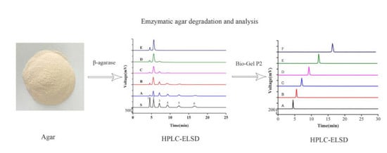
2.3. Preparation of NAOS Products with Different Enzymolysis Time

3 g agar was completely dissolved in 1 L of 0.1 M Tris-HCl solution by heating and then cooled to 45 °C. Subsequently, the solution was treated with the β-agarase for 4 h, 6 h, 8 h, 10 h or 12 h to obtain products A, B, C, D and E, respectively. After inactivation by incubation in a boiling water bath for 15 min and centrifugation for 30 min (12,000 rpm), the insoluble agar was removed and the NAOS in the supernatant was filtered with a 0.22 μm membrane (Millipore, Cork, Ireland). The crude products resulting from the different enzymolysis times were finally obtained by evaporation, concentration and lyophilization and stored at −20 °C until needed. Each product was measured in triplicate.

2.4. Analysis of NAOS Products by HPLC-ELSD

The NAOS products were analyzed by an HPLC-ELSD system which consisted of a Waters e2695 HPLC system (Waters, Milford, MA, USA) equipped with an evaporative light scattering detector (Waters 2424, USA). Separation was performed on an Asahipak NH2P-50 4E multi-mode analytical column (250 mm × 4.6 mm, 5 μm) with the column temperature set at 30 °C. Isocratic elution was conducted with acetonitrile-water (65:35) as the mobile phase with a flow rate of 1 mL/min. The injection volume was set at 10 μL and the detector nebulizer temperature was 75 °C.

2.5. Purification of NAOS

For the purification of NAOS, 1 g of crude NAOS product powder was resuspended in 200 mL distilled water, followed by the addition of 10 g of activated carbon. The mixture was stirred for 2 h and then the NAOS were washed with 30% ethanol solution, and ethanol was removed by evaporation. Then the remainder was freeze-dried and detected.
GPC was applied to separate the NAOS. A sample of 250 mg of products A was resuspended in 1 mL of NH4HCO3 (0.1 M), and the solutions were loaded onto the Bio-Gel P2 column (1.8 × 150 cm, Bio-Rad Laboratories, Hercules, CA, USA). NH4HCO3 solution was used as the eluent at a flow rate of 0.4 mL/min and a tube of 4 mL were collected. Then the collected fractions were detected by TLC on a Silica Gel 60 plate (Merck, Darmstadt, Germany) and developed with a solvent of isopropanol/water/ammonium hydroxide (30:15:2, v/v/v). After being sprayed by anisaldehyde and heated for 10 min, the products could be visualized as spots and could merge the same components.

2.6. Identification of NAOS

The structure and the molecular mass of the isolated NAOS were elucidated by FTIR, ESI-TOF-MS and NMR. The FTIR spectra were obtained on an FTIR-84 spectrophotometer (Shimadzu, Japan) on KBr pellets; the NAOS were dried at 105 °C for 2 h prior to recordings. Scans were performed over the range of 4000–400 cm−1 with a resolution of 4 cm−1 for 32 scans. ESI-TOF-MS analysis was carried out on a Q Exactive Hybrid Quadrupole Orbitrap mass spectrometer (Thermo, Bremen, Germany) coupled with an ESI source in positive ion mode. The recorded mass range was m/z 200–2000. 1H-NMR and 13C-NMR spectra were measured in D2O on an Avance 500 spectrometer (Bruker, Avance III, Switzerland, 500 MHz 1H, 125 MHz 13C) at room temperature and C3D6O was added as an internal standard, its chemical shifts were reported at δ = 2.05 ppm for 1H-NMR, δ = 29.84 ppm for 13C-NMR.

3. Results and Discussion

3.1. Optimization of Hydrolysis Parameters by Single Factor Experiments

Single factor experiments were carried out to optimize the enzymatic hydrolysis process. Figure 1A depicts the influence of temperature on the yield of reducing sugar. Temperature is an important factor in the enzymatic hydrolysis process [22]. Within a certain temperature range, the increased temperature is beneficial to the enzymatic reaction. As shown in Figure 2a, the yield of reducing sugar was increased with the elevation of temperature and it reached a maximum of 45.2% at 45 °C, after that the yield was decreased significantly with increased temperature. Therefore, the optimal temperature was determined to be 45 °C. The pH is another vital parameter in the enzymatic hydrolysis process because enzyme activity is greatly affected by the pH [28]. Generally, an enzyme can maintain high enzyme activity at a suitable pH. As shown in Figure 2b, the yield of reducing sugar was increased with the increase of pH from 6.0 to 8.5, the largest yield of reducing sugar was 42.7% at pH 8.5. Above pH 8.5, the yield was decreased, which may be attributed to the inhibition of the β-agarase activity. As for the substrate concentration, low substrate concentration leads to low enzyme utilization; however, a too high substrate concentration hinders the diffusion of molecules and thereby reduces the enzymatic reaction rate. As shown in Figure 2c, when the substrate concentration was set to 0.3%, the yield of reducing sugar increased to 43.3%. In addition, not only the enzyme amount but also enzymolysis time has prominent effects on the yield of reducing sugar [47,48]. The enzyme amounts were also investigated in the present study and using 100 U/g turned out to be optimal, affording a 42.4% yield of reducing sugar (Figure 2d). Furthermore, the yield of reducing sugar was not increased by the addition of more enzymes. Figure 2e indicated that the yield of reducing sugar increased with the extension of enzymolysis time up to 20 h. When the enzymolysis time was 20 h, the reducing sugar yield was highest at 43.3%, and increasing the enzymolysis time further did not increase the yield. Therefore, the optimized conditions were determined as follows: a temperature of 45 °C, a pH of 8.5, a substrate concentration of 0.3%, an enzyme amount of 100 U/g and an enzymolysis time of 20 h.

3.2. Preparation of NAOS with Different DP

The β-agarase obtained from different strains degrade agar to gain the NAOS with different DP [28,32,36,37]. It has been reported that the immobilized β-agarase could prepare NAOS with different DP by controlling the enzyme hydrolysis time [24]. But, the regular changes of NAOS with different DP at different enzymolysis time were found in the present study (Figure 3), which was not previously reported. The enzymolysis products were determined by HPLC-ELSD by comparing with authentic standard substances, and the content of each oligosaccharide was calculated from the regression equations, as shown in Figure 2, Table 1 and Table 2. The enzymolysis products were found to be composed of NA2, NA4, NA6, NA8, NA10 and NA12 with yields of 5.0, 38.9, 18.1, 16.8, 13.8 and 2.5 mg/100 mg, respectively after the agar was hydrolyzed by β-agarase for 4 h. When the hydrolysis time was 6 h, NA12 was completely hydrolyzed and the products obtained were NA2, NA4, NA6, NA8, and NA10 with yields of 6.0, 52.7, 15.2, 13.7, and 11 mg/100 mg, respectively. As the hydrolysis time was extended to 8 h, NA10 was not detected in the product mixture and the yields of NA2, NA4, NA6 and NA8 were 9.3, 59.5, 13.7 and 11.7 mg/100 mg, respectively. With the extension of the hydrolysis time, the DP of NAOS obtained were decreased. When the hydrolysis time was 10 h, the product mixture consisted of NA2, NA4, and NA6 with yields of 13.8, 68.4 and 9.6 mg/100 mg, respectively. Once the hydrolysis time reached 12 h, only NA4 and NA2 were left in the product mixture with the yields of 21.6 mg/100 mg and 71.1 mg/100 mg, respectively. Beyond 12 h, the product components were no longer changed with the extension of time.
At present, there are increasing numbers of reports on the preparation of NAOS by the enzymatic method, and agarase is mainly derived from the secretions of marine bacteria. Frequently, β-agarase is found to degrade agar to generate NA2, NA4, or NA6 as the main products. DagA secreted β-agarase hydrolyzes agar to gain NA2, NA4 and NA6 [49]. β-agarase from Stenotrophomonas sp. NTa degrades agar to yield NA2, NA4 and NA6 as the predominant products with a small amount of 3,6-anhydro-α-l-galactose; notably the products did not change with the change of the hydrolysis time [37]. At the same time, the composition of the agarolytic product did not change over time due to some β-agarase, which included four even-numbered NAOS with DP of 2–8, and the amount of NA4 was greater than for the others [27]. However, these methods found in the literature were used in preparing NAOs mainly with DP of no more than eight [27,37,49]. Interestingly, we found in our study that the DP of the NAOS was reduced regularly with increased enzymolysis time, and the final product was composed of NA4 and NA2. Therefore, desired NOAS with different DP could be obtained by controlling the enzymolysis time, which may be beneficial to the further studies on NAOS.

3.3. Separation of NAOS

Separation and purification of NAOS was carried out using a Bio-Gel P2 column and detected by TLC, and the results are shown in Figure 4. For product A, fractions 18 to 22 contained NA12 with a yield of 3.2%; fractions 24 to 27 contained NA10 with a yield of 4.2%; fractions 30 to 35 contained NA8 with a yield of 7.5%; fractions 37 to 40 contained NA6 with a yield of 10.2%; fractions 44 to 56 contained NA4 with a yield of 35.8%; and fractions 61 to 64 contained NA2 with a yield of 23.2%. After detection by HPLC-ELSD, the purity of NA2, NA4, NA6, NA8, NA10 and NA12 were 99.3%, 98.9%, 98.0%, 97.6%, 97.3% and 97.4%, respectively (Figure 5).
GPC and HPAEC are commonly used methods for the separation and purification of NAOS, and Bio-Gel P-2 and Bio-Gel P-6 are frequently applied to purify NAOS [6,40,41,42,43,44,45]. However, these two separations methods are usually combined to isolate NAOS with diverse DP. Notably, in the present study, one chromatographic step using Bio-Gel P-2 column chromatography allowed us to obtain NAOS of DP2-12 with purities of greater than 97%, suggesting that it is a simple method for the preparation of NAOS.

3.4. Characterization of NAOS

The structures and the molecular mass of the purified NAOS were determined by FTIR and MS analysis. Figure 6 shows the results of the FTIR analyses. In all six obtained oligosaccharides, the disappearance of an absorption band around 1260 cm−1 indicated the elimination of a sulfate group in the degradation process. There was a broad absorption band around 3400 cm−1, which may be assigned to hydroxyl groups. The region around 2950 cm−1 and 2900 cm−1 were assigned as C-H groups. The band around 1640 cm−1 suggested the existence of a C-C sugar ring. The fingerprint region, including many FTIR absorptions of specific characteristic bonds, was a region of lower wave numbers. There was an absorption band around 1159 cm−1 which was assigned as the stretch vibration of C-O within C-O-H. Absorption bands appeared at 1072 cm−1, indicating the presence of C-O within C-O-C bond. A well-defined peak was shown at about 930 cm−1 corresponding to 3,6-anydro-d-galactose.
The ESI-TOF-MS analysis results are displayed in Table 3, confirming that the purified oligosaccharides were NA2, NA4, NA6, NA8, NA10 and NA12. The NA4 is shown in Figure 7 and other monomers were in Figures S1, S2 and S3.
The structural information of six monomers was determined by 1H-NMR and 13C-NMR spectroscopy. Assignments of 1H-NMR and 13C-NMR spectroscopy were determined by comparison to literature values, and the interpretation of these signals was shown in Figure 8 and Table 4 [1,43,44]. The spectra indicated the presence of twelve major anomeric carbon signals (G and A), which we assigned as the major repeating unit of NA2, and the signals (Gnr and Anr) were the residues towards the nonreducing end of the NAOS. Resonances at around 96.6 and 92.6 ppm were characteristic of β and α anomeric form of galactose residues at the reducing end of the NAOS [42,50], respectively. The 13C-NMR spectrum of NA4 is shown in Figure 9. 1H-NMR and 13C-NMR of other monomers are shown in Figure S2, respectively.

4. Conclusions

In summary, the present study has developed a feasible approach for the preparation of desired NAOS with different DP by regulating the enzymolysis time of β-agarase. Furthermore, the NAOS of diverse DP were simply isolated by Bio-gel P2 column chromatography with purities greater than 97%, which may be useful for further evaluating their bioactivity potential.

Supplementary Materials

The following are available online at https://www.mdpi.com/2227-9717/7/5/267/s1, Figure S1: The ESIMS spectra of NA2, NA6, NA8, NA10 and NA12, Figure S2: 1H NMR spectra of NA2, NA6, NA8, NA10 and NA12, Figure S3: 13C-NMRspectra of NA2, NA6, NA8, NA10 and NA12.

Author Contributions

Conceptualization, Y.Y. and M.X.; methodology, F.L.; software, Y.Y.; validation, J.Y., Y.H and Y.Y.; resources, M.X.; writing-original draft preparation, F.L.; writing-review and editing, Y.Y. and M.X.; supervision, Y.Y.; project administration, Y.H. and X.M.; funding acquisition, M.X.

Funding

This research was funded by the Public Science and Technology Research Funs Projects of Ocean, grant numbers 20130515-2, 201505026-5, and the Quanzhou Science and Technology Plan Project, grant number 2015Z140.

Acknowledgments

This work was supported by the Public Science and Technology Research Funs Projects of Ocean [grant numbers 20130515-2, 201505026-5]; Subsidized Project for Postgraduates’ Innovative Fund in Scientific Research of Huaqiao University and Quanzhou Science and Technology Plan Project [grant number 2015Z140]. We thank James Murray, PhD, from Edanz Group (www.edanzediting.com/ac) for editing a draft of this manuscript.

Conflicts of Interest

The authors declare no conflict of interest.

References

  1. Hamer, G.K.; Bhattacharjee, S.S.; Yaphe, W. Analysis of the enzymlc hydmlysls products of agahose by C-NMR. Carbohydr. Res. 1977, 54, C7–C10. [Google Scholar] [CrossRef]
  2. Jensen, A. Present and future needs for algae and algal products. Hydrobiologia 1993, 260–261, 15–23. [Google Scholar] [CrossRef]
  3. Chi, W.J.; Chang, Y.K.; Hong, S.K. Agar degradation by microorganisms and agar-degrading enzymes. Appl. Microbiol. Biotechnol. 2012, 94, 917–930. [Google Scholar] [CrossRef]
  4. Chen, H.M.; Zheng, L.; Yan, X.J. The preparation and bioactivity research of agaro-oligosaccharides. Food Technol. Biotech. 2005, 43, 29–36. [Google Scholar]
  5. Chen, H.M.; Yan, X.J. Antioxidant activities of agaro-oligosaccharides with different degrees of polymerization in cell-based system. Biochim. Biophys. Acta 2005, 1722, 103–111. [Google Scholar] [CrossRef]
  6. Wu, S.C.; Wen, T.N.; Pan, C.L. Algal-oligosaccharide-lysates prepared by two bacterial agarases stepwise hydrolyzed and their anti-oxidative properties. Fish. Sci. 2005, 71, 1149–1159. [Google Scholar] [CrossRef]
  7. Chen, H.; Yan, X.; Zhu, P.; Lin, J. Antioxidant activity and hepatoprotective potential of agaro-oligosaccharides in vitro and in vivo. Nutr. J. 2006, 5, 31–42. [Google Scholar] [CrossRef] [PubMed]
  8. Kang, O.L.; Ghani, M.; Hassan, O.; Rahmati, S.; Ramli, N. Novel agaro-oligosaccharide production through enzymatic hydrolysis: Physicochemical properties and antioxidant activities. Food Hydrocolloids 2014, 42, 304–308. [Google Scholar] [CrossRef]
  9. Xu, S.Y.; Huang, X.; Cheong, K.L. Recent advances in marine algae polysaccharides: Isolation, structure, and activities. Mar. Drugs 2017, 15, 388. [Google Scholar] [CrossRef] [PubMed]
  10. Higashimura, Y.; Naito, Y.; Takagi, T.; Uchiyama, K.; Mizushima, K.; Ushiroda, C.; Ohnogi, H.; Kudo, Y.; Yasui, M.; Inui, S.; et al. Protective effect of agaro-oligosaccharides on gut dysbiosis and colon tumorigenesis in high-fat diet-fed mice. Am. J. Physiol-Gastr. Liver Physiol. 2016, 310, G367–G375. [Google Scholar] [CrossRef]
  11. Higashimura, Y.; Baba, Y.; Inoue, R.; Takagi, T.; Mizushima, K.; Ohnogi, H.; Honda, A.; Matsuzaki, Y.; Naito, Y. Agaro-oligosaccharides regulate gut microbiota and adipose tissue accumulation in mice. J. Nutr. Sci. Vitaminol. 2017, 63, 269–276. [Google Scholar] [CrossRef] [PubMed]
  12. Enoki, T.; Tanabe, M.; Shimomura, M.; Ohnogi, H. Induction mechanism of heme oxygenase-1 and anti-inflammatory activity by agaro-oligosaccharides. Nippon Shokuhin Kogyo Gakkaishi 2010, 57, 157–162. [Google Scholar] [CrossRef]
  13. Enoki, T.; Tominaga, T.; Takashima, F.; Ohnogi, H.; Sagawa, H.; Kato, I. Anti-tumor-promoting activities of agaro-oligosaccharides on two-stage mouse skin carcinogenesis. Biol. Pharm. Bull. 2012, 35, 1145–1149. [Google Scholar] [CrossRef] [PubMed]
  14. Higashimura, Y.; Naito, Y.; Takagi, T.; Tanimura, Y.; Mizushima, K.; Harusato, A.; Fukui, A.; Yoriki, H.; Handa, O.; Ohnogi, H.; et al. Preventive effect of agaro-oligosaccharides on non-steroidal anti-inflammatory drug-induced small intestinal injury in mice. J. Gastroen. Hepatol. 2014, 29, 310–317. [Google Scholar] [CrossRef]
  15. Kobayashi, R.; Takisada, M.; Suzuki, T.; Kirimuraab, K.; Usamiab, S. Neoagarobiose as a novel moisturizer with whitening effect. Biosci. Biotechnol. Biochem. 1997, 61, 162–163. [Google Scholar] [CrossRef] [PubMed]
  16. Kim, J.H.; Yun, E.J.; Yu, S.; Kim, K.H.; Kang, N.J. Different levels of skin whitening activity among 3,6-Anhydro-l-galactose, Agarooligosaccharides, and Neoagarooligosaccharides. Mar. Drugs 2017, 15, 321. [Google Scholar] [CrossRef] [PubMed]
  17. Potin, P.; Richard, C.; Rochas, C.; Kloareg, B. Purification and characterization of the α-agarase from Alteromonas agarlyticus (Cataldi) comb. nov., strain GJ1B. Eur. J. Biochem. 1993, 214, 559–607. [Google Scholar] [CrossRef]
  18. Hassairi, I.; Amar, R.B.; Nonus, M.; Gupta, B.B. Production and separation of α-agarase from Altermonas agarlyticus strain GJ1B. Bioresour. Technol. 2001, 79, 47–51. [Google Scholar] [CrossRef]
  19. Ohta, Y.; Hatada, Y.; Miyazaki, M.; Nogi, Y.; Ito, S.; Horikoshi, K. Purification and characterization of a novel alpha-agarase from a Thalassomonas sp. Curr. Microbiol. 2005, 50, 212–216. [Google Scholar] [CrossRef] [PubMed]
  20. Kirimura, K.; Masuda, N.; Iwasaki, Y.; Nakagawa, H.; Kobayashi, R.; Usami, S. Purification and characterization of a novel β-agarase from an alkalophilic bacterium, Alteromonas sp. E-1. J. Biosci. Bioeng. 1999, 87, 436–441. [Google Scholar] [CrossRef]
  21. Kang, N.Y.; Choi, Y.L.; Cho, Y.S.; Kim, B.K.; Jeon, B.S.; Cha, J.Y.; Kim, C.H.; Lee, Y.C. Cloning, expression and characterization of a β-agarase gene from a marine bacterium, Pseudomonas sp. SK38. Biotechnol. Lett. 2003, 25, 1165–1170. [Google Scholar] [CrossRef] [PubMed]
  22. Fu, W.; Han, B.; Duan, D.; Liu, W.; Wang, C. Purification and characterization of agarases from a marine bacterium Vibrio sp. F-6. J. Ind. Microbiol. Biotechnol. 2008, 35, 915–922. [Google Scholar] [CrossRef] [PubMed]
  23. Bhattarai, Y.; Kashyap, P.C. Agaro-oligosaccharides: A new frontier in the fight against colon cancer? Am. J. Physiol-Gastr. Liver Physiol. 2016, 310, G335–G336. [Google Scholar] [CrossRef]
  24. Xiao, Q.; Liu, C.; Ni, H.; Zhu, Y.; Jiang, Z.; Xiao, A. beta-Agarase immobilized on tannic acid-modified Fe3O4 nanoparticles for efficient preparation of bioactive neoagaro-oligosaccharide. Food Chem. 2019, 272, 586–595. [Google Scholar] [CrossRef] [PubMed]
  25. Hu, B.; Gong, Q.; Wang, Y.; Ma, Y.; Li, J.; Yu, W. Prebiotic effects of neoagaro-oligosaccharides prepared by enzymatic hydrolysis of agarose. Anaerobe 2006, 12, 260–266. [Google Scholar] [CrossRef]
  26. Wang, W.; Liu, P.; Hao, C.; Wu, L.; Wan, W.; Mao, X. Neoagaro-oligosaccharide monomers inhibit inflammation in LPS-stimulated macrophages through suppression of MAPK and NF-kappaB pathways. Sci. Rep. 2017, 7, 44252. [Google Scholar] [CrossRef]
  27. Xu, X.Q.; Su, B.M.; Xie, J.S.; Li, R.K.; Yang, J.; Lin, J.; Ye, X.Y. Preparation of bioactive neoagaroligosaccharides through hydrolysis of Gracilaria lemaneiformis agar: A comparative study. Food Chem. 2018, 240, 330–337. [Google Scholar] [CrossRef]
  28. Shi, Y.L.; Lu, X.Z.; Yu, W.G. A new β-agarase from marine bacterium Janthinobacterium sp. SY12. World J. Microbiol. Biotechnol. 2008, 24, 2659–2664. [Google Scholar] [CrossRef]
  29. Fu, X.T.; Lin, H.; Kim, S.M. Purification and characterization of a novel beta-agarase, AgaA34, from Agarivorans albus YKW-34. Appl. Microbiol. Biotechnol. 2008, 78, 265–273. [Google Scholar] [CrossRef]
  30. Sugano, Y.; Terada, I.; Arita, M.; Noma, M.; Matsumoto, T. Purification and characterization of a new agarase from a marine bacterium, Vibrio sp. strain JT0107. Appl. Environ. Micro. 1993, 59, 1549–1554. [Google Scholar]
  31. Su, Q.; Jin, T.; Yu, Y.; Yang, M.; Mou, H.; Li, L. Extracellular expression of a novel beta-agarase from Microbulbifer sp. Q7, isolated from the gut of sea cucumber. AMB Express 2017, 7, 220. [Google Scholar] [CrossRef] [PubMed]
  32. Wang, J.; Mou, H.; Jiang, X.; Guan, H. Characterization of a novel beta-agarase from marine Alteromonas sp. SY37-12 and its degrading products. Appl. Microbiol. Biotechnol. 2006, 71, 833–839. [Google Scholar] [CrossRef] [PubMed]
  33. Lu, X.; Chu, Y.; Wu, Q.; Gu, Y.; Han, F.; Yu, W. Cloning, expression and characterization of a new agarase-encoding gene from marine Pseudoalteromonas sp. Biotechnol. Lett. 2009, 31, 1565–1570. [Google Scholar] [CrossRef] [PubMed]
  34. Long, M.; Yu, Z.; Xu, X. A novel beta-agarase with high pH stability from marine Agarivorans sp. LQ48. Mar. Biotechnol. 2010, 12, 62–69. [Google Scholar] [CrossRef] [PubMed]
  35. Oh, C.; Nikapitiya, C.; Lee, Y.; Whang, I.; Kim, S.J.; Kang, D.H.; Lee, J. Cloning, purification and biochemical characterization of beta agarase from the marine bacterium Pseudoalteromonas sp. AG4. J. Ind. Microbiol. Biotechnol. 2010, 37, 483–494. [Google Scholar] [CrossRef]
  36. Lee, D.G.; Jang, M.K.; Lee, O.H.; Kim, N.Y.; Ju, S.A.; Lee, S.H. Over-production of a glycoside hydrolase family 50 beta-agarase from Agarivorans sp. JA-1 in Bacillus subtilis and the whitening effect of its product. Biotechnol. Lett. 2008, 30, 911–918. [Google Scholar] [CrossRef] [PubMed]
  37. Zhu, Y.; Zhao, R.; Xiao, A.; Li, L.; Jiang, Z.; Chen, F.; Ni, H. Characterization of an alkaline beta-agarase from Stenotrophomonas sp. NTa and the enzymatic hydrolysates. Int. J. Biol. Macromol. 2016, 86, 525–534. [Google Scholar] [CrossRef]
  38. Yang, M.; Mao, X.; Liu, N.; Qiu, Y.; Xue, C. Purification and characterization of two agarases from Agarivorans albus OAY02. Process. Biochem. 2014, 49, 905–912. [Google Scholar] [CrossRef]
  39. Ma, C.; Lu, X.; Shi, C.; Li, J.; Gu, Y.; Ma, Y.; Chu, Y.; Han, F.; Gong, Q.; Yu, W. Molecular cloning and characterization of a novel beta-agarase, AgaB, from marine Pseudoalteromonas sp. CY24. J. Biol. Chem. 2007, 282, 3747–3754. [Google Scholar] [CrossRef]
  40. Osumi, Y.; Kawai, M.; Amano, H.; Noda, H. Purification and structure of oligosaccharides from porphyran degradated by enzymes from Arthrobacter sp. S-22. Nippon Suisan Gakkaishi 1998, 64, 88–97. [Google Scholar] [CrossRef]
  41. Chen, H.M.; Zheng, L.; Lin, W.; Yan, X.J. Product monitoring and quantitation of oligosaccharides composition in agar hydrolysates by precolumn labeling HPLC. Talanta 2004, 64, 773–777. [Google Scholar] [CrossRef]
  42. Li, J.; Han, F.; Lu, X.; Fu, X.; Ma, C.; Chu, Y.; Yu, W. A simple method of preparing diverse neoagaro-oligosaccharides with beta-agarase. Carbohydr. Res. 2007, 342, 1030–1033. [Google Scholar] [CrossRef]
  43. Morrice, L.M.; McLean, M.W.; Long, W.F.; Williamson, F.B. β-Agarases I and II from Pseudomonas atlantica. Eur. J. Biochem. 1983, 116–117, 576–579. [Google Scholar] [CrossRef]
  44. Kazlowski, B.; Pan, C.L.; Ko, Y.T. Separation and quantification of neoagaro-and agaro-oligosaccharide products generated from agarose digestion by beta-agarase and HCl in liquid chromatography systems. Carbohydr. Res. 2008, 343, 2443–2450. [Google Scholar] [CrossRef]
  45. Kazlowski, B.; Pan, C.L.; Ko, Y.T. Monitoring and preparation of neoagaro- and agaro-oligosaccharide products by high performance anion exchange chromatography systems. Carbohydr. Polym. 2015, 122, 351–358. [Google Scholar] [CrossRef]
  46. Han, J.P.; Huang, Y.Y.; Ye, J.; Xiao, M.T. Screening and identification of a bacterium capable of converting agar to neoagaro oligosaccharides. Acta Microbiol. Sin. 2015, 55, 1126–1132. [Google Scholar]
  47. Fan, J.M.; Xie, C.Q.; Jia, J.; Yin, X.U.; Zhang, C.X. Opitimization of extraction processing on polysaccharides from Pleurotus eryngii by cellulase enzymolysis. Food. Sci. Tech. 2013, 38, 192–196. [Google Scholar] [CrossRef]
  48. Liu, J.B.; Zhao, S.N.; Lin, S.Y.; Zhang, Y.; Ren, M. Process optimization on extracting collagen with enzymolysis method from black fungus. Trans. Chin. Soc. Agric. Eng. 2012, 28, 282–286. [Google Scholar] [CrossRef]
  49. Hong, S.J.; Lee, J.H.; Kim, E.J.; Yang, H.J.; Park, J.S.; Hong, S.K. Toxicological evaluation of neoagarooligosaccharides prepared by enzymatic hydrolysis of agar. Regul. Toxicol. Pharm. 2017, 90, 9–21. [Google Scholar] [CrossRef]
  50. Mei, J.; Shao, J.; Wang, Q.; Wang, H.; Yi, Y.; Ying, G. Separation and quantification of neoagaro-oligosaccharides. J. Food. Sci. Tech. Mys. 2013, 50, 1217–1221. [Google Scholar] [CrossRef]
Figure 1. Structure of agarobiose (A) and neoagarobiose (B).
Processes 07 00267 g001
Figure 2. Effects of enzymolysis condition on reducing sugar yield.
Processes 07 00267 g002
Figure 3. Analysis of enzymatic hydrolysates with different enzymolysis times, S-NAOS standards, 1-NA2, 2-NA4, 3-NA6, 4-NA8, 5-NA10, 6-NA12; A 4 h, B 6 h, C 8 h, D 10 h, E 12 h.
Processes 07 00267 g003
Figure 4. TLC analysis of purified NAOS. The ladder of NAOS with different DP (2-12); S: NAOS standards; Fractions 18–22: NA12, Fractions 24–27: NA10, Fractions 30–35: NA8, Fractions 37–40: NA6, Fractions 44–56: NA4, Fractions 61–64: NA2.
Processes 07 00267 g004
Figure 5. High liquid chromatograms of purified products.
Processes 07 00267 g005
Figure 6. FTIR spectrum of NAOS.
Processes 07 00267 g006
Figure 7. The ESIMS spectra of NA4.
Processes 07 00267 g007
Figure 8. 1H NMR spectra of NA4, Peak labels: A, 3,6 anhydrogalactose; G, galactose; nr and r refer to the non-reducing and reducing ends; α, β refer to positions of protons on reducing ends; numbers from 1 to 6 refer to place of protons.
Processes 07 00267 g008
Figure 9. 13C NMR spectra of NA4.
Processes 07 00267 g009
Table 1. The regression equations of NAOS.
OligosaccharideEquationR2
NA2y = 6.29305x − 2.210220.992
NA4y = 6.05888x − 3.181310.996
NA6y = 5.54881x − 3.719280.997
NA8y = 4.85490x − 4.420570.996
NA10y = 4.48493x − 3.885470.994
NA12y = 4.25971x − 3.752220.992
x: the concentration of NAOS (mg/mL), y: peak area × 10−6.
Table 2. The percentage of monomers at different hydrolysis times (mg/100 mg).
ABCDE
NA25.06.09.313.821.6
NA438.952.759.568.471.1
NA618.115.213.79.6nd
NA816.813.711.7ndnd
NA1013.811.0ndndnd
NA122.5ndndndnd
A, B, C, D and E are the products that resulted from agar upon treatment with β-agarase for 4 h, 6 h, 8 h, 10 h and 12 h, respectively. nd: the monomers were not detected.
Table 3. The ESI-TOF-MS analysis of the purified products.
Purified ProductsMS Signals (m/z)
[M + Na]+
Calculated Molecular Weights
NA2347.0949347.0954
NA4653.1886653.1905
NA6959.2866959.2856
NA81265.37521265.3807
NA101571.47521571.4757
NA121877.57511877.5708
Table 4. Chemical shift assignments for 13C-NMR spectra of NAOS.
UnitC1C2C3C4C5C6
Gnr102.269.982.067.875.161.3
Grβ96.669.982.568.675.161.3
Grα92.667.879.969.269.961.4
Anr98.369.480.869.677.368.7
Arα98.169.579.977.375.369.0

