Article

Targeting Dermatophyte Biofilms: Effects of Lavandula stoechas subsp. luisieri Essential Oil

1
Faculty of Pharmacy, University Coimbra, Azinhaga de S. Comba, 3000-548 Coimbra, Portugal
2
Postgraduate Program in Development and Technological Innovation in Medicines (PPgDITM), Federal University of Ceará (UFC), Fortaleza 60430-160, CE, Brazil
3
Chemical Engineering and Renewable Resources for Sustainability (CERES), Department of Chemical Engineering, University Coimbra, 3030-790 Coimbra, Portugal
4
Coimbra Institute for Clinical and Biomedical Research—Center for Innovative Biomedicine and Biotechnology (iCBR-CIBB), Faculty of Medicine, University Coimbra, 3000-548 Coimbra, Portugal
5
Clinical Academic Centre of Coimbra (CACC), 3004-531 Coimbra, Portugal
*
Author to whom correspondence should be addressed.
Processes 2026, 14(16), 2655; https://doi.org/10.3390/pr14162655
Submission received: 13 July 2026 / Revised: 7 August 2026 / Accepted: 17 August 2026 / Published: 20 August 2026

Abstract

The increasing prevalence of dermatophytosis and biofilm-associated infections highlights the need for new therapeutic approaches. This study evaluated the potential of Lavandula stoechas subsp. luisieri essential oil against clinically relevant dermatophytes. The essential oil obtained by hydrodistillation was chemically characterized by gas chromatography–mass spectrometry (GC–MS). Antifungal activity was assessed through determination of minimum inhibitory concentration (MIC) and minimum fungicidal concentration (MFC), while antibiofilm activity against Epidermophyton floccosum was evaluated by measuring biofilm biomass, extracellular matrix (ECM) deposition, and cell viability. An ex vivo model of Trichophyton rubrum-induced skin infection was used to assess efficacy under tissue-relevant conditions. The essential oil was mainly composed of oxygenated monoterpenes, with trans-α-necrodyl acetate (19.1%), lavandulyl acetate (13.6%), camphor (8.8%), 1,8-cineole (6.1%), and trans-α-necrodol (5.3%) as major constituents. The oil exhibited antifungal activity against all tested dermatophytes (MIC: 12.5–100 μg/mL), induced hyphal morphological alterations, significantly inhibited E. floccosum biofilm formation, particularly ECM deposition, and reduced fungal dissemination in the ex vivo skin model. These findings support the antidermatophytic and antibiofilm potential of L. stoechas subsp. luisieri essential oil, although further studies are required.

1. Introduction

Cutaneous fungal infections, particularly dermatophytosis, represent a global health challenge, affecting millions of people worldwide with a well-documented upward trend in recent years [1]. These infections, caused by keratinophilic fungi that colonize skin, hair, and nails, persist due to frequent relapses and treatment difficulties, amplified by limitations of standard antifungal drugs [2,3,4]. Dermatophytes exhibit distinct morphological and physiological states depending on whether they exist as free-living planktonic cells or as organized biofilms adhered to keratinized surfaces [5]. In contrast to planktonic cells, which consist of individual conidia or hyphae suspended in a medium and constitute the conventional target for standardized susceptibility testing, fungal biofilms are highly organized multicellular communities that confer substantial protection against antifungal agents, thereby contributing to treatment failure and recurrent onychomycosis [6,7,8,9,10].
Fungal biofilms are highly structured, three-dimensional communities of hyphae and conidia embedded within a self-produced extracellular matrix (ECM) [11,12]. Composed of polysaccharides, proteins, extracellular DNA, and lipids, the ECM provides both physical protection and physiological support, promoting adhesion to biotic (nails, hair, and skin) and abiotic surfaces while reducing antifungal penetration [5,13,14,15]. Biofilm formation by dermatophytes follows a sequential developmental pathway. During the initial adhesion phase, conidia and hyphal fragments attach to surfaces within 3–4 h through adhesin-mediated interactions with the substrate [5,10]. This is followed by a maturation stage, typically reaching its peak between 72 and 120 h, characterized by hyphal germination, the establishment of a dense mycelial network, and extensive ECM production [16,17]. Dermatophyte infection of the skin proceeds through a similarly structured invasive sequence. Following initial firm adherence to the stratum corneum, mediated by specialized surface adhesins, the conidia germinate to form hyphae that extend around corneocytes [18,19,20,21]. These hyphae subsequently initiate keratin degradation via sulfitolysis, a chemical reduction of disulfide bonds that facilitates the hydrolytic action of secreted keratinases [22,23]. By breaking down keratin into nutrients, the fungus penetrates deeper into the epidermal layers, establishing a persistent infection and facilitating the dissemination of new fungal propagules [19,24,25].
Given the pronounced antifungal resistance to conventional drugs, natural products, particularly aromatic plants and their essential oils, emerge as promising alternatives or complementary antifungal agents, as their chemical diversity enables multi-target activity against these structures [26,27,28,29,30]. Specifically, terpenes and phenolic compounds disrupt these biofilms through multiple mechanisms, namely by inhibiting hyphal adhesion, disrupting quorum sensing, destabilizing the ECM and eradicating persister cells [31]. Essential oils, complex mixtures of volatile compounds, are widely recognized for their antimicrobial properties [32], making them attractive candidates for diverse biomedical and industrial applications. In the pharmaceutical field, they have been investigated as alternative or complementary antimicrobial agents, particularly for their ability to inhibit microbial growth, disrupt biofilms, and enhance the efficacy of conventional therapies [33,34]. Furthermore, essential oils show considerable potential in cosmetology and food preservation, where they can effectively control microbial contamination while maintaining relatively low toxicity and a limited environmental footprint [35,36,37,38].
The genus Lavandula (Lamiaceae) comprises approximately 40 species of aromatic shrubs, naturally distributed from the North Atlantic and the Mediterranean basin to southern Asia, with its center of diversity located in the western Mediterranean [39,40,41,42]. Lavandula stoechas subsp. luisieri (Rozeira) Rozeira, endemic to the Iberian Peninsula, stands out for its unique phytochemical profile, featuring exclusive necrodanes [43,44,45], alongside proven antimicrobial [46,47,48,49], antioxidant [46,49] and anti-inflammatory [48,50] effects. In traditional medicine, infusions prepared from the aerial parts are taken orally to alleviate heartburn, motion sickness, improve blood circulation, and exert sedative effects, whereas external preparations from fresh or dried material are applied for the treatment of skin disorders and as a nasal decongestant [51]. While L. stoechas subsp. luisieri is well-recognized for its biological properties, the current scientific literature primarily focuses on its efficacy against planktonic microbial states [45,47]. Despite the demonstrated antibiofilm potential for other Lavandula species [52,53,54], specific research evaluating the antibiofilm activity of L. stoechas subsp. luisieri essential oil remains unexplored.
Indeed previous investigations have evaluated the antifungal activity of the essential oil by determining its minimum inhibitory and minimum fungicidal concentrations, as well as its ability to inhibit germ tube formation in Candida albicans. However, whether the essential oil can interfere with dermatophyte biofilm formation and fungal colonization under conditions that more closely mimic the infected tissue environment remains unknown. Addressing these knowledge gaps is particularly important, as biofilm-associated growth and tissue colonization are key determinants of persistent infection and therapeutic failure that are not adequately captured by conventional planktonic susceptibility assays. By integrating these findings, this work aims to advance the development of sustainable therapeutic strategies derived from Iberian endemic resources for the management of dermatophytosis and onychomycosis.

2. Materials and Methods

2.1. Plant Material and Essential Oil Analysis

The plant material used in this study consisted of the aerial parts of L. stoechas subsp. luisieri, collected in Serra da Malcata, Portugal. The collected plant material was allowed to air-dry before essential oil extraction by hydrodistillation for 2 h in a Clevenger-type apparatus, according to the European Pharmacopoeia guidelines [55]. The chemical composition of the essential oil was determined by gas chromatography–mass spectrometry (GC–MS) using a SCION-SQ 456 GC/MS system (Bruker Corporation, Billerica, MA, USA), as previously reported [56].

2.2. Antifungal Activity

2.2.1. Fungal Strains

The antifungal activity of L. stoechas subsp. luisieri essential oil was evaluated against a panel of clinically relevant dermatophytes. Seven fungal strains, including collection and clinical strains, were used, namely Trichophyton mentagrophytes FF7, T. mentagrophytes var. interdigitale CECT 2958, T. rubrum CECT 2794, and T. verrucosum CECT 2992, Microsporum canis FF1, M. gypseum CECT 2908, and Epidermophyton floccosum FF9.
The strains were maintained under appropriate laboratory conditions and cultured on Sabouraud Dextrose Agar (SDA) (Oxoid, Basingstoke, UK) or Potato Dextrose Agar (PDA) (Oxoid, Basingstoke, UK), at 30 °C until sufficient growth was achieved for experimental assays.

2.2.2. Minimum Inhibitory and Minimum Fungicidal Concentrations

The antifungal activity of L. stoechas subsp. luisieri essential oil was evaluated by determining the minimum inhibitory concentration (MIC) and the minimum fungicidal concentration (MFC), using a broth microdilution method in accordance with the guidelines of the European Committee on Antimicrobial Susceptibility Testing (EUCAST), with adaptations for dermatophytes [57].
Fungal inocula were prepared from fresh cultures by suspending the fungal material in a sterile saline solution (0.9% NaCl; B. Braun, Melsungen, Germany). The suspension was adjusted to match the turbidity of a 0.5 McFarland standard using a turbidimeter. Subsequently, a 1:10 dilution in sterile water was performed to obtain a final inoculum concentration of 1–5 × 10⁵ CFU/mL. Then, 100 µL of the standardized inoculum was added to each well of the microtiter plate, resulting in an appropriate final cell density for the antifungal assays. Serial two-fold dilutions of the essential oil were prepared from a stock solution in dimethyl sulfoxide (DMSO; Sigma-Aldrich, St. Louis, MO, USA), yielding final concentrations ranging from 800 to 1.56 µg/mL. Appropriate controls were included in each assay, namely a growth control (inoculated medium without essential oil) and a sterility control (medium without fungal inoculum). The absence of inhibitory effects of DMSO at the highest concentration used was also confirmed.
Plates were incubated at 30 °C for 5 days, according to the EUCAST-adapted protocol. The MIC, recorded after 5 days of incubation, was defined as the lowest concentration of essential oil that completely inhibited visible fungal growth. For MFC determination, 20 µL aliquots from wells showing no visible growth were subcultured onto SDA plates and incubated again at 30 °C for the time required for dermatophyte growth. The MFC was defined as the lowest concentration resulting in no visible fungal regrowth after subculture.

2.3. Antibiofilm Effect on Epidermophyton floccosum

2.3.1. Biofilm Formation and Disruption

The antibiofilm activity of L. stoechas subsp. luisieri essential oil was evaluated against Epidermophyton floccosum through biofilm formation and mature biofilm disruption assays, using a concentration range of 800 to 1.56 µg/mL. The assays were performed in 96-well polystyrene microtiter plates, following a methodology based on previously described protocols by Ali et al. [58], with minor modifications optimized by Alves-Silva et al. [52].
For both assays, fungal conidia were initially allowed to adhere to the wells for 3 h at 37 °C. Following removal of non-adherent cells by washing with PBS, biofilms were allowed to develop for 72 h at 37 °C. In the biofilm formation inhibition assay, the essential oil was added immediately after the adhesion phase and maintained throughout the biofilm development period. In the mature biofilm disruption assay, established 72 h-old biofilms were exposed to the same concentration range of the essential oil for an additional 24 h at 37 °C to evaluate its ability to disrupt pre-formed biofilm structures.
In both experimental conditions, three parameters were assessed: biofilm biomass, ECM deposition, and fungal cell viability. All assays were performed in triplicate and repeated in three independent experiments.
Biofilm Biomass Quantification
Biofilm biomass was quantified using the crystal violet staining method under consistent experimental conditions for all samples. After removal of the culture medium, wells were washed with PBS to eliminate non-adherent cells, and residual liquid was carefully removed. Biofilms were then fixed with methanol for 10 min. Subsequently, 100 µL of 0.5% crystal violet (Sigma-Aldrich, St. Louis, MO, USA) solution was added to each well and incubated for 15 min. Excess stain was removed by washing each well twice with sterile water. The bound dye was then solubilized using 150 µL of 33% (v/v) acetic acid. The resulting solution was transferred to new wells, and absorbance was measured at 620 nm using a Multiskan™ FC microplate spectrophotometer (Thermo Fisher Scientific, Waltham, MA, USA).
Biofilm biomass was expressed relative to control as: Biomass (%) = Abs treatment/Abs Control × 100, where Abs treatment corresponds to treated biofilms and Abs Control to untreated biofilms.
Extracellular Matrix Deposition Quantification
ECM deposition was assessed using safranin staining. After medium removal, wells were washed with PBS and residual liquid was eliminated. Safranin solution (0.5%) (Sigma-Aldrich, St. Louis, MO, USA) was then added to stain the ECM components of the biofilm. Following incubation, excess dye was removed by washing, and the bound stain was solubilized with 33% (v/v) acetic acid. Absorbance was measured at 520 nm using a Multiskan™ FC microplate spectrophotometer (Thermo Fisher Scientific, Waltham, MA, USA).
ECM deposition was expressed relative to control as: ECM (%) = Abs treatment/Abs Control × 100, where Abs treatment represents treated biofilms and Abs Control untreated biofilms.
Fungal Cell Viability Assessment
Metabolic activity of Epidermophyton floccosum biofilms was assessed using the XTT reduction assay. After incubation, the medium was removed and wells were washed with PBS. Subsequently, 100 µL of freshly prepared XTT solution (1 mg/mL supplemented with 4 µM menadione; PanReac AppliChem, Barcelona, Spain) was added to each well. Plates were incubated at 37 °C for 3 h to allow reduction of XTT by metabolically active cells. Absorbance was measured at 450 nm using a MultiskanTM FC microplate spectrophotometer (Thermo Fisher Scientific Inc., Waltham, MA, USA) with the resulting colorimetric signal proportional to fungal metabolic activity.
Viability was expressed relative to control as: Viability (%) = Abs treatment/Abs Control × 100, where Abs treatment corresponds to treated biofilms and Abs Control to untreated biofilms.

2.4. Essential Oil Safety Profile

2.4.1. Cell Culture

Mouse fibroblasts NIH/3T3 (ATCC CRL-1658, Manassas, VA, USA) were used to evaluate the safety profile of the essential oil from L. stoechas subsp. luisieri. Cells were cultured in Dulbecco’s Modified Eagle Medium (DMEM; Gibco, Thermo Fisher Scientific, Waltham, MA, USA) supplemented with 10% (v/v) heat-inactivated fetal bovine serum (FBS; Sigma-Aldrich, St. Louis, MO, USA) and 1% (v/v) penicillin–streptomycin solution (Sigma-Aldrich, St. Louis, MO, USA).
Cell cultures were maintained in 75 cm2 culture flasks at 37 °C in a humidified atmosphere containing 5% CO2. The culture medium was renewed every 2–3 days, and cells were subcultured at approximately 80% confluence using TrypLE Express (Gibco) for cell detachment. Cell morphology and growth were routinely monitored using an inverted light microscope (VWR VisiScope IT417PH, VWR International, Leuven, Belgium).

2.4.2. Cell Viability

The resazurin-based metabolic assay was used, following previously described methodologies with minor modifications [52,59]. Fibroblasts were seeded into 96-well plates and incubated overnight at 37 °C to allow cell adhesion and stabilization. Subsequently, cells were exposed to serial concentrations of the essential oil (800–6.25 µg/mL) for 24 h. Treatment solutions were prepared from a DMSO stock solution, ensuring that the final solvent concentration remained below 0.5% (v/v).
After the incubation period, the culture medium was removed and cells were gently washed with PBS. Fresh medium containing resazurin solution (500 µM; 1:10 dilution in DMEM) was added, for 2 h at 37 °C. All experiments included untreated controls, solvent controls, and blank wells containing medium without cells. Blank values were subtracted from all experimental conditions prior to data analysis.
Absorbance was measured at 570 nm with 620 nm as the reference wavelength using a MultiskanTM FC microplate spectrophotometer (Thermo Fisher Scientific Inc., Waltham, MA, USA).
Cell viability was expressed as a percentage relative to the untreated control according to the following equation: Cell viability (%) = Abs_Exp/Abs_CT × 100, where Abs_Exp corresponds to the absorbance difference (570–620 nm) of treated cells, and Abs_CT corresponds to the absorbance difference in untreated cells.

2.5. Ex Vivo Skin Infection Model

Human skin explants were obtained from Biopredic International (Saint-Grégoire, France). Following thawing, 1 cm2 explant sections were transferred to 12-well culture plates and maintained at the air–liquid interface in Dulbecco’s Modified Eagle Medium (DMEM; 12800-017, Gibco, Thermo Fisher Scientific, Waltham, MA, USA) supplemented with fetal bovine serum (FBS; Sigma-Aldrich, St. Louis, MO, USA) and penicillin–streptomycin (Sigma-Aldrich, St. Louis, MO, USA). Cultures were incubated at 37 °C in a humidified atmosphere with 5% CO2. Tissue surface was mildly disrupted using sterile needle punctures (9 punctures per sample) to promote fungal adherence to the tissue [60].
To induce infection, 10 µL of a standardized T. rubrum conidial suspension (1–5 × 10⁶ conidia/mL) was applied directly to the epidermal surface of each skin explant. Non-inoculated explants served as negative controls. During the course of the experiment, the culture medium was renewed daily, ensuring maintenance of the air–liquid interface.
Two experimental models were established. In the preventive model, the essential oil was added daily to the culture medium at a concentration of 100 µg/mL starting at the time of infection and continuing for 10 consecutive days. In the therapeutic model, explants were first infected and maintained for 10 days before treatment was initiated; thereafter, essential oil was added daily to the culture medium for an additional 10 day-period. Throughout the experiment, the culture medium was renewed daily while maintaining air–liquid interface conditions. For scanning electron microscopy (SEM), samples were fixed in glutaraldehyde 2.5% prepared in cacodylate buffer 0.1 M, washed with the same buffer and mounted on aluminum stubs using double-sided carbon adhesive tabs. Observations were performed using a FlexSEM 1000 (Hitachi, Tokyo, Japan) operating in variable-pressure mode and equipped with a cryo-stage. Images were acquired at an accelerating voltage of 15 kV.

3. Results

3.1. Phytochemical Profile of L. stoechas subsp. luisieri Essential Oil

The essential oil was characterized by high amounts of trans-α-necrodyl acetate (19.1%), followed by lavandulyl acetate (13.6%), camphor (8.8%), 1,8-cineole (6.1%), and trans-α-necrodol (5.3%). These results indicate that the oil is mainly characterized by oxygenated monoterpenes, particularly necrodyl derivatives and acetate esters, as previously reported by our team [56].

3.2. Antifungal Effect of L. stoechas subsp. luisieri Essential Oil on Planktonic Fungi

3.2.1. Minimum Inhibitory Concentration and Minimum Fungicidal Concentration

The essential oil of L. stoechas subsp. luisieri exhibited relevant antifungal activity against all tested dermatophyte strains, with MIC values ranging from 12.5 to 100 µg/mL and MFC values between 12.5 and 200 µg/mL (Table 1). Trichophyton verrucosum CECT 2992 was the most susceptible strain, showing both MIC and MFC values of 12.5 µg/mL, indicating a strong fungicidal effect. High susceptibility was also observed for Epidermophyton floccosum FF9 and Trichophyton mentagrophytes var. interdigitale CECT 2958, with MIC values ranging from 25 to 50 µg/mL. For E. floccosum, MIC values were identical to MFC values (25–50 µg/mL), suggesting a fungicidal effect of the essential oil against this strain.
The remaining dermatophytes, including Microsporum canis FF1, M. gypseum CECT 2908, T. mentagrophytes FF7, and T. rubrum CECT 2794, showed MIC values between 50 and 100 µg/mL and MFC values between 100 and 200 µg/mL. Overall, these results demonstrate a consistent and dose-dependent antidermatophytic activity of L. stoechas subsp. luisieri essential oil.

3.2.2. Fungal Morphological Alterations

Optical microscopy analysis was performed to further evaluate the effects of L. stoechas subsp. luisieri essential oil on the morphological structure of the tested dermatophytes. Representative images of untreated (DMSO), and treated fungi with sub-inhibitory concentration (MIC/2), and inhibitory concentrations (MIC) of the essential oil were obtained for all seven fungal species analyzed.
As observed in Figure 1, untreated fungi exhibited the typical morphology of dermatophytes, characterized by well-developed, continuous, and organized hyphal structures forming dense and structured mycelial networks. On the other hand, fungi exposed to the essential oil at MIC/2 displayed morphological abnormalities, including diminished mycelial density and the formation of irregular, and partially collapsed hyphal structures. At MIC values of the essential oil, as expected, a marked inhibitory effect was observed, with a pronounced reduction or complete absence of visible fungal growth compared to the control. In most cases, the mycelial architecture was severely compromised, preventing the formation of organized hyphal networks. These observations are consistent with the antifungal activity previously determined in the MIC and MFC assays, confirming a concentration-dependent morphological response of dermatophytes to L. stoechas subsp. luisieri essential oil.

3.3. Antibiofilm Effect of L. stoechas subsp. luisieri Essential Oil on Epidermophyton floccosum

3.3.1. Antifungal Effects of L. stoechas subsp. luisieri Essential Oil on Immature Biofilms

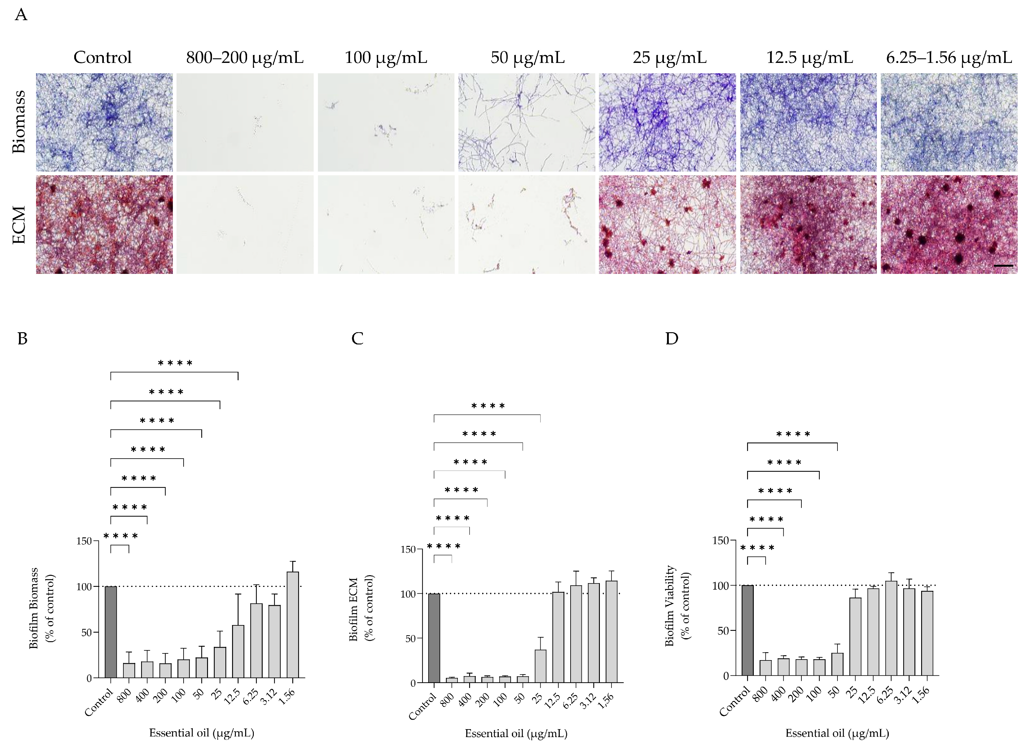
The effect of the essential oil on immature E. floccosum biofilms was evaluated by quantifying biomass formation, ECM deposition, and fungal viability across a concentration range from 800 to 1.56 µg/mL, allowing comparison of the antibiofilm activity across the tested concentration range. The essential oil exerted a concentration-dependent effect on biofilm development, with a differential response observed among the evaluated parameters. Representative microscopic images of biofilm structure across the tested concentrations are shown in Figure 2A, illustrating the progressive recovery of biofilm development at lower concentrations.
Regarding biofilm biomass a significant reduction was observed up to 25 µg/mL (reductions exceeding approximately 70%). Significant inhibitory effects were also observed at 25 and 12.5 µg/mL, with statistically significant differences compared to the untreated control. From 25 µg/mL onwards, a progressive recovery was observed, although values remained below the control (Figure 2B).
Interestingly, ECM was highly affected, with near-complete inhibition observed between 800 and 50 µg/mL (approximately 80–95% reduction). At 25 µg/mL, a significant reduction was still detected (~50% of the control), whereas at lower concentrations ECM progressively approached control values (Figure 2C).
Fungal cell viability also exhibited a concentration-dependent response, with a marked reduction observed up to 50 µg/mL. From 25 µg/mL and below, cell viability was similar to that observed in untreated biofilms (Figure 2D). Overall, L. stoechas subsp. luisieri essential oil demonstrated a concentration-dependent inhibitory effect on the development of immature E. floccosum biofilms, with ECM deposition being the most sensitive parameter.

3.3.2. Antifungal Effects of L. stoechas subsp. luisieri Essential Oil on Mature Biofilms

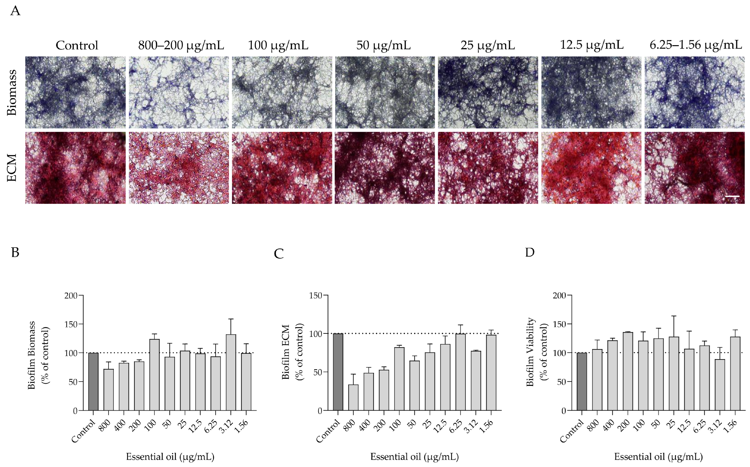
The potential of the essential oil to interfere with mature E. floccosum biofilms was evaluated using the same concentration range previously tested in the biofilm formation assay (800 to 1.56 µg/mL), allowing comparison of its efficacy at different stages of biofilm development. Overall, the essential oil exhibited limited activity in disrupting mature biofilms, with most evaluated parameters remaining close to control levels across the tested concentrations (Figure 3). Nevertheless, ECM deposition was the most affected parameter, showing a more evident reduction at higher concentrations, although a progressive recovery was observed at intermediate and lower concentrations (Figure 3C). On the other hand, biofilm biomass showed only a slight reduction at the highest concentration tested, whereas at the remaining conditions values were comparable to those of the control. Cell viability remained unchanged under all tested conditions, suggesting that the essential oil does not significantly impair the metabolic activity of cells within mature biofilms. Overall, these results indicate that L. stoechas subsp. luisieri essential oil has limited ability to disrupt mature E. floccosum biofilms, contrasting with the more pronounced inhibitory effects observed during the biofilm formation stage.

3.4. Potential of L. stoechas subsp. luisieri Essential Oil on an Ex Vivo Model of Trichophyton rubrum-Induced Dermatophytosis

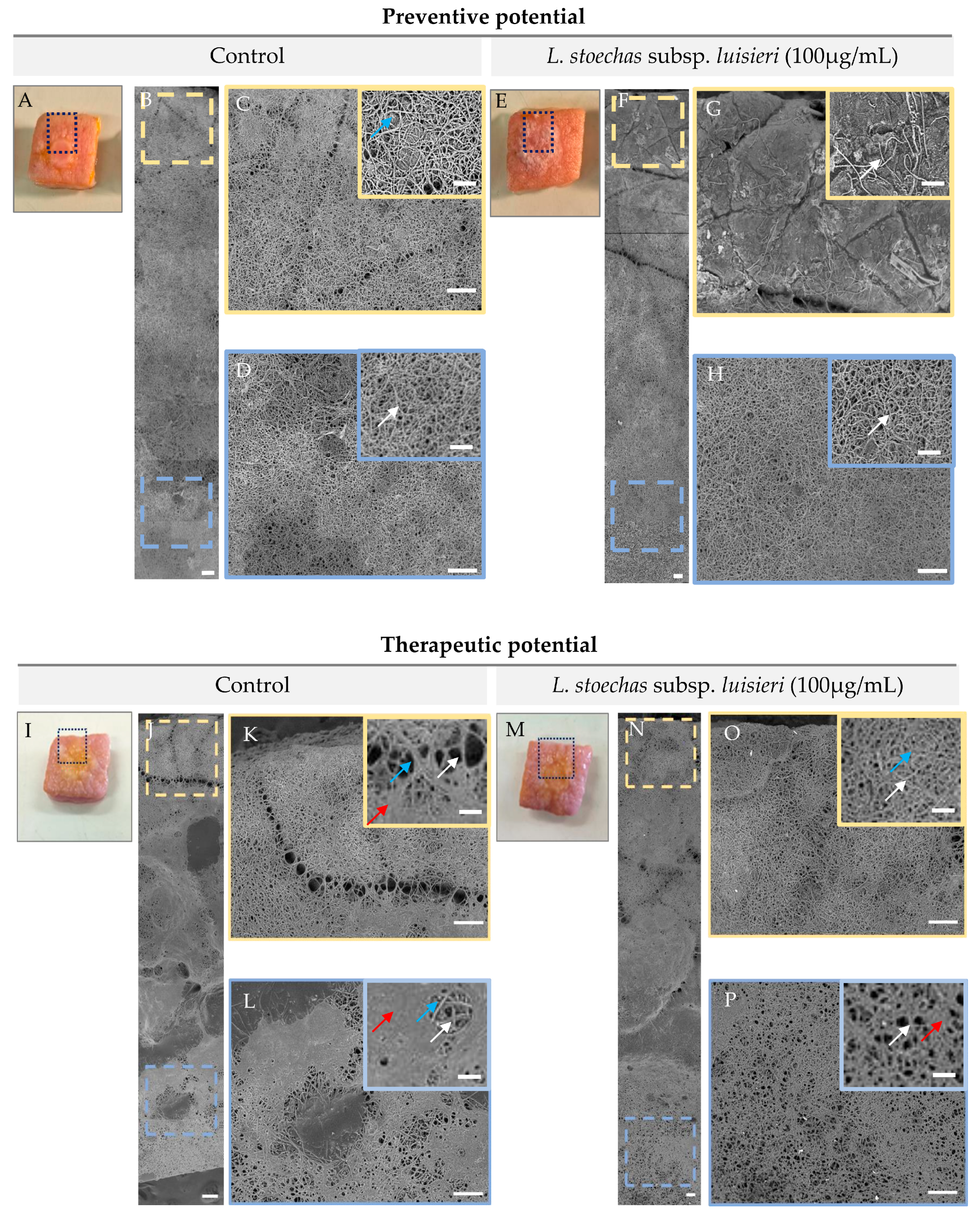
Two experimental approaches were conducted: a preventive assay, in which the essential oil was applied at the same time as infection, and a therapeutic assay, in which fungal growth was allowed to proceed for 10 days before the essential oil was added for an additional treatment period. In the first, macroscopic and microscopic analysis of the skin (Figure 4) revealed marked differences between control and treated samples. In untreated controls, a dense hyphal network was observed extending from the central infection site to the peripheral border regions of the skin, indicating extensive fungal dissemination across the tissue. In contrast, samples treated with the essential oil showed hyphal growth that remained dense in the central infected area but did not extend towards the surrounding border regions. This pattern suggests a reduction in fungal spread and an inhibitory effect on dissemination within the skin model.
In the second assay (Figure 4) although hyphal growth was still observed extending to the peripheral regions, a less pronounced extracellular matrix deposition was observed in the central infection site, suggesting a partial effect of the treatment on biofilm organization.

3.5. Safety Profile of L. stoechas subsp. luisieri Essential Oil on Skin Fibroblasts

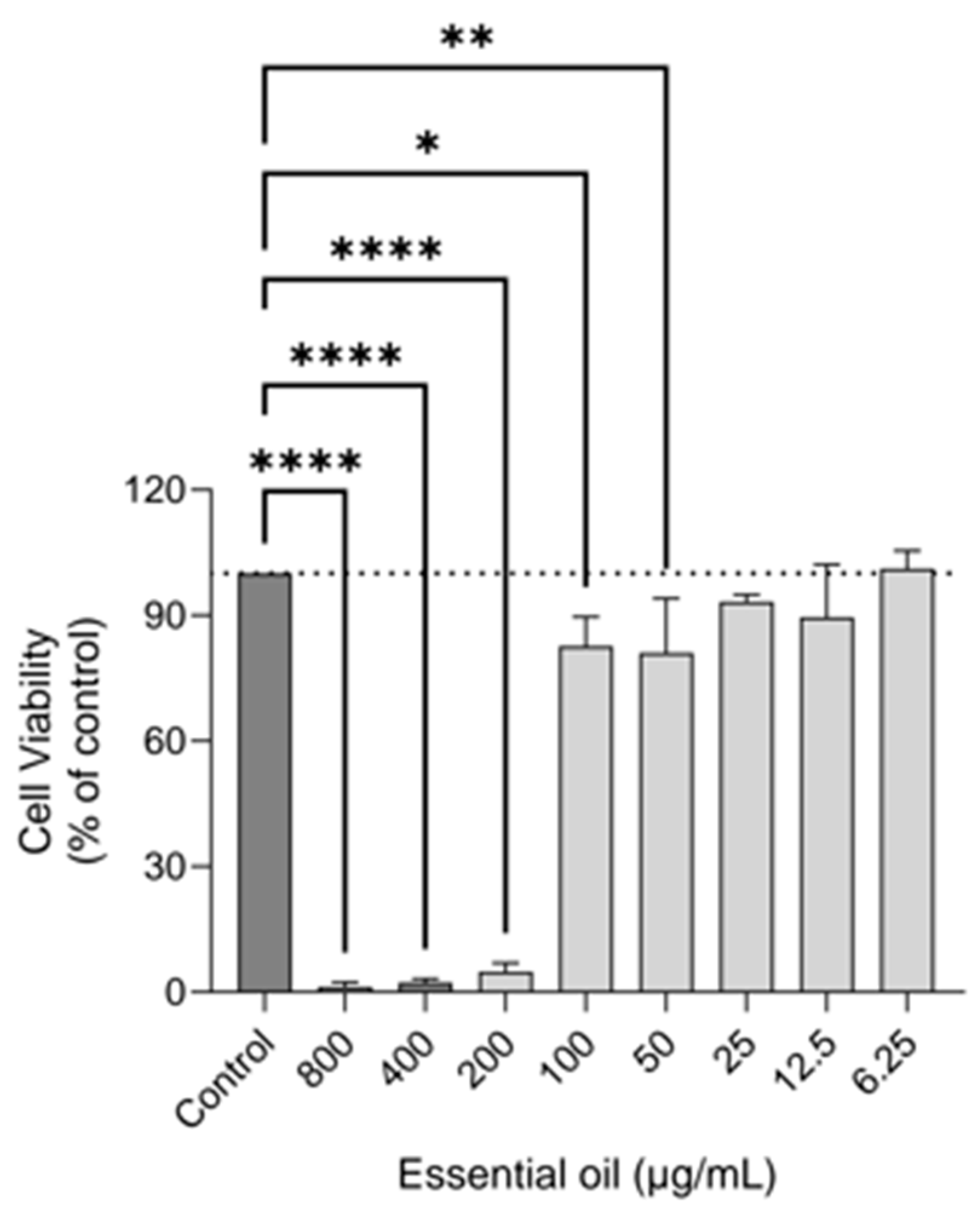
Cell viability was assessed using the resazurin assay after exposing 3T3 fibroblasts to the essential oil (800–6.25 µg/mL). Although statistically significant reductions were observed up to 50 µg/mL, cell viability remained above 80% at concentrations ≤100 µg/mL, which is indicative of low cytotoxicity. More pronounced, dose-dependent decreases in viability were only observed at higher concentrations (200–800 µg/mL; Figure 5).

4. Discussion

Species belonging to the Lavandula genus have long been recognized for their historical, medicinal, and economic importance, being widely used since antiquity for food preservation, flavoring, and the treatment of several health conditions, including wound healing and microbial infections [61,62]. The increasing global demand for lavender-derived products has intensified agronomic interest in their cultivation, particularly regarding the optimization of biomass production, essential oil yield, and chemical quality through breeding and cultivation strategies [63].
While some lavender species like Lavandula angustifolia are globally recognized and widely used in cosmetic, aromatherapy, and pharmaceutical products due to their well-established safety profile and favorable organoleptic properties [64], other species like L. stoechas subsp. luisieri remain comparatively underexplored. Indeed, this species is still relatively understudied despite its distinctive phytochemical composition, particularly its richness in necrodane derivatives associated with promising biological activities [65].
In the present study, chemical analysis revealed a predominance of oxygenated monoterpenes, particularly trans-α-necrodyl acetate, lavandulyl acetate, camphor, 1,8-cineole, and trans-α-necrodol. This composition is consistent with previous studies on this species, in which necrodane derivatives were identified as characteristic constituents of L. stoechas subsp. luisieri essential oil [47,48,66].
Interestingly, trans-α-necrodyl acetate, a major constituent of L. stoechas subsp. luisieri essential oil, has recently been identified as a minor sex pheromone component of the invasive mealybug Delottococcus aberiae, exhibiting male attractant activity with potential applications in citrus pest management [67]. This represents an example of a plant-derived secondary metabolite simultaneously functioning as an insect semiochemical [68], reinforcing the biochemical relevance of necrodyl derivatives and suggesting that this Lavandula species may constitute a viable botanical source for the isolation and further bioprospection of this compound.
Regarding antifungal activity, the essential oil exhibited MIC values ranging from 12.5 to 100 µg/mL and MFC values ranging from 12.5 to 200 µg/mL against the dermatophytes tested. Among the species evaluated, Trichophyton verrucosum showed the highest susceptibility, presenting identical MIC and MFC values (12.5 µg/mL), suggesting a particularly effective fungicidal action. High susceptibility was also observed for Epidermophyton floccosum and Trichophyton mentagrophytes var. interdigitale.
Compared with the standard antifungal drug, fluconazole, the antifungal activity of the essential oil against dermatophytes is particularly relevant [69]. Indeed, fluconazole has shown MIC values of 128 µg/mL against Microsporum canis FF1, M. gypseum CECT 2908, and Trichophyton mentagrophytes var. interdigitale CECT 2958, whereas the essential oil exhibited substantially lower MIC values ranging from 50 to 100 µg/mL for the same strains. Similarly, T. verrucosum CECT 2992 showed MIC and MLC values of 12.5 µg/mL for the essential oil, whereas fluconazole presented MIC values higher than 128 µg/mL, indicating lower susceptibility to the azole standard antifungal [69].
L. stoechas subsp. luisieri essential oil also demonstrated a relevant fungicidal profile, as evidenced by the MFC/MIC ratios obtained for most dermatophyte strains. According to the commonly accepted criterion in which MFC/MIC ratios ≤4 are indicative of fungicidal activity [70], the essential oil exhibited fungicidal effects against all tested dermatophytes. In particular, T. verrucosum CECT 2992 showed an MFC/MIC ratio of 1, with identical MIC and MLC values (12.5 µg/mL), while E. floccosum FF9 and T. mentagrophytes FF7 also presented ratios ranging from 1 to 2, supporting a fungicidal mechanism.
These findings are consistent with our previous study demonstrating that L. stoechas subsp. luisieri essential oil containing higher levels of trans-α-necrodyl acetate exhibited enhanced antifungal activity against dermatophytes, as evidenced by lower MIC values (0.16–0.32 µL mL−1) compared with samples containing lower concentrations of this compound [71]. Overall, these findings suggest that elevated levels of necrodyl derivatives in the essential oil, particularly trans-α-necrodyl acetate and trans-α-necrodol, may contribute not only to the antidermatophytic activity but also to the inhibitory effect on biofilm formation. Given that essential oils are complex mixtures comprising numerous constituents, this interpretation should be approached with caution, as the contribution of other compounds and potential synergistic or antagonistic interactions among them cannot be ruled out. Indeed, the biological activity of essential oils is generally regarded as the result of a combined action of several constituents rather than that of a single compound. Previous studies have suggested that synergistic interactions may play an important role in essential oils’ antidermatophytic effects [72,73,74]. In particular, essential oils rich in oxygenated monoterpenes have been consistently associated with antifungal activity against filamentous fungi, particularly due to their ability to interfere with fungal growth, hyphal organization, and cellular integrity [75,76,77]. Among these compounds, 1,8-cineole deserves particular attention due to its reported antifungal properties. Shahina et al. (2023) have demonstrated that this monoterpene can interfere with fungal cellular organization by affecting cytoskeletal dynamics and morphogenetic processes, which may impair hyphal development and reduce the ability of fungal cells to adhere and form structured biofilms [78].
The morphological alterations observed by optical microscopy were consistent with the results obtained in the susceptibility assays. Exposure to the essential oil led to a progressive reduction in mycelial development, particularly evident at the MICs, where marked inhibition of fungal growth and loss of the typical organization of hyphal structures were observed. These findings corroborate the antifungal activity demonstrated by the MIC and MFC assays and indicate a concentration-dependent response. Since hyphal organization is required for dermatophyte colonization and invasion of keratinized tissues [79,80,81], the structural disorganization observed at the MICs may also reflect reduced fungal virulence and proliferation capacity.
The ability of dermatophytes to form biofilms has been widely recognized as an important factor associated with infection persistence and reduced efficacy of conventional antifungal treatments [10,82,83,84]. Epidermophyton floccosum was selected as the experimental model because it is a clinically relevant dermatophyte with a well-documented ability to form homogeneous and reproducible biofilms in vitro, making it particularly suitable for standardized antibiofilm assays [85,86]. As this was a proof-of-concept study, the antibiofilm activity of L. stoechas subsp. luisieri essential oil was evaluated in a single, well-characterized dermatophyte model. Therefore, these findings should be interpreted specifically for E. floccosum, and future studies should determine whether similar effects are observed in other clinically relevant dermatophyte species.
The results demonstrated that L. stoechas subsp. luisieri essential oil exerted a marked effect during the biofilm formation stage. ECM deposition was the most affected parameter, followed by biomass and cell viability. Between 800 and 50 µg/mL, substantial reductions in these parameters were observed, highlighting the ability of the essential oil to interfere with the early stages of biofilm development. These findings suggest that the essential oil may be particularly effective in preventing the establishment of these organized fungal communities. However, the antibiofilm effects did not directly correlate with the MIC values, as MIC assays evaluate inhibition of planktonic fungal growth, whereas biofilm assays assess more complex processes, including biomass accumulation, ECM production, and biofilm-associated cell viability. In the present study, inhibition of biofilm formation required slightly higher concentrations than those affecting planktonic growth, which is consistent with the increased tolerance of biofilm-associated cells and the structural complexity of fungal biofilms. Therefore, the antifungal activity observed in planktonic conditions cannot be directly extrapolated to predict antibiofilm efficacy.
In contrast, the activity observed against mature biofilms was considerably lower. Although some reduction in ECM deposition was detected at the highest concentrations tested, the effects on biomass and cell viability were limited. A similar trend has been described for dermatophyte biofilms, in which established biofilms exhibit lower susceptibility to antifungal agents than developing biofilms [10,87]. These results therefore reinforce the importance of intervention during the initial stages of fungal colonization, before biofilm-associated mechanisms are fully established.
The lower susceptibility observed in mature biofilms may be partially explained by the presence of a structured extracellular matrix, which acts as a physical barrier limiting the penetration of antifungal compounds [88,89]. Furthermore, biofilm-associated stress-response mechanisms have been reported to contribute to the increased tolerance of mature fungal biofilms to antimicrobial agents [90,91,92]. This pattern is consistent with observations that plant essential oils generally exert greater efficacy during the preventive stage of biofilm development than in the therapeutic disruption of already structured biofilms [26,59,93].
To the best of our knowledge, no studies have specifically evaluated the antibiofilm activity of L. stoechas subsp. luisieri essential oil against dermatophytes. Nevertheless, recent studies have shown that other species of the genus Lavandula are capable of interfering with microbial biofilm formation. Alves-Silva et al. [52] reported significant antibiofilm activity for Lavandula multifida essential oil, highlighting the potential of this botanical genus as a source of bioactive compounds for the prevention of biofilm-associated infections. Similarly, Santos et al. [94] demonstrated that Lavandula dentata essential oil exhibited greater efficacy during the early stages of biofilm development than against mature biofilms, with significant inhibition of biofilm formation observed at concentrations ranging from MIC/2 to MIC × 2, whereas a substantially higher concentration (MIC × 5) was required to affect pre-formed biofilms.
Therefore, the present findings contribute to expanding the current knowledge regarding the antifungal potential of L. stoechas subsp. luisieri, particularly in the context of dermatophyte biofilms. Although obtained under in vitro conditions, the ability of the essential oil to interfere with biofilm establishment on inanimate surfaces suggests potential applications beyond direct antifungal therapy. Previous works have demonstrated that essential oils can be incorporated into antifungal coatings for medical devices and hospital-contact abiotic surfaces, reducing risks of fungal biofilm colonization [95,96,97,98], which is of particular interest in the prevention of hospital-associated infections.
In the ex vivo skin infection model, T. rubrum was selected due to its clinical relevance as the predominant causative agent of human dermatophytosis and onychomycosis, as well as its well-established ability to colonize keratinized tissues. Overall, L. stoechas subsp. luisieri essential oil demonstrated a clear impact on fungal behavior within a tissue-relevant environment. In the preventive approach, the oil effectively limited radial dissemination of T. rubrum across the epidermal surface, with hyphal growth largely confined to the central inoculation site, suggesting interference with early colonization events and fungal propagation. In contrast, in the therapeutic model, where infection was already established, the essential oil showed a more limited effect on fungal extension, although a noticeable reduction in ECM deposition was observed at the infection site. Importantly, these effects occurred at concentrations that were non-cytotoxic to mammalian fibroblasts, supporting the safety profile of the essential oil. Together, these findings reinforce the relevance of early intervention and suggest higher efficacy in preventing rather than reversing established dermatophytic infection.
Although human skin was available and employed in the present study, porcine skin models have been widely used in dermatological research due to the limited availability of human samples and ethical constraints associated with human experimentation [99]. However, the use of human ex vivo skin is particularly relevant for T. rubrum, an anthropophilic dermatophyte adapted to human hosts, as it better reproduces its natural infection environment [100]. Current evidence on essential oils in antifungal therapy remains largely restricted to in vitro studies, whereas ex vivo and in vivo models are still scarce [101]. Therefore, the present study addresses this gap by demonstrating, in a human ex vivo model, both preventive effects on fungal dissemination and partial therapeutic effects on established infection.
Although Prasad et al. [102] used an in vivo guinea pig model, comparison remains relevant, particularly regarding fungal maturity and treatment response. In the present study, therapy was initiated after 10 days of fungal growth, when dense hyphal networks and ECM deposition were already established, conditions associated with increased tolerance. In contrast, Prasad et al. [102] initiated treatment earlier and maintained therapy for longer periods, which may explain their stronger antifungal response.
Importantly, the concentration of 100 µg/mL did not exhibit cytotoxic effects in fibroblasts while maintaining antifungal activity, highlighting the potential of L. stoechas subsp. luisieri essential oil as a promising pharmaceutical ingredient. These results partially contrast with previous reports of higher cytotoxicity in NHDF cells exposed to oils rich in necrodane derivatives, such as trans-α-necrodyl acetate [45]. In the present study, despite the presence of this compound (≈19%), no significant reduction in fibroblast viability was observed, which may be attributed to differences in chemical composition, cell models, and methodological approaches (resazurin vs. MTT assays). Although 3T3 fibroblasts represent a relevant mammalian model for cytotoxicity evaluation, further studies using human skin-derived cell models would be valuable to confirm its safety for topical applications.
The results obtained demonstrate that L. stoechas subsp. luisieri essential oil possesses relevant antidermatophytic activity, particularly during the early stages of biofilm development. The combination of fungal growth inhibition and interference with biofilm formation further supports the interest in this Iberian endemic species as a source of bioactive compounds with potential relevance for the management of dermatophytosis. Although fluconazole was used as the reference antifungal to enable comparison with previously published susceptibility data for the same dermatophyte strains, future studies should include first-line antifungal agents, particularly terbinafine and itraconazole, to provide a more clinically relevant assessment of the therapeutic potential of L. stoechas subsp. luisieri essential oil.
Nevertheless, despite these promising biological properties and their potential therapeutic applications, the translation of essential oils from laboratory research to large-scale industrial use still faces important technological and economic challenges. Their chemical composition is strongly influenced by factors such as geographical origin, climatic conditions, harvesting season, and extraction methods, which may compromise batch-to-batch reproducibility and biological consistency [103]. Furthermore, essential oils are highly volatile and susceptible to oxidation and degradation by light, oxygen, and temperature, reducing their stability during storage and formulation [104,105,106]. An additional limitation is the relatively low essential oil yield, which may increase production costs and hinder large-scale manufacturing [107,108]. Therefore, standardized cultivation and extraction procedures, as well as quality control protocols, together with comprehensive toxicological and stability studies, remain essential before these natural products can be widely implemented in industrial settings.
The industrial application of essential oils for biofilm control faces further challenges, including regulatory constraints and the scarcity of studies evaluating their efficacy under real industrial conditions. Factors such as scalability, long-term stability, compatibility with manufacturing processes, and the reproducibility of antibiofilm activity must still be thoroughly investigated. Consequently, although essential oils represent promising alternatives for controlling microbial biofilms, further technological development and validation studies are required to support their widespread industrial adoption.
Future studies incorporating objective metrics for fungal burden assessment, together with in vivo models of dermatophytosis, will be important to further understand the antifungal potential of L. stoechas subsp. luisieri essential oil.

5. Conclusions

The essential oil of L. stoechas subsp. luisieri displays relevant antidermatophytic activity against all tested dermatophytes, as well as the ability to interfere with immature biofilm formation. The observed activity was superior to fluconazole for several strains, supporting the potential of this oil as a natural alternative for dermatophytosis treatment, aligning with the traditional use of lavender species for skin care, and wound healing.
The phytochemical profile, dominated by oxygenated monoterpenes, particularly necrodyl acetate and necrodol, confirms the chemical uniqueness of the species and suggests that these constituents contribute to its biological activity. Morphological alterations in hyphae and inhibition of the biofilm ECM indicate mechanisms of action that affect both fungal viability and colonization capacity.
Moreover, this study demonstrates that the essential oil exerts relevant antifungal effects in a skin infection model, with a clear impact on fungal dissemination in the preventive approach and a partial but notable effect on biofilm architecture in the post-infection setting. Importantly, these effects were observed at concentrations that were considered safe, as they did not compromise fibroblast viability. Together, these findings highlight the potential of the essential oil as a promising natural agent for the control of dermatophytic infections, particularly in the early stages of infection and in limiting tissue colonization.
Despite the economic and scientific potential for phytocosmetic and dermatological products, further studies to translate its traditional skin applications into drug delivery systems are warranted. Future research should focus on formulation development, stability, and safety assessments, including incorporation into systems such as cyclodextrin structures, nanotechnology-based carriers, and other topical delivery platforms to enhance efficacy, controlled release, and skin penetration for health applications. In addition, the combination of essential oils with conventional antifungal drugs or nanoparticle-based systems represents a promising strategy to improve therapeutic efficacy and facilitate their pharmaceutical and industrial application.

Author Contributions

Conceptualization, L.S. and M.Z.; methodology, T.M. and I.L.S.; validation, M.Z.; investigation, T.M. and I.L.S.; resources, L.S. and M.Z.; writing—original draft preparation, T.M.; writing—review and editing, I.L.S., L.S. and M.Z.; supervision, L.S. and M.Z.; funding acquisition, L.S. and M.Z. All authors have read and agreed to the published version of the manuscript.

Funding

This work was supported by national funds through FCT—Fundação para a Ciência e a Tecnologia, I.P., under Base and Programmatic Funding (UID/00102/2025; https://doi.org/10.54499/UID/00102/2025), the strategic project of the Center for Innovative Biomedicine and Biotechnology (CIBB) (UID/04539/2025; https://doi.org/10.54499/UID/04539/2025), the Recovery and Resilience Plan (PRR) (UID/PRR/00102/2025; https://doi.org/10.54499/UID/PRR/00102/2025). Additional funding was provided by the Promove Program of the “la Caixa” Foundation, in collaboration with BPI and the Foundation for Science and Technology (FCT) under the project VITAGE (Ref. PD25-00049). Support from PPBI-Portuguese Platform of BioImaging: POCI-01–0145-FEDER-022122 was also provided.

Data Availability Statement

The original contributions presented in this study are included in the article.

Acknowledgments

The authors thank Joana Domingues (Centre of Beira Interior, Castelo Branco, Portugal) and Fernanda Delgado (Polytechnic University of Castelo Branco-School of Agriculture, Castelo Branco, Portugal) for providing the essential oil used in this study.

Conflicts of Interest

The authors declare no conflicts of interest.

Abbreviations

The following abbreviations are used in this manuscript:
CECTColección Española de Cultivos Tipo
CFUsColony-Forming Units
DMEMDulbecco’s Modified Eagle Medium
DMSODimethyl Sulfoxide
ECMExtracellular Matrix
EPSsExtracellular Polymeric Substances
EUCASTEuropean Committee on Antimicrobial Susceptibility Testing
FBSFetal Bovine Serum
GC–MSGas Chromatography–Mass Spectrometry
MFCMinimum Fungicidal Concentration
MICMinimum Inhibitory Concentration
MTT3-(4,5-Dimethylthiazol-2-yl)-2,5-Diphenyltetrazolium Bromide
NHDFsNormal Human Dermal Fibroblasts
OCTOptimal Cutting Temperature
PASPeriodic Acid–Schiff
PBSPhosphate-Buffered Saline
PDAPotato Dextrose Agar
RIRetention Index
SDASabouraud Dextrose A
SEMScanning Electron Microscopy
XTT2,3-Bis(2-Methoxy-4-Nitro-5-Sulfophenyl)-2H-Tetrazolium-5-Carboxanilide

References

  1. Wang, H.; Sun, F.; Wang, C.; Ye, J.; Xia, P.; Wang, W.; Wu, Y. A Systematic Analysis of the Global, Regional, and National Burden of Fungal Skin Diseases from 1990 to 2021. Front. Epidemiol. 2024, 4, 1489148. [Google Scholar] [CrossRef] [Scilit] [PubMed]
  2. Barac, A.; Stjepanovic, M.; Krajisnik, S.; Stevanovic, G.; Paglietti, B.; Milosevic, B. Dermatophytes: Update on Clinical Epidemiology and Treatment. Mycopathologia 2024, 189, 101. [Google Scholar] [CrossRef] [Scilit] [PubMed]
  3. Hill, R.C.; Caplan, A.S.; Elewski, B.; Gold, J.A.W.; Lockhart, S.R.; Smith, D.J.; Lipner, S.R. Expert Panel Review of Skin and Hair Dermatophytoses in an Era of Antifungal Resistance. Am. J. Clin. Dermatol. 2024, 25, 359–389. [Google Scholar] [CrossRef] [Scilit] [PubMed]
  4. Valdez-Martinez, A.; Arenas, R.; Moreno-Salinas, A.; Perez-Tristan, M.; Gomez-Rico, M.J.; Torres-Olguín, I.; Fuentes-Venado, C.E.; Bastida-González, F.; Martínez-Herrera, E.; Pinto-Almazán, R. Resistance and Recalcitrance in Dermatophytosis: Mechanistic and Clinical Considerations for Keratinized Tissues. Antibiotics 2026, 15, 634. [Google Scholar] [CrossRef] [Scilit] [PubMed]
  5. Arantes, B.B.A.; Cabral, A.K.L.F.; dos Santos, K.S.; Mendonça, M.B.; dos Santos, R.C.; Bugalho, B.C.M.; Fernandes, L.D.S.; Martinez, L.R.; Fusco-Almeida, A.M.; Mendes-Giannini, M.J.S. Characterization of Biofilm Formation by the Dermatophyte Nannizzia gypsea. J. Fungi 2025, 11, 455. [Google Scholar] [CrossRef] [Scilit] [PubMed]
  6. Gupta, A.K.; Elewski, B.; Joseph, W.S.; Lipner, S.R.; Daniel, C.R.; Tosti, A.; Guenin, E.; Ghannoum, M. Treatment of Onychomycosis in an Era of Antifungal Resistance: Role for Antifungal Stewardship and Topical Antifungal Agents. Mycoses 2024, 67, e13683. [Google Scholar] [CrossRef] [Scilit] [PubMed]
  7. Cañete-Gibas, C.F.; Mele, J.; Patterson, H.P.; Sanders, C.J.; Ferrer, D.; Garcia, V.; Fan, H.; David, M.; Wiederhold, N.P. Terbinafine-Resistant Dermatophytes and the Presence of Trichophyton indotineae In North America. J. Clin. Microbiol. 2023, 61, e0056223. [Google Scholar] [CrossRef] [Scilit] [PubMed]
  8. Brescini, L.; Fioriti, S.; Morroni, G.; Barchiesi, F. Antifungal Combinations in Dermatophytes. J. Fungi 2021, 7, 727. [Google Scholar] [CrossRef] [Scilit] [PubMed]
  9. Kruithoff, C.; Gamal, A.; McCormick, T.S.; Ghannoum, M.A. Dermatophyte Infections Worldwide: Increase in Incidence and Associated Antifungal Resistance. Life 2024, 14, 1. [Google Scholar] [CrossRef] [Scilit] [PubMed]
  10. Markantonatou, A.M.; Samaras, K.; Vyzantiadis, T.A. Dermatophytic Biofilms: Characteristics, Significance and Treatment Approaches. J. Fungi 2023, 9, 228. [Google Scholar] [CrossRef] [Scilit] [PubMed]
  11. Pruitt, H.M.; Zhu, J.C.; Riley, S.P.; Shi, M. The Hidden Fortress: A Comprehensive Review of Fungal Biofilms with Emphasis on Cryptococcus neoformans. J. Fungi 2025, 11, 236. [Google Scholar] [CrossRef] [Scilit] [PubMed]
  12. Mannan, M.; Nabeela, S.; Mishra, R.; Uppuluri, P. Host Immune Response against Fungal Biofilms. Curr. Opin. Microbiol. 2024, 81, 102520. [Google Scholar] [CrossRef] [Scilit] [PubMed]
  13. Balducci, E.; Papi, F.; Capialbi, D.E.; Del Bino, L. Polysaccharides’ Structures and Functions in Biofilm Architecture of Antimicrobial-Resistant (AMR) Pathogens. Int. J. Mol. Sci. 2023, 24, 4030. [Google Scholar] [CrossRef] [Scilit] [PubMed]
  14. Le, P.H.; Linklater, D.P.; Medina, A.A.; MacLaughlin, S.; Crawford, R.J.; Ivanova, E.P. Impact of Multiscale Surface Topography Characteristics on Candida albicans Biofilm Formation: From Cell Repellence to Fungicidal Activity. Acta Biomater. 2024, 177, 20–36. [Google Scholar] [CrossRef] [Scilit] [PubMed]
  15. Tuan, D.A.; Uyen, P.V.N. Mechanisms and Species-Specific Patterns of ECM-Mediated Antifungal Resistance in Candida Biofilms: A Systematic Review and Exploratory Quantitative Synthesis. Future Microbiol. 2026, 21, 645–656. [Google Scholar] [CrossRef] [Scilit] [PubMed]
  16. Rehman, A.; Ismail, F.; Safdar, A.; Imran, M. In Vitro Characterization of Trichophyton rubrum Biofilm by Combined Anti-Biofilm Enzymes. PLoS ONE 2025, 20, e0331291. [Google Scholar] [CrossRef] [Scilit] [PubMed]
  17. Gupta, A.K.; Wang, T.; Piguet, V.; Chowdhary, A. New Insights into Host–Dermatophyte Interactions: Pathogenesis, Host Defense Mechanisms, and Emerging Clinical Challenges. Curr. Opin. Microbiol. 2026, 90, 102711. [Google Scholar] [CrossRef] [Scilit] [PubMed]
  18. Baldo, A.; Monod, M.; Mathy, A.; Cambier, L.; Bagut, E.T.; Defaweux, V.; Symoens, F.; Antoine, N.; Mignon, B. Mechanisms of Skin Adherence and Invasion by Dermatophytes. Mycoses 2012, 55, 218–223. [Google Scholar] [CrossRef] [Scilit] [PubMed]
  19. Faway, É.; Lambert de Rouvroit, C.; Poumay, Y. In Vitro Models of Dermatophyte Infection to Investigate Epidermal Barrier Alterations. Exp. Dermatol. 2018, 27, 915–922. [Google Scholar] [CrossRef] [Scilit] [PubMed]
  20. Duek, L.; Kaufman, G.; Ulman, Y.; Berdicevsky, I. The Pathogenesis of Dermatophyte Infections in Human Skin Sections. J. Infect. 2004, 48, 175–180. [Google Scholar] [CrossRef] [Scilit] [PubMed]
  21. Martínez-Herrera, E.; Moreno-Coutiño, G.; Fuentes-Venado, C.E.; Hernández-Castro, R.; Arenas, R.; Pinto-Almazán, R.; Rodríguez-Cerdeira, C. Main Phenotypic Virulence Factors Identified in Trichophyton rubrum. J. Biol. Regul. Homeost. Agents 2023, 37, 2345–2356. [Google Scholar] [CrossRef] [Scilit]
  22. Ruffin, P.; Andrieu, S.; Biserte, G.; Biguet, J. Sulphitolysis in Keratinolysis. Biochem. Proof. Med. Mycol. 1976, 14, 181–184. [Google Scholar] [CrossRef] [Scilit] [PubMed]
  23. Mercer, D.K.; Stewart, C.S. Keratin Hydrolysis by Dermatophytes. Med. Mycol. 2019, 57, 13–22. [Google Scholar] [CrossRef] [Scilit] [PubMed]
  24. Deng, R.; Wang, X.; Li, R. Dermatophyte Infection: From Fungal Pathogenicity to Host Immune Responses. Front. Immunol. 2023, 14, 1285887. [Google Scholar] [CrossRef] [Scilit] [PubMed]
  25. Das, A.; Dutta, B.; Padovese, V.; Hay, R.J.; Khan, S.S. The Changing Face of Dermatophytosis: From Common Skin Disease to Emerging Resistant Epidemic. Clin. Exp. Dermatol. 2026, 51, 805–813. [Google Scholar] [CrossRef] [Scilit] [PubMed]
  26. Alves-Silva, J.M.; Gonçalves, M.J.; Silva, A.; Cavaleiro, C.; Cruz, M.T.; Salgueiro, L. Chemical Profile, Anti-Microbial and Anti-Inflammaging Activities of Santolina rosmarinifolia L. Essential Oil from Portugal. Antibiotics 2023, 12, 179. [Google Scholar] [CrossRef] [Scilit] [PubMed]
  27. Gündüz, M.; Bekteş, A.; Karabıyıklı Çiçek, Ş.; Kilinç, V. Bioactive Properties and Mechanisms of Effect of Phenolic Compounds. Manas J. Agric. Vet. Life Sci. 2025, 15, 146–160. [Google Scholar] [CrossRef] [Scilit]
  28. Dantas, T.d.S.; Machado, J.C.B.; Ferreira, M.R.A.; Soares, L.A.L. Bioactive Plant Compounds as Alternatives Against Antifungal Resistance in the Candida Strains. Pharmaceutics 2025, 17, 687. [Google Scholar] [CrossRef] [Scilit] [PubMed]
  29. Torres Tediole, G.; Torres Resende, M.E.; De Castro Costa, A.B.; Duarte-Almeida, J.M.; Paiva, M.C. Promising Potential of the Antifungal Action of Essential Oils and Its Challenges: A Systematic Review. Rev. Colomb. Cienc. Quím.-Farm. 2025, 54, 442–459. [Google Scholar] [CrossRef] [Scilit]
  30. Li, K.; Guo, J.; Liang, R.; Wang, X.; Chen, Q.; Wang, J.; Ge, X.; Chen, C.; Sun, J.; Zhao, C.; et al. A Review on Antifungal Activity of Plant Essential Oils. Phytother. Res. 2025, 39, 3736–3761. [Google Scholar] [CrossRef] [Scilit] [PubMed]
  31. Jeyachandran, S.; Sekar, S. Natural Anti-Biofilm Agents: A Comprehensive Review and Future Perspectives. Curr. Microbiol. 2026, 83, 115. [Google Scholar] [CrossRef] [Scilit] [PubMed]
  32. de Sousa, D.P.; Damasceno, R.O.S.; Amorati, R.; Elshabrawy, H.A.; de Castro, R.D.; Bezerra, D.P.; Nunes, V.R.V.; Gomes, R.C.; Lima, T.C. Essential Oils: Chemistry and Pharmacological Activities. Biomolecules 2023, 13, 1144. [Google Scholar] [CrossRef] [Scilit] [PubMed]
  33. Nurzyńska-Wierdak, R. Antibiofilm Potential of Natural Essential Oils. Appl. Sci. 2025, 15, 5847. [Google Scholar] [CrossRef] [Scilit]
  34. Soulaimani, B. Comprehensive Review of the Combined Antimicrobial Activity of Essential Oil Mixtures and Synergism with Conventional Antimicrobials. Nat. Prod. Commun. 2025, 20, 1–22. [Google Scholar] [CrossRef] [Scilit]
  35. Ben Miri, Y. Essential Oils: Chemical Composition and Diverse Biological Activities: A Comprehensive Review. Nat. Prod. Commun. 2025, 20, 1–29. [Google Scholar] [CrossRef] [Scilit]
  36. Tanasă, F.; Nechifor, M.; Teacă, C.A. Essential Oils as Alternative Green Broad-Spectrum Biocides. Plants 2024, 13, 3442. [Google Scholar] [CrossRef] [Scilit] [PubMed]
  37. Diogo Gonçalves, S.; Paiva-Cardoso, M.d.N.; Caramelo, A. Green Preservation Strategies: The Role of Essential Oils in Sustainable Food Preservatives. Sustainability 2025, 17, 7326. [Google Scholar] [CrossRef] [Scilit]
  38. Yusupova, U.; Tojiboeva, D.; Mukhamatkhanova, R.; Dusmatova, D. Essential Oils as an Alternative to Synthetic Antiseptics in Cosmetics and Personal Care Products. Chem. Biodivers. 2026, 23, e03449. [Google Scholar] [CrossRef] [Scilit] [PubMed]
  39. Héral, B.; Stierlin, É.; Fernandez, X.; Michel, T. Phytochemicals from the Genus Lavandula: A Review. Phytochem. Rev. 2021, 20, 751–771. [Google Scholar] [CrossRef] [Scilit]
  40. Habán, M.; Korczyk-Szabó, J.; Čerteková, S.; Ražná, K. Lavandula Species, Their Bioactive Phytochemicals, and Their Biosynthetic Regulation. Int. J. Mol. Sci. 2023, 24, 8831. [Google Scholar] [CrossRef] [Scilit] [PubMed]
  41. Dobros, N.; Zawada, K.D.; Paradowska, K. Phytochemical Profiling, Antioxidant and Anti-Inflammatory Activity of Plants Belonging to the Lavandula Genus. Molecules 2023, 28, 256. [Google Scholar] [CrossRef] [Scilit] [PubMed]
  42. Salehi, B.; Mnayer, D.; Özçelik, B.; Altin, G.; Kasapoǧlu, K.N.; Daskaya-Dikmen, C.; Sharifi-Rad, M.; Selamoglu, Z.; Acharya, K.; Sen, S.; et al. Plants of the Genus Lavandula: From Farm to Pharmacy. Nat. Prod. Commun. 2018, 13. [Google Scholar] [CrossRef] [Scilit]
  43. Julio, L.F.; Barrero, A.F.; Herrador Del Pino, M.M.; Arteaga, J.F.; Burillo, J.; Andres, M.F.; Díaz, C.E.; González-Coloma, A. Phytotoxic and Nematicidal Components of Lavandula luisieri. J. Nat. Prod. 2016, 79, 261–266. [Google Scholar] [CrossRef] [Scilit] [PubMed]
  44. Baldovini, N.; Lavoine-Hanneguelle, S.; Ferrando, G.; Dusart, G.; Lizzani-Cuvelier, L. Necrodane Monoterpenoids from Lavandula luisieri. Phytochemistry 2005, 66, 1651–1655. [Google Scholar] [CrossRef] [Scilit] [PubMed]
  45. Domingues, J.; Goulão, M.; Delgado, F.; Gonçalves, J.C.; Gonçalves, J.; Pintado, C.S. Essential Oils of Two Portuguese Endemic Species of Lavandula as a Source of Antifungal and Antibacterial Agents. Processes 2023, 11, 1165. [Google Scholar] [CrossRef] [Scilit]
  46. Baptista, R.; Madureira, A.M.; Jorge, R.; Adão, R.; Duarte, A.; Duarte, N.; Lopes, M.M.; Teixeira, G. Antioxidant and Antimycotic Activities of Two Native Lavandula Species from Portugal. Evid.-Based Complement. Altern. Med. 2015, 2015, 570521. [Google Scholar] [CrossRef] [Scilit] [PubMed]
  47. Domingues, J.; Delgado, F.; Gonçalves, J.C. Cristina Santos Pintado. Essential Oils of Lavandula stoechas subsp. luisieri as Antifungal Agent against Fungi from Strawberry Tree Fruit. J. Pharm. Pharmacol. 2021, 9, 98–106. [Google Scholar] [CrossRef] [Scilit]
  48. Arantes, S.; Candeias, F.; Lopes, O.; Lima, M.; Pereira, M.; Tinoco, T.; Cruz-Morais, J.; Martins, M.R. Pharmacological and Toxicological Studies of Essential Oil of Lavandula stoechas subsp. luisieri. Planta Med. 2016, 82, 1266–1273. [Google Scholar] [CrossRef] [Scilit] [PubMed]
  49. Pombal, S.; Rodrigues, C.F.; Araújo, J.P.; Rocha, P.M.; Rodilla, J.M.; Diez, D.; Granja, Á.P.; Gomes, A.C.; Silva, L.A. Antibacterial and Antioxidant Activity of Portuguese Lavandula luisieri (Rozeira) Rivas-Martinez and Its Relation with Their Chemical Composition. Springerplus 2016, 5, 1711. [Google Scholar] [CrossRef] [Scilit] [PubMed][Green Version]
  50. Zuzarte, M.; Sousa, C.; Cavaleiro, C.; Cruz, M.T.; Salgueiro, L. The Anti-Inflammatory Response of Lavandula luisieri and Lavandula pedunculata Essential Oils. Plants 2022, 11, 3. [Google Scholar] [CrossRef] [Scilit] [PubMed]
  51. Novais, M.H.; Santos, I.; Mendes, S.; Pinto-Gomes, C. Studies on Pharmaceutical Ethnobotany in Arrabida Natural Park (Portugal). J. Ethnopharmacol. 2004, 93, 183–195. [Google Scholar] [CrossRef] [Scilit] [PubMed]
  52. Alves-Silva, J.; Zuzarte, M.; Cavaleiro, C.; Salgueiro, L. Antibiofilm Effect of Lavandula multifida Essential Oil: A New Approach for Chronic Infections. Pharmaceutics 2023, 15, 2142. [Google Scholar] [CrossRef] [Scilit] [PubMed]
  53. Castagliuolo, G.; Badalamenti, N.; Ilardi, V.; Fontana, G.; Antonini, D.; Varcamonti, M.; Bruno, M.; Zanfardino, A. Lavandula pedunculata subsp. atlantica: A Multifunctional Essential Oil for Potentially Combating Microbial Infections and Inflammatory Processes. Molecules 2025, 30, 2267. [Google Scholar] [CrossRef] [Scilit] [PubMed]
  54. El-Naggar, N.E.A.; Eltarahony, M.; Hafez, E.E.; Bashir, S.I. Green Fabrication of Chitosan Nanoparticles Using Lavendula angustifolia, Optimization, Characterization and In-vitro Antibiofilm Activity. Sci. Rep. 2023, 13, 11127. [Google Scholar] [CrossRef] [Scilit] [PubMed]
  55. European Directorate for the Quality of Medicines & HealthCare (EDQM). European Pharmacopoeia, 11th ed.; Council of Europe: Strasbourg, France, 2023. [Google Scholar]
  56. Alves-Silva, J.M.; Domingues, J.; Delgado, F.; Resina, T.R.; Cruz, M.T.; Salgueiro, L.; Girão, H.; Zuzarte, M. Targeting Skin Aging: Anti-Senescent Potential and Key Enzyme Modulation of Lavandula Essential Oils. Front. Pharmacol. 2026, 17, 1796715. [Google Scholar] [CrossRef]
  57. Arendrup, M.C.; Kahlmeter, G.; Guinea, J.; Meletiadis, J.; Arendrup, M.C.; Meletiadis, J.; Guinea, J.; Kahlmeter, G.; Arikan-Akdagli, S.; Friberg, F.; et al. How to: Perform Antifungal Susceptibility Testing of Microconidia-Forming Dermatophytes Following the New Reference EUCAST Method E.Def 11.0, Exemplified by Trichophyton. Clin. Microbiol. Infect. 2021, 27, 55–60. [Google Scholar] [CrossRef] [Scilit] [PubMed]
  58. Ali, I.; Satti, N.K.; Dutt, P.; Prasad, R.; Khan, I.A. Hydroxychavicol: A Phytochemical Targeting Cutaneous Fungal Infections. Sci. Rep. 2016, 6, 37867. [Google Scholar] [CrossRef] [Scilit] [PubMed]
  59. Soares, I.L.; Sá, K.; Canuto, K.M.; Bandeira, M.A.; Salgueiro, L.; Zuzarte, M. Multifunctional Activity of Lippia gracilis Schauer Essential Oil Against Skin Infections. Plants 2026, 15, 1681. [Google Scholar] [CrossRef] [Scilit] [PubMed]
  60. Corzo-León, D.E.; Munro, C.A.; MacCallum, D.M. An Ex Vivo Human Skin Model to Study Superficial Fungal Infections. Front. Microbiol. 2019, 10, 1172. [Google Scholar] [CrossRef] [Scilit] [PubMed]
  61. Batiha, G.E.S.; Teibo, J.O.; Wasef, L.; Shaheen, H.M.; Akomolafe, A.P.; Teibo, T.K.A.; Al-kuraishy, H.M.; Al-Garbeeb, A.I.; Alexiou, A.; Papadakis, M. A Review of the Bioactive Components and Pharmacological Properties of Lavandula Species. Naunyn. Schmiedebergs. Arch. Pharmacol. 2023, 396, 877–900. [Google Scholar] [CrossRef] [Scilit] [PubMed]
  62. Khan, S.U.; Hamza, B.; Mir, R.H.; Fatima, K.; Malik, F. Lavender Plant: Farming and Health Benefits. Curr. Mol. Med. 2023, 24, 702–711. [Google Scholar] [CrossRef] [Scilit] [PubMed]
  63. Karakaş, İ. A Comprehensive Review of Lavender (Lavandula spp.): Taxonomy, Pollination Biology, Essential Oil Applications and Breeding Developments. Manas J. Agric. Vet. Life Sci. 2025, 15, 251–270. [Google Scholar] [CrossRef] [Scilit]
  64. Crișan, I.; Ona, A.; Vârban, D.; Muntean, L.; Vârban, R.; Stoie, A.; Mihăiescu, T.; Morea, A. Current Trends for Lavender (Lavandula angustifolia Mill.) Crops and Products with Emphasis on Essential Oil Quality. Plants 2023, 12, 357. [Google Scholar] [CrossRef] [Scilit] [PubMed]
  65. Domingues, J.; Delgado, F.; Gonçalves, J.C.; Zuzarte, M.; Duarte, A.P. Mediterranean Lavenders from Section Stoechas: An Undervalued Source of Secondary Metabolites with Pharmacological Potential. Metabolites 2023, 13, 337. [Google Scholar] [CrossRef] [Scilit] [PubMed]
  66. Márquez-García, F.; García-Alonso, D.; del Carmen García-Custodio, M.; Vázquez-Pardo, F.M. Lavandula Stoechas subsp. luisieri (Rozeira) Rozeira: Variability of Chemical Composition of Essential Oil in Wild Populations. Plants 2025, 14, 3435. [Google Scholar] [CrossRef] [Scilit] [PubMed]
  67. Bargues, J.M.; Vacas, S.; Fuertes, I.N.; López-Puertollano, D.; Primo, J.; Abad-Somovilla, A.; Navarro-Llopis, V. Trans-α-Necrodyl Acetate: Minor Sex Pheromone Component of the Invasive Mealybug Delottococcus aberiae (De Lotto). Insects 2025, 16, 318. [Google Scholar] [CrossRef] [Scilit] [PubMed]
  68. Zou, Y.; Daane, K.M.; Bentley, W.J.; Millar, J.G. Synthesis and Bioassay of Racemic and Chiral Trans -α-Necrodyl Isobutyrate, the Sex Pheromone of the Grape Mealybug Pseudococcus maritimus. J. Agric. Food Chem. 2010, 58, 4977–4982. [Google Scholar] [CrossRef] [Scilit] [PubMed]
  69. González-Vázquez, M.; Guerrero, A.Q.; Zuzarte, M.; Salgueiro, L.; Alves-Silva, J.; González-Rodríguez, M.L.; Puerta, R.D. la Phytochemical Analysis and Therapeutic Potential of Tuberaria lignosa (Sweet) Samp. Aqueous Extract in Skin Injuries. Plants 2025, 14, 2299. [Google Scholar] [CrossRef] [Scilit] [PubMed]
  70. Hazen, K.C. Fungicidal versus Fungistatic Activity of Terbinafine and Itraconazole: An in Vitro Comparison. J. Am. Acad. Dermatol. 1998, 38, S37–S41. [Google Scholar] [CrossRef] [Scilit] [PubMed]
  71. Zuzarte, M.; Gonçalves, M.J.; Cruz, M.T.; Cavaleiro, C.; Canhoto, J.; Vaz, S.; Pinto, E.; Salgueiro, L. Lavandula luisieri Essential Oil as a Source of Antifungal Drugs. Food Chem. 2012, 135, 1505–1510. [Google Scholar] [CrossRef] [Scilit] [PubMed]
  72. Jalendiran, H.; Roy, A.; Sathiyanarayanan, S.; Geethanarayanan, S.; Selvam, A.G.K.; Vijayasekaran, H.; Ramaiah, S.; Anbarasu, A. Essential Oils as Nature-Engineered Antifungals: A Comprehensive Review of Pharmacological Mechanisms, Drug Synergism, and Advanced Formulation Strategies. Front. Pharmacol. 2026, 17, 1799031. [Google Scholar] [CrossRef] [Scilit] [PubMed]
  73. Sayed, M.A.; Ghazy, N.M.; El Sayed, H.; El-Bassuony, A.A.H. Synergistic Potential of Essential Oil Combinations against Microsporum, Trichophyton, and Epidermophyton. Int. Microbiol. 2025, 28, 811–827. [Google Scholar] [CrossRef] [Scilit] [PubMed]
  74. Ahmad, A.; Van Vuuren, S.; Viljoen, A. Unravelling the Complex Antimicrobial Interactions of Essential Oils—The Case of Thymus vulgaris (Thyme). Molecules 2014, 19, 2896–2910. [Google Scholar] [CrossRef] [Scilit] [PubMed]
  75. Miron, D.; Battisti, F.; Silva, F.K.; Lana, A.D.; Pippi, B.; Casanova, B.; Gnoatto, S.; Fuentefria, A.; Mayorga, P.; Schapoval, E.E.S. Antifungal Activity and Mechanism of Action of Monoterpenes against Dermatophytes and Yeasts. Rev. Bras. Farmacogn. 2014, 24, 660–667. [Google Scholar] [CrossRef] [Scilit]
  76. Nardoni, S.; Giovanelli, S.; Pistelli, L.; Mugnaini, L.; Profili, G.; Pisseri, F.; Mancianti, F. In Vitro Activity of Twenty Commercially Available, Plant-Derived Essential Oils against Selected Dermatophyte Species. Nat. Prod. Commun. 2015, 10, 1473–1478. [Google Scholar] [CrossRef] [Scilit]
  77. Dias, N.; Dias, M.C.; Cavaleiro, C.; Sousa, M.C.; Lima, N.; Machado, M. Oxygenated Monoterpenes-Rich Volatile Oils as Potential Antifungal Agents for Dermatophytes. Nat. Prod. Res. 2017, 31, 460–464. [Google Scholar] [CrossRef] [Scilit] [PubMed]
  78. Shahina, Z.; Yennamalli, R.M.; Dahms, T.E.S. Key Essential Oil Components Delocalize Candida albicans Kar3p and Impact Microtubule Structure. Microbiol. Res. 2023, 272, 127373. [Google Scholar] [CrossRef] [Scilit] [PubMed]
  79. Brand, A. Hyphal Growth in Human Fungal Pathogens and Its Role in Virulence. Int. J. Microbiol. 2012, 2012, 517529. [Google Scholar] [CrossRef] [Scilit] [PubMed]
  80. Martinez-Rossi, N.M.; Peres, N.T.A.; Rossi, A. Pathogenesis of Dermatophytosis: Sensing the Host Tissue. Mycopathologia 2017, 182, 215–227. [Google Scholar] [CrossRef] [Scilit] [PubMed]
  81. Kaufman, G.; Horwitz, B.; Duek, L.; Ullman, Y.; Berdicevsky, I. Infection Stages of the Dermatophyte Pathogen Trichophyton: Microscopic Characterization and Proteolytic Enzymes. Med. Mycol. 2007, 45, 149–155. [Google Scholar] [CrossRef] [Scilit] [PubMed]
  82. Costa-Orlandi, C.B.; Sardi, J.C.O.; Santos, C.T.; Fusco-Almeida, A.M.; Mendes, M. In Vitro Characterization of Trichophyton rubrum and T. mentagrophytes Biofilms. Biofouling 2014, 30, 719–727. [Google Scholar] [CrossRef] [Scilit] [PubMed]
  83. Nogueira Brilhante, R.S.; Correia, E.E.M.; De Melo Guedes, G.M.; Pereira, V.S.; De Oliveira, J.S.; Bandeira, S.P.; De Alencar, L.P.; De Andrade, A.R.C.; De Souza Collares Maia Castelo-Branco, D.; De Aguiar Cordeiro, R.; et al. Quantitative and Structural Analyses of the in Vitro and Ex Vivo Biofilm-Forming Ability of Dermatophytes. J. Med. Microbiol. 2017, 66, 1045–1052. [Google Scholar] [CrossRef] [Scilit] [PubMed]
  84. Pereira, F.D.O. A Review of Recent Research on Antifungal Agents against Dermatophyte Biofilms. Med. Mycol. 2021, 59, 313–326. [Google Scholar] [CrossRef] [Scilit] [PubMed]
  85. Nokdhes, Y.N.; Leeyaphan, C.; Jirawattanadon, P.; Pongkittilar, B.; Sereeaphinan, C.; Bunyaratavej, S. Prevalence and Characteristics of Epidermophyton floccosum Skin Infections: A 12-Year Retrospective Study. Mycoses 2024, 67, e13702. [Google Scholar] [CrossRef] [Scilit] [PubMed]
  86. Besrour, R.; Mtibaa, L.; Rabhi, F.; Baccouchi, N.; Dhaoui, A.; Jemli, B. Epidermophyton floccosum, an Etiological Agent of Tinea Pedis and Tinea Unguium: About Two Cases. Pan Afr. Med. J. 2025, 50, 102. [Google Scholar] [CrossRef] [PubMed]
  87. Brilhante, R.S.N.; Correia, E.E.M.; de Melo Guedes, G.M.; de Oliveira, J.S.; de Souza Collares Maia Castelo-Branco, D.; de Aguiar Cordeiro, R.; de Queiroz Pinheiro, A.; Chaves, L.J.Q.; de Aquino Pereira Neto, W.; Sidrim, J.J.C.; et al. In Vitro Activity of Azole Derivatives and Griseofulvin against Planktonic and Biofilm Growth of Clinical Isolates of Dermatophytes. Mycoses 2018, 61, 449–454. [Google Scholar] [CrossRef] [Scilit] [PubMed]
  88. Mitchell, K.F.; Zarnowski, R.; Andes, D.R. The Extracellular Matrix of Fungal Biofilms. Adv. Exp. Med. Biol. 2016, 931, 21–35. [Google Scholar] [CrossRef] [Scilit] [PubMed]
  89. Touati, A.; Mairi, A.; Ibrahim, N.A.; Idres, T. Essential Oils for Biofilm Control: Mechanisms, Synergies, and Translational Challenges in the Era of Antimicrobial Resistance. Antibiotics 2025, 14, 503. [Google Scholar] [CrossRef] [Scilit] [PubMed]
  90. Van Acker, H.; Coenye, T. The Role of Efflux and Physiological Adaptation in Biofilm Tolerance and Resistance. J. Biol. Chem. 2016, 291, 12565–12572. [Google Scholar] [CrossRef] [Scilit] [PubMed]
  91. Hayes, B.M.E.; Anderson, M.A.; Traven, A.; van der Weerden, N.L.; Bleackley, M.R. Activation of Stress Signalling Pathways Enhances Tolerance of Fungi to Chemical Fungicides and Antifungal Proteins. Cell. Mol. Life Sci. 2014, 71, 2651–2666. [Google Scholar] [CrossRef] [Scilit] [PubMed]
  92. Van Acker, H.; Van Dijck, P.; Coenye, T. Molecular Mechanisms of Antimicrobial Tolerance and Resistance in Bacterial and Fungal Biofilms. Trends Microbiol. 2014, 22, 326–333. [Google Scholar] [CrossRef] [Scilit] [PubMed]
  93. Furtado, C.; González-Vázquez, M.; Kılıç, C.S.; Salgueiro, L.; Zuzarte, M. In Vitro Antifungal and Wound-Healing Potential of Ferulago cassia and Ferulago silaifolia Essential Oils in Skin Candidiasis. Antibiotics 2026, 15, 471. [Google Scholar] [CrossRef] [Scilit] [PubMed]
  94. Santos, A.A.; Oliveira-Filho, A.A.; Teixeira, B.A.; Borchardt, H.; Galvão, J.L.F.M.; Medeiros, M.A.A.; Alves, M.S.; Barbosa, D.H.X.; Mafra, R.P.; Nascimento, Y.M.; et al. Lavandula Dentata L. Essential Oil: A Promising Antifungal and Antibiofilm Agent against Oral Candida albicans. Braz. J. Biol. 2024, 84, e283646. [Google Scholar] [CrossRef] [Scilit] [PubMed]
  95. Grumezescu, A.M.; Chifiriuc, M.C.; Saviuc, C.; Grumezescu, V.; Hristu, R.; Mihaiescu, D.E.; Stanciu, G.A.; Andronescu, E. Hybrid Nanomaterial for Stabilizing the Antibiofilm Activity of Eugenia carryophyllata Essential Oil. IEEE Trans. Nanobioscience 2012, 11, 360–365. [Google Scholar] [CrossRef] [Scilit] [PubMed]
  96. Sahal, G.; Woerdenbag, H.J.; Hinrichs, W.L.J.; Visser, A.; van der Mei, H.C.; Bilkay, I.S. Candida Biofilm Formation Assay on Essential Oil Coated Silicone Rubber. Bio. Protoc. 2021, 11, e3941. [Google Scholar] [CrossRef] [Scilit] [PubMed]
  97. Sahal, G.; Woerdenbag, H.J.; Hinrichs, W.L.J.; Visser, A.; Tepper, P.G.; Quax, W.J.; van der Mei, H.C.; Bilkay, I.S. Antifungal and Biofilm Inhibitory Effect of Cymbopogon citratus (Lemongrass) Essential Oil on Biofilm Forming by Candida tropicalis Isolates; an in Vitro Study. J. Ethnopharmacol. 2020, 246, 112188. [Google Scholar] [CrossRef] [Scilit] [PubMed]
  98. Chifiriuc, C.; Grumezescu, V.; Grumezescu, A.M.; Saviuc, C.; Lazăr, V.; Andronescu, E. Hybrid Magnetite Nanoparticles/Rosmarinus officinalis Essential Oil Nanobiosystem with Antibiofilm Activity. Nanoscale Res. Lett. 2012, 7, 209. [Google Scholar] [CrossRef] [Scilit] [PubMed]
  99. Basso, K.M.; Bracarense, A.P.F.R.L. Explantes Teciduais: Um Modelo Redescoberto Na Experimentação Animal. Semin. Cienc. Agrar. 2013, 34, 6. [Google Scholar] [CrossRef] [Scilit]
  100. Peres, N.T.D.A.; Da Silva, L.G.; Da Silva Santos, R.; Jacob, T.R.; Persinoti, G.F.; Rocha, L.B.; Falcão, J.P.; Rossi, A.; Martinez-Rossi, N.M. In Vitro and Ex Vivo Infection Models Help Assess the Molecular Aspects of the Interaction of Trichophyton rubrum with the Host Milieu. Med. Mycol. 2016, 54, 420–427. [Google Scholar] [CrossRef] [Scilit] [PubMed]
  101. Rashed, A.A.; Rathi, D.N.G.; Nasir, N.A.H.A.; Rahman, A.Z.A. Antifungal Properties of Essential Oils and Their Compounds for Application in Skin Fungal Infections: Conventional and Nonconventional Approaches. Molecules 2021, 26, 1093. [Google Scholar] [CrossRef] [Scilit] [PubMed]
  102. Prasad, C.S.; Shukla, R.; Kumar, A.; Dubey, N.K. In Vitro and in Vivo Antifungal Activity of Essential Oils of Cymbopogon martini and Chenopodium ambrosioides and Their Synergism against Dermatophytes. Mycoses 2010, 53, 123–129. [Google Scholar] [CrossRef] [Scilit] [PubMed]
  103. Abdelmohsen, U.R.; Elmaidomy, A.H. Exploring the Therapeutic Potential of Essential Oils: A Review of Composition and Influencing Factors. Front. Nat. Prod. 2025, 4, 1490511. [Google Scholar] [CrossRef] [Scilit]
  104. Ganosi, E.; Barda, C.; Grafakou, M.E.; Rallis, M.C.; Skaltsa, H. An In-Depth Stability Study of the Essential Oils from Mentha × piperita, Mentha spicata, Origanum vulgare, and Thymus vulgaris: The Impact of Thermal and Storage Conditions. Separations 2023, 10, 488. [Google Scholar] [CrossRef] [Scilit]
  105. Zhu, R.; Morkos, B.; Liu, L. Therapeutic Potentials and Encapsulation Strategies of Essential Oils. Processes 2026, 14, 335. [Google Scholar] [CrossRef] [Scilit]
  106. Gcabashe, N.H.; Silue, Y.; Fawole, O.A. Innovative Approaches for Enhancing the Stability and Functionality of Essential Oils in Food Systems: A Critical and Bibliometric Review. Plants 2026, 15, 1811. [Google Scholar] [CrossRef] [Scilit] [PubMed]
  107. Aqeel, U.; Aftab, T.; Khan, M.M.A.; Naeem, M. Regulation of Essential Oil in Aromatic Plants under Changing Environment. J. Appl. Res. Med. Aromat. Plants 2023, 32, 100441. [Google Scholar] [CrossRef] [Scilit]
  108. Rehman, R.; Asif Hanif, M. Biosynthetic Factories of Essential Oils: The Aromatic Plants. Nat. Prod. Chem. Res. 2016, 4, 4. [Google Scholar] [CrossRef]
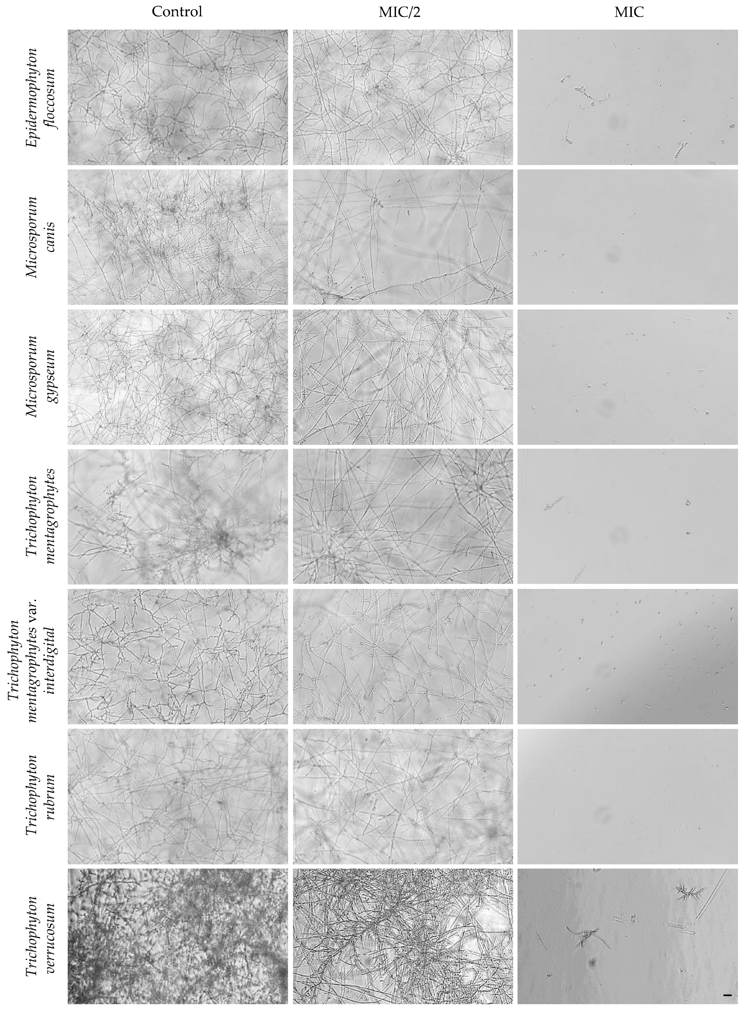
Figure 1. Optical microscopy images of dermatophyte strains exposed to L. stoechas subsp. luisieri essential oil, illustrating a concentration-dependent morphological response. The untreated control corresponds to fungal cells exposed to DMSO at the highest concentration used in essential oil dilution, while MIC/2 represents sub-inhibitory exposure and MIC the inhibitory concentration as determined in Table 1. Scale bar: 20 µm.
Processes 14 02655 g001
Figure 2. Immature biofilms of Epidermophyton floccosum, untreated (control) or exposed to L. stoechas subsp. luisieri essential oil. (A) Representative microscopic images of biofilms across the tested concentrations. (B) Quantification of biofilm biomass. (C) Quantification of extracellular matrix deposition. (D) Assessment of cell viability. Results are expressed as mean ± standard error of the mean of at least three independent experiments. Statistical analysis is performed using one-way ANOVA followed by Dunnett’s multiple comparisons test. Statistical significance is indicated as **** p < 0.0001, compared to the control group. Scale bar = 100 µm.
Processes 14 02655 g002
Figure 3. Mature biofilms of Epidermophyton floccosum, untreated (control) or exposed to L. stoechas subsp. luisieri essential oil. (A) Representative microscopic images of biofilms across the tested concentrations. (B) Biofilm biomass after treatment. (C) Extracellular matrix production. (D) Cell viability. Results are expressed as mean ± standard error of the mean of at least three independent experiments. Statistical analysis is performed using one-way ANOVA followed by Dunnett’s multiple comparisons test. Scale bar = 100 µm.
Processes 14 02655 g003
Figure 4. Preventive and therapeutic effects of L. stoechas subsp. luisieri essential oil (100 µg/mL) against Trichophyton rubrum infection in an ex vivo skin model. Macroscopic analysis of infected skin sections: untreated controls after 10 days (A) and 20 days (I), and samples treated with the essential oil either during the first 10 consecutive days of infection (E) or during the last 10 days of the 20-day infection period (M). Scanning electron microscopy (SEM) images of reconstructed skin sections, showing untreated controls after 10 days (B) and 20 days (J), and samples treated with the essential oil either during the first 10 consecutive days of infection (F) or during the last 10 days of the 20-day infection period (N); scale bars = 150 nm. Zoomed-in views of the central infection site and surrounding border region under the referred experimental conditions, including untreated controls after 10 days (C,D) and 20 days (K,L), and essential oil-treated samples during the first (G,H) or last (O,P) 10 days of the 20-day infection period; bars = 150 nm. The cropped images are enlarged views of zoom-ins: white arrow—fungal hypha, red arrow—extracellular matrix deposition; blue arrow—interwoven hyphal network; and bars = 50 nm.
Processes 14 02655 g004
Figure 5. Effect of L. stoechas subsp. luisieri essential oil on 3T3 fibroblast viability assessed by the resazurin assay. Results are expressed as mean ± standard error of the mean from at least three independent experiments. Statistical analysis is performed using one-way ANOVA followed by Dunnett’s multiple comparisons test to compare each treatment group with the untreated control. Statistical significance is indicated as * p < 0.05, ** p < 0.01, and **** p < 0.0001 versus control. The dashed horizontal line represents the untreated control, normalized to 100% cell viability.
Processes 14 02655 g005
Table 1. Antifungal activity of L. stoechas subsp. luisieri essential oil.
StrainsEssential Oil
MICMFC
Epidermophyton floccosum FF925–5025–50
Microsporum canis FF150–100200
Microsporum gypseum CECT 290850–100100–200
Trichophyton mentagrophytes FF750–100100–200
Trichophyton mentagrophytes var. interdigitale CECT 295825–50100
Trichophyton rubrum CECT 279450–100100–200
Trichophyton mverrucosum CECT 299212.512.5
Data are expressed in µg/mL. MIC, minimum inhibitory concentration; MFC, minimum fungicidal concentration. Values represent the range obtained from independent experiments (n ≥ 3). When concordant MIC/MFC values were obtained among replicates, a single value was assigned; when no unique endpoint could be established after confirmation assays, the observed concentration range was reported.
Disclaimer/Publisher’s Note: The statements, opinions and data contained in all publications are solely those of the individual author(s) and contributor(s) and not of MDPI and/or the editor(s). MDPI and/or the editor(s) disclaim responsibility for any injury to people or property resulting from any ideas, methods, instructions or products referred to in the content.