Article

Sustainable Laccase Production by Schizophyllum commune TMF3 on Agro-Industrial Waste for Efficient Dye Degradation and Comprehensive Toxicity Assessment


by
Nevena Ilić
1,
Anja Antanasković
2,
Jelena Filipović Tričković
3,
Miona Miljković
4,
Ana Milivojević
4,
Marija Milić
4 and
Katarina Mihajlovski
4,*
1
Innovation Centre of Faculty of Technology and Metallurgy, Karnegijeva 4, 11000 Belgrade, Serbia
2
Institute for Technology of Nuclear and Other Mineral Raw Materials, Bulevar Franš d’Eperea 86, 11000 Belgrade, Serbia
3
National Institute of the Republic of Serbia, Vinča Institute of Nuclear Sciences, University of Belgrade, Mike Petrovića Alasa 12-14, 11351 Belgrade, Serbia
4
Faculty of Technology and Metallurgy, University of Belgrade, Karnegijeva 4, 11000 Belgrade, Serbia
*
Author to whom correspondence should be addressed.
Processes 2026, 14(10), 1531; https://doi.org/10.3390/pr14101531
Submission received: 17 April 2026 / Revised: 2 May 2026 / Accepted: 7 May 2026 / Published: 9 May 2026

Abstract

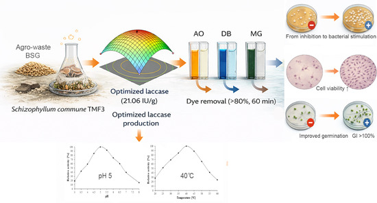
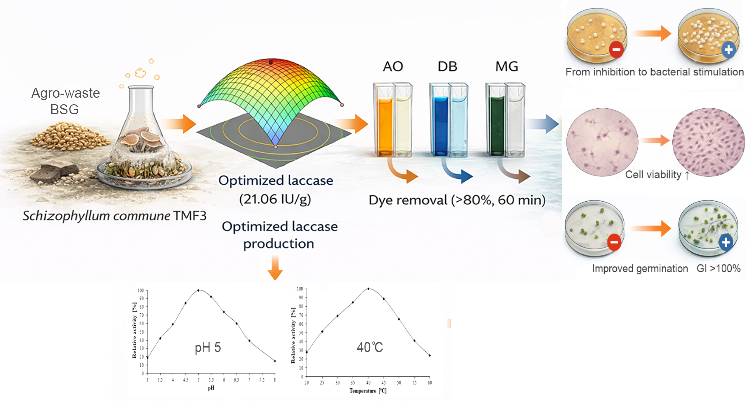
This study addresses the need for sustainable approaches in textile wastewater treatment by investigating laccase production with the white-rot fungus Schizophyllum commune TMF3 using agro-industrial waste as a substrate. Laccase was produced via solid-state fermentation on brewery spent grain under optimized conditions (1.75 g malt extract, 75% moisture, 7 days, 25 °C), reaching a maximum activity of 21.06 IU/g dry substrate. The crude enzyme was applied for the decolorization of azo and triphenylmethane dyes (50 mg/L). Decolorization efficiencies above 80% were achieved within 60 min without redox mediators, while chemical oxygen demand (COD) was reduced by more than 50% for all tested dyes. HPLC analysis showed parent dye peaks decreasing and the transformation products’ appearance. Antimicrobial activity testing showed no increase in inhibitory effects against Escherichia coli, Lactobacillus rhamnosus, Candida albicans, and Saccharomyces cerevisiae, while slight growth stimulation was observed in selected cases. Phytotoxicity assays using Triticum aestivum showed no inhibitory effects, with germination index values of 77–124%. Cytotoxicity assessment showed no effects for azo dyes, while cytotoxicity of the triphenylmethane dye decreased by 30% after treatment. These findings support the potential of agro-industrial laccase production as an effective approach for dye removal in sustainable wastewater strategies.

Graphical Abstract

1. Introduction

Rapid urbanization, population growth, and accelerated industrialization have significantly intensified environmental pollution worldwide [1]. Among the major environmental challenges, the discharge of industrial wastewater represents a serious threat to aquatic ecosystems and human health [2]. The textile industry is considered one of the main contributors to water pollution due to the extensive use of synthetic dyes during dyeing and finishing processes. It has been estimated that this sector accounts for nearly 20% of global industrial wastewater pollution, while more than 7 million tons of synthetic dyes are produced annually worldwide [3,4]. Synthetic dyes are widely employed not only in textiles but also in printing, papermaking, plastics, leather processing, cosmetics, and food-related industries. Based on their chromophoric structures, they are classified into several groups, including azo, anthraquinone, indigoid, triphenylmethane, and phthalocyanine dyes, with azo and triphenylmethane dyes being among the most commonly used in industrial applications [4]. Due to their complex aromatic structures and high chemical stability, these compounds often exhibit strong resistance to natural degradation processes. Consequently, dye-containing wastewater is typically persistent and may exert toxic, mutagenic, or carcinogenic effects even at low concentrations [5]. In addition, prolonged exposure to synthetic dyes and their transformation products has been associated with adverse health effects, including organ toxicity and potential endocrine disruption, primarily through contaminated water and food chains [2,6]. In particular, azo dyes such as Acid Orange 7 (AO) and Direct Blue 1 (DB) may undergo reductive cleavage under environmental or biological conditions, leading to the formation of aromatic amines, many of which are recognized as potentially carcinogenic compounds. Similarly, triphenylmethane dyes such as Malachite Green (MG) are well known for their pronounced cytotoxic, genotoxic, and bioaccumulative effects in aquatic organisms and humans. These characteristics significantly increase the environmental and human health risks associated with their persistence in wastewater systems.
In aquatic environments, the discharge of dye-laden effluents reduces light penetration, disrupts photosynthetic processes, and negatively affects aquatic ecosystems [6]. Furthermore, even low concentrations of dyes can significantly alter key water quality parameters, such as color, chemical oxygen demand (COD), and dissolved oxygen levels, thereby impairing overall water usability [7]. Such changes reduce water transparency and limit oxygen transfer, leading to broader ecological imbalances in affected aquatic systems.
Among commonly used industrial dyes, MG, DB, and AO are of particular environmental concern due to their widespread use and persistence. MG is a cationic triphenylmethane dye known for its high toxicity and potential carcinogenic effects [8]. DB and AO belong to the group of azo dyes frequently applied in textile dyeing processes and are characterized by high water solubility and environmental persistence [9].
Conventional physicochemical treatment methods used for dye removal from industrial effluents include coagulation–flocculation, adsorption, membrane filtration, and chemical oxidation. Although these techniques can be effective, they often involve high operational costs, incomplete pollutant mineralization, and the generation of secondary sludge that requires further treatment [10,11]. In contrast, biological strategies are based on enzymatic and microbial activity capable of breaking down complex dye structures into less toxic or fully mineralized products under environmentally benign conditions [2,6]. While physicochemical methods primarily rely on phase transfer or chemical transformation processes that may generate secondary pollution, biological treatments offer a more sustainable solution by enabling direct biodegradation and detoxification of contaminants [7]. Moreover, biological approaches typically operate under milder conditions, require lower energy input, and produce fewer harmful by-products, making them more environmentally compatible alternatives. These limitations have stimulated increasing interest in sustainable and environmentally friendly biological approaches for dye removal.
Among biological strategies, enzyme-based treatment systems have attracted considerable attention due to their high catalytic efficiency and ability to operate under mild reaction conditions [12]. Laccase (EC 1.10.3.2), a multicopper oxidase, catalyzes the oxidation of a broad range of phenolic and non-phenolic compounds using molecular oxygen as the final electron acceptor while producing water as the only by-product [2,13]. Owing to its broad substrate specificity, laccase has demonstrated significant potential for the degradation and decolorization of structurally diverse synthetic dyes [14,15]. Beyond decolorization, laccase-mediated reactions can transform complex dye molecules into simpler and less toxic metabolites, thereby reducing their environmental impact [15].
White-rot fungi are recognized as important natural producers of oxidative enzymes capable of transforming complex aromatic compounds, including synthetic dyes [13,16]. In recent years, increasing attention has been directed toward the production of fungal enzymes using agro-industrial residues as fermentation substrates, since this approach can reduce enzyme production costs while simultaneously contributing to the valorization of agricultural waste and the development of circular bioeconomy strategies [17,18]. In this context, the use of agro-industrial by-products is particularly relevant for sustainable waste management and environmentally safer wastewater treatment processes [17].
Within this context, S. commune is a basidiomycete whose enzymatic potential for dye degradation remains relatively underexplored compared with extensively studied white-rot fungi such as Trametes and Pleurotus [18,19,20]. In particular, studies focusing on laccase production from S. commune using agro-industrial residues and the application of crude enzyme preparations for rapid dye degradation remain limited. Furthermore, while numerous studies focus primarily on dye decolorization, relatively few investigations evaluate the environmental safety of the degradation products through comprehensive toxicity assessment.
Therefore, this study aimed to develop a sustainable approach for laccase production by S. commune TMF3 using brewery spent grain (BSG) supplemented with malt extract. This strategy was designed to overcome the inherently limited laccase-producing potential of S. commune, which remains insufficiently explored both for efficient enzyme production and its application in the degradation of structurally diverse dyes such as MG, DB, and AO. Accordingly, this study evaluates whether the obtained crude laccase can effectively degrade these dyes while reducing COD and toxicity, thereby assessing the environmental applicability of the proposed approach.

2. Materials and Methods

2.1. Sample Material and Chemicals

Brewer’s spent grain (BSG) was obtained as a byproduct from a local brewery in Serbia. Tryptic soy broth (TSB), agar, yeast extract, malt extract broth, and MRS broth were procured from Torlak (Belgrade, Serbia). Acid Orange 7 (AO) and Malachite Green (MG) were purchased from Sigma-Aldrich (Merck KGaA, Darmstadt, Germany) as commercial dye formulations with specified dye content (≥98% and ≥90%, respectively). Direct Blue 1 (DB) was purchased from MedChemExpress LLC (Monmouth Junction, NJ, USA, 95% purity). Guaiacol (99+%) was purchased from Fisher Scientific (Acros Organics, Geel, Belgium).

2.2. Microorganisms

The fungal strain S. commune TMF3 (accession number MW327505), Gram-positive Lactobacillus rhamnosus ATCC 7469, Gram-negative Escherichia coli ATCC 25922, yeast Candida albicans ATCC 10259, and waste brewer’s yeast Saccharomyces cerevisiae were from the culture collection from the Department of Biochemical Engineering and Biotechnology of the Faculty of Technology and Metallurgy, Belgrade, Serbia [21]. Wheat seeds (Triticum aestivum) were purchased from a local market.

2.3. Inoculum Preparation

The S. commune TMF3 culture was maintained on malt extract agar (20 g/L malt extract and 15 g/L agar) and incubated at 25 °C in the dark for 6–7 days. This incubation period ensured the development of actively growing mycelium, which was subsequently used as inoculum for solid-state fermentation.

2.4. Solid-State Fermentation

Solid-state fermentation (SSF) was performed in 200 mL Erlenmeyer flasks containing 20 g of dried BSG. Incubation time (days), malt extract (g), and moisture content (%) were varied according to the experimental design described in Table 1. After adjustment of moisture with sterile distilled water, the substrates were sterilized at 121 °C for 15 min. Once cooled, each flask was inoculated with five 1 × 1 cm agar plugs of actively growing S. commune TMF3 mycelium and incubated statically at 25 °C in the dark for the defined cultivation period. Laccase extraction from each flask was carried out according to Ilić et al. [22]. The supernatant obtained after centrifugation was considered as the crude laccase extract and was subsequently subjected to partial purification and concentration steps previously described in Ilić et al. [22].
Laccase activity was determined spectrophotometrically by monitoring guaiacol oxidation at 470 nm using a UV/Vis Ultrospec 3300 Pro spectrophotometer (Amersham Bioscience, Little Chalfont, Buckinghamshire, UK). The reaction mixture consisted of 0.25 mL of 1 mM guaiacol, 0.75 mL of sodium acetate buffer (pH 5.0), and 0.25 mL of crude fungal laccase extract. The control sample contained 1.00 mL of sodium acetate buffer (pH 5.0) and 0.25 mL of crude enzyme. All assays were incubated at 40 °C, corresponding to the previously established optimal temperature for the crude laccase preparation. The laccase activity (IU/mL and IU/g) was calculated according to the equations described in the previous study by Ilić et al. [22].

Experimental Design—RSM

For optimization of laccase production by S. commune TMF3, RSM was applied using Design-Expert software (version 12.0.3.0). Three independent variables were selected: incubation time (A, days), malt extract (B, g), and moisture content (C, %). A three-level Box–Behnken design was employed to evaluate the individual and interactive effects of these parameters on laccase activity (Table 1).
A total of 17 experimental runs were conducted, including five replicates at the central point. The central point replicates were used to estimate pure experimental error and to assess model adequacy through lack-of-fit analysis. The experimental data were fitted to a second-order polynomial equation, and the statistical significance of the model and its coefficients was evaluated by analysis of variance (ANOVA), with p-values < 0.05 considered statistically significant.

2.5. Characterization of Laccase

2.5.1. Optimum Temperature and pH of Laccase

The optimal pH of laccase was evaluated spectrophotometrically using guaiacol as the substrate, with buffer solutions ranging from pH 3 to 7 at 30 °C. Subsequently, the optimal temperature for laccase was determined in the range of 20–60 °C, employing guaiacol in 0.1 M sodium acetate buffer (pH 5). The laccase activity was calculated according to the equations described in the previous study by Ilić et al. [23] and enzymatic activity was expressed in relative terms, with the maximum activity normalized to 100%. All experiments were performed in triplicates with standard deviation less than ±5% and all results represent the mean value of three measurements.

2.5.2. Thermostability and pH Stability of Laccase

To investigate thermostability, laccase was incubated in 0.1 M sodium acetate buffer (pH 5) at various temperatures (20–60 °C) for 240 min. In addition, pH stability was evaluated by incubating the enzyme in buffers with pH values ranging from 3 to 7 for the same incubation time at 40 °C. The laccase activity was calculated according to the equations described in the previous study by Ilić et al. [23], and enzymatic activity was expressed in residual terms, with the maximum activity normalized to 100%. All experiments were performed in triplicates with standard deviation less than ±5% and all results represent the mean value of three measurements.

2.6. Dyes Degradation with Laccase from S. commune TMF3

In this study, the applicability of crude extracellular laccase produced by S. commune was assessed for the degradation of selected synthetic dyes in aqueous media. The enzymatic assays were carried out using MG, AO, and DB as model textile dyes, each prepared at an initial concentration of 50 mg/L in a final reaction volume of 10 mL.
The effect of environmental conditions on dye removal efficiency was systematically examined. Decolorization experiments were performed at pH values of 3.0, 4.0, 4.5, 5.0, 5.5, 6.0, and 7.0, while the reaction temperature was varied between 30 and 60 °C. All assays were conducted for a total period of 60 min to evaluate both the extent and kinetics of dye degradation. All experiments were performed in triplicates with standard deviation less than ±5% and all results represent the mean value of three measurements.
Dye decolorization was monitored by measuring the reduction in absorbance at the characteristic maximum wavelength of each dye using a UV–Vis spectrophotometer. Samples were withdrawn at 10-min intervals throughout the reaction. Control treatments, consisting of dye solutions incubated under identical experimental conditions but without the addition of laccase, were included in each experimental set to account for non-enzymatic dye removal.
The experimental procedure, including sample handling and spectrophotometric analysis, as well as the method used to calculate the efficiency of decolorization, was performed in accordance with the protocol described in our previous study [22]. The efficiency of decolorization was determined based on changes in absorbance relative to the initial value, as previously reported [22].
Decolorization efficiency (%) = ((A0 − At)/A0) × 100,
where A0 is initial absorbance of dye, At is absorbance of dye after laccase degradation.

2.7. HPLC Analysis

Samples were analyzed using a Dionex Ultimate 3000 HPLC system (ThermoScientific, Waltham, MA, USA) equipped with a Zorbax Eclipse Plus C18 reversed-phase column (150 mm × 4.6 mm, particle size 5 μm) (Agilent Technologies, Santa Clara, CA, USA). The mobile phases consisted of (A) H2O with 0.1% formic acid and (B) acetonitrile. Isocratic elution was performed using 80%, 60%, and 40% acetonitrile for MG, AO, and DB, respectively. The flow rate was 0.8 mL/min and the column temperature was maintained at 30 °C. The injection volume was 5–40 µL depending on sample. Detection of dyes and their degradation products was carried out using a UV/Vis detector at 600 and 254 nm for MG, 480 and 248 nm for AO, and 594 and 248 nm for DB.

2.8. Determination of Chemical Oxygen Demand

Chemical oxygen demand (COD) was determined using a Lovibond MD 610 spectrophotometer (Tintometer GmbH, Dortmund, Germany). For the analysis, 2 mL of each sample was added to digestion vials containing 10 mL of pre-measured reagents, corresponding to two concentration ranges: a low range (0–150 mg O2/L) and a medium range (0–1500 mg O2/L). Blank samples were prepared with deionized water for both concentration ranges. The vials were tightly sealed and inverted several times to ensure proper mixing of the sample with the reagents, and then placed in a Lovibond® RD 125 thermoreactor (Tintometer GmbH, Dortmund, Germany) for thermal digestion at 150 °C for 2 h. After digestion, the vials were removed from the reactor and allowed to cool to approximately 60 °C, gently inverted several times to homogenize the contents, and subsequently cooled to room temperature prior to spectrophotometric measurement.

2.9. Cytotoxicity Test

2.9.1. Cell Culture and Treatment

Commercially available human fetal lung fibroblast cell line MRC-5 (CCL-171ATCC, ATCC, Manassas, VA, USA) was used for the cytotoxicity assessment. The cells were grown in Dulbecco’s Modified Eagle’s Medium (DMEM, Gibco™, Thermo Fisher Scientific, Waltham, MA, USA) supplemented with 10% fetal bovine serum (FBS, Capricorn Scientific, Ebsdorfergrund, Germany) and 1% antibiotic-antimycotic (Gibco™ Antibiotic-Antimycotic, Thermo Fisher Scientific, Waltham, MA, USA), under the standard cell culture conditions, 37 °C and 5% CO2.

2.9.2. Cytotoxicity Assessment

The cytotoxicity was evaluated by 2,3-Bis-(2-Methoxy-4-Nitro-5-Sulfophenyl)-2H-Tetrazolium-5-Carboxanilide (XTT) assay according to the standard procedure [24]. Briefly, cells were seeded in the 24-well plates at density of 0.05 × 10⁶ cells per well, and allowed to adhere overnight. Following that, the cell cultures were treated with 20% v/v of the test samples for 24 h. Upon the treatment, the cell culture media was discharged, and the cells were washed with 0.9% NaCl, supplemented with XTT reagent activated with phenazine methosulfate (PMS) and incubated at 37 °C, until the color developed. Absorbance was measured at 470 nm on a microplate reader (Sunrise, Tecan Group Ltd., Männedorf, Switzerland). The results are presented as a percentage of viable cells compared to untreated control (100% of viable cells). To confirm the XTT assay results, the cellular morphology was monitored by trinocular inverted microscope (Optech, Gmbh, Munchen, Germany) with 20× magnification objective, equipped with OptikamB5 C-B5 digital camera and Optika Proview imaging software (Version 4.1) (Optika microscopes, Ponteranica, Italy).

2.9.3. Statistical Analysis

Results are presented as mean value ± standard error of mean. All experiments were set up in duplicate, and repeated twice. Statistical analysis was performed using One-way ANOVA statistical test from SPSS 10 statistical package for Windows (IBM, Armonk, NY, USA). p values < 0.05 were accepted as the level of significance.

2.10. Assessment of Microbial Responses to Dyes and Their Laccase-Derived Metabolites

To evaluate the biological effects of three synthetic dyes (MG, AO and DB) and their transformation products generated following laccase-mediated decolorization, selected microbial strains were employed. The assessment was carried out using L. rhamnosus ATCC 7469, E. coli ATCC 25922, C. albicans ATCC 10259, and residual brewer’s yeast S. cerevisiae. The experimental design and testing procedure were conducted following the methodology previously reported by Ilić et al. [23]. Dye solutions and their laccase-treated samples were prepared using sterile media and handled under aseptic conditions prior to antimicrobial testing.

2.11. Assessment of Phytotoxic Effects of Laccase-Treated Dyes

The phytotoxic potential of dyes (MG, AO and DB) and their enzymatic degradation products was investigated using wheat seeds (T. aestivum). The study aimed to determine how laccase-mediated decolorization influences plant growth and development. The experimental setup and procedures were conducted following the approach described by Ilić et al. [22].

3. Results

3.1. Laccase Production

3.1.1. Model Fitting and Evaluation of Process Variables

To optimize laccase production by S. commune TMF3, a Box–Behnken experimental design was applied to evaluate the effects of three independent variables on enzyme activity. Incubation time (A), malt extract supplementation (B), and moisture content (C) were systematically varied to assess their individual and combined effects on laccase activity during solid-state fermentation on dried BSG (Table 2).
Based on the experimental results, the data were fitted to a second-order polynomial model, expressed in coded variables as follows:
Y = 20.36 + 2.77A + 1.08B − 2.87C − 0.86AB − 3.43AC + 1.71BC − 7.38A2 − 6.70B2 − 7.33C2
where Y represents laccase activity (IU/g), A is incubation time (day), B is malt extract (g), and C moisture content (%).
ANOVA results (Table 3) confirmed that the model was highly significant (F = 197.94, p < 0.0001). The high coefficient of determination (R2 = 0.9961), together with the adjusted (0.9911) and predicted R2 (0.9704), demonstrates excellent agreement between experimental and predicted values. The lack of fit was not significant (p = 0.4980), confirming that the model adequately describes the system. The Adeq. Precision value of 35.744 indicates a satisfactory signal-to-noise ratio and further supports the robustness of the model.
As shown in Table 3, all linear terms (A, B, C), interaction terms (AB, AC, BC), and quadratic terms (A2, B2, C2) were statistically significant (p < 0.05), highlighting the complex and non-linear nature of the system.

3.1.2. Influence of Selected Process Variables on Laccase Production by S. commune TMF3

Laccase activity in S. commune TMF3 was significantly influenced by cultivation parameters, particularly incubation time, malt extract concentration, and moisture content (Table 2 and Table 3). The perturbation plot (Supplementary Material Figure S1) indicated that moisture content exerted the strongest effect, followed by incubation time and malt extract. The observed curvature confirmed the presence of quadratic effects and well-defined optima rather than simple linear relationships.
Numerical optimization predicted a maximum laccase activity of 21.06 IU/g at 9.54 days of incubation, 1.80 g malt extract, and 71.38% moisture content (desirability = 1). This value is substantially higher than the previously reported activity of 2.79 IU/g obtained under non-optimized conditions using BSG as the sole substrate [25].
The interaction between incubation time and malt extract (A × B) showed a statistically significant but moderate effect (Table 3). Laccase activity initially increased with simultaneous increases in both variables, followed by a decline at higher levels (Figure 1a). Incubation time exhibited a pronounced quadratic effect, with activity increasing to a maximum and then decreasing.
The interaction between incubation time and moisture content (A × C) was highly significant (Table 3). Increased incubation time enhanced laccase production only within a defined moisture range, whereas prolonged cultivation at higher moisture levels reduced enzyme activity (Figure 1b).
The interaction between malt extract and moisture content (B × C) was also significant (Table 3). At 1.75 g malt extract, laccase activity increased from 14.89 IU/g at 60% moisture to 21.06 IU/g at 75%, followed by a sharp decrease to 2.20 IU/g at 90% moisture (Table 2; Figure 1c).

3.2. Characterization of Laccase from S. commune TMF3

3.2.1. Optimum pH and Temperature

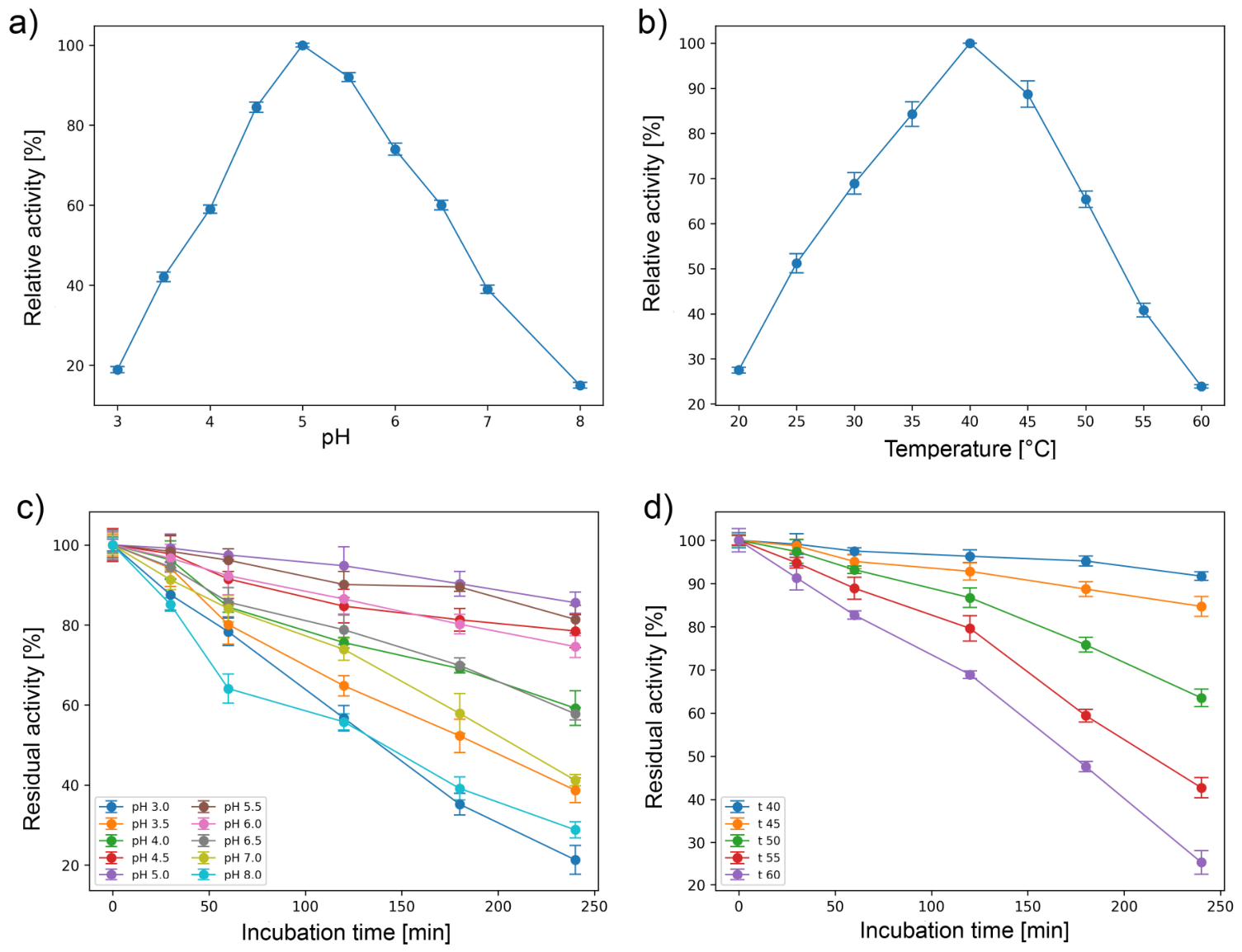
The influence of pH on the activity of laccase produced by S. commune TMF3 was examined over a pH range of 3.0–8.0 using guaiacol as the phenolic substrate. The enzyme displayed a clear bell-shaped activity profile (Figure 2a). At pH 3.0, activity was low (18.7%), increasing progressively to 42.3% at pH 3.5 and 59.1% at pH 4.0, followed by a marked rise to 84.6% at pH 4.5. Maximum activity (100%) was observed at pH 5.0, indicating the optimal pH for S. commune TMF3 laccase activity.
Beyond the optimum, activity gradually declined, reaching 92.1% at pH 5.5, 73.8% at pH 6.0, and 60.1% at pH 6.5, with a sharp decrease at higher pH values (39.2% at pH 7.0 and 15.1% at pH 8.0).
In addition to pH, temperature is another critical factor influencing laccase activity. The effect of temperature on the relative activity of laccase produced by S. commune TMF3 was evaluated over the range of 20–60 °C using guaiacol as the substrate (Figure 2b). The enzyme showed a clear temperature-dependent activity pattern, characterized by a gradual increase in activity with rising temperature up to an optimum, followed by a sharp decline at higher temperatures.
At 20 °C, laccase activity was relatively low (27.5%), but increased substantially at 25 °C (51.2%) and 30 °C (68.9%). A further rise in temperature resulted in enhanced activity, reaching 84.3% at 35 °C. Maximum relative activity (100%) was observed at 40 °C, identifying this temperature as optimal for laccase activity from S. commune TMF3. Beyond this point, enzyme activity decreased progressively, with relative activities of 88.7% at 45 °C and 65.4% at 50 °C. At higher temperatures, a pronounced loss of activity was detected, with only 40.8% activity remaining at 55 °C and 23.9% at 60 °C, indicating thermal sensitivity of the enzyme above its optimum.

3.2.2. pH and Thermostability of Laccase

The pH stability of laccase produced by S. commune TMF3 was evaluated by incubating the enzyme at different pH values (3.0–8.0) for up to 240 min, after which residual activity was measured under standard assay conditions (Figure 2c). At the beginning of incubation (0 min), the enzyme retained full activity (100%) at all tested pH values. However, prolonged incubation revealed clear differences in stability depending on pH.
At strongly acidic pH 3.0, laccase activity decreased rapidly over time, with residual activity dropping to 78.3% after 60 min, 56.7% after 120 min, and only 21.3% remaining after 240 min. A similar but slightly less pronounced trend was observed at pH 3.5, where 38.7% residual activity was detected after 240 min. In contrast, laccase incubated at moderately acidic pH values (4.0–5.5) exhibited markedly higher stability. At pH 5.0, which corresponded to the optimal pH for enzyme activity, the laccase retained 94.8% of its initial activity after 120 min and 85.6% after 240 min. Comparable stability was observed at pH 4.5 and 5.5, where more than 78% and 81% residual activity, respectively, remained after 240 min of incubation.
At near-neutral pH values, a gradual decline in stability was observed. At pH 6.0, residual activity decreased to 74.6% after 240 min, while at pH 6.5 and 7.0, the enzyme retained 57.8% and 41.2% activity, respectively. The least stability among the tested conditions was observed at pH 8.0, where residual activity dropped to 28.8% after 240 min of incubation. These results indicate that S. commune TMF3 laccase is most stable in the mildly acidic pH range, particularly between pH 4.5 and 5.5, while prolonged exposure to strongly acidic or alkaline conditions leads to progressive enzyme inactivation.
Following the evaluation of pH stability, the thermal stability of laccase produced by S. commune TMF3 was investigated by incubating the enzyme at different temperatures (40–60 °C) for up to 240 min and subsequently determining the residual activity under standard assay conditions. At the start of incubation, the enzyme retained full activity (100%) at all tested temperatures (Figure 2d). However, prolonged exposure revealed a temperature- and time-dependent decrease in activity.
At 40 °C, which corresponded to the optimal temperature for enzyme activity, the laccase exhibited high stability, retaining 96.3% of its initial activity after 120 min and 91.7% after 240 min of incubation. A similarly high level of stability was observed at 45 °C, with 92.8% residual activity after 120 min and 84.7% after 240 min. At 50 °C, the enzyme showed a moderate decline in stability, retaining 86.7% of its activity after 120 min and 63.5% after 240 min.
A more pronounced loss of activity was detected at higher temperatures. At 55 °C, residual activity decreased to 79.6% after 120 min and further dropped to 42.7% after 240 min. The lowest thermostability was observed at 60 °C, where only 68.9% activity remained after 120 min, and prolonged incubation led to a substantial reduction to 25.4% after 240 min. These results indicate that S. commune TMF3 laccase is relatively stable at moderate temperatures but undergoes significant thermal inactivation upon extended exposure to temperatures above 50 °C.

3.3. Dyes Decolorization with S. commune TMF3 Laccase

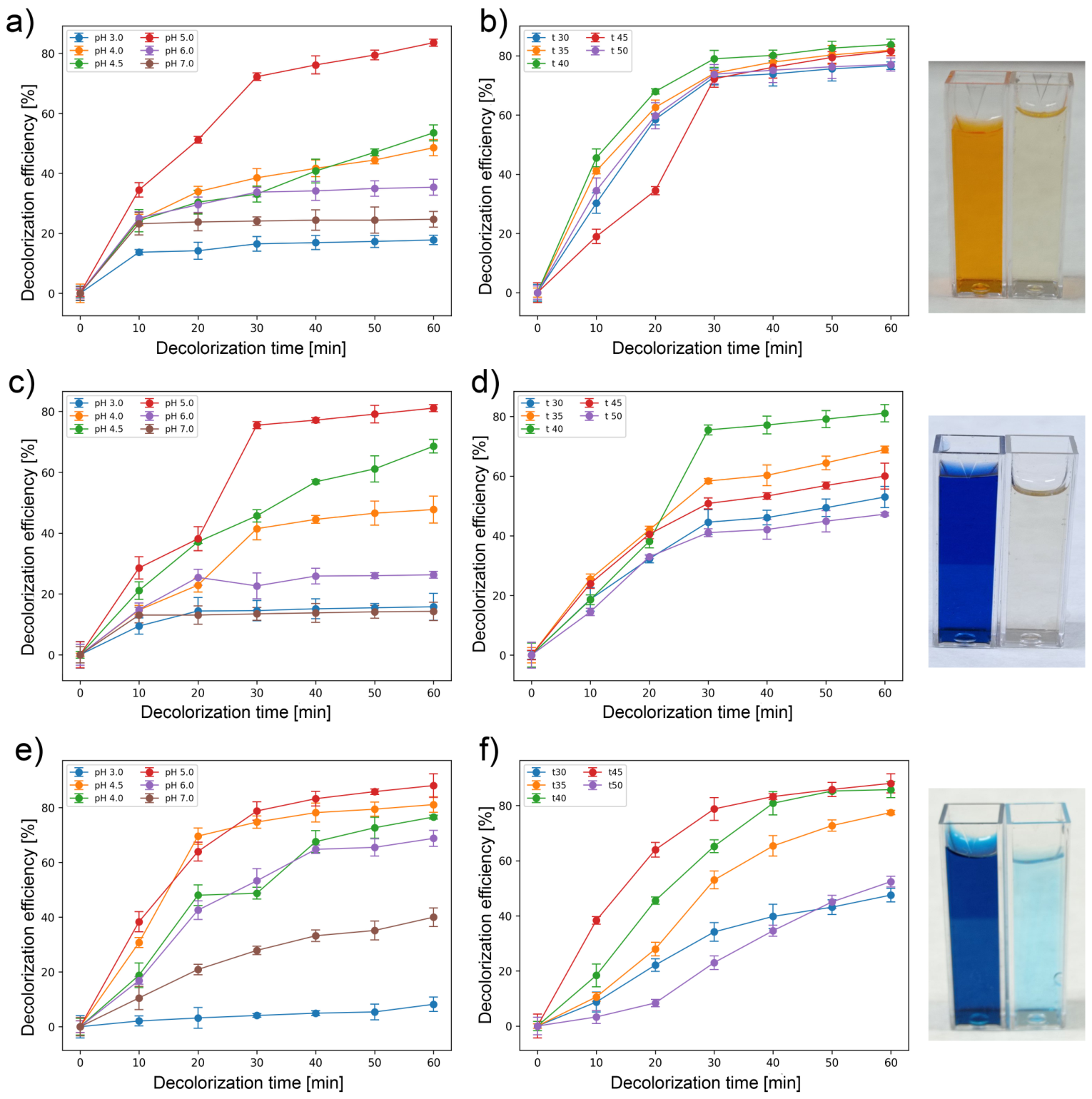
The effects of temperature and pH on the decolorization efficiency of laccase from S. commune TMF3 were evaluated using AO, DB, and MG at an initial dye concentration of 50 mg/L (Figure 3). Decolorization was assessed after 30 and 60 min, enabling direct comparison with previously reported laccase-based systems that generally require significantly longer treatment times.
For the azo dyes AO and DB, optimal decolorization was observed at 40 °C and pH 5. DB reached 75.5% decolorization within 30 min and 81.1% after 60 min, while the lowest removal (14.25%) occurred at pH 7 and 30 °C (Figure 3c,d). AO showed a comparable pattern, reaching 72.23% decolorization after 30 min and 83.57% after 60 min, whereas minimal removal (17.8%) was recorded at pH 3 and 30 °C (Figure 3a,b). For MG, maximum decolorization was achieved at 45 °C and pH 5, reaching 78.79% after 30 min and 88.07% after 60 min, whereas only 8.18% decolorization was observed at pH 3 after 60 min (Figure 3e,f).

3.4. HPLC Analysis

The HPLC chromatograms of MG recorded at different wavelengths indicate a clear decrease in the parent dye after laccase treatment. At 600 nm, which is characteristic for MG, the control sample shows a dominant peak, while this peak is strongly reduced in the treated sample (Supplementary Material Figure S2). This confirms that a significant portion of the dye was removed. At 254 nm, several additional peaks appear in the treated sample, suggesting the formation of degradation products. However, these signals should be interpreted with caution, since compounds originating from the enzyme preparation may also contribute.
For AO, a similar trend was observed. At 480 nm, a clear decrease in the main peak is visible after treatment, together with the appearance of new peaks (Supplementary Material Figure S3), indicating transformation of the dye. At 248 nm, peaks are shifted toward shorter retention times, which may suggest the formation of more polar products. As in the case of MG, interpretation at this wavelength is less specific due to possible matrix interference.
In the case of DB, chromatograms recorded at 594 nm show a strong reduction in the main peak after treatment, confirming effective degradation of the dye (Supplementary Material Figure S4). At the same time, additional peaks appear, especially at longer retention times, which likely correspond to intermediate compounds. Chromatograms recorded at 248 nm support this observation, although they are less selective and may include signals from the enzyme matrix.

3.5. Chemical Oxygen Demand

In the present study, the effectiveness of laccase from S. commune TMF3 in reducing the organic load of dye-containing solutions was evaluated through COD measurements following enzymatic decolorization. For MG, the initial COD value of 1472 mg/L was reduced to 239.4 mg/L after laccase treatment, corresponding to an 84.0% reduction. This substantial decrease indicates not only effective color removal but also extensive degradation of organic compounds contributing to the pollution load.
For the azo dyes, initial COD values of 1632 mg/L for AO and 2002 mg/L for DB were measured prior to treatment. After laccase-mediated decolorization, COD values decreased to 810 mg/L and 618 mg/L, corresponding to COD reductions of 50.37% for AO and 69.13% for DB, respectively. These results demonstrate that laccase from S. commune TMF3 is capable of significantly lowering the organic burden of azo dye solutions, although the extent of COD reduction varied depending on dye structure.

3.6. Cytotoxicity

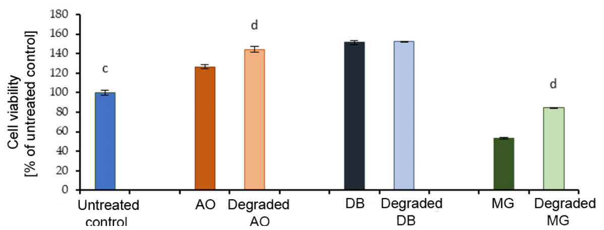
In the present study, the tested dyes and their degradation products showed distinct effects on cell viability (Figure 4 and Supplementary Material Figure S5). AO, degraded AO, DB, and degraded DB exhibited a stimulatory effect compared to the untreated control, whereas MG and its degradation products significantly reduced cell viability. Among all tested samples, MG showed the strongest cytotoxic effect, decreasing viability by nearly 50% (p < 0.001). Although laccase treatment significantly reduced MG toxicity, increasing cell viability by nearly 30% (p < 0.001), residual toxicity remained, indicating that some transformation products may still retain biological activity. The increase in cell viability observed for AO and DB, particularly after laccase treatment, may be attributed to the transformation of complex dye molecules into less toxic, low-molecular-weight compounds. This effect was especially pronounced for degraded AO, which increased viability by approximately 20% compared to the untreated dye (p < 0.001). In contrast, no significant difference was observed between DB and its degradation products, suggesting that the transformation did not substantially alter its cytotoxic profile under the tested conditions.

3.7. Antimicrobial Test

In this study, the effects of selected dyes (AO, DB, and MG), before and after laccase-mediated degradation, were evaluated on the growth of E. coli, L. rhamnosus, C. albicans, and S. cerevisiae. Results are expressed as percentages of growth inhibition and stimulation (Table 4).
AO exhibited an inhibitory effect on all tested microorganisms, with the highest inhibition observed for E. coli (21.86%) and the lowest for C. albicans (5.94%). Following degradation, this effect changed, resulting in slight growth stimulation in E. coli (2.75%) and a pronounced stimulatory effect in S. cerevisiae (34.21%).
DB showed moderate inhibition of E. coli (7.58%) and C. albicans (9.25%). After degradation, the effect shifted from inhibition to stimulation in all tested microorganisms, including E. coli (4.79%), L. rhamnosus (9.41%), C. albicans (5.15%), and S. cerevisiae (9.68%).
MG demonstrated the strongest inhibitory activity among the tested dyes, particularly against L. rhamnosus (98.14%) and C. albicans (96.89%), while inhibition of E. coli reached 58.19%. After degradation, the inhibitory effect was substantially reduced, and growth stimulation was observed for L. rhamnosus (24.43%) and S. cerevisiae (17.37%), with weaker stimulation in E. coli (4.79%) and C. albicans (3.83%).

3.8. Phytotoxicity

In the present study, phytotoxicity was evaluated using the germination index (GI), where GI values between 25–65% indicate phytotoxicity, values above 65% indicate non-phytotoxicity, and values exceeding 101% suggest a phytostimulatory effect [22].
Before enzymatic treatment, all tested dyes showed phytotoxic effects. Wheat seeds treated with AO and DB exhibited GI values of 27.91% and 59.06%, respectively, while MG showed a GI of 43.62%, confirming its inhibitory effect on germination (Supplementary Material Figure S5).
After treatment with crude laccase from S. commune TMF 3, a pronounced reduction in phytotoxicity was observed. The GI for AO increased to 77.57%, indicating complete loss of phytotoxicity. Furthermore, DB and MG degradation products exhibited phytostimulatory effects, with GI values of 124.09% and 103.12%, respectively (Supplementary Material Figure S5). These findings demonstrate that crude laccase treatment not only detoxifies dyes but can also generate metabolites that promote seed germination.

4. Discussion

S. commune is a wood-decaying basidiomycete capable of producing ligninolytic enzymes, including laccases [19,25]; however, it remains less explored compared to genera such as Trametes and Pleurotus [19,20]. Available studies addressing laccase production by S. commune under solid-state fermentation are relatively limited and frequently involve the use of chemical inducers to stimulate enzyme synthesis [26]. On the other hand, the present results demonstrate that BSG can serve as an effective substrate for laccase production without chemical inducers, yielding a maximum activity of 21.06 IU/g under optimized conditions (incubation time 9.54 days, 1.80 g malt extract, 71.38% moisture) (Table 2).
The substantial increase in laccase activity under optimized conditions compared to previous findings highlights the importance of process optimization in SSF systems. For example, under non-optimized conditions on BSG (30 °C, 6 days, no malt extract), S. commune TMF3 produced only 2.79 IU/g [25]. The observed dependence of enzyme production on incubation time reflects typical fungal growth-associated kinetics, where laccase synthesis is linked to active colonization and declines during later stages due to nutrient depletion [23].
Although BSG itself provides a nutrient base for fungal growth, its composition alone does not necessarily ensure optimal enzyme synthesis. Malt extract was therefore introduced as a readily assimilable carbon source to support fungal growth and laccase production [25]. Malt extract supplementation exhibited a nonlinear effect: moderate concentrations (~1.80 g) enhanced laccase production, while higher levels caused inhibition (Figure 1c). This behavior is consistent with carbon catabolite repression mechanisms reported in basidiomycetes, where excess readily metabolizable carbon suppresses secondary metabolism, including ligninolytic enzyme synthesis [20].
Moisture content was identified as the most critical factor affecting enzyme production [27,28]. Optimal hydration (~71–75%) ensured sufficient nutrient solubility and fungal metabolism while maintaining substrate porosity and oxygen transfer. Excessive moisture (e.g., 90%) sharply reduced enzyme activity to 2.20 IU/g (Figure 1b), likely due to restricted aeration, which is essential for laccase activity as molecular oxygen serves as the terminal electron acceptor [29,30].
The significant interactions between process variables further emphasize the complexity of SSF systems. The strong A × C interaction (incubation time × moisture) indicates that the effect of incubation time is highly dependent on moisture levels, while the B × C interaction (malt extract × moisture) suggests that the efficiency of carbon supplementation is conditioned by substrate hydration. For instance, at 1.75 g malt extract, laccase activity increased from 14.89 IU/g at 60% moisture to 21.06 IU/g at 75% moisture, then sharply declined at 90% moisture (Figure 1c). These findings confirm that balanced process conditions are required to achieve maximal laccase production.
Comparison with literature data reveals considerable variability depending on substrate type, cultivation mode, and use of inducers. For example, laccase activity was not detected during SSF on Jerusalem artichoke stalk [31], while only low activity (1–4 IU/mL) was observed under submerged cultivation on alkaline-pretreated lignocellulosic substrates [32], and no activity was seen on pure cellulose (Avicel-PH101). Higher activities (up to 367 IU/mL after 6 days) in SSF of banana stalk have been reported, but only in the presence of inducers such as copper and ABTS [26].
The present study demonstrates that competitive laccase production (21.06 IU/g) can be achieved under inducer-free conditions on BSG, supporting the potential of agro-industrial waste valorization within sustainable bioprocessing strategies.
The effect of pH on laccase activity is a critical parameter influencing enzyme performance and its potential application in biotechnological processes. The obtained results are consistent with previous reports indicating that fungal laccases typically exhibit maximum activity toward phenolic substrates within an acidic pH range (3.0–5.5), followed by a rapid decline at neutral and alkaline pH [23,33]. A comparative summary of pH optima and activity profiles of S. commune laccases reported in the literature is presented in Table 5.
A similar pH-dependent activity profile has been observed for laccases from other S. commune strains, although the exact optimal pH varies depending on strain, cultivation conditions, and substrate. For instance, laccase from S. commune IBL-06 showed maximal activity at pH 6.0 when ABTS was used as a substrate, with reduced activity at higher pH values [34]. In contrast, laccase produced by S. commune NI07 exhibited a narrow optimal range between pH 4.43 and 4.46 under submerged fermentation conditions, with a sharp loss of activity above pH 4 [11]. Additionally, immobilized laccase from the same strain showed maximum activity at pH 5.5 using ABTS [11]. These findings confirm that laccase pH optima are strongly dependent on both enzyme origin and substrate type.
The observed pH profile of S. commune TMF3 laccase (Figure 2a) can be explained by pH-dependent changes in enzyme catalysis. Under acidic conditions, the redox potential difference between the phenolic substrate and the type 1 copper center facilitates efficient electron transfer [23,35]. With increasing pH, hydroxide ions interact with the type 2/type 3 copper cluster, disrupting internal electron transfer and reducing catalytic efficiency [23,35]. Furthermore, changes in redox potential between the substrate and the enzyme’s active site contribute to the decline in activity at higher pH values [35].
The observed temperature–activity profile is consistent with the typical behavior of fungal laccases (Figure 2b), where catalytic efficiency increases with temperature due to enhanced molecular mobility and collision frequency between enzyme and substrate, up to a threshold beyond which thermal denaturation and structural destabilization occur [36]. The optimal temperature of 40 °C determined for S. commune TMF3 laccase suggests moderate thermostability (Figure 2b,d), which is advantageous for applications performed under mild processing conditions.
Comparable temperature optima have been reported for laccases produced by other S. commune strains, although considerable variability exists depending on the strain and cultivation strategy [11,34]. Laccase from S. commune NI07 produced under submerged fermentation conditions showed maximal activity at 30 °C, whereas the same strain, when immobilized, exhibited enhanced thermal tolerance across a broader temperature range (25–65 °C), with optimal activity at 35 °C [11]. Similarly, the purified laccase from S. commune IBL-06 displayed an optimal temperature of 40 °C [34], closely matching the value observed in the present study.
While many fungal laccases are reported to exhibit temperature optima between 50 and 70 °C, several enzymes with lower temperature optima have also been described, particularly among basidiomycete-derived laccases [37]. The relatively lower optimal temperature observed for S. commune TMF3 laccase (Figure 2b) may reflect structural characteristics specific to this strain, as well as the use of the phenolic substrate guaiacol, which can influence apparent temperature optima.
The observed temperature profile, with maximal activity at moderate temperatures and a decline at higher temperatures, together with the acidic pH optimum, suggests potential applicability of this enzyme in processes involving phenolic compound oxidation under mild conditions.
In addition to its effect on catalytic activity, pH can also significantly influence enzyme stability by altering the ionization state of amino acid residues that are crucial for maintaining the native conformation of the protein [38]. Exposure to extreme pH conditions may lead to hydrolysis or chemical modification of the enzyme molecule, resulting in partial or complete loss of activity [38]. As previously reported, fungal laccases can remain structurally stable over a relatively broad pH range, even though they may be catalytically inactive at certain pH values [23]. The pH stability profile observed in this study (Figure 2c) is consistent with previous reports on laccases from S. commune. For instance, purified laccase from S. commune IBL-06 was reported to remain stable within the pH range of 5.0–8.0 after 1 h of incubation, although extended exposure to more alkaline conditions resulted in substantial activity loss, with only about 22% residual activity remaining at pH 9.0 after 24 h at 35 °C [34]. Similarly, laccase produced by immobilized S. commune NI07 exhibited relatively high pH tolerance and greater stability compared to the enzyme obtained from submerged cultures [11]. Short-term incubation studies showed that this enzyme retained high activity at mildly acidic pH, while significant losses were observed under strongly acidic and alkaline conditions [11].
Although optimal pH values for laccase activity differ among S. commune strains and depend on the substrate used, the available literature consistently indicates that laccases from this species display greater stability in acidic to near-neutral pH ranges [11,34]. The results obtained for S. commune TMF3 support this observation and demonstrate that, despite reduced catalytic activity at higher pH values, the enzyme maintains a considerable degree of structural stability over a broad pH range.
The thermal stability profile observed in this study (Figure 2d) is consistent with the general characteristics of fungal laccases, whose thermostability is often correlated with the temperature range preferred by the producing organism. In contrast to bacterial laccases, which frequently display high thermostability, fungal laccases typically lose activity rapidly at temperatures exceeding 60 °C. It has been reported that many fungal laccases exhibit half-lives of less than one hour at 70 °C and only a few minutes at 80 °C, highlighting their limited resistance to high-temperature stress [20,39].
Comparable variations in thermostability have been reported among laccases from different S. commune strains. For instance, laccase produced by S. commune NI07 under submerged culture conditions showed optimal activity at 30 °C and lost more than 50% of its activity with relatively small temperature deviations [11]. In contrast, immobilized S. commune NI07 exhibited enhanced thermotolerance over a wider temperature range (25–65 °C), retaining high residual activity even after short-term exposure to elevated temperatures [11]. On the other hand, laccase from S. commune IBL-06 displayed low thermostability, retaining only approximately one-third of its initial activity after 1 h of incubation at 60 °C [34]. Compared to these reports, S. commune TMF3 laccase demonstrates moderate thermostability, maintaining substantial activity at temperatures up to 50 °C but becoming increasingly susceptible to thermal inactivation at higher temperatures (Figure 2d).
When the pH and thermal stability profiles are considered together, S. commune TMF3 laccase can be characterized as an enzyme that is most stable under mildly acidic conditions and moderate temperatures. The enzyme retained high residual activity at pH 4.5–5.5 and temperatures of 40–45 °C over prolonged incubation periods, while exposure to extreme pH values or elevated temperatures resulted in accelerated loss of activity (Figure 2c,d). This behavior is typical of fungal laccases and reflects the sensitivity of their three-dimensional structure to environmental stressors that affect intramolecular interactions and copper center integrity.
Collectively, the pH and thermal stability characteristics of S. commune TMF3 laccase demonstrate that this enzyme is well suited for biotechnological applications that require sustained catalytic performance under mildly acidic conditions and moderate temperature regimes. Such characteristics are particularly advantageous for processes involving the oxidation of phenolic compounds, where long-term enzyme activity and operational stability are critical.
The characterized laccase from S. commune TMF3 was subsequently applied for the decolorization of three dyes (AO, DB and MG), demonstrating its practical potential in biotechnological dye removal processes (Figure 3). Compared to literature data, the laccase from S. commune TMF3 showed faster decolorization kinetics. For instance, AO removal of 87.9% has been reported only after 48 h using laccase from T. trogii in the presence of the redox mediator HBT [40]. Similarly, DB removal typically requires extended reaction times or mediator-assisted systems, with reported values of ~40% using immobilized laccase [41] or ~81% after 24 h with crude laccase from T. versicolor [42]. In contrast, in the present study, comparable decolorization levels for both AO and DB (>80%) were achieved within 60 min, at a higher dye concentration (50 mg/L) and without the addition of any redox mediators (Figure 3a–d), emphasizing the enhanced catalytic efficiency of the S. commune TMF3 laccase. Literature reports indicate that MG decolorization values (90–91.6%) are typically achieved after 120–180 min using higher enzyme activities at dye concentrations of approximately 110 mg/L, using higher laccase activities and redox mediators such as HBT [43]. Additionally, optimal temperatures reported for MG removal often reach 55 °C, which is less favorable from an energy-efficiency perspective [43]. Notably, a study has also demonstrated that the presence of HBT increases MG decolorization to approximately 90% within 1 h, whereas in mediator-free systems decolorization is significantly slower or incomplete [43].
Comparable or slightly higher decolorization efficiency have been reported for laccase from Cerrena sp. However, these results were obtained under more demanding conditions. Specifically, a maximum MG decolorization efficiency of 91.6% was predicted using 2.8 IU/mL laccase, an initial dye concentration of 109.9 mg/L, and a reaction time of approximately 172 min [44]. In contrast, the S. commune TMF3 laccase achieved nearly 90% MG removal in 60 min without mediator addition and at a lower temperature, highlighting a key advantage of this enzyme (Figure 3e,f). To further highlight the performance of the obtained enzyme, a comparative overview of dye decolorization efficiencies relative to literature reports is presented in Table 6.
The high decolorization efficiency observed in this study, particularly under mediator-free conditions, may be attributed not only to the intrinsic catalytic properties of the laccase but also to the lignocellulosic substrate used for its production. BSG is rich in lignin and phenolic compounds, including ferulic and p-coumaric acids, which can be released during fungal growth and lignin modification [45,46]. These low-molecular-weight phenolic compounds may act as natural redox mediators, facilitating electron transfer during laccase-catalyzed reactions [37,47]. This may further enhance electron transfer efficiency and support effective dye degradation even in the absence of externally added mediators. Similar mediator-like behavior of lignin-derived phenolics has been reported in lignocellulosic systems [48].
The degradation products of all three dyes were analyzed by HPLC to further elucidate the transformation process. The reduction in the main peaks at dye-specific wavelengths, together with the appearance of additional signals, points to structural changes in the dyes and the formation of intermediate products (Supplementary Material Figures S2–S4). Laccase-catalyzed dye degradation is primarily based on one-electron oxidation reactions that generate highly reactive radical intermediates. These intermediates undergo further non-enzymatic transformations, leading to the cleavage of chromophoric groups, including azo bonds, and the breakdown of complex aromatic structures. As a result, dye molecules are converted into smaller, more polar compounds with reduced affinity for cellular membranes and macromolecules, which explains the observed decrease in cytotoxic and antimicrobial effects. This trend is consistent with the experimental data obtained in this study (Table 4), where higher decolorization efficiencies (>80% within 60 min) corresponded to a more pronounced reduction in antimicrobial activity. This suggests that enzymatic treatment not only removes color but also alters functional groups responsible for microbial inhibition. The oxidative cleavage of chromophoric structures, including azo bonds (–N=N–) and conjugated aromatic systems, leads to the formation of smaller and more polar compounds with reduced toxicity and a lower ability to interact with microbial cell membranes and intracellular targets. In some cases, this transformation may result in a shift from inhibitory effects to slight growth stimulation. This behavior is in agreement with previously described laccase-mediated degradation pathways [49,50].
These findings on dye transformation at the molecular level prompted further evaluation of the process efficiency through changes in overall organic load, as reflected by COD removal. Textile wastewater is characterized by a complex composition that includes high concentrations of dyes, organic matter, suspended solids, and various auxiliary chemicals, which collectively result in elevated COD [23]. COD is widely recognized as a key indicator of organic pollution and is commonly used to evaluate the effectiveness of wastewater treatment processes by comparing values before and after treatment [23]. When compared with literature data, the COD reduction efficiencies obtained in this study are highly competitive, particularly considering the short treatment time (≤60 min) and mediator-free conditions. For instance, COD removal of 95.95% has been reported for MG at a higher dye concentration (150 mg/L) using Pseudomonas plecoglossicida MG2 [51]. However, this removal was achieved through bacterial degradation processes that typically require longer incubation periods. Similarly, very high COD reductions (up to 98.84%) have been reported using complex hybrid systems involving immobilized laccase combined with metal oxide materials, where enzymatic degradation is coupled with adsorption processes, often at much lower dye concentrations (around 5 mg/L) [52].
In contrast, the laccase from S. commune TMF3 achieved an 84% COD reduction for MG and up to 69.13% for azo dyes at a higher dye concentration (50 mg/L), within only one hour, and without the use of additional adsorbents, immobilization matrices, or redox mediators. This highlights the efficiency of the enzyme as a standalone biocatalyst.
Further comparison with microbial systems producing ligninolytic enzymes also underscores the advantages of the S. commune TMF3 laccase. For example, degradation of textile dyes by Bacillus aryabhattai DC100 resulted in a maximum COD reduction in only 43% after 48 h, which is significantly lower and markedly slower than the reductions observed in the present study [52]. Moreover, COD reductions of 93.47% and 92.17% have been reported for CI Acid Black 210 and CI Acid Black 234 dyes, respectively, but only after 96 h of treatment using crude bacterial enzyme preparations [53]. In comparison, the laccase from S. commune TMF3 achieved substantial COD removal within 60 min, demonstrating enhanced superior process kinetics and efficiency.
These findings indicate that laccase from S. commune TMF3 enables rapid and effective reduction in both color and organic load in dye-containing solutions. The combination of short treatment time, relatively high COD removal efficiency, and the absence of additional treatment components distinguishes this enzyme from many previously reported systems, indicating promising potential for future wastewater treatment applications. In addition to these improvements in physicochemical parameters, it is essential to assess the biological impact of treated dye solutions in order to fully evaluate the environmental relevance of the process. Synthetic dyes, particularly azo and triphenylmethane dyes, are known to affect cell viability due to their persistence and ability to induce oxidative stress and cellular damage [54,55,56]. Azo dyes may undergo reductive cleavage, leading to the formation of aromatic amines associated with cytotoxic and genotoxic effects, while triphenylmethane dyes can interact with cellular components such as membranes, proteins, and nucleic acids, thereby disrupting normal cellular functions [54,56]. MG exhibited pronounced cytotoxicity, likely due to its interaction with cellular components and its ability to induce oxidative stress [56]. These findings indicate that laccase treatment can reduce dye toxicity, although complete detoxification is not always achieved, particularly in the case of structurally complex dyes such as MG. Synthetic dyes can affect microorganisms due to their chemical structure and potential toxicity, which may lead to growth inhibition or altered cellular activity. In the present study, all tested dyes exhibited inhibitory effects prior to treatment, while laccase-mediated degradation led to a noticeable reduction in this inhibition. Differences in microbial response can also be related to the structural and physiological characteristics of the tested organisms. E. coli, as a Gram-negative bacterium, possesses an outer membrane that can act as a permeability barrier, reducing the uptake of toxic compounds. In contrast, L. rhamnosus, a Gram-positive bacterium, lacks this outer membrane and is therefore often more susceptible to external agents. Yeasts such as C. albicans and S. cerevisiae, being eukaryotic microorganisms, have more complex cellular organization, which can influence their response to both dyes and their degradation products. These structural differences may partly explain the higher sensitivity of L. rhamnosus and C. albicans to malachite green observed in this study. In addition, some microorganisms, particularly lactic acid bacteria and yeasts, are capable of utilizing low-molecular-weight organic compounds, which may explain the observed transition from inhibition to growth stimulation after laccase treatment. In the case of E. coli, the initial inhibitory effect was markedly reduced after degradation, while a slight growth stimulation was observed. This may indicate that the degradation products were less toxic and could serve as metabolically accessible substrates. Phytotoxicity evaluation is an important step in assessing the environmental impact of dyes and their degradation products [23]. Seed germination assays are commonly used due to their sensitivity and relevance, as germination represents an early stage of plant development [57]. Similar trends have been reported in the literature. For example, laccase-treated AO51 showed increased germination compared to untreated dye; however, GI values remained relatively low (8.8%, 10.8%, and 29.0% at dye concentrations of 100, 75, and 20 mg/L, respectively), indicating only partial detoxification [40]. In addition, although the application of the mediator HBT improved decolorization efficiency, it also led to increased phytotoxicity, as reflected by lower GI values. This highlights a potential limitation of mediator-assisted systems [57]. In contrast, the results obtained in the present study indicate a more pronounced reduction in phytotoxicity under mediator-free conditions.
Further support for the effectiveness of biological detoxification comes from in vivo studies on MG. Untreated MG solutions have been shown to reduce seed germination to approximately 20%. After treatment with either crude or purified laccase from Pseudomonas putida, germination increased to around 40% [58]. Additionally, biodegradation of MG by Bacillus pacificus ROC1 resulted in germination rates of only 60% at 50 ppm and 13% at 1000 ppm in untreated samples, whereas ROC1-treated samples exhibited germination rates above 80% across all tested concentrations, along with improved root and shoot growth [59].
Compared to these reports, the results obtained in the present study using non-purified laccase are particularly noteworthy, as GI values exceeding 100% were achieved for MG, indicating a clear phytostimulatory effect rather than mere detoxification. In some cases, these transformation products may also become more bioavailable and metabolically accessible, which could contribute to the observed growth stimulation effects.
The results obtained in this study, particularly the high GI values observed after treatment, indicate effective reduction in phytotoxicity and suggest that crude laccase can generate transformation products with lower environmental impact. At the same time, the absence of redox mediators represents an additional advantage, as such compounds may contribute to increased toxicity in some systems.
Although the present study demonstrates high efficiency of crude S. commune TMF3 laccase in dye degradation, COD reduction, and phytotoxicity removal, enzyme stability and reusability were not evaluated. These parameters are important for practical and large-scale applications.
In this context, enzyme immobilization represents a well-established strategy to improve operational stability, enhance reusability, and enable repeated application of laccase in wastewater treatment systems. Various immobilization approaches, particularly those based on polymeric matrices, have been reported to improve biocatalyst stability, resistance to environmental stress, and long-term applicability. For example, polymer-based systems such as PVP/PEG/agar matrices have been successfully applied in wastewater treatment, demonstrating improved stability and reusability under operational conditions [60].
Future studies could therefore focus on the development of immobilized laccase systems in order to further enhance the operational performance and applicability of the enzymatic process investigated in this work.

5. Conclusions

The results of this study demonstrate that agro-industrial waste can serve as an effective substrate for laccase production by S. commune TMF3, supporting the valorization of BSG within a circular bioeconomy framework. High enzyme activity was achieved under optimized solid-state fermentation conditions.
The produced crude laccase exhibited optimal activity at pH 5.0 and 40 °C and enabled rapid decolorization of both azo and triphenylmethane dyes, achieving over 80% removal within a short time and without the addition of redox mediators. The treatment also reduced the chemical oxygen demand of dye solutions, indicating effective degradation of organic compounds.
Toxicological assessment showed no increase in antimicrobial or cytotoxic effects after treatment, while phytotoxicity tests confirmed the absence of inhibitory effects on wheat seed germination, with slight stimulatory responses observed in some cases.
These findings indicate that mediator-free laccase production on agro-industrial waste can be applied as a sustainable approach for dye removal, integrating waste valorization with environmentally safer treatment of dye-contaminated wastewater.

Supplementary Materials

The following supporting information can be downloaded at: https://www.mdpi.com/article/10.3390/pr14101531/s1, Figure S1: Perturbation plot of tested variables on laccase production by S. commune TMF3, Figure S2: HPLC analysis of MG; Figure S3: HPLC analysis of AO, Figure S4: HPLC analysis of DB; Figure S5: Representative photomicrographs of (a) untreated MRC-5 cells, and cells treated with 20% vol/vol of (b) AO, (c) degraded AO, (d) DB, (e) degraded DB, (f) MG, and (g) degraded MG and Figure S6: Phytotoxic effects of untreated and laccase-treated dyes (AO, DB, MG) on seed germination.

Author Contributions

Conceptualization, N.I. and K.M.; methodology, N.I., M.M. (Marija Milić), A.M.; software, K.M.; validation, N.I., M.M. (Marija Milić) and K.M.; formal analysis, A.A., J.F.T., M.M. (Miona Miljković) and A.M.; investigation, N.I. and M.M. (Miona Miljković); resources, K.M.; data curation, M.M. (Marija Milić); writing—original draft preparation, N.I. and K.M.; writing—review and editing, K.M., M.M. (Marija Milić) and N.I.; visualization, N.I.; supervision, K.M.; project administration, K.M. All authors have read and agreed to the published version of the manuscript.

Funding

This work was supported by the Ministry of Education, Science and Technological Development (Contract No. 451-03-33/2026-03/200287, 451-03-34/2026-03/200135, 451-03-33/2026-03/200017, 451-03-33/2026-03/200135).

Data Availability Statement

The original contributions presented in this study are included in the article/Supplementary Materials. Further inquiries can be directed to the corresponding author.

Acknowledgments

During the preparation of this manuscript, the authors used an AI tool (ChatGPT 5.1) for the purpose of generating some visual components in the graphical abstract. The authors have reviewed and edited the output and take full responsibility for the content of this publication.

Conflicts of Interest

The authors declare no conflicts of interest.

Abbreviations

The following abbreviations are used in this manuscript:
AOAcid Orange 7
BSGBrewer’s spent grain
CODChemical oxygen demand
DBDirect Blue 1
FBSFetal Bovine serum
MGMalachite Green
PMSPhenoline methosulfate
SSFSolid-state fermentation
TSBTryptic soy broth

References

  1. Li, H.; Zhao, X.; Ding, X.; Zhang, R. The Impact of Rapid Urbanization on the Efficiency of Industrial Green Water Use in Urban Agglomerations around Poyang Lake. Sustainability 2024, 16, 8698. [Google Scholar] [CrossRef]
  2. Singh, B.J.; Chakraborty, A.; Sehgal, R. A systematic review of industrial wastewater management: Evaluating challenges and enablers. J. Environ. Manag. 2023, 348, 119230. [Google Scholar] [CrossRef]
  3. Dutta, P. Effects of textile dyeing effluent on the environment and its treatment: A review. Eng. Appl. Sci. Lett. 2022, 5, 1–17. [Google Scholar] [CrossRef]
  4. Othman, A.M.; Flaifil, A.G. Characterization and evaluation of the immobilized laccase enzyme potential in dye degradation via one factor and response surface methodology approaches. Sci. Rep. 2025, 15, 735. [Google Scholar] [CrossRef]
  5. Mnyango, J.I.; Nyoni, B.; Phiri, C.; Fouda-Mbanga, B.G.; Amusat, S.O.; Maringa, A.; Yalala-Ndlovu, B.; Hlabano-Moyo, B.; Tywabi-Ngeva, Y.; Hlangothi, S.P. Sustainable wastewater treatment: Mechanistic, environmental, and economic insights into biochar for synthetic dye removal. Next Mater. 2025, 9, 100974. [Google Scholar] [CrossRef]
  6. Periyasamy, A.P. A review of bioremediation of textile dye containing wastewater. Clean. Water 2025, 4, 100092. [Google Scholar] [CrossRef]
  7. Forgacs, E.; Cserháti, T.; Oros, G. Removal of synthetic dyes from wastewaters: A review. Environ. Int. 2004, 30, 953–971. [Google Scholar] [CrossRef]
  8. Lu, X.; Che, Q.; Niu, X.; Zhang, Y.; Chen, Y.; Han, Q.; Li, M.; Wang, S.; Lan, J. Catalytic Degradation of Triphenylmethane Dyes with an Iron Porphyrin Complex as a Cytochrome P450 Model. Molecules 2023, 28, 5401. [Google Scholar] [CrossRef]
  9. Tripathi, M.; Pathak, N.; Yadav, G.; Pathak, S.; Singh, P.; Singh, P.K.; Garg, D. Effective decolorization of Direct blue-1 dye by Bacillus cereus isolate for its possible application in environmental sustainability. Sustain. Process. Connect 2025, 1, 6. [Google Scholar] [CrossRef]
  10. Du, L.; Wu, H.; Li, G.; Wei, Y.; Wang, F.; Xu, L.; Dong, X. Efficient degradation and decolorization of triphenylmethane dyes by Serratia sp. WKD under extreme environmental conditions and the mechanism. Int. Biodeterior. Biodegrad. 2023, 179, 105565. [Google Scholar] [CrossRef]
  11. Kumar, V.P.; Naik, C.; Sridhar, M. Production, purification and characterization of novel laccase produced by Schizophyllum commune NI-07 with potential for delignification of crop residues. Appl. Biochem. Microbiol. 2015, 51, 432–441. [Google Scholar] [CrossRef]
  12. Dai, R.; Zhou, B.; Zeng, J.; Qian, P.; Sun, Y.; Zhou, Y.; Guo, Z. Boosting catalytic dye degradation in Fe2O3 nanozyme through Cu-doping mediated phase transformation. J. Environ. Chem. Eng. 2026, 14, 120625. [Google Scholar] [CrossRef]
  13. Younus, H.; Khan, M.A.; Khan, A.; Alhumaydhi, F.A. Eco-Friendly Biocatalysts: Laccase Applications, Innovations, and Future Directions in Environmental Remediation. Catalysts 2025, 15, 921. [Google Scholar] [CrossRef]
  14. Mishra, S.; Maiti, A. Applicability of enzymes produced from different biotic species for biodegradation of textile dyes. Clean Technol. Environ. Policy 2019, 21, 763–781. [Google Scholar] [CrossRef]
  15. Ge, M.; Deng, W.; Wang, Z.; Weng, C.; Yang, Y. Effective Decolorization and Detoxification of Single and Mixed Dyes with Crude Laccase Preparation from a White-Rot Fungus Strain Pleurotus eryngii. Molecules 2024, 29, 669. [Google Scholar] [CrossRef]
  16. Ma, Y.; Liu, M.; Gu, R.; Zhang, R.; Ji, X.; Zhang, J.; Wen, W.; Peng, Z. Optimization of the scale-up production process for high-yield laccase from white-rot fungi. Front. Bioeng. Biotechnol. 2025, 13, 1631687. [Google Scholar] [CrossRef] [PubMed]
  17. Pérez-Contreras, S.; Avalos-de la Cruz, D.A.; Lizardi-Jiménez, M.A.; Herrera-Corredor, J.A.; Baltazar-Bernal, O.; Hernández-Martínez, R. Production of Ligninolytic and Cellulolytic Fungal Enzymes for Agro-Industrial Waste Valorization: Trends and Applicability. Catalysts 2025, 15, 30. [Google Scholar] [CrossRef]
  18. Adebami, G.E.; Adebayo-Tayo, B.C.; Kuila, A.; Babalola, B.A.; Abiala, M.A. Biosourcing and optimization of fungal lipase production from cheap agro waste via solid state fermentation. Sci. Rep. 2025, 15, 20967. [Google Scholar] [CrossRef]
  19. Rodríguez Couto, S.; Toca Herrera, J.L. Industrial and biotechnological applications of laccases: A review. Biotechnol. Adv. 2006, 24, 500–513. [Google Scholar] [CrossRef]
  20. Baldrian, P. Fungal laccases-occurrence and properties. FEMS Microbiol. Rev. 2006, 30, 215–242. [Google Scholar] [CrossRef] [PubMed]
  21. Mihajlovski, K.; Radovanović, Ž.; Carević, M.; Dimitrijević-Branković, S. Valorization of damaged rice grains: Optimization of bioethanol production by waste brewer’s yeast using an amylolytic potential from the Paenibacillus chitinolyticus CKS1. Fuel 2018, 224, 591–599. [Google Scholar] [CrossRef]
  22. Ilić, N.; Davidović, S.; Milić, M.; Lađarević, J.; Onjia, A.; Dimitrijević-Branković, S.; Mihajlovski, K. Green biocatalyst for decolorization of azo dyes from industrial wastewater: Coriolopsis trogii 2SMKN laccase immobilized on recycled brewer’s spent grain. Environ. Sci. Pollut. Res. 2024, 31, 32072–32090. [Google Scholar] [CrossRef]
  23. Ilić, N.; Tričković, J.F.; Milić, M.; Mihajlovski, K. Harnessing the hidden environmental power of Bjerkandera adusta laccase: Sustainable production, green immobilization, and eco-friendly decolorization of mixed azo dyes. Sustain. Chem. Pharm. 2024, 42, 101747. [Google Scholar] [CrossRef]
  24. Roehm, N.W.; Rodgers, G.H.; Hatfield, S.M.; Glasebrook, A.L. An improved colorimetric assay for cell proliferation and viability utilizing the tetrazolium salt XTT. J. Immunol. Methods 1991, 142, 257–265. [Google Scholar] [CrossRef]
  25. Ilić, N.; Davidović, S.; Milić, M.; Rajilić-Stojanović, M.; Pecarski, D.; Ivančić-Šantek, M.; Mihajlovski, K.; Dimitrijević-Branković, S. Valorization of lignocellulosic wastes for extracellular enzyme production by novel Basidiomycetes: Screening, hydrolysis, and bioethanol production. Biomass Convers. Biorefin. 2023, 13, 17175–17186. [Google Scholar] [CrossRef]
  26. Anwar, Z.; Gulfraz, M.; Irshad, M. Agro-industrial lignocellulosic biomass a key to unlock the future bio-energy: A brief review. J. Radiat. Res. Appl. Sci. 2014, 7, 163–173. [Google Scholar] [CrossRef]
  27. Glassey, J.; Ward, A.C. Solid state fermentation. In Diversity, Dynamics and Functional Role of Actinomycetes on European Smear Ripened Cheeses; Springer: Cham, Switzerland, 2015; Volume 13, pp. 217–225. [Google Scholar] [CrossRef]
  28. Mihajlovski, K.; Buntić, A.; Milić, M.; Rajilić-Stojanović, M.; Dimitrijević-Branković, S. From Agricultural Waste to Biofuel: Enzymatic Potential of a Bacterial Isolate Streptomyces fulvissimus CKS7 for Bioethanol Production. Waste Biomass Valorization 2021, 12, 165–174. [Google Scholar] [CrossRef]
  29. Xin, F.; Geng, A. Utilization of horticultural waste for laccase production by Trametes versicolor under solid-state fermentation. Appl. Biochem. Biotechnol. 2011, 163, 235–246. [Google Scholar] [CrossRef]
  30. Pandey, A. Solid-state fermentation. Biochem. Eng. J. 2003, 13, 81–84. [Google Scholar] [CrossRef]
  31. Zhu, N.; Liu, J.; Yang, J.; Lin, Y.; Yang, Y.; Ji, L.; Li, M.; Yuan, H. Comparative analysis of the secretomes of Schizophyllum commune and other wood-decay basidiomycetes during solid-state fermentation reveals its unique lignocellulose-degrading enzyme system. Biotechnol. Biofuels 2016, 9, 42. [Google Scholar] [CrossRef]
  32. Sornlake, W.; Rattanaphanjak, P.; Champreda, V.; Eurwilaichitr, L.; Kittisenachai, S.; Roytrakul, S.; Fujii, T.; Inoue, H. Characterization of cellulolytic enzyme system of Schizophyllum commune mutant and evaluation of its efficiency on biomass hydrolysis. Biosci. Biotechnol. Biochem. 2017, 81, 1289–1299. [Google Scholar] [CrossRef]
  33. Yin, Q.; Zhou, G.; Peng, C.; Zhang, Y.; Kües, U.; Liu, J.; Xiao, Y.; Fang, Z. The first fungal laccase with an alkaline pH optimum obtained by directed evolution and its application in indigo dye decolorization. AMB Express 2019, 9, 151–155. [Google Scholar] [CrossRef]
  34. Irshad, M.; Asgher, M.; Sceikh, M.A.; Nawaz, H. Purification and Characterization of Laccase produced by Schyzophylum commune IBL-06 in solid state culture of banana stalks. BioResources 2011, 6, 2861–2873. [Google Scholar] [CrossRef]
  35. Vršanská, M.; Voběrková, S.; Jiménez Jiménez, A.M.; Strmiska, V.; Adam, V. Preparation and optimisation of cross-linked enzyme aggregates using native isolate white rot fungi Trametes versicolor and Fomes fomentarius for the decolourisation of synthetic dyes. Int. J. Environ. Res. Public Health 2018, 15, 23. [Google Scholar] [CrossRef]
  36. López-Cruz, J.I.; Viniegra-González, G.; Hernández-Arana, A. Thermostability of native and pegylated Myceliophthora thermophila laccase in aqueous and mixed solvents. Bioconjug. Chem. 2006, 17, 1093–1098. [Google Scholar] [CrossRef]
  37. Loi, M.; Glazunova, O.; Fedorova, T.; Logrieco, A.F.; Mulè, G. Fungal laccases: The forefront of enzymes for sustainability. J. Fungi 2021, 7, 1048. [Google Scholar] [CrossRef] [PubMed]
  38. Dixon, M.; Webb, E.C. Enzymes; Academic Press: London, UK, 1979; pp. 207–215. [Google Scholar]
  39. Claus, H. Laccases and their occurrence in prokaryotes. Arch. Microbiol. 2003, 179, 145–150. [Google Scholar] [CrossRef]
  40. Daasi, D.; Zovari-Mechichi, H.; Fricha, F.; Jesus Martinez, M.; Nasri, M.; Mechichi, T. Decolorization of the azo dye Acid Orange 51 by laccase produced in solid culture of a newly isolated Trametes trogii strain. 3 Biotech 2013, 3, 115–125. [Google Scholar] [CrossRef] [PubMed]
  41. Abadulla, E.; Tzanov, T.; Costa, S.; Cavaco, A.; Gübitz, G. Decolorization and Detoxification of Textile Dyes with a Laccase from Trametes hirsuta. Appl. Environ. Microbiol. 2000, 66, 3357–3362. [Google Scholar] [CrossRef]
  42. Ramasamy, U.; Lakshmanan, R.; Ravichandran, M.; Periasamy, P.; Sengodan, S. Ameliorating Direct Blue Dye Degradation Using Trametes versicolor Derived Laccase Enzyme Optimized through Box–Behnken Design (BBD) via Submerged Fermentation. J. Exp. Biol. Agric. Sci. 2022, 10, 818–830. [Google Scholar] [CrossRef]
  43. Maalej-Kammoun, M.; Zouari-Mechichi, H.; Belbahri, L.; Woodward, S.; Mechichi, T. Malachite green decolourization and detoxification by the laccase from a newly isolated strain of Trametes sp. Int. Biodeterior. Biodegrad. 2009, 63, 600–606. [Google Scholar] [CrossRef]
  44. Yang, J.; Yang, X.; Lin, Y.; Ng, T.B.; Lin, J.; Ye, X. Laccase-catalyzed decolorization of malachite green: Performance optimization and degradation mechanism. PLoS ONE 2015, 10, e0127714. [Google Scholar] [CrossRef]
  45. Giacobbe, S.; Piscitelli, A.; Raganati, F.; Lettera, V.; Sannia, G.; Marzocchella, A.; Pezzela, C. Butanol production from laccase-pretreated brewer’s spent grain. Biotechnol. Biofuels 2019, 12, 47. [Google Scholar] [CrossRef]
  46. Verni, M.; Pontonio, E.; Krona, A.; Jacob, S.; Pinto, D.; Rinaldi, F.; Verardo, V.; Diazele-Cerio, E.; Coda, R.; Rizello, C.G. Bioprocessing of Brewers’ Spent Grain Enhances Its Antioxidant Activity: Characterization of Phenolic Compounds and Bioactive Peptides. Front. Microbiol. 2020, 11, 1831. [Google Scholar] [CrossRef]
  47. Strong, P.J.; Claus, H. Laccase: A review of its past and its future in bioremediation. Crit. Rev. Environ. Sci. Technol. 2011, 41, 373–434. [Google Scholar] [CrossRef]
  48. Datta, R.; Kelkar, A.; Baraniya, D.; Molaei, A.; Moulick, A.; Meena, R.S.; Formanek, P. Enzymatic degradation of lignin in soil: A review. Sustainability 2017, 9, 1163. [Google Scholar] [CrossRef]
  49. Shanmugam, S.; Ulaganathan, P.; Swaminathan, K.; Sadhasivam, S.; Wu, Y.R. Enhanced biodegradation and detoxification of malachite green by Trichoderma asperellum laccase: Degradation pathway and product analysis. Int. Biodeterior. Biodegrad. 2017, 125, 258–268. [Google Scholar] [CrossRef]
  50. Tauber, M.M.; Gübitz, G.M.; Rehorek, A. Degradation of azo dyes by oxidative processes-Laccase and ultrasound treatment. Bioresour. Technol. 2008, 99, 4213–4220. [Google Scholar] [CrossRef]
  51. Bendary, E.; Cell, M.; Bendary, M.A.; Fawzy, M.E.; Abdelraof, M.; El Sedik, M.; Allam, M.A. Efficient malachite green biodegradation by Pseudomonas plecoglossicide MG2: Process optimization, application in bioreactors, and degradation pathway. Microb. Cell Fact. 2023, 22, 192. [Google Scholar] [CrossRef]
  52. Antecka, K.; Zdarta, J.; Siwinska-Stefanska, K.; Sztuk, G.; Jankowska, E.; Oleskowicz-Popiel, P.; Jesinowski, T. Synergistic Degradation of Dye Wastewaters Using Binary or Ternary Oxide Systems with Immobilized Laccase. Catalysts 2018, 8, 402. [Google Scholar] [CrossRef]
  53. Kanagaraj, J.; Senthilvelan, T.; Panda, R.C. Degradation of azo dyes by laccase: Biological method to reduce pollution load in dye wastewater. Clean Technol. Environ. Policy 2015, 17, 1443–1456. [Google Scholar] [CrossRef]
  54. Li, Q.; Feng, Y.; Zhuang, S.; Kang, L.; Yang, Y. Decolorization and Detoxification of Azo and Triphenylmethane Dyes Damaging Human Health by Crude Laccase from White-Rot Fungus Pleurotus ostreatus Yang1 and Molecular Docking Between Laccase and Structurally Diverse Dyes. Int. J. Mol. Sci. 2025, 26, 8363. [Google Scholar] [CrossRef]
  55. Kyhoiesh, H.A.K.; Al-Adilee, K.J. Synthesis, spectral characterization, antimicrobial evaluation studies and cytotoxic activity of some transition metal complexes with tridentate (N,N,O) donor azo dye ligand. Results Chem. 2021, 3, 100245. [Google Scholar] [CrossRef]
  56. Abdelraof, M.; Karim, G.S.A.A.; Elsayed, A.M.; Othman, A.M. Boosting laccase production by Penicillium commune s6 via solid-state fermentation on phenolic agri-wastes: Statistical enhancement and bioremediation application in dye decolorization. BMC Biotechnol. 2026, 26, 42. [Google Scholar] [CrossRef]
  57. Charles, J.; Sancey, B.; Morin-Crini, N.; Badot, P.M.; Degiorgi, F.; Trunfio, G.; Crini, G. Ecotoxicology and Environmental Safety Evaluation of the phytotoxicity of polycontaminated industrial effluents using the lettuce plant (Lactuca sativa) as a bioindicator. Ecotoxicol. Environ. Saf. 2011, 74, 2057–2064. [Google Scholar] [CrossRef]
  58. Verma, A.; Dhiman, K.; Shirkot, P. Biodegradation of Malachite Green by Extracellular Bacterial Laccase and Its Phytotoxicity Studies. Int. J. Pure Appl. Biosci. 2017, 5, 913–922. [Google Scholar] [CrossRef]
  59. Bibi, S.; Breeze, C.W.; Jadoon, V.; Fareed, A.; Syed, A.; Frkic, R.L.; Zaffar, H.; Ali, M.; Zeb, I.; Jackson, C.J.; et al. Isolation, identification, and characterisation of the malachite green detoxifying bacterial strain Bacillus pacificus ROC1 and the azoreductase AzrC. Sci. Rep. 2025, 15, 3499. [Google Scholar] [CrossRef]
  60. Hamimed, S.; Ben Ammar, N.E.; Slimi, H.; Asses, N.; Hamzaoui, A.H.; Chatti, A. Innovative entrapped Yarrowia lipolytica within polyvinylpyrrolidone (PVP)/polyethylene glycol (PEG)/agar for improving olive mill wastewater bioremediation. J. Clean. Prod. 2024, 449, 141828. [Google Scholar] [CrossRef]
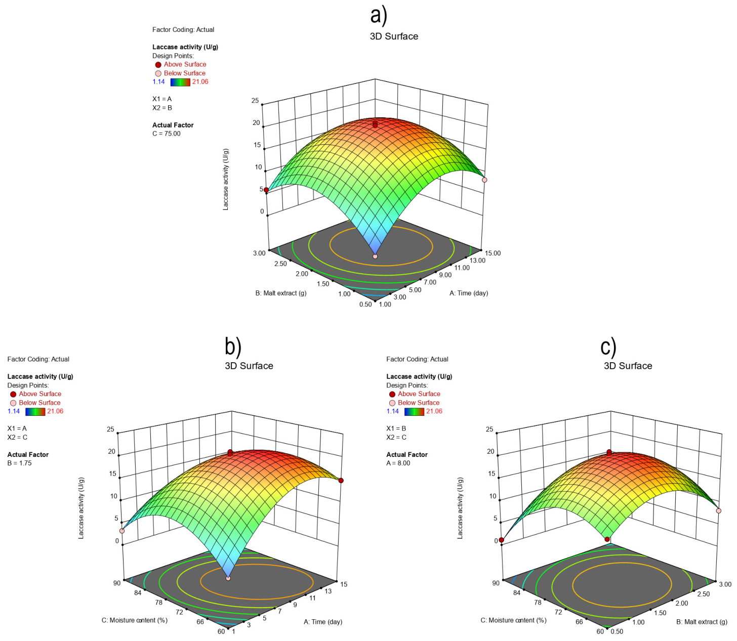
Figure 1. Three-dimensional response surface plots illustrating the interactive effects of interactions: (a) AB; (b) AC; (c) BC on laccase activity.
Processes 14 01531 g001
Figure 2. Optimum pH and temperature, and pH and thermal stability of laccase: (a) effect of pH; (b) effect of temperature; (c) pH stability; (d) thermal stability.
Processes 14 01531 g002
Figure 3. Effect of pH and temperature on the decolorization efficiency of AO, DB, and MG by laccase from S. commune TMF3: (a,b) AO; (c,d) DB; (e,f) MG.
Processes 14 01531 g003
Figure 4. The viability of MRC-5 cells upon 24-h treatment with 20% vol/vol of samples. The results are expressed as a percentage of untreated control. c p < 0.001, all treatments vs. untreated control; d p < 0.001, degraded AO compared to AO, and degraded MG compared to MG.
Processes 14 01531 g004
Table 1. Independent variables and their levels.
Independent
Variables
Level
−10+1
A: Incubation time, day1815
B: Malt extract, g0.30.91.5
C: Moisture
content, %
607590
Table 2. Box–Behnken experimental design matrix with actual factor levels and observed laccase activity.
RunIndependent VariablesResponse
Y
(Laccase Activity, U/g)
A
(Incubation Time, Day)
B
(Malt Extract, g)
C
(Moisture Content, %)
18.01.7575.020.16
21.00.5075.01.28
38.01.7575.020.46
48.01.7575.020.88
515.03.075.09.55
615.01.7560.014.89
78.03.060.08.11
815.00.575.08.29
91.03.075.05.99
108.01.7575.019.22
111.01.7590.03.26
128.00.5090.01.14
138.01.7575.021.06
148.03.090.05.90
151.01.7560.02.24
1615.01.7590.02.20
178.00.5060.010.18
Table 3. Analysis of variance (ANOVA) for the quadratic model of laccase production.
SourceY
F-Valuep-Value
Prob > F
Model197.94<0.0001 a
A119.13<0.0001 a
B18.190.0037 a
C127.44<0.0001 a
AB5.770.0472
AC91.20<0.0001 a
BC22.630.0021
A2445.28<0.0001 a
B2366.47<0.0001 a
C2438.66<0.0001 a
Lack of fit 0.4980 b
A: incubation time; B: malt extract; C: moisture content and Y: laccase activity. a Significant coefficient (p < 0.05); b Insignificant coefficient (p > 0.05).
Table 4. Effect of initial dyes and their degraded products on microorganisms.
SampleE. coliL. rhamnosusC. albicansS. cerevisiae
AO21.86 a12.16 a5.94 a8.07 a
Degraded AO2.75 b//34.21 b
DB7.58 a/9.25 a/
Degraded DB4.79 b9.41 b5.15 b9.68 b
MG58.19 a98.14 a96.89 a/
Degraded MG4.79 b24.43 a3.83 b17.37 b
a inhibition percentage, b stimulation percentage.
Table 5. Effect of pH on laccase activity and comparison with reported S. commune laccases.
Enzyme
Source
SubstratepH RangeOptimal pHActivity ProfileReference
S. commune TMF3Guaiacol3.0–8.05.0Bell-shaped profile; low activity at pH 3.0 (18.7%), gradual increase to maximum at pH 5.0, followed by decline; sharp decrease above pH 6.5This study
S. commune IBL-06ABTS3.0–9.06.0Maximum activity at pH 6.0; reduced activity at higher pH[34]
S. commune NI07ABTS3.0–10.04.46Narrow optimum; sharp decline above pH 4.7[11]
S. commune NI07
(immobilized)
ABTS3.0–10.05.5Maximum activity at pH 5.5; improved stability near optimum[11]
Table 6. Comparison of dye decolorization by laccases from S. commune TMF3 and literature reports.
DyeEnzyme SourceConditions (pH, T, Mediator)Decolorization (%)TimeInitial Dye Conc. (mg/L)Reference
AOS. commune TMF3 pH 5, 40 °C, no mediator72.23% (30 min); 83.57% (60 min)30–60 min50This study
AOT. trogiimediator (HBT); pH 5, 50 °C87.9%48 h60.38[40]
DBS. commune TMF3 pH 5, 40 °C, no mediator75.5% (30 min); 81.1% (60 min)30–60 min50This study
DBT. hirsuta BT2566pH 5, 30 °C, no mediator~4%10 h~257[41]
DBT. versicolorpH 4, 40 °C, no mediator~81%24 h40[42]
MGS. commune TMF3pH 5, 45 °C, no mediator78.79% (30 min); 88.07% (60 min)30–60 min50This study
MGTrametes sp.mediator (HBT); pH 6, 55 °C90%2.5 h50[43]
MGCerrena sp.pH 6, 28 °C, no mediator91.6%~172 min109.9[44]
Disclaimer/Publisher’s Note: The statements, opinions and data contained in all publications are solely those of the individual author(s) and contributor(s) and not of MDPI and/or the editor(s). MDPI and/or the editor(s) disclaim responsibility for any injury to people or property resulting from any ideas, methods, instructions or products referred to in the content.