Article

Microwave-Assisted Synthesis of Antimicrobial Silver Nanoparticles Using Propolis Extracts

1
Lithuanian Research Centre for Agriculture and Forestry, Institute of Horticulture, LT-54333 Babtai, Lithuania
2
Department of Pharmacognosy, Faculty of Pharmacy, Lithuanian University of Health Sciences, LT-50162 Kaunas, Lithuania
3
Institute of Pharmaceutical Technologies, Faculty of Pharmacy, Lithuanian University of Health Sciences, LT-50162 Kaunas, Lithuania
4
Department of Drug Technology and Social Pharmacy, Faculty of Pharmacy, Lithuanian University of Health Sciences, LT-50161 Kaunas, Lithuania
5
Research Institute of Natural and Technological Sciences, Vytautas Magnus University, 46324 Kaunas, Lithuania
6
Laboratory of Pharmaceutics and Phytopharmaceutical Drug Development, Faculty of Pharmaceutical Sciences, University of Kinshasa, Kinshasa B.P. 212, Democratic Republic of the Congo
7
Department of Pharmacy, Faculty of Medecine and Pharmacy, University of Kisangani, Kisangani B.P. 2012, Democratic Republic of the Congo
8
Department of Pharmacy, Faculty of Pharmaceutical Sciences and Public Health, Official University of Bukavu, Bukavu B.P. 570, Democratic Republic of the Congo
9
Centre de Recherche et d’Innovation Technologique en Environnement et en Sciences de la Santé (CRITESS), University of Kinshasa, Kinshasa B.P. 212, Democratic Republic of the Congo
*
Author to whom correspondence should be addressed.
Processes 2025, 13(9), 2861; https://doi.org/10.3390/pr13092861
Submission received: 28 May 2025 / Revised: 6 August 2025 / Accepted: 5 September 2025 / Published: 7 September 2025

Abstract

Silver nanoparticle (AgNP) biosynthesis using propolis extracts as capping and reducing agents presents multiple opportunities for solving many biological challenges. This study demonstrates a sustainable, low toxicity, and high production cost phytochemical synthesis of AgNPs using propolis extracts and microwaves. Biosynthesized with propolis, AgNPs are analyzed by various methods: phytochemical, physicochemical, and morphological, such as TEM. The determined antimicrobial and antioxidant activity of propolis extracts is compared with the activity of green synthesized AgNPs with propolis. The green synthesized AgNPs are spherically shaped with an average size ranging from 6 to 40 nm. The biosynthesized AgNPs demonstrate potential antibacterial activity on tested microorganism strains two folds higher than pure propolis extracts.

1. Introduction

The increasing demand for innovative and natural solutions in agriculture, food, pharmaceutical, and cosmetic industries has drastically heightened attention in propolis and natural extracts [1]. It is characterized by rapid production, environmental friendliness, simplicity, and low cost [2,3]. Nanoparticles with propolis extract have a wide potential for applications in drug delivery and biomedical imaging [4,5,6]. The natural extract of green propolis is a resinous substance collected by bees from exudates and plant buds [7]. For centuries, there has been a wide demonstration of the bioactive potential of propolis [8]. Bioactivity of this resin includes antimicrobial, antitumor, antiviral, anti-inflammatory, antimicrobial, and antiparasitic actions [9,10,11]. In the study of Silveira et al. [12], the authors showed antiviral action against COVID-19 using propolis in the treatment of kidney damage and the reduction of hospitalization time. Sokolonski et al. [13] analyzed its antifungal action against isolates of Candida albicans. Silva-Beltan et al. [14] confirm its efficiency against coronavirus 229E. The cause of this bioactivity is the confirmation of phenolic compounds (esters group of phenolic acid and flavonoids) in its composition [10]. Different types of propolis have different bioactive compounds and have different solvents and different types of extraction techniques [15,16], which result in different extracts and chemical profiles [17]. Conventional methods like Soxhlet extraction tend to require a longer time for analysis, the decaying of bioactive compounds because of high temperature, and high solvent consumption. Perhaps non-conventional methods would require a shorter time, less environmental impact, and would provide a greater purity [18,19].
Like various plant extracts, a high concentration of phenolic compound epically artepilin C is present, which contains antitumor, anti-inflammatory, and antimicrobial properties. The literature has shown that the antioxidant capacity of propolis is much higher than common plant extracts such as grape seed and green tea [20]. Green propolis has strong reducing agents which facilitate the conversion of Ag+ to Ag0, forming Ag nanoparticles. The synthesis of nanoparticles occurs mainly in three phases: (1) The activation phase: In this phase, metal ions reduce, followed by a coagulation of that metal ion, while green extract behaves as reducing agent by oxidizing itself. (2) The growth phase: It is characterized by (Ostwald ripening) rapid aggregation of small pieces to larger ones. (3) The stabilization phase: In this phase, the extract behaves as a stabilizing agent [21]. These phases are intended to have decreased pollution, a low toxicity of nanoparticles, a fast synthesis, and a wide spectrum efficacy. For achieving these goals, ultrasound-assisted and microwave-assisted synthesis can be adopted. These techniques lower the duration of the synthesis of nanoparticles, thus ensuring physiological activity and the smaller size of nanoparticles [22,23].
Plants, algae, bacteria, yeast, fungi, and various other organisms can be used for the synthesis of AgNPs; they have various advantages as compared to chemical and physical approaches. Out of all these methods involving yeast, viruses, microorganisms, marine source, fungi, and bacteria, metal nano particle synthesis with plant-based extracts is strongly preferred [24]. The synthesis of nanoparticles by utilizing microorganisms is not a steady process and only produces a few sizes and shapes. Highly advanced conditions and equipment are needed to form clear filtrates in colloidal broths for the synthesis of NPs. Extensive research and large applications are required with the host microbe in the case of using viruses for the synthesis of NPs [25]. Plants such as Rumex rosus [26], Vernonia amygdalina [27], Bauhinia purpurea [26], Mussaenda glabrata [28], Pueraria tuberosa [26], and Sanvitalia procumbens [29] are used for synthesis of AgNPs. It is stated that green synthesized AgNPs are ecologically friendly, effective, inexpensive, and nontoxic. All plants have different chemical phenomena, and hence significant differences in shape, sizes, and the biological and physiochemical properties of AgNPs. As mostly conventional heating methods used in the synthesis of AgNPs can require long periods of time and release huge amounts of energy, microwave-assisted AgNPs facilitate faster nanoparticle development by accelerating the nucleation process with uniform and controlled energy distribution [29]. The literature shows the results of nanoparticle formation with conventional and microwave heating, and proves that NPs with microwave heating have smaller size dispersion, high crystallinity, and greater shape morphogenesis. Various other studies also facilitate microwave-assisted nanoparticles over conventional methods [30]. It consumes lower energy with less time, and provides better conditions of synthesis to reactants, with the interaction of solvent and radiation [31,32,33,34].
Propolis, as a natural raw material, due to its unique composition and diverse effects (antioxidant, regenerative, anti-inflammatory, antibacterial), can become an alternative active ingredient in the development of medical products, starting from the prevention of cardiovascular diseases, oral health care, wound healing, and another diseases [35,36,37,38]. Antimicrobial effects have been widely reported among the many biological activities of propolis extracts. Ethanolic propolis extracts are effective against a broad range of bacteria, especially against Gram-positive bacteria species [39]. The antimicrobial activity of European propolis has been related to its contained phenolics, flavonoids, and caffeic acid derivatives [40,41]. It is increasingly being discussed and studied that propolis has specific properties as a food preservative, which is ensured by its bactericidal and bacteriostatic properties [40]. Propolis contains more than 300 biologically active compounds, and its chemical composition varies widely depending on its botanical and geographical origin. Therefore, propolis is an attractive natural material that does not induce antibiotic resistance [41]. Furthermore, propolis preparations may act synergistically with antibiotics [42]. However, there is a challenge in selecting solvents and creating preparations from propolis with a high yield of biologically active compounds. Ethanol is one of the best solvents and is commonly used to extract propolis, but at the same time this limits the broader application of ethanolic extracts. Our previous research confirmed that non-ethanolic propolis extracts (NEP) prepared by the developed technology could be an alternative to ethanolic propolis extract, since it contains many antioxidants, namely flavonoids and phenolic acids [43], comparable to that of ethanolic extract. Predominantly identified compounds were phenolic acids, contributing ca. 40% of the total radical scavenging activity.
Moreover, the investigated non-ethanolic extracts inhibited the growth and reproduction of all tested microorganisms. The antimicrobial activity of some extracts was equal to or exceeded the antimicrobial effect of the ethanolic extract [44]. Therefore, the development and application of alternative non-ethanol extract technologies of propolis have become excellent alternatives for the broader application of propolis [45]. Considering that, in the literature, no one studied propolis with microwaves of different ratios on its effectiveness, for the first time, the author uses a microwave-assisted extract with different proportions for the determination of antioxidant and antimicrobial activity. This study focused on synthesizing AgNPs with propolis, based on a microwave approach, and evaluating morphological and phytochemical properties and antimicrobial activity.

2. Materials and Methods

2.1. Propolis Material and Preparation of Non-Ethanolic Propolis Extracts

Preparation of non-ethanolic propolis extracts (NEP): Raw propolis was obtained from the beekeeper company UAB Bitute (Vilnius, Lithuania). Prior to analysis, propolis samples were kept in a fridge in the dark. Crude propolis was ground into powder. The NEP was prepared by adding a system of solvents, namely a mixture of water and 30% polyethylene glycol 400 (makrogolum) at 1:10 (w/v), at 1:15 (w/v), and at 1:20 (w/v) sample-to-solvent ratio. Extractions were conducted by shaking for 10 min at 70 °C temperature in an ultrasonic bath of 35 kHz frequency.
After extraction, extracts of propolis were filtered through Whatman No. 1 filter paper. Solutions were clear, yellow liquids and remained stable when stored, i.e., the color remained unchanged, no precipitate was observed, and they did not turn white.

2.2. Preparation of AgNPs

An amount of 0.34 g of silver nitrate (AgNO3) was dissolved in etylenglycol and the obtained solution was added to 100 mL of NEP of propolis extract. After completely dissolving AgNO3, we exposed the solution to microwaves at 450 W for 120 s by using a commercial microwave with some modifications. The solution was placed into a big size round bottom flask. The flask was further connected with a condenser. After completion of 120 s, the extract was immediately filtered. The resulting solution was left to settle at room temperature for 24 h. Due to the formed AgNPs, the solution acquired a yellowish-brown color.

2.3. Reagents

The reagents used in the analysis satisfied all quality requirements and were of analytical grade. The following substances were used in the study: ethanol 96% (v/v) (manufactured by AB MV GROUP Production, Vilnius, Lithuania), the Folin–Ciocalteu reagent, gallic acid, acetic acid, aluminium chloride hexahydrate, hexamethylenetetramine, rutin, DMCA (4-(dimethylamino)-cinnamaldehyde), hydrochloric acid, (-)-epicatechin, sodium nitrite, sodium molybdate, sodium hydroxide, chlorogenic acid, DPPH (2,2-diphenyl-1-picrylhydrazyl), ABTS (2,20’-azino-bis(3-ethylbenzothiazoline-6-sulfonic acid), potassium persulfate, ammonium acetate, copper (II) chloride dihydrate, neocuproine, Trolox ((±)-6-hydroxy-2,5,7,8-tetramethylchromano-2-carboxylic acid) (Sigma-Aldrich Steinheim, Germany), TPTZ (2,4,6-Tris(2-pyridyl)-s-triazine) (Carl Roth GmbH, Karlsruhe, Germany), and iron (III) chloride hexahydrate (Vaseline-Fabrik Rhenania, Bonn, Germany). Purified deionized water was produced using the Milli-Q® 180 water purification system (Millipore, Bedford, MA, USA).

2.4. Spectrophotometric Studies

Determination of the Total Content of Phenolic Compounds, Flavonoids, Proanthocyanidins and Hydroxycinnamic Acid Derivatives

All spectrophotometric measurements were carried out using a spectrophotometer M550 (Spectronic CamSpec, Garforth, United Kingdom). The total phenolic content in the extracts was determined by the Folin–Ciocalteu assay proposed by Bobinaitė et al. [46] and expressed as mg gallic acid equivalent (GAE) per gram of dry weight. The total flavonoid content was determined using the described methodology by Urbonavičiūtė et al. [47] and expressed as mg rutin equivalent (RE) per gram of dry weight. The total proanthocyanidin content was determined by the DMCA assay proposed by Heil et al. [37] and expressed as mg (−)-epicatechin equivalent (EE) per gram of dry weight. The total hydroxycinnamic acid derivatives content was determined using the described methodology by Didier et al. [16] and expressed as mg ChAE/g DW.

2.5. Evaluation of Antioxidant Activity

ABTS•+ (2,2′-azino-bis(3-ethylbenzthiazoline-6-sulphonic acid)) radical cation scavenging assay was applied by taking 7 mM aqueous solution of ABTS. Radical cation (•+) was prepared from stock solution of ABTS, having a final concentration of 2.45 mM K2S2O8. This solution was kept in the dark before use for about 12—16 h. Because potassium persulfate and ABTS react stoichiometrically with a ratio of 0.5:1, this results in partial oxidation of ABTS. The formation of the radical began immediately, but required 6 h minimum to reach stability. Thus, it can keep its stability for two days or more in dark conditions [48].
The DPPH• (2,2-diphenyl- 1-picrylhydrazyl)) free radical scavenging activity was determined using the free radical of DPPH. For all antioxidants, various concentrations were tried and tested. Methanol (0.1 mL) in aqueous solution was added up till 3.9 mL in DPPH• methanol solution with a concentration of 6 × 10⁻⁵ mol/L [49].
The reduction activity of extracts was determined by taking a freshly prepared blank M1 at 593 nm; 300 μL of FRAP reagent having 37 °C; 30 μL of distilled water was added with 10 μL of sample with final dilution ratio of 34/1. Absorbance values were measured after every 0.5 s and 15 s during the monitoring period, and the average was noted. The maximum monitoring time was up to eight min [50], and the cupric reducing antioxidant capacity (CUPRAC) assay was carried out by making a solution of copper (II) chloride after dissolving 0.4262 g of CuCl2·2H2O in 250 mL distilled water. A solution of neocuproine (Nc) was prepared by taking 0.039 g of Nc at 7.5 × 10⁻³ M in 96% EtOH, and then diluted to 25 mL using ethanol. Buffer solution (pH-7) of ammonium acetate (NH4Ac) was prepared by taking 19.27 g of NH4Ac in 250 mL of distilled water [51]. The antioxidant activity of the extracts was expressed as mmol of the Trolox equivalent (TE) per one gram of raw material.

2.6. Microscopy

The morphology of green synthesis AgNPs was studied from images obtained by Transmission Electron Microscopy (TEM) Tecnai G2 F20 X-TWIN (FEI) equipped with a field emission electron gun at the accelerating voltage of 200 kV. For microscopic studies, the diluted samples were deposited on the TEM grids. The sizes of AgNPs were measured by software ImageJ v.1.54d.

2.7. Antimicrobial Activity

The extracts’ and synthesized nanoparticles’ antimicrobial activity was investigated against Gram-negative and Gram-positive bacteria cultures. The agar diffusion was chosen for the evaluation of antibacterial activity. The antimicrobial activity of extracts was tested via an agar well diffusion assay. For this purpose, using sterile cotton swabs, a 0.5 McFarland Unit density suspension (~108 CFU mL−1) of each pathogenic bacterial strain was inoculated onto the surface of the cooled Mueller Hinton Agar (Oxoid, UK). Wells of 6 mm diameter were punched in the agar and filled with 50 µL of the tested AgNPs-propolis material. The experiments were repeated three times, and the average size of the inhibition zones was calculated. The antimicrobial activities against the tested bacteria were determined by measuring the diameter of the inhibition zones (mm). The antimicrobial activity of extracts was determined against Staphylococcus aureus ATCC 25923, beta-hemolytic streptococcus group b ATCC 15185, Staphylococcus epidermidis ATCC 12228, Enterococcus faecalis ATCC 29212, Escherichia coli ATCC 25922, Klebsiella pneumoniae ATCC 13883, Pseudomonas aeruginosa ATCC 27853, Proteus vulgaris ATCC 8427, Bacillus cereus ATCC 11778, Listeria monocytogenes ATCC 19115, and Candida albicans ATCC 10231 in Lithuanian University of Health Sciences (Kaunas, Lithuania).

2.8. Color Measurement of Propolis/AgNPs

The extracts and solutions of the formed AgNPs were evaluated according to the color coordinates, which confirmed that the synthesis of silver nanoparticles took place. Color indices in the CIEL*a* b* space were measured with a MiniScan XE Plus spectrophotometer (Hunter Associates Laboratory, Inc., Reston, VA, USA) and chroma (C = (a*2 + b*2)1/2 and the hue angle was calculated (h° = arctan (b*/a*)).

2.9. Statistical Analysis

The statistical analysis was performed using SPSS v.20 software (SPSS Inc., Chicago, IL, USA). All the data were prepared in triplicate (n = 3), and the results are shown as mean ± standard error mean (SEM) of all calculated values. Differences at p < 0.05 were statistically significant.

3. Results

3.1. Color Change of Propolis and Propolis/AgNPs

The flourishing green synthesis of AgNPs using the propolis extracts was confirmed by color change and spectroscopic studies of the reaction medium and nanoparticles. After a mixture of propolis extracts and silver nitrate, it showed an apparent color change from light cream to dark brown (Figure 1). Considering the different types of propolis concentration, 10%, 15%, and 20% extracts were the lightest, and the darkest extracts in each category were 10% AgNPs, 15% AgNPs, and 20% AgNPs. The literature data suggest that the darker propolis typically has a higher concentration of polyphenols, higher antioxidants, and antimicrobial activities [52].
AgNPs exhibits surface plasmon resonance phenomenon in which free electrons on specific wavelength resonate with light, which results in a shift in color change from yellowish to dark brown or blackish. Color change is the first sight indicator. Darker color indicates a reduction in Ag ions from Ag+ to Ag0. The color coordinates (L, a, b) were also determined to assess changes (Table 1). The lightness (L) coordinate ranges from 0 (dark) to 100 (light). Considering ten types of propolis extracts, 10%, 15%, and 20% concentrations of propolis were the lightest, and the darkest products in each sample were 10%-1 AgNPs, 15%-1 AgNPs, and 20%-2 AgNPs.
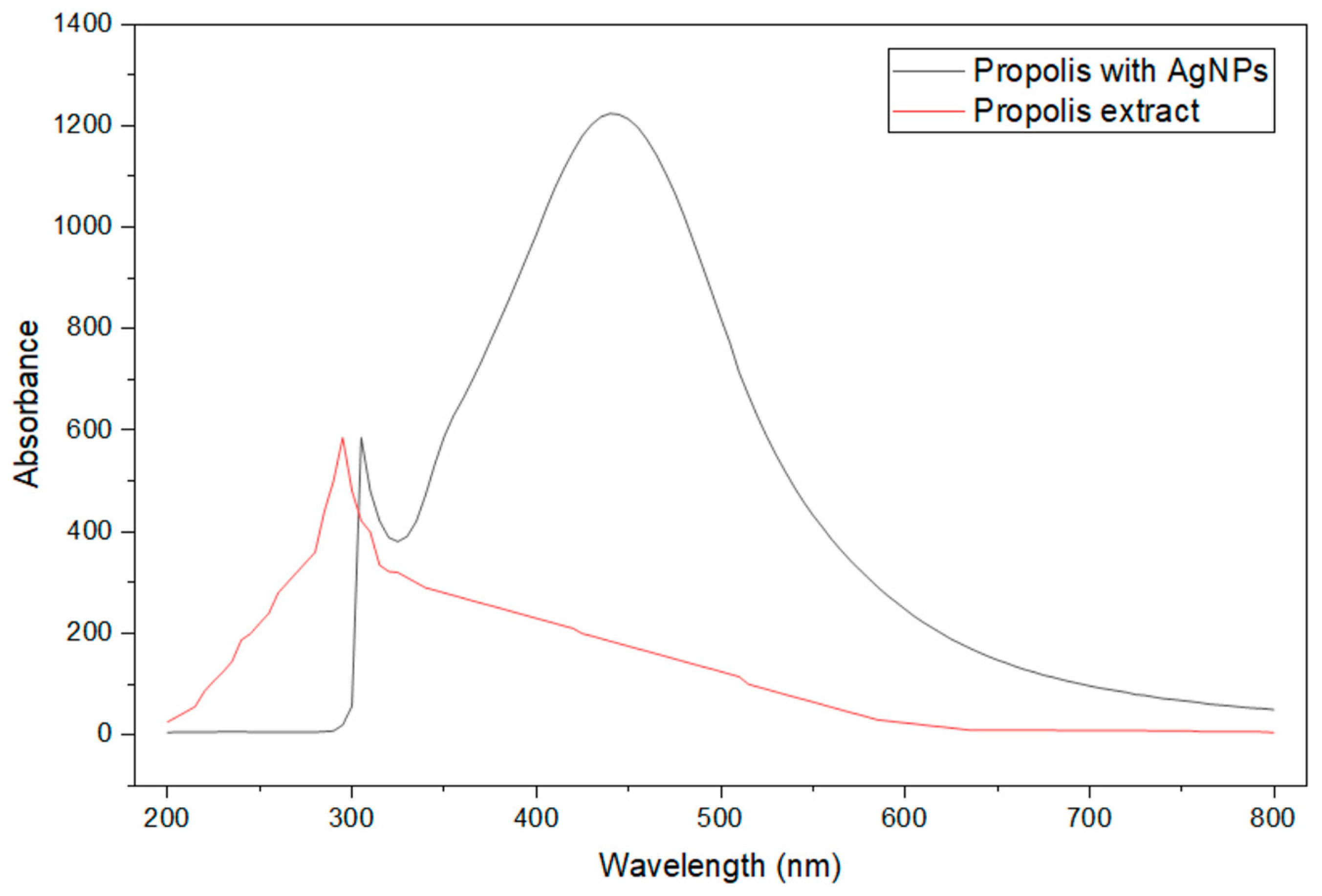
The UV-Vis spectrophotometric analysis of the total composition of phenolic compounds, flavonoids, proanthocyanidins, and hydroxycinnamic acid derivatives in propolis extracts and propolis/AgNPs was performed to evaluate the phytochemical composition. The obtained results are summarized in Table 2 and Figure 2 and Figure 3. The UV-Vis graph shows the silver peak around 400 nm and, for the presence of bioactive compounds, the propolis extract showed an absorbance peak around 290 nm.
The highest total phenolic content (145.69 ± 2.91 mg GAE/g DW, p < 0.05) was determined in 20-2 propolis extract. The highest total proantocyanidin content (4.98 ± 0.50 mg EE/g DW and 5.23 ± 0.52 mg EE/g DW), the total hydroxycinnamic acid derivatives content 7.28 ± 0.36 mg ChE/g DW and 6.81 ± 0.34 mg ChE/g DW, and the total flavonoid content (31.02 ± 0.62 mg RE/g DW and 31.89 ± 0.64 mg RE/g DW) were found [ML1] in propolis extracts 20-1 and 20-2, respectively. All results of the phytochemical analysis of propolis extracts and biosynthesized AgNPs are shown in Table 2.
There are many studies on total phenolic content in propolis. Some of them are as follows: Socha et al. [32] in Poland: 150.05–190.97 mg GAE/g DW; Salleh et al. [33] in Malaysia: 7.60–13.21 mg GAE/g; Altuntaş et al. [34] in Turkey: 16.73–125.83 mg GAE/g; Ibrahim & Alqurash [54] in Egypt: 210.33–321 mg GAE/g; Fikri et al. [36] in Indonesia: 10.30–28.65 mg GAE/g; Moreira et al. [37] in Portugal: 151.00–329.00 mg GAE/g; Nina et al. [38] in Bolivia: 43.0–176.0 mg GAE/g. Zhang et al. determined that the total phenolic content in Chinese propolis extract was 192.80 ± 10.85 mg GAE/g and the total flavonoid content was 297.24 ± 10.32 mg RE/g [55]. The total phenolic content and total flavonoid content found in our tested propolis extracts were lower. These results may be influenced by different extraction, sample preparation, propolis composition, climate, and other conditions.
Such a decrease in phenolic compounds after synthesizing silver nanoparticles is regular. Proanthocyanidin, phenolic, flavonoid compounds, and hydroxycinnamic acid derivatives are active compounds in the synthesis reaction. They act as reducers, so there is no need to use any chemical compounds, stabilizers, or anticoagulants [56,57].
The results showed that the total amount of proanthocyanidins, hydroxycinnamic acid derivatives, phenolic content, and flavonoids decreased after synthesizing silver nanoparticles in all extracts (Table 2). Notably, propolis crude extract and propolis/AgNPs total phenolic content in all tested cases decreased ~9.5 times.

3.2. Determination of Antioxidant Properties of Propolis and Propolis/AgNPs

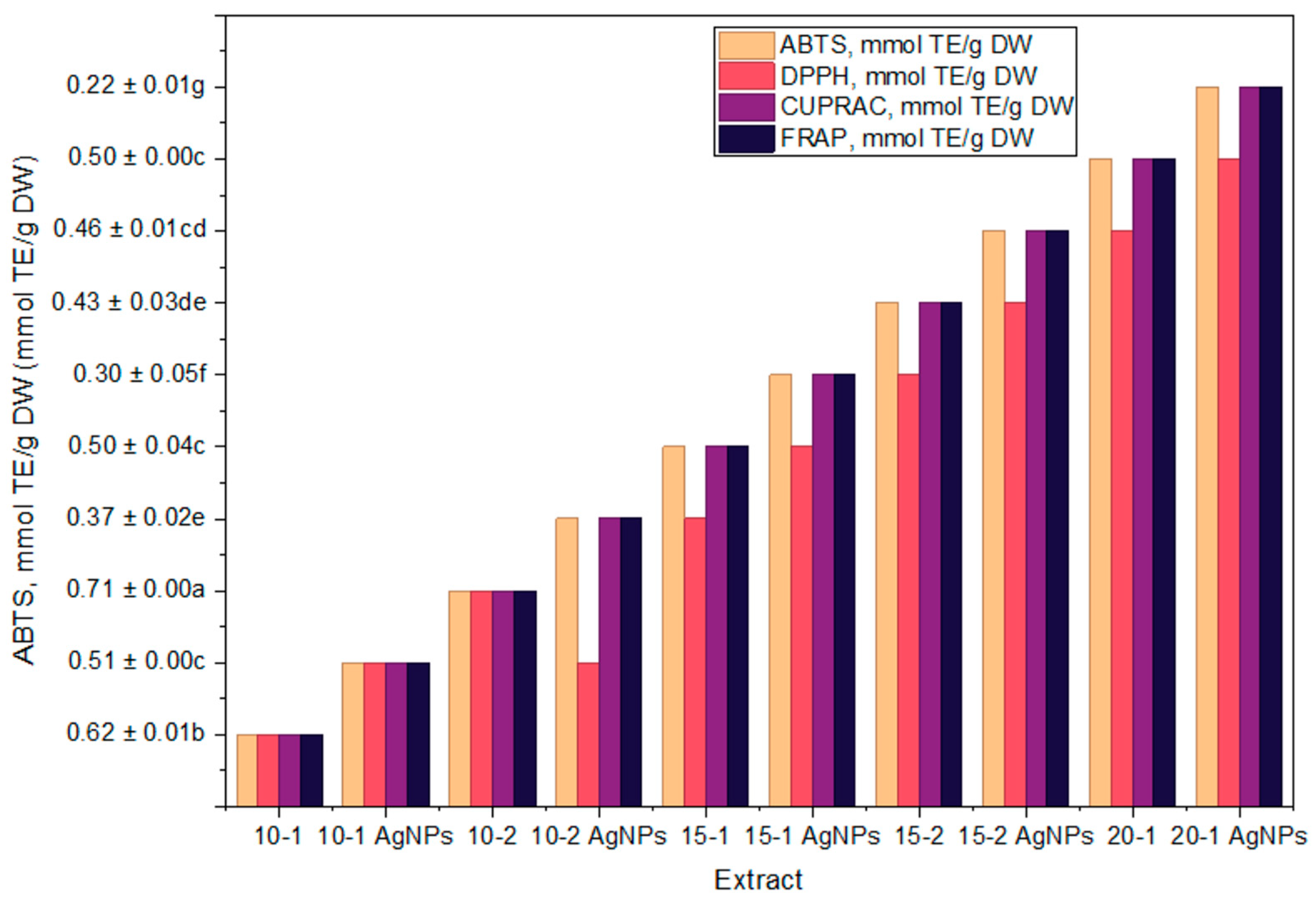
ABTS, DPPH, CUPRAC, and FRAP UV-Vis spectrophotometry methods to the in vitro studies of selected propolis extracts’ antioxidant activity were applied. The results are summarized in Table 3.
The evaluation of the antioxidant activity of propolis preparations is of crucial importance. This provides a scientific basis for the use of propolis preparations for the prevention of damage caused by oxidative stress. Table 4 and Figure 4 show the results of the antioxidant activity of propolis extracts, measured by ABTS, DPPH, CUPRAC, and FRAP UV-Vis spectrophotometry methods.
The tested propolis extracts show an intense antioxidant activity in vitro. Determination of selected propolis extracts using four different in vitro spectrophotometric methods showed that the strongest in vitro antiradical activity, assessed by ABTS and DPPH methods, and in vitro reducing activity, determined by the FRAP method, were characterized by the extract of the 10-2 propolis sample. Bertotto et al. determined that antiradical activity in vitro tested by the DPPH assay of the Brazilian-type propolis extract was 28.55 ± 0.03 mmol TE/g [42]. The antiradical activity in vitro tested by the DPPH assay of our European-type propolis was weaker. These results could be due to differences in propolis type, composition, extraction, climatic conditions, and other factors. Şengül and Seda found that reducing activity in vitro of 10% water extract of propolis from Türkiye determined by the FRAP assay was 1.78 ± 0.07 mmol TE/g [43]. These data correspond with our results of reducing activity in vitro of European-type propolis collected in Lithuania.
Silver nanoparticles were successfully obtained using propolis extracts. Further, the antioxidant activity of biosynthesized AgNPs was compared to the antioxidant activity of propolis extracts without silver nanoparticles. Data obtained by ABTS, DPPH, and FRAP in vitro assays show that biosynthesized AgNPs possess about a two-fold lower antioxidant potential. This could be caused by the decrease of biologically active compounds in AgNP samples. The decrease in antioxidant activity confirms the formation of silver nanoparticles. These compounds participate in the green synthesis reaction as active agents that act as reducing agents, anticoagulants, and stabilizers [58,59].

3.3. Transmission Electron Microscopy (TEM) of Propolis/AgNPs

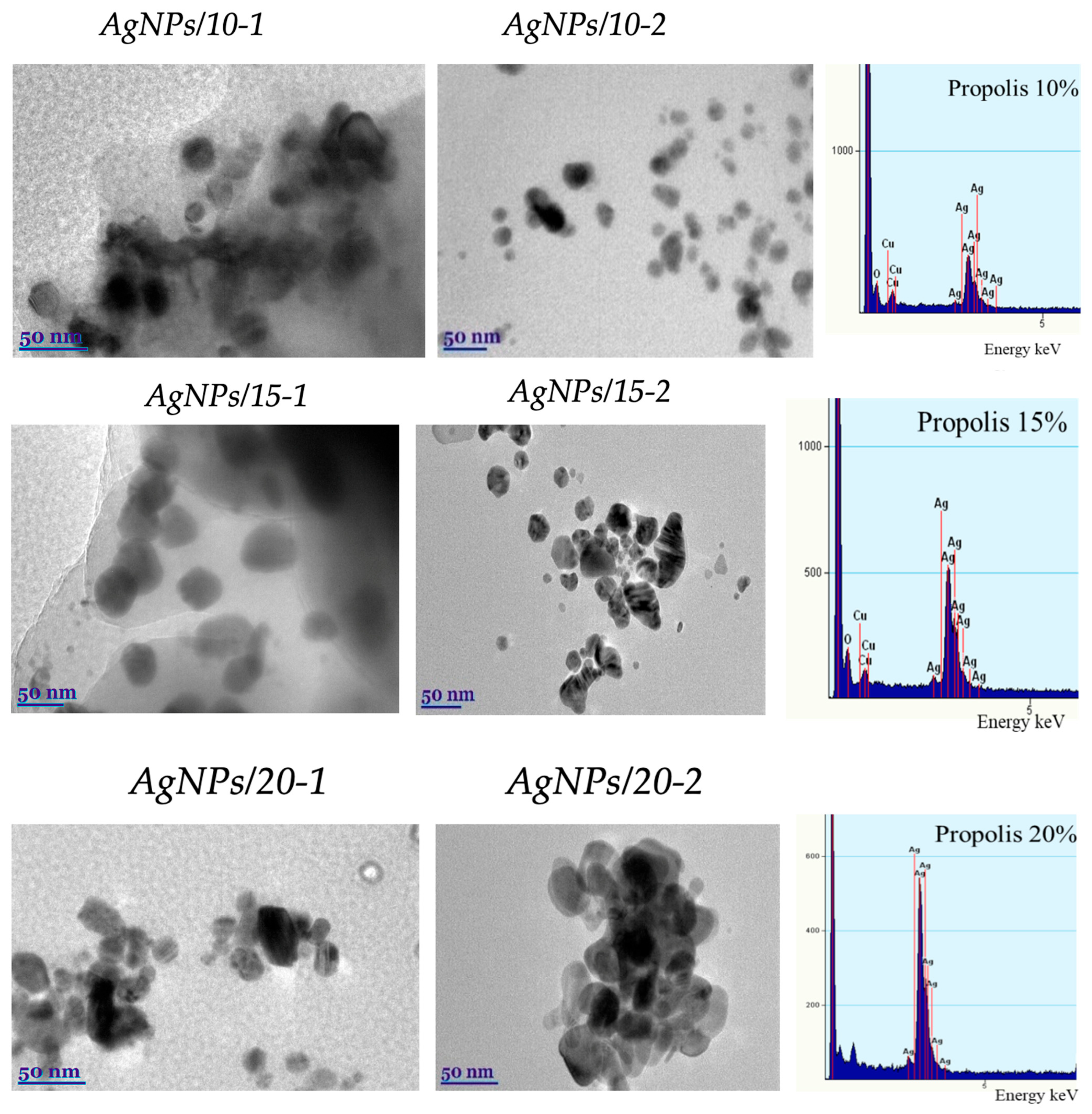
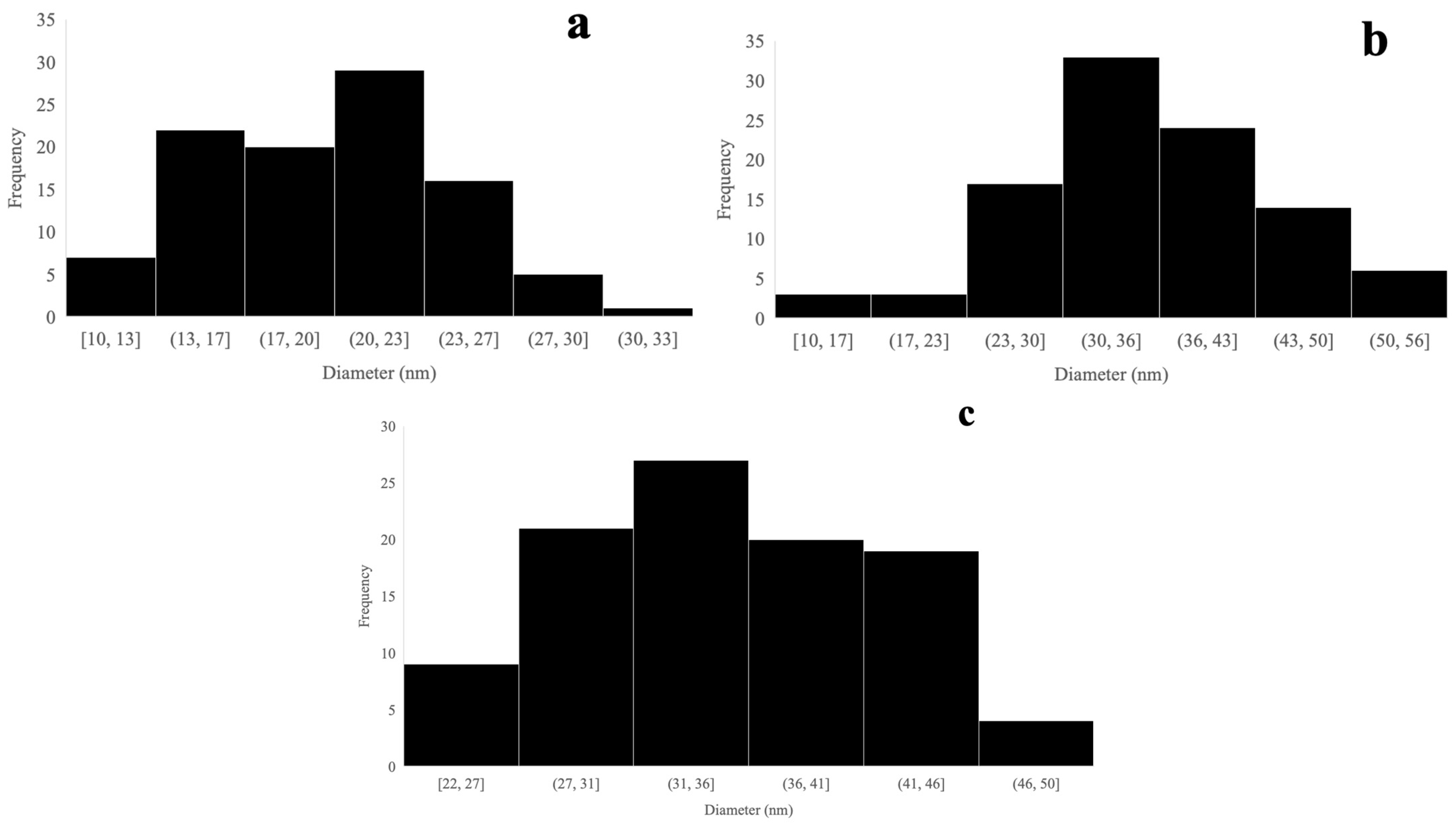
TEM and TEM-EDS examined the morphology, shape, and size of synthesized propolis/AgNPs. It can be found in the scientific literature that the type of plant extract, or other natural reducing agent, and its concentration influence the morphology of the nanoparticles formed. In contrast, the pH and temperature of the extract medium control the growth and size of the nanoparticles [60]. TEM images clearly show that the synthesized silver nanoparticles are predominantly spherical and below 50 nm in size (Figure 5). We can observe that the type of extract and its concentration affect the shape of silver nanoparticles. Particle size distribution histogram of different concentration of propolis is presented in Figure 6.

3.4. Antimicrobial Activity

Ten propolis extracts and AgNPs synthesized in these extracts were investigated to evaluate their antibacterial activity against ten different pathogenic and opportunistic bacteria strains using the disc diffusion method. Based on the current literature, the possible mechanisms include the release of Ag+ ions that disrupt protein functions by binding to thiol groups, the generation of reactive oxygen species (ROS) that cause oxidative stress and damage cellular components, and the direct interaction of silver nanoparticles with bacterial cell membranes, leading to increased permeability and cell lysis. Additionally, silver nanoparticles can interfere with bacterial DNA replication and transcription, cause ribosome disassembly and protein synthesis inhibition, and lead to ATP depletion. They may also affect quorum sensing, inhibit signal transduction, and prevent biofilm formation. These mechanisms may act individually or synergistically to exert strong antibacterial effects. The evaluation of the antibacterial activity of these propolis extracts and AgNPs was recorded in Table 4. The results revealed that all propolis extracts could effectively suppress microbial growth bacteria with variable potency. The results revealed that 20-1 and 10-1 extracts are potentially effective in suppressing bacterial growth with a range of inhibition zones from 6.20 to 11.90 mm. Accordingly, propolis extracts were found to show slightly higher antibacterial activity against Gram-positive bacteria than Gram-negative bacteria (p > 0.05) due to the variation in their cell wall structure [60,61]. The 10-2 propolis extract exhibits a more potent inhibitory effect against four pathogens, except S. aureus, P. aeruginosa, P. vulgaris, B. cereus, E. faecalis, and C. albicans. Notably, a high inhibition ability of propolis was detected against K. pneumoniae and P. aeruginosa, which cause various infections, including pneumonia [62].
Propolis extracts and AgNPs synthesized in these extracts were investigated using the disc diffusion method to evaluate their antibacterial activity against ten different opportunistic pathogens strains. The evaluation of the antibacterial activity of these propolis extracts and AgNPs was recorded in Table 4. The results revealed that all propolis extracts could effectively suppress microbial growth bacteria with variable potency. The results revealed that 20-1 and 10-1 extracts are potentially effective in suppressing bacterial growth with a range of inhibition zones from 6.20 to 11.90 mm. Accordingly, propolis extracts were found to show slightly higher antibacterial activity against Gram-positive bacteria than Gram-negative bacteria (p > 0.05) due to the variation in their cell wall structure [63,64]. The 10-2 propolis extract exhibits a more potent inhibitory effect against four pathogens, except S. aureus, P. aeruginosa, P. vulgaris, B. cereus, E. faecalis, and C. albicans. Notably, a high inhibition ability of propolis was detected against K. pneumoniae and P. aeruginosa, which cause various infections, including pneumonia [65]. Antibacterial effects of propolis were investigated against Gram-negative bacteria K. pneumoniae in mouse blood. The inhibitory activity of propolis extracts against Gram-positive bacteria, especially Staphylococcus aureus and Candida albicans, has been widely reported in the literature [66,67,68,69,70]. The antibacterial action of propolis against bacteria needs to be clearly understood. Different studies show that propolis’s antibacterial potential depends on its geographic location. It is also believed that flavonoids can reduce bacterial resistance to various antibacterial compounds via binding to the bacterial cell wall, resulting in their lysis and death [71].
As can be seen from Table 4, biosynthesized propolis extracts demonstrate a 30–60% higher antibacterial activity against all tested pathogenic microorganisms compared to pure propolis extracts. Comparing antibacterial properties between nanoparticles, propolis extracts/AgNPs more effectively prevent the growth of the bacteria tested, with a range of inhibition zone from 11.48 to 22.20 mm.
In the literature, there have been several antimicrobial studies of propolis and AgNPs and their combined form in a bacterial growth-dependent manner. In addition, the antimicrobial activity of AgNPs also depends on the nanoparticle hydrodynamic parameters and particle size [72]. Authors Mançano et al. prove size 35–80 nm AgNPs prepared from green propolis, having quasi-spherical shapes, showed antibacterial, antioxidant, anti-inflammatory, and anticancer activity against E. coli, S. aureus, and are used in recovering wounds [1]. Authors Islam et al. report that AgNPs synthesized from red propolis, with a size and shape of 50–90 nm, spherical, showed antioxidant, wound healing, antifungal, and anticancerous activity, having application in photocatalysis of malachite dye and occlusive dressings [73]. Another study on black propolis showed a 10–30 nm size and spherical shape of nanoparticles. It showed antimicrobial, anti-inflammatory, and neuroprotective biological activity, having strong neurodegenerative therapy cytotoxic and antioxidant activity [1]. In a study from Iran, propolis showed antioxidant, antibacterial, and geno-protective biological activity, while NP with size ~45 nm and spherical shape showed low cytotoxicity and free radical scavenging application [74]. Another type of propolis, “Apis mellifera”, showed cytoprotective and antimicrobial activity, while 20–50 nm sized nanoparticles have a strong activity against fungi and oral bacteria [75].

4. Conclusions

In this work, the authors synthesized silver nanoparticles using two types of propolis and three concentrations of propolis, 10, 15 and 20%, as reducing, capping, and anticoagulant agents, and observed the effect on the particle size, antioxidant, and antimicrobial activity. The size of AgNPs mediated by two types of propolis extract was found to depend on the concentration of propolis, but no significant increase in particle size was observed, even in the presence of 20% propolis. Phenolic and flavonoid compounds, proanthocyanidins, hydroxycinnamic acid derivatives, and capped AgNPs in propolis extracts are responsible for its biological activity. The synthesized green AgNPs exerted notable antibacterial and antioxidant activity properties against both tested Gram-positive and Gram-negative bacteria strains. Moreover, the biological evaluation of AgNPs revealed strong bactericidal properties against Staphylococcus aureus, Klebsiella pneumoniae, Escherichia coli, and Pseudomonas aeruginosa, pathogens commonly involved in infectious diseases. The values of varied in the range of 0.15 to 2.04 mg CGA g−1 DW; after the synthesis of silver nanoparticles, these values decreased in the range of 0.09 to 1.78 mg CGA g−1 DW.

Author Contributions

Conceptualization, A.B. and J.V.; methodology, A.B. and J.V.; software, J.V.; validation, A.B., J.V. and P.V.; formal analysis, A.B., S.H.Z. and M.L.; investigation, J.V., S.S., P.B.M., M.L., D.K.I., J.V. and A.B.; resources, P.V.; data curation, J.V., L.K., J.K., S.S., S.T., A.B. and J.V.; writing—original draft preparation, A.B., S.S., J.K., P.B.M., D.K.I., J.V., M.L. and S.H.Z.; writing—review and editing, A.B., S.H.Z., S.S., L.K., J.K. and J.V.; visualization, A.B., S.H.Z. and J.V.; supervision, P.V. All authors have read and agreed to the published version of the manuscript.

Funding

The authors would like to thank the Research Council of Lithuania, (LMTLT) agreement No. S-MIP-24-56, for the financial support given to achieve this work.

Institutional Review Board Statement

Not applicable.

Informed Consent Statement

Not applicable.

Data Availability Statement

All data is included in this article.

Acknowledgments

The authors thank the Research Council of Lithuania (LMTLT) for supporting this research.

Conflicts of Interest

The authors declare no conflicts of interest.

References

  1. Mançano, R.R.; Matheus, L.R.; Castro, L.E.N.; Barroso, T.L.C.T.; Da Rosa, R.G.; Ferreira, V.C.; Forster-Carneiro, T.; Colpini, L.M.S. Extraction Techniques for Propolis and Its Utilization in Silver Nanoparticle Synthesis: A Comprehensive Review. Eur. Food Res. Technol. 2025, 251, 1397–1433. [Google Scholar] [CrossRef]
  2. Zehra, S.H.; Ramzan, K.; Viskelis, J.; Viskelis, P.; Balciunaitiene, A. Advancements in Green Synthesis of Silver-Based Nanoparticles: Antimicrobial and Antifungal Properties in Various Films. Nanomaterials 2025, 15, 252. [Google Scholar] [CrossRef] [PubMed]
  3. Balciunaitiene, A.; Zehra, S.H.; Liaudanskas, M.; Zvikas, V.; Viskelis, J.; Nuapia, Y.B.; Siukscius, A.; Singh, P.K.; Janulis, V.; Viskelis, P. Biosynthesis of Silver Nanoparticles via Medusomyces Gisevii Fermentation with Origanum Vulgare L. Extract: Antimicrobial Properties, Antioxidant Properties, and Phytochemical Analysis. Molecules 2025, 30, 1706. [Google Scholar] [CrossRef]
  4. Alavi, M.; Rai, M. Recent Advances in Antibacterial Applications of Metal Nanoparticles (MNPs) and Metal Nanocomposites (MNCs) against Multidrug-Resistant (MDR) Bacteria. Expert Rev. Anti-Infect. Ther. 2019, 17, 419–428. [Google Scholar] [CrossRef]
  5. Dantas, K.N.M.; Andrade, L.R.; Lisboa, E.; Santana, V.L.; Santos, A.L.S.; Mello, T.P.; Sangenito, L.S.; Lima, Á.S.; Fricks, A.T.; Begnami, A.F.; et al. Antimycotic Nail Polish Based on Humic Acid-coated Silver Nanoparticles for Onychomycosis. J. Chem. Tech. Biotech. 2021, 96, 2208–2218. [Google Scholar] [CrossRef]
  6. Santos, T.S.; Passos, E.M.D.; Seabra, M.G.D.J.; Souto, E.B.; Severino, P.; Mendonça, M.D.C. Entomopathogenic Fungi Biomass Production and Extracellular Biosynthesis of Silver Nanoparticles for Bioinsecticide Action. Appl. Sci. 2021, 11, 2465. [Google Scholar] [CrossRef]
  7. Bankova, V. Chemical Diversity of Propolis and the Problem of Standardization. J. Ethnopharmacol. 2005, 100, 114–117. [Google Scholar] [CrossRef]
  8. Zulhendri, F.; Chandrasekaran, K.; Kowacz, M.; Ravalia, M.; Kripal, K.; Fearnley, J.; Perera, C.O. Antiviral, Antibacterial, Antifungal, and Antiparasitic Properties of Propolis: A Review. Foods 2021, 10, 1360. [Google Scholar] [CrossRef]
  9. Kuropatnicki, A.K.; Szliszka, E.; Krol, W. Historical Aspects of Propolis Research in Modern Times. Evid.-Based Complement. Altern. Med. 2013, 2013, 964149. [Google Scholar] [CrossRef]
  10. Silva-Beltrán, N.P.; Umsza-Guez, M.A.; Ramos Rodrigues, D.M.; Gálvez-Ruiz, J.C.; De Paula Castro, T.L.; Balderrama-Carmona, A.P. Comparison of the Biological Potential and Chemical Composition of Brazilian and Mexican Propolis. Appl. Sci. 2021, 11, 11417. [Google Scholar] [CrossRef]
  11. Ghisalberti, E.L. Propolis: A Review. Bee World 1979, 60, 59–84. [Google Scholar] [CrossRef]
  12. Duarte Silveira, M.A.; De Jong, D.; Dos Santos Galvão, E.B.; Ribeiro, J.C.; Silva, T.C.; Berretta, A.A.; Amorim, T.C.; San Martin, R.L.A.; Rebelo Da Conceição, L.F.M.; Dantas Gomes, M.M.; et al. Efficacy of Propolis as an Adjunct Treatment for Hospitalized COVID-19 Patients: A Randomized, Controlled Clinical Trial. MedRxiv 2021, 138, 111526. [Google Scholar] [CrossRef]
  13. Sokolonski, A.R.; Fonseca, M.S.; Machado, B.A.S.; Deegan, K.R.; Araújo, R.P.C.; Umsza-Guez, M.A.; Meyer, R.; Portela, R.W. Activity of Antifungal Drugs and Brazilian Red and Green Propolis Extracted with Different Methodologies against Oral Isolates of Candida spp. BMC Complement. Med. Ther. 2021, 21, 286. [Google Scholar] [CrossRef] [PubMed]
  14. Silva-Beltrán, N.P.; Galvéz-Ruíz, J.C.; Ikner, L.A.; Umsza-Guez, M.A.; De Paula Castro, T.L.; Gerba, C.P. In Vitro Antiviral Effect of Mexican and Brazilian Propolis and Phenolic Compounds against Human Coronavirus 229E. Int. J. Environ. Health Res. 2023, 33, 1591–1603. [Google Scholar] [CrossRef] [PubMed]
  15. Yıldırım, H.K. Assessment of Propolis Treated by Different Extraction Methods. Braz. Arch. Biol. Technol. 2022, 65, e22210251. [Google Scholar] [CrossRef]
  16. Lin, H.; Zheng, Z.; Yuan, J.; Zhang, C.; Cao, W.; Qin, X. Collagen Peptides Derived from Sipunculus Nudus Accelerate Wound Healing. Molecules 2021, 26, 1385. [Google Scholar] [CrossRef]
  17. Contieri, L.S.; De Souza Mesquita, L.M.; Sanches, V.L.; Chaves, J.; Pizani, R.S.; Da Silva, L.C.; Viganó, J.; Ventura, S.P.M.; Rostagno, M.A. Recent Progress on the Recovery of Bioactive Compounds Obtained from Propolis as a Natural Resource: Processes, and Applications. Sep. Purif. Technol. 2022, 298, 121640. [Google Scholar] [CrossRef]
  18. Teixeira, T.D.; Machado, B.A.S.; Barreto, G.D.A.; Dos Anjos, J.P.; Leal, I.L.; Nascimento, R.Q.; Hodel, K.V.S.; Umsza-Guez, M.A. Extraction of Antioxidant Compounds from Brazilian Green Propolis Using Ultrasound-Assisted Associated with Low- and High-Pressure Extraction Methods. Molecules 2023, 28, 2338. [Google Scholar] [CrossRef] [PubMed]
  19. Madkour, L.H. Ecofriendly Green Biosynthesized of Metallic Nanoparticles: Bio-Reduction Mechanism, Characterization and Pharmaceutical Applications in Biotechnology Industry. Glob. Drugs Ther. 2017, 3, 1–11. [Google Scholar] [CrossRef]
  20. Kustov, L.; Vikanova, K. Synthesis of Metal Nanoparticles under Microwave Irradiation: Get Much with Less Energy. Metals 2023, 13, 1714. [Google Scholar] [CrossRef]
  21. Badrillah, N.; Susanti, D.; Kamil, T.K.T.M.; Swandiny, G.F.; Widyastuti, Y.; Zaini, E.; Taher, M. Silver Nanoparticles Biogenically Synthesised Using Maclurodendron Porteri Extract and Their Bioactivities. Heliyon 2024, 10, e25454. [Google Scholar] [CrossRef] [PubMed]
  22. Kowalczyk, P.; Szymczak, M.; Maciejewska, M.; Laskowski, Ł.; Laskowska, M.; Ostaszewski, R.; Skiba, G.; Franiak-Pietryga, I. All That Glitters Is Not Silver—A New Look at Microbiological and Medical Applications of Silver Nanoparticles. Int. J. Mol. Sci. 2021, 22, 854. [Google Scholar] [CrossRef] [PubMed]
  23. Shahverdi, A.R.; Minaeian, S.; Shahverdi, H.R.; Jamalifar, H.; Nohi, A.-A. Rapid Synthesis of Silver Nanoparticles Using Culture Supernatants of Enterobacteria: A Novel Biological Approach. Process Biochem. 2007, 42, 919–923. [Google Scholar] [CrossRef]
  24. Satpathy, S.; Patra, A.; Ahirwar, B.; Delwar Hussain, M. Antioxidant and Anticancer Activities of Green Synthesized Silver Nanoparticles Using Aqueous Extract of Tubers of Pueraria tuberosa. Artif. Cells Nanomed. Biotechnol. 2018, 46, 71–85. [Google Scholar] [CrossRef]
  25. Balciunaitiene, A.; Viskelis, P.; Viskelis, J.; Streimikyte, P.; Liaudanskas, M.; Bartkiene, E.; Zavistanaviciute, P.; Zokaityte, E.; Starkute, V.; Ruzauskas, M.; et al. Green Synthesis of Silver Nanoparticles Using Extract of Artemisia Absinthium L., Humulus Lupulus L. and Thymus Vulgaris L., Physico-Chemical Characterization, Antimicrobial and Antioxidant Activity. Processes 2021, 9, 1304. [Google Scholar] [CrossRef]
  26. Vijayan, R.; Joseph, S.; Mathew, B. Anticancer, Antimicrobial, Antioxidant, and Catalytic Activities of Green-Synthesized Silver and Gold Nanoparticles Using Bauhinia Purpurea Leaf Extract. Bioprocess Biosyst. Eng. 2019, 42, 305–319. [Google Scholar] [CrossRef] [PubMed]
  27. Francis, S.; Joseph, S.; Koshy, E.P.; Mathew, B. Green Synthesis and Characterization of Gold and Silver Nanoparticles Using Mussaenda Glabrata Leaf Extract and Their Environmental Applications to Dye Degradation. Environ. Sci. Pollut. Res. 2017, 24, 17347–17357. [Google Scholar] [CrossRef] [PubMed]
  28. Aslam, M.; Fozia, F.; Gul, A.; Ahmad, I.; Ullah, R.; Bari, A.; Mothana, R.A.; Hussain, H. Phyto-Extract-Mediated Synthesis of Silver Nanoparticles Using Aqueous Extract of Sanvitalia Procumbens, and Characterization, Optimization and Photocatalytic Degradation of Azo Dyes Orange G and Direct Blue-15. Molecules 2021, 26, 6144. [Google Scholar] [CrossRef]
  29. Ashraf, H.; Anjum, T.; Riaz, S.; Naseem, S. Microwave-Assisted Green Synthesis and Characterization of Silver Nanoparticles Using Melia Azedarach for the Management of Fusarium Wilt in Tomato. Front. Microbiol. 2020, 11, 238. [Google Scholar] [CrossRef]
  30. Abboud, Y.; Eddahbi, A.; El Bouari, A.; Aitenneite, H.; Brouzi, K.; Mouslim, J. Microwave-Assisted Approach for Rapid and Green Phytosynthesis of Silver Nanoparticles Using Aqueous Onion (Allium Cepa) Extract and Their Antibacterial Activity. J. Nanostruct. Chem. 2013, 3, 84. [Google Scholar] [CrossRef]
  31. Bano, S.; Nazir, S.; Nazir, A.; Munir, S.; Mahmood, T.; Afzal, M.; Ansari, F.L.; Mazhar, K. Microwave-Assisted Green Synthesis of Superparamagnetic Nanoparticles Using Fruit Peel Extracts: Surface Engineering, T2 Relaxometry, and Photodynamic Treatment Potential. Int. J. Nanomed. 2016, 11, 3833–3848. [Google Scholar] [CrossRef]
  32. Bhat, R.; Sharanabasava, V.G.; Deshpande, R.; Shetti, U.; Sanjeev, G.; Venkataraman, A. Photo-Bio-Synthesis of Irregular Shaped Functionalized Gold Nanoparticles Using Edible Mushroom Pleurotus Florida and Its Anticancer Evaluation. J. Photochem. Photobiol. B Biol. 2013, 125, 63–69. [Google Scholar] [CrossRef]
  33. Petruzzi, L.; Rosaria Corbo, M.; Campaniello, D.; Speranza, B.; Sinigaglia, M.; Bevilacqua, A. Antifungal and Antibacterial Effect of Propolis: A Comparative Hit for Food-Borne Pseudomonas, Enterobacteriaceae and Fungi. Foods 2020, 9, 559. [Google Scholar] [CrossRef]
  34. Oršolić, N.; Landeka Jurčević, I.; Đikić, D.; Rogić, D.; Odeh, D.; Balta, V.; Perak Junaković, E.; Terzić, S.; Jutrić, D. Effect of Propolis on Diet-Induced Hyperlipidemia and Atherogenic Indices in Mice. Antioxidants 2019, 8, 156. [Google Scholar] [CrossRef]
  35. Zulhendri, F.; Felitti, R.; Fearnley, J.; Ravalia, M. The Use of Propolis in Dentistry, Oral Health, and Medicine: A Review. J. Oral Biosci. 2021, 63, 23–34. [Google Scholar] [CrossRef] [PubMed]
  36. Oryan, A.; Alemzadeh, E.; Moshiri, A. Potential Role of Propolis in Wound Healing: Biological Properties and Therapeutic Activities. Biomed. Pharmacother. 2018, 98, 469–483. [Google Scholar] [CrossRef]
  37. Rana, M.S.; Lee, S.Y.; Kang, H.J.; Hur, S.J. Reducing Veterinary Drug Residues in Animal Products: A Review. Food Sci. Anim. Resour. 2019, 39, 687–703. [Google Scholar] [CrossRef]
  38. Centers for Disease Control and Prevention (U.S.). Antibiotic Resistance Threats in the United States, 2019; Centers for Disease Control and Prevention (U.S.): Atlanta, GA, USA, 2019.
  39. Keum, G.B.; Kim, E.S.; Cho, J.; Song, M.; Oh, K.K.; Cho, J.H.; Kim, S.; Kim, H.; Kwak, J.; Doo, H.; et al. Analysis of Antibiotic Resistance Genes in Pig Feces during the Weaning Transition Using Whole Metagenome Shotgun Sequencing. J. Anim. Sci. Technol. 2023, 65, 175–182. [Google Scholar] [CrossRef] [PubMed]
  40. Tosi, E.A.; Ré, E.; Ortega, M.E.; Cazzoli, A.F. Food Preservative Based on Propolis: Bacteriostatic Activity of Propolis Polyphenols and Flavonoids upon Escherichia Coli. Food Chem. 2007, 104, 1025–1029. [Google Scholar] [CrossRef]
  41. Banskota, A.H.; Tezuka, Y.; Kadota, S. Recent Progress in Pharmacological Research of Propolis. Phytother. Res. 2001, 15, 561–571. [Google Scholar] [CrossRef]
  42. Salatino, A. Perspectives for Uses of Propolis in Therapy against Infectious Diseases. Molecules 2022, 27, 4594. [Google Scholar] [CrossRef]
  43. Liaudanskas, M.; Kubilienė, L.; Žvikas, V.; Trumbeckaitė, S. Comparison of Ethanolic and Aqueous-Polyethylenglycolic Propolis Extracts: Chemical Composition and Antioxidant Properties. Evid.-Based Complement. Altern. Med. 2021, 2021, 5557667. [Google Scholar] [CrossRef]
  44. Kubiliene, L.; Laugaliene, V.; Pavilonis, A.; Maruska, A.; Majiene, D.; Barcauskaite, K.; Kubilius, R.; Kasparaviciene, G.; Savickas, A. Alternative Preparation of Propolis Extracts: Comparison of Their Composition and Biological Activities. BMC Complement. Altern. Med. 2015, 15, 156. [Google Scholar] [CrossRef]
  45. Sforcin, J.M.; Bankova, V. Propolis: Is There a Potential for the Development of New Drugs? J. Ethnopharmacol. 2011, 133, 253–260. [Google Scholar] [CrossRef] [PubMed]
  46. Chiesa, L.M.; DeCastelli, L.; Nobile, M.; Martucci, F.; Mosconi, G.; Fontana, M.; Castrica, M.; Arioli, F.; Panseri, S. Analysis of antibiotic residues in raw bovine milk and their impact toward food safety and on milk starter cultures in cheese-making process. LWT 2020, 131, 1097831. [Google Scholar] [CrossRef]
  47. Lee, S.Y.; Lee, D.Y.; Kang, H.J.; Yun, S.H.; Mariano, E., Jr.; Lee, J.; Kim, J.H.; Hur, S.J. Analysis of changes in antibiotic resistance in the human body using an in vitro digestion model incorporating human gut microbiota. Heliyon 2023, 9, e16128. [Google Scholar] [CrossRef] [PubMed]
  48. Logeswari, P.; Silambarasan, S.; Abraham, J. Synthesis of silver nanoparticles using plants extract and analysis of their antimicrobial property. J. Saudi. Chem. Soc. 2015, 19, 311–317. [Google Scholar] [CrossRef]
  49. Nadagouda, M.N.; Speth, T.; Varma, R.S. Microwave-assisted green synthesis of silver nanostructures. Acc. Chem. Res. 2011, 44, 469–478. [Google Scholar] [CrossRef]
  50. Jadoun, S.; Arif, R.; Jangid, N.K.; Meena, R.K. Green synthesis of nanoparticles using plant extracts: A review. Environ. Chem. Let. 2021, 19, 355–374. [Google Scholar] [CrossRef]
  51. Apak, R.; Güçlü, K.; Özyürek, M.; Karademi˙ r, S.E.N.; Altun, M. Total antioxidant capacity assay of human serum using copper (II)-neocuproine as chromogenic oxidant: The CUPRAC method. Free Radic Res. 2005, 39, 949–961. [Google Scholar] [CrossRef] [PubMed]
  52. Revilla, I.; Vivar-Quintana, A.M.; González-Martín, I.; Escuredo, O.; Seijo, C. The potential of near infrared spectroscopy for determining the phenolic, antioxidant, color and bactericide characteristics of raw propolis. Michrochem. J. 2017, 134, 211–217. [Google Scholar] [CrossRef]
  53. Kasote, D.M.; Pawar, M.V.; Bhatia, R.S.; Nandre, V.S.; Gundu, S.S.; Jagtap, S.D.; Kulkani, M.V. HPLC, NMR based chemical profiling and biological characterisation of Indian propolis. Fitoterapia 2017, 122, 52–60. [Google Scholar] [CrossRef] [PubMed]
  54. Socha, R.; Gałkowska, D.; Bugaj, R.; Juszczak, L. Phenolic composition and antioxidant activity of propolis from various regions of Poland. Nat. Prod. Res. 2014, 29, 416–422. [Google Scholar] [CrossRef] [PubMed]
  55. Salleh, S.N.A.S.; Hanapiah, N.A.M.; Johari, W.L.W.; Ahmad, H.; Osman, N.H. Analysis of bioactive compounds and chemical composition of Malaysian stingless bee propolis water extracts. Saudi J. Biol. Sci. 2021, 28, 6705–6710. [Google Scholar] [CrossRef] [PubMed]
  56. Altuntaş, Ü.; Güzel, İ.; Özçelik, B. Phenolic Constituents, Antioxidant and Antimicrobial Activity and Clustering Analysis of Propolis Samples Based on PCA from Different Regions of Anatolia. Molecules 2023, 28, 1121. [Google Scholar] [CrossRef]
  57. Ibrahim, M.E.E.; Alqurashi, R.M. Anti-fungal and antioxidant properties of propolis (bee glue) extracts. Int. J. Food Microbiol. 2022, 361, 109463. [Google Scholar] [CrossRef]
  58. Fikri, A.M.; Sulaeman, A.; Marliyati, S.A.; Fahrudin, M. Antioxidant Activity and Total Phenolic Content of Stingless Bee Propolis from Indonesia. J. Api. Sci. 2019, 63, 139–147. [Google Scholar] [CrossRef]
  59. Moreira, L.; Dias, L.G.; Pereira, J.A.; Estevinho, L. Antioxidant properties, total phenols and pollen analysis of propolis samples from Portugal. Food Chem. Toxicol. 2008, 46, 3482–3485. [Google Scholar] [CrossRef]
  60. Nina, N.; Quispe, C.; Jiménez-Aspee, F.; Theoduloz, C.; Giménez, A.; Schmeda-Hirschmann, G. Chemical profiling and antioxidant activity of Bolivian propolis. J. Sci. Food Agric. 2016, 96, 2142–2153. [Google Scholar] [CrossRef] [PubMed]
  61. Zhang, J.; Shen, X.; Wang, K.; Cao, X.; Zhang, C.; Zheng, H.; Hu, F. Antioxidant activities and molecular mechanisms of the ethanol extracts of Baccharis propolis and Eucalyptus propolis in RAW64. 7 cells. Pharm. Biol. 2016, 54, 2220–2235. [Google Scholar] [CrossRef]
  62. Balciunaitiene, A.; Puzeryte, V.; Radenkovs, V.; Krasnova, I.; Memvanga, P.B.; Viskelis, P.; Streimikyte, P.; Viskelis, J. Sustainable–green synthesis of silver nanoparticles using aqueous Hyssopus Officinalis and Calendula Officinalis extracts and their antioxidant and antibacterial activities. Molecules 2022, 27, 7700. [Google Scholar] [CrossRef]
  63. Luzala, M.M.; Muanga, C.K.; Kyana, J.; Safari, J.B.; Zola, E.N.; Mbusa, G.V.; Nuapia, Y.B.; Liesse, J.I.; Nkanga, C.I.; Krause, R.W.M.; et al. A critical review of the antimicrobial and antibiofilm activities of green-synthesized plant-based metallic nanoparticles. Nanomaterials 2022, 12, 1841. [Google Scholar] [CrossRef] [PubMed]
  64. Bertotto, C.; Bilck, A.P.; Yamashita, F.; Anjos, O.; Siddique, M.A.B.; Harrison, S.; Brunton, N.P.; Carpes, S.T. Development of a biodegradable plastic film extruded with the addition of a Brazilian propolis by-product. LWT 2022, 157, 113124. [Google Scholar] [CrossRef]
  65. Şengül, M.; Seda, U.F.U.K. The effects of different extraction methods and solvents on antioxidant properties of propolis. BSJ Agri. 2023, 6, 386–393. [Google Scholar]
  66. Balčiūnaitienė, A.; Liaudanskas, M.; Puzerytė, V.; Viškelis, J.; Janulis, V.; Viškelis, P.; Griškonis, E.; Jankauskaitė, V. Eucalyptus globulus and Salvia officinalis extracts mediated green synthesis of silver nanoparticles and their application as an antioxidant and antimicrobial agent. Plants 2022, 11, 1085. [Google Scholar] [CrossRef] [PubMed]
  67. Balčiūnaitienė, A.; Štreimikytė, P.; Puzerytė, V.; Viškelis, J.; Štreimikytė-Mockeliūnė, Ž.; Maželienė, Ž.; Sakalauskienė, V.; Viškelis, P. Antimicrobial activities against opportunistic pathogenic bacteria using green synthesized silver nanoparticles in plant and lichen enzyme-assisted extracts. Plants 2022, 11, 1833. [Google Scholar] [CrossRef] [PubMed]
  68. El-Seedi, H.R.; El-Shabasy, R.M.; Khalifa, S.A.; Saeed, A.; Shah, A.; Shah, R.; Iftikhar, F.J.; Abdel-Daim, M.M.; Omri, A.; Hajrahand, N.H. Metal nanoparticles fabricated by green chemistry using natural extracts: Biosynthesis, mechanisms, and applications. RSC Adv. 2019, 9, 24539–24559. [Google Scholar] [CrossRef] [PubMed]
  69. Kędzia, B.; Hołderna-Kędzia, E. Aktywność antybiotyczna propolisu krajowego i europejskiego. The antibiotic activity of native and European propolis. Post. Fitoter. 2013, 2, 97–107. [Google Scholar]
  70. Sforcin, J.M. Biological properties and therapeutic applications of propolis. Phytother. Res. 2016, 30, 894–905. [Google Scholar] [CrossRef]
  71. Wahyuni, T.D.; Hatta, M.; Bukhari, A.; Santoso, A.; Massi, M.N. Increasing Natural Resistance Associated Macrophage Protein 1 serum level after Miana treatment in BALB/c induced Klebsiella pneumoniae experimental research. Ann. Med. Surg. 2021, 65, 102262. [Google Scholar] [CrossRef]
  72. Taufik, F.F.; Natzir, R.; Patellongi, I.; Santoso, A.; Hatta, M.; Junita, A.R.; Syukri, A.; Primaguna, M.R.; Dwiyanti, R.; Febrianti, A. In vivo and in vitro inhibition effect of propolis on Klebsiella pneumoniae: A review. Ann. Med. Surg. 2022, 81, 104388. [Google Scholar] [CrossRef] [PubMed]
  73. Islam, S.; Hussain, E.A.; Shujaat, S.; Rasheed, M.A. Green Synthesis of Propolis Mediated Silver Nanoparticles with Antioxidant, Antibacterial, Anti-Inflammatory Properties and Their Burn Wound Healing Efficacy in Animal Model. Biomed. Phys. Eng. Express 2025, 11, 015050. [Google Scholar] [CrossRef] [PubMed]
  74. Karimitabar, Z.; Farmani, A.; Azimzadeh, M.; Alikhani, M.S.; Moghadam Shakib, M.; Alikhani, M.Y. The Antimicrobial Activity of Propolis Ethanolic Extract and Silver Nanoparticles Synthesized by Green Method on Gram-Positive and Negative Bacteria. Avicenna J. Clin. Microbiol. Infect. 2023, 10, 131–136. [Google Scholar] [CrossRef]
  75. Singh, S.; Nirala, S.K.; Kumar, D.; Shukla, S.; Shrivastava, S.; Bhadauria, M. Physicochemical Characterization and In Vitro Biological Activity of Silver Nanoparticles of Propolis Extract. BioNanoScience 2024, 14, 2181–2196. [Google Scholar] [CrossRef]
Figure 1. Color changes in propolis extracts before and after silver nanoparticles synthesis.
Processes 13 02861 g001
Figure 2. Visualization of phytochemical analysis of propolis extracts and propolis/AgNPs. Lowercase letters indicate the significant differences statistically between the total proantocyanidin, hydroxycinnamic acid derivatives and total phenolic content of plant extracts (p < 0.05).
Processes 13 02861 g002
Figure 3. UV-Visible spectra of propolis and propolis with silver nanoparticles.
Processes 13 02861 g003
Figure 4. Visualization of antioxidant activity of propolis and propolis/AgNPs. Lowercase letters indicate the significant differences statistically between the different antioxidant activities of plant extracts (p < 0.05).
Processes 13 02861 g004
Figure 5. TEM micrographs of AgNPs obtained using propolis extracts.
Processes 13 02861 g005
Figure 6. Particle size distribution histogram of (a) AgNPs-propolis 10%, (b) AgNPs-propolis 15%, (c) and AgNPs-propolis 12%.
Processes 13 02861 g006
Table 1. The color coordinates (L*, a*, b*) of the propolis and propolis/AgNPs.
SampleL*a*b*
10%-154.231.739.48
10%-1 AgNPs31.522.452.68
15%-155.541.7217.12
15%-1 AgNPs29.770.961.28
15%-242.861.7316.52
15%-2 AgNPs42.861.7316.52
20%-154.241.3420.71
20%-1 AgNPs33.931.516.31
20%-241.542.8917.36
20%-2 AgNPs29.713.762.73
Note: The redness–greenness (a*) coordinate of all extracts ranged from 0.96 (15%-1 AgNPs) to 3.76 (20%-2 AgNPs). Three extracts (10%-1, 15%-2, 15%-2 AgNPs) showed the same (1.73) color. The blueness–yellowness (b) coordinates of the propolis extracts ranged from 1.28 (15%-1 AgNPs) to 20.71 (20%-1). Two extracts (15%-2 and 15%-2 AgNPs) showed the same color (16.52). Five different colored propolis extracts (brown, green, green–brown, red, red brown) were reported among Indian samples; most displayed a chemical composition like those originating in the middle latitudes [53].
Table 2. Phytochemical analysis of propolis extracts and propolis/AgNPs.
ExtractThe Total Proantocyanidin Content, mg EE/g DWThe Total Hydroxycinnamic Acid Derivatives Content, mg ChAE/g DWThe Total Phenolic Content, mg GAE/g DWThe Total Flavonoid Content, mg RE/g DW
10-13.46 ± 0.69 c4.47 ± 0.45 d92.93 ± 3.72 d15.73 ± 0.63 c
10-1 AgNPs1.17 ± 0.23 e0.68 ± 0.14 g6.66 ± 0.67 f3.58 ± 0.36 fg
10-23.28 ± 0.66 c4.27 ± 0.43 d93.30 ± 3.73 d16.02 ± 0.64 c
10-2 AgNPs1.05 ± 0.21 e0.78 ± 0.16 fg7.21 ± 0.72 f3.11 ± 0.31 g
15-13.27 ± 0.56 c5.95 ± 0.45 bc122.42 ± 3.67 b20.16 ± 0.60 b
15-1 AgNPs1.71 ± 0.26 de0.87 ± 0.13 efg12.63 ± 0.95 f6.18 ± 0.46 e
15-23.84 ± 0.58 bc5.79 ± 0.43 c111.58 ± 3.35 c21.07 ± 0.63 b
15-2 AgNPs1.68 ± 0.25 de0.98 ± 0.15 efg12.96 ± 0.97 f5.01 ± 0.38 ef
20-14.98 ± 0.50 ab7.28 ± 0.36 a114.39 ± 2.29 c31.02 ± 0.62 a
20-1 AgNPs2.63 ± 0.26 cd1.76 ± 0.18 e21.71 ± 1.09 e10.30 ± 0.52 d
20-25.23 ± 0.52 a6.81 ± 0.34 ab145.69 ± 2.91 a31.89 ± 0.64 a
20-2 AgNPs2.91 ± 0.29 cd1.62 ± 0.16 ef22.20 ± 1.11 e9.56 ± 0.48 d
Note: data are expressed as average value ± standard deviation of three replicates and the different letters in each column indicate significant differences (p < 0.05).
Table 3. Antioxidant activity of propolis and propolis/AgNPs.
Extract ABTS,
mmol TE/g DW
DPPH,
mmol TE/g DW
CUPRAC,
mmol TE/g DW
FRAP,
mmol TE/g DW
10-10.62 ± 0.01 b0.19 ± 0.00 cd0.29 ± 0.00 c1.93 ± 0.01 b
10-1 AgNPs0.51 ± 0.00 c0.14 ± 0.00 ef0.16 ± 0.00 fg1.78 ± 0.01 c
10-20.71 ± 0.00 a0.26 ± 0.00 a0.35 ± 0.00 b2.04 ± 0.04 a
10-2 AgNPs0.37 ± 0.02 e0.14 ± 0.00 ef0.24 ± 0.01 d1.70 ± 0.02 d
15-10.50 ± 0.04 c0.18 ± 0.01 cde0.22 ± 0.03 de1.23 ± 0.02 f
15-1 AgNPs0.30 ± 0.05 f0.09 ± 0.00 g0.19 ± 0.00 ef1.16 ± 0.01 g
15-20.43 ± 0.03 de0.20 ± 0.00 b0.17 ± 0.00 f1.29 ± 0.01 e
15-2 AgNPs0.46 ± 0.01 cd0.14 ± 0.03 ef0.19 ± 0.01 ef1.05 ± 0.00 h
20-10.50 ± 0.00 c0.17 ± 0.03 cde0.39 ± 0.01 a1.20 ± 0.00 fg
20-1 AgNPs0.22 ± 0.01 g0.10 ± 0.02 fg0.13 ± 0.02 g0.69 ± 0.01 i
20-20.43 ± 0.01 de0.15 ± 0.02 de0.33 ± 0.02 b1.17± 0.04 g
20-2 AgNPs0.27 ± 0.00 fg0.09 ± 0.01 g0.09 ± 0.01 h0.68 ± 0.00 i
Note: Data are expressed as average value ± standard deviation of three replicates and the different letters in each column indicate significant differences (p < 0.05).
Table 4. Inhibition zones of the propolis and propolis/AgNPs against pathogenic opportunistic microorganisms.
SamplesReference (Standard) Cultures of Microorganisms
Staphylococcus aureusß-streptococcusStaphylococcus epidermidisEscherichia
coli
Klebsiella pneumoniaePseudomonas aeruginosaProteus
vulgaris
Bacillus
cereus
Enterococcus faecalisCandida
albicans
12345678910
10-110.00 ± 0.10 de11.80 ± 0.10 d11.65 ± 0.55 e11.90 ± 0.10 d10.10 ± 0.55 d9.50 ± 0.10 de8.87 ± 0.00 e6.75 ± 0.05 e8.55 ± 0.01 c7.40 ± 0.45 e
10-1
AgNPs
19.10 ± 0.10 b17.9 ± 0.10 bc15.90 ± 0.10 c20.8 ± 0.35 ab22.20 ± 0.01 a20.00 ± 0.10 a15.55 ± 0.04 c12.55 ± 0.55 b16.25 ± 0.00 a12.00 ± 0.1 d
10-210.40 ± 0.15 d9.70 ± 0.10 ef9.05 ± 0.25 f10.15 ± 0.10 e9.75 ± 0.45 d10.3 ± 0.25 d9.00 ± 0.44 e7.00 ± 0.00 de9.05 ± 0.15 c8.15 ± 0.39 e
10-2
AgNPs
20.50 ± 0.15 a20.40 ± 0.70 a19.70 ± 0.01 a21.70 ± 0.15 a20.35 ± 0.15 b20.00 ± 0.90 a19.04 ± 0.00 a11.55 ± 0.10 c14.00 ± 0.45 b16.00 ± 0.1 c
15-19.80 ± 0.00 e10.00 ± 0.50 e8.55 ± 0.10 fg10.00 ± 0.01 e9.85 ± 0.40 d8.95 ± 0.65 ef8.50 ± 0.75 e7.85 ± 0.15 d9.45 ± 0.87 c8.00 ± 0.55 e
15-1
AgNPs
20.00 ± 0.20 a19.10 ± 0.55 ab14.65 ± 0.25 d17.25 ± 0.60 c21.10 ± 0.90 ab16.50 ± 0.10 bc17.44 ± 0.55 b12.50 ± 0.25 b13.85 ± 0.20 b18.40 ± 0.2 a
20-18.50 ± 0.50 f8.50 ± 0.50 f7.80 ± 0.00 g10.60 ± 0.30 e9.73 ± 0.60 d8.55 ± 0.00 ef6.50 ± 0.01 f6.95 ± 0.00 e9.35 ± 0.85 c6.20 ± 0.01 f
20-1
AgNPs
17.50 ± 0.05 c18.00 ± 0.90 bc16.50 ± 0.45 c21.10 ± 0.09 ab20.00 ± 0.55 bc17.35 ± 0.50 b11.48 ± 0.00 d13.00 ± 0.0 b14.00 ± 0.95 b17.32 ± 0.6 b
20-28.00 ± 0.10 f8.75 ± 0.02 ef7.87 ± 0.85 g10.85 ± 0.15 e8.80 ± 0.01 d8.02 ± 0.05 f6.74 ± 0.02 f7.01 ± 0.55 de8.55 ± 0.01 c7.65 ± 0.10 e
20-2
AgNPs
18.00 ± 0.25 c17.50 ± 0.50 c18.20 ± 0.10 b20.45 ± 0.75 b18.75 ± 0.00 c15.85 ± 0.05 c15.45 ± 0.25 c14.00 ± 0.45 a15.33 ± 0.54 ab16.95 ± 0.00 bc
Note: Superscript letters indicate the significant differences statistically between different cultures of microorganisms (p < 0.05).
Disclaimer/Publisher’s Note: The statements, opinions and data contained in all publications are solely those of the individual author(s) and contributor(s) and not of MDPI and/or the editor(s). MDPI and/or the editor(s) disclaim responsibility for any injury to people or property resulting from any ideas, methods, instructions or products referred to in the content.