Article

5-Fluorouracil Encapsulation in PLA Films: The Role of Chitosan Particles in Modulating Drug Release and Film Properties

by
Sofia Milenkova
* and
Maria Marudova
Department of Physics, University of Plovdiv “Paisii Hilendarski”, 24 Tzar Assen Str., 4000 Plovdiv, Bulgaria
*
Author to whom correspondence should be addressed.
Processes 2025, 13(7), 1961; https://doi.org/10.3390/pr13071961
Submission received: 26 May 2025 / Revised: 16 June 2025 / Accepted: 19 June 2025 / Published: 21 June 2025
(This article belongs to the Special Issue Development and Characterization of Advanced Polymer Nanocomposites)

Abstract

The development of effective drug delivery systems, in terms of their application route and release profile, is crucial for improving the therapeutic outcomes of all bioactive compounds. In this study, we explored the encapsulation of 5-fluorouracil, a commonly used chemotherapeutic agent, in poly(lactic acid) films for the first time and the role of chitosan particles in the structure, as no previous studies have examined their potential for this purpose. The objective is to enhance the sustained release of 5-FU and minimise the burst release step while leveraging the biocompatibility and biodegradability of these polymers. PLA films were fabricated using a solvent casting method, and 5-FU was encapsulated either directly within the PLA matrix or loaded into chitosan particles, which were then incorporated into the film. The physicochemical properties of the films, including morphology, wettability, phase state of the drug, thermal stability, drug loading efficiency, and release kinetics, were evaluated along with their barrier and mechanical properties. The results indicate a change in morphology after the addition of the drug and/or particles compared to the empty film. Additionally, the strain value at break decreased from nearly 400% to below 15%. Young’s modulus also changes from 292 MPa to above 500 MPa. The addition of chitosan particles lowered the permeability and vapour transmission rate slightly, while dissolving 5-FU increased them to 241 g/m2·24 h and 1.56 × 10⁻¹³ g·mm/m2·24 h·kPa, respectively. Contact angle and surface energy values went from 71° and 34 mJ/m2 for pure PLA to below 53° and around 58 mJ/m2 for the composite structures, respectively. Drug release tests, conducted for 8 h, indicated a nearly 2-fold decrease in the amount of drug released from the film with particles within this period, from around 45% for bare particles and PLA film to 25% for the combined structure, indicating the potential of this system for sustained release of 5-FU.

1. Introduction

5-fluorouracil (5-FU) is a pyrimidine analogue that was first used in the 1970s for the topical treatment of superficial cancer lesions. This drug has high antimetabolic activity, suppresses the growth of fibroblasts, and interferes with the breakdown of RNA and DNA. It is mainly administered orally or intravenously due to its short half-life and low selectivity; however, this leads to drug accumulation in non-desired sites along with high toxicity and adverse side effects [1]. Among topical agents, 5-fluorouracil is a well-established antineoplastic drug widely used for the treatment of various skin conditions, such as non-melanoma skin cancers, vitiligo, and keloid scars [2,3,4]. However, conventional topical formulations of 5-FU, including creams and solutions, face several clinical challenges, including poor penetration through the skin’s outermost barrier, rapid drug degradation, and the need for frequent application, which can compromise patient compliance. A possible approach to tackle these obstacles and improve therapeutic effectiveness is the application of polymeric carriers as delivery systems. They can load or interact with the drug and improve its penetration and stability, restrict their application only to the desired site, and release it in a controlled manner over longer periods [5,6]. As the main target of application will be the skin, a suitable cargo system may be a patch-like structure that can ensure sufficient surface area of application and absorption in a non-invasive manner.
Poly (lactic acid) is a suitable candidate for this purpose. It is a biocompatible and biodegradable polymer that is particularly attractive for fabricating films with tunable mechanical and barrier properties [7]. PLA-based films can also act as drug reservoirs, providing sustained and controlled release of 5-FU directly to the skin surface. These films maintain intimate contact with the application site, ensuring accurate dosing and prolonged drug availability, which are critical for effective topical therapy [8,9]. These types of films also show great potential for the encapsulation of nano- and microparticles. The addition of these factors to the patch may further enhance the delivery platform.
The incorporation of another biopolymer, chitosan, may positively influence the properties of the passive patch. It is a biopolymer with low toxicity, high biocompatibility, biodegradability, and mucoadhesive properties [6]. Moreover, chitosan particles can modulate the release kinetics of 5-FU by acting as drug reservoirs within the film matrix, enabling a more controlled and sustained drug-release profile. This is particularly important in topical applications, where maintaining therapeutic drug levels over extended periods can improve treatment efficacy and reduce the frequency of application [10]. Khan et al. [11] proposed oil-in-water emulsions with chitosan for 5-FU transdermal delivery. The resultant structure exhibited a sustained drug release profile and improved skin drug retention and permeation in in vivo tests compared to 5-FU alone. Sabitha et al. [12] suggest that due to the presence of natural positive charge in these particles, they can strongly interact with the stratum corneum and accumulate the drug in the depth of the skin. Despite these advantages, drug delivery systems based solely on chitosan suffer from major drawbacks, such as fast swelling, resulting in burst release behaviour, as reported by many authors [10,13,14,15]. Our research group investigated the potential of different cross-linkers (sodium trypolyphosphate and glutaraldehyde) to suppress the burst effect [16]. All obtained particles were characterised in terms of size, yield, loading efficiency, morphology, chemical interactions, thermal stability, and drug release profiles. Despite this effort, non-cross-linked particles had the lowest amount of released drug. They also had the smallest size (13.8 ± 0.2 µm), yield above 90%, and loading efficiency above 88%. Therefore, in terms of these expectations, they are a suitable platform for future functionalisation and structural upgrading. Although many articles have focused on chitosan-based structures for 5-FU encapsulation, few have explored the potential of PLA in combination with other polymers. No articles studying bare PLA films and PLA films with chitosan particles regarding their potential as a delivery system for 5-FU were found by the authors during the preparation of this manuscript.
Based on the aforementioned, the aim of this investigation is to develop 5-fluorouracil delivery systems based on PLA film and chitosan 5-FU-loaded particles into the PLA film matrix. The focus is to characterise these prepared structures in terms of their mechanical and barrier properties, along with their morphology. As burst release was mentioned earlier as an undesired effect, drug release profiles of the three types of structures (chitosan particles, PLA film, and chitosan particles in PLA film) were obtained and compared. To describe the mechanism responsible for the release process, all release data were fitted to an applicable mathematical model.

2. Materials and Methods

2.1. Materials

Poly (D-lactic acid) (PLA) and Sorbitan Trioleate C60H108O8 (Span 85) were purchased from Sigma Aldrich (St. Louis, MI, USA), low molecular weight chitosan (deacetylation degree ≥ 85%) was provided by Glentham Life Sciences (Corsham, UK), and 5-fluorouracil was purchased from Thermo Fisher Scientific (Waltham, MA, USA). All chemicals and solvents used were of analytical grade, and no further modifications were made.

2.2. Methods

2.2.1. Preparation of PLA and 5-FU-PLA Films

PLA films were formed using the casting method. A 2% polymer solution of chloroform was poured into a Petri dish and dried overnight. PLA film with 5-FU (PLA+ 5-FU) was prepared by mixing a methanol solution of 5-FU (8 mg/mL) and PLA solution using a homogeniser DLAB D-160 (DLAB SCIENTIFIC, Beijing, China) for 10 min at 10,000 rpm, and the mixture was cast and dried overnight. The amount of loaded 5-FU was calculated to be equivalent to that loaded into the chitosan particles in the chosen PLA: particle weight ratio.

2.2.2. Preparation Protocol of PLA Films Loaded with Chitosan Particles

Chitosan particles with or without 5-FU were formed by water-in-oil emulsification with a solvent evaporation step, following a previously described protocol [16]. A film containing drug-loaded particles (PLA + CH + 5-FU) was prepared by adding dry particles to a PLA solution at a 1:5 mass ratio. The mixture was then homogenised for 15 min at 10,000 rpm. The final solution was cast and allowed to dry.

2.3. Characterisation of PLA, PLA + 5-FU and PLA + CH + 5-FU Structures

2.3.1. Film Thickness

The thicknesses of the bare films and films loaded with drugs or particles were measured at ten points on each sample and calculated as the average value with standard deviation using a digital micrometer No. 293-5 (Mitutoyo, Kawasaki City, Japan). This value was used for further characterisation.

2.3.2. Mechanical Properties

A breakage test with a speed of tension of 1 mm/s was used to characterise these structures using Lloyd Instrument Universal Testing Machines (Lloyd Instrument LRX Plus, Lloyd Instruments Ltd., An AMATEK Company, Leicester, UK) according to standard ASTM D882-91 [17] To conduct this test, identical samples with a width of 10 mm and length of 40 mm were cut from the prepared films. These stripes were fastened using rubber-sealed pneumatic clamps with a gauge distance of 20 mm. The stress and strain at break, along with the Young’s modulus (YM) of these samples, were examined. The YM was calculated as the slope of the linear range of the stress-strain curve. All results shown are averaged over 10 repetitions and their standard deviations.

2.3.3. Water-Vapour Transmission Rate (WVTR) and Permeability (P)

The rate of water evaporation through the films was tested using an instrument W3/031 (Neu-Isenburg, Germany) in the proportional mode. The working conditions were set to 38 °C, a constant humidity difference of 85%, and a time of point collection of 60 min. For each type of sample, three specimens were tested, and the WVTR and P values are presented as averages with standard deviations.

2.3.4. Morphology

The surfaces of the obtained samples were studied using optical microscopy with a Leica model DM4 P microscope equipped with a Leica digital camera MC190HD (Leica Microsystems CMS GmbH, Wetzlar, Germany). Scanning electron microscopy (SEM) was used to obtain cross-sectional images of the obtained samples at an electron beam energy of 10 kV and 1000× magnification (Prisma E SEM, Thermo Scientific, Waltham, MA, USA).

2.3.5. Water Contact Angle and Surface Energy Measurement

The wettability and hydrophilicity of the samples were evaluated using the static water contact angle method. Droplets of volume 2 µL were slowly and carefully added to the surfaces of the different films using a precise micro syringe (Innovative Labour System GmbH, Ilmenau, Germany). A total of 6 droplets of polar and 6 droplets of non-polar liquids were deposited. The contact angle values were determined using the tangent value of the drop profile obtained using a high-resolution camera and the public domain ImageJ software (ImageJ v1.51k software, National Institutes of Health, Bethesda, MD, USA). All tests were performed under atmospheric conditions and at room temperature. The contact angle values were used to calculate the surface free energy. All calculations were performed based on the theory of Owens and Wendt [18] using the following formula:
γlv(1 + cosθ) = 2(γsdγlvd)1/2 + 2(γspsγlvp)1/2,
where: γlv = γdlv + γplv.
The surface free energy of a solid surface, γs, can be calculated as a sum of contributions from γds and γps components. Both of these can be evaluated from the contact angle data of polar and non-polar liquids with known dispersion, γdlv, and polar, γplv, parts of their interfacial energy.

2.3.6. Phase State and Thermal Stability

To evaluate the phase state and crystallinity of 5-FU before and after loading it into the developed polymeric structures, differential scanning calorimetry was performed using a DSC 204F1 Phoenix model (Netzsch Gerätebau GmbH, Selb, Germany). Instrument calibration was performed using an indium standard (Tm = 156.6 °C, ΔHm = 28.5 J/g) regarding both heat flow and temperature sensitivity. The temperature interval chosen for this test was from room temperature (around 25 °C) to 350 °C with a heating step of 10 K/min.

2.3.7. Drug Release Test

An in vivo drug release test was performed for PLA + 5FU and PLA + CH + 5FU samples. Three specimens of each sample type with an area of 4 cm2 were submerged in 30 mL of buffer simulating skin conditions (pH = 5.5, T = 32 °C). At preselected time intervals, 3 mL of the release media was removed, and an equivalent amount of buffer was added back. Samples were spectrophotometrically analysed at λ = 261 nm using a spectrophotometre (Metertech SP-8001; Metertech Inc., Nangang, Taipei, Taiwan) to determine the amount of 5-FU released according to the preliminary calibration curve.

2.3.8. Mathematical Modelling

After processing the data from the release experiment, to find an appropriate mathematical model, non-linear regression was performed using TableCurve™ 2D (version 5.01, Sigma-Aldrich, St. Louis, MO, USA) against different mathematical models suitable for describing drug release processes. The values used were after 3 repetitions. Other statistical evaluations were performed using MS Excel (version 2016, Microsoft Corporation, Redmond, WA, USA).

3. Results and Discussion

A comprehensive in vitro characterisation of the developed drug release system in terms of its morphology, phase state, and drug release kinetics is essential before proceeding to in vivo studies and testing. As the structures prepared in this study are intended to be applied in a film form, their mechanical and barrier properties should be considered. The objective of this study is to investigate the potential of PLA films as a material for film preparation. Three types of samples were investigated: pure PLA film, PLA film loaded with 5-FU, and PLA film embedded with chitosan microparticles loaded with 5-FU.

3.1. Tensile Properties Test

To evaluate the potential of the as-prepared films as passive patches and the effect of additives on the pure PLA film, their mechanical properties were examined. For this purpose, all three types of structures were tested in a tension regime, and the results are shown in Table 1.
The pure PLA film had a thickness of around 63 µm, and the addition of the drug or drug-loaded particles did not change it drastically. All films exhibited a thickness around or below 70 µm. This parameter is crucial because it influences the mechanical and barrier properties. The samples showed similar values for one other parameter, namely, stress at break. Between PLA and PLA-5-FU, the difference was only in the standard deviation, meaning that the drug did not significantly influence the resistance of the film. When chitosan particles were added to the film structure, this value decreased to 24 MPa. This is rather expected because the particles have sizes in the micrometre range, namely 13 µm. In addition, the distribution of the particles in the PLA film matrix leads to void reduction within its chains and increased intramolecular interactions, as observed by other authors [19]. This hypothesis corresponds well with the values for the strain at break. The Bare PLA film showed an elongation of nearly 400% from its original size, while the other two samples were more brittle and were able to elongate by less than 15% of their starting sizes. This trend can be observed in the values of Young’s modulus. The PLA film had the lowest value, followed by PLA + CH + 5FU. The highest value was observed for PLA + 5-FU. Usually, films containing a drug in a crystalline or partially crystalline phase state exhibit higher Young’s modulus values owing to the lower molecular dispersity within the film, making it more brittle [20]. Therefore, the drug phase state is important not only for its bioavailability but also for the mechanical properties of the delivery system (Figure 1).

3.2. Barrier Properties and Permeability
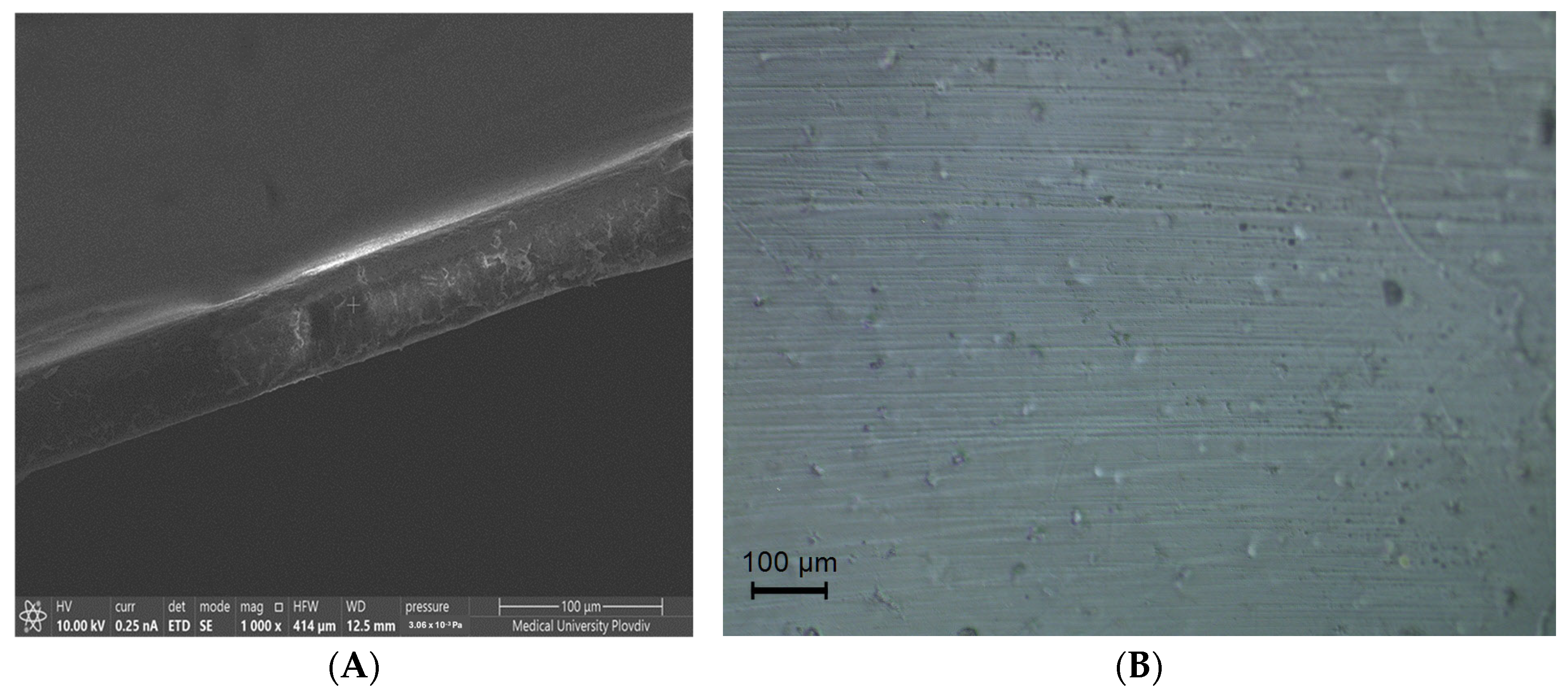
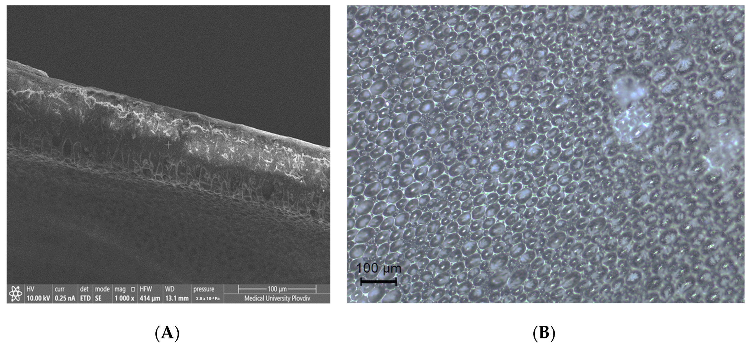
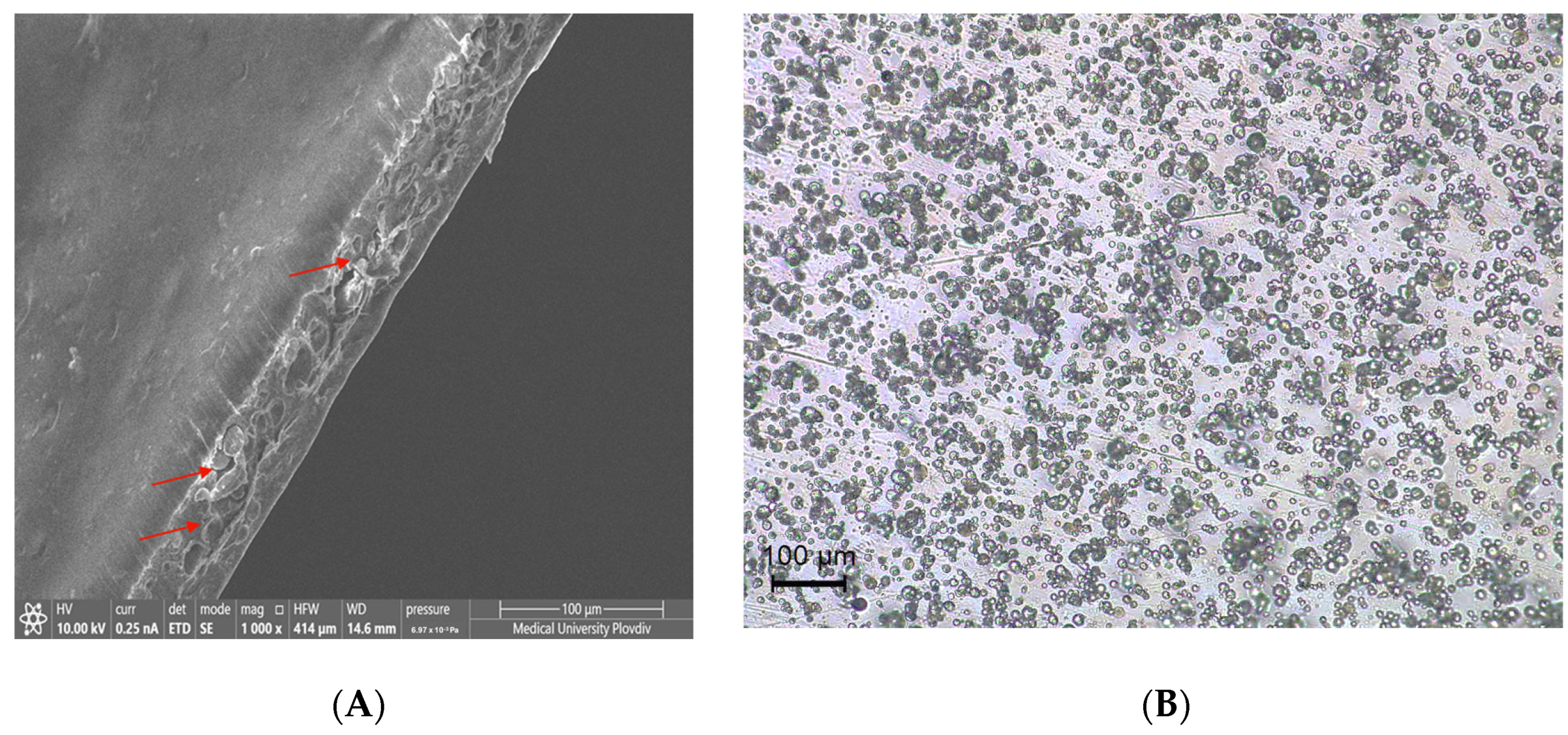
Regarding the topical application of drugs, the polymeric film plays a role not only as a carrier but also as a barrier. It can protect the treated spot from friction, moisture, and outside contamination while maintaining a healthy environment in terms of humidity and penetration of active substances [21,22]. Therefore, when aiming for topical applications, it is a crucial part of the research to obtain barrier properties, such as water vapour transmission (WVTR) and permeability (P). The values for the structures obtained in this study are presented in Table 2.
Given that PLA is hydrophobic by nature, its low permeability and water vapour transmission rate are expected, and the obtained values are in good correspondence with previously obtained results by Guivier et al. [23]. When 5-FU was added to the film, its permeability increased along with the evaporation rate of water. This can be attributed to the hydrophilicity of the drug and its effect on the structural morphology, which will be further discussed. The lowest values for both parameters were observed for the films loaded with chitosan microparticles. Despite the hydrophilic nature of this biopolymer, when it is formed into particles and embedded in the structure, it leads to a lower rate of evaporation. This is a consequence of two major factors: increased compactness of the film due to the lower number of voids within its volume and the particles acting as obstructions for the penetrating water molecules, resulting in a longer path to cross through the film [9,24].

3.3. Morphology

The morphology of each micro- and nanostructure is critically important because it directly influences the physical and chemical properties, stability, and overall behaviour of the material. Understanding the detailed structural features, namely the surface texture and porosity, provides valuable insights into how these structures interact with their environment. In the present study, information regarding the topography of the films was collected using an optical microscope, and images of the microstructure were obtained as cross-sectional images using a scanning electron microscope. All images are shown in Figure 2, Figure 3 and Figure 4. As shown in Figure 2, the PLA film has a rather uniform surface, and its cross-sectional image reveals a smooth texture on the surface and inside the film. When 5-FU was dissolved in methanol and added to the film, a significant change in the surface morphology was observed. Even though chloroform and methanol are miscible, they have different evaporation rates, with chloroform having nearly twice the evaporation rate of methanol [25]. When this solvent (chloroform) evaporated, the film structure formed and dried out, while the drug was still partly in the form of a methanol solution; therefore, it slowly migrated to the surface until the methanol evaporated. Completely. As a result of this, we observed pocket-like voids on the surface of the film with a homogeneous distribution, resulting in a structure with a porous surface. This type of morphology explains the lower strain at break and higher water vapour transmission rate. When chitosan particles were loaded into the PLA film in a mass ratio of 1:5 in favour of the film-forming concentration, the obtained structure had a smooth surface and even distribution of the particles within the volume of the film, according to the cross-sectional photo. This homogenous and even particle distribution is confirmed by the optical microscope photo, confirming the successful preparation of a uniform and smooth PLA film with chitosan microparticles.
Figure 2, Figure 3 and Figure 4 SEM (left-handside) and optical microscope images (right-handside) in the illuminating mode of the three samples. All scale bars are set to 100 µm.

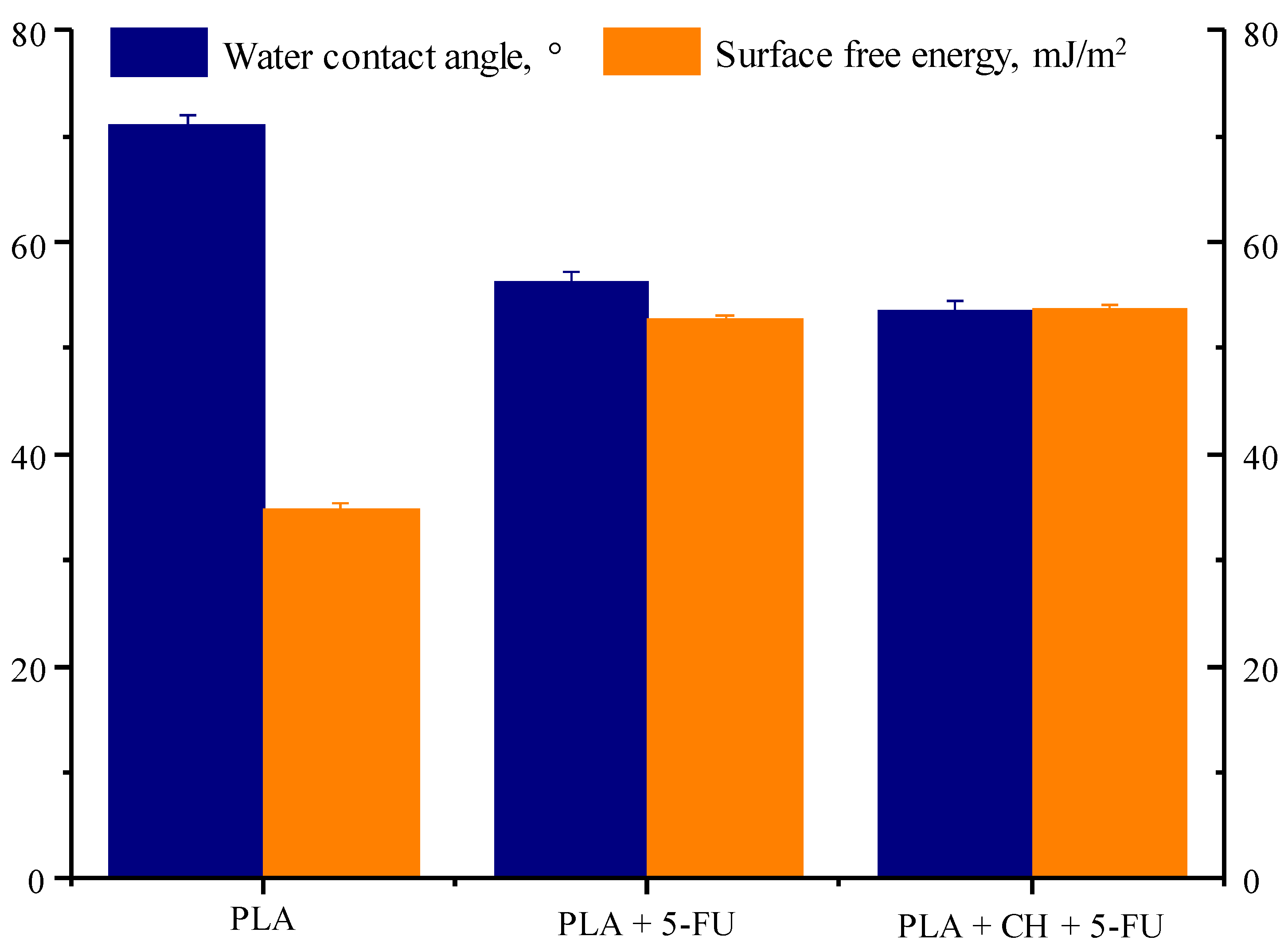
3.4. Water Contact Angle and Surface Free Energy

A structure’s wettability, adhesion, and successful and uniform application on the skin is crucial. All of these factors are influenced by the contact angle of the prepared formulation. The water contact angles and surface free energies of each obtained sample are shown in Figure 5.
PLA is a hydrophobic polymer with a typical contact angle of up to 85°, depending on the measurement conditions and surface roughness [26]. In the present case, this value is slightly above 70°, which is in good correspondence with other authors’ findings. Because of its poor wetting capabilities, its surface energy is generally considered to be moderate to low. Suitable values for these parameters in terms of proper adhesion and wettability are well below 90° for the contact angle and above 27 mJ/m2 for the surface free energy, based on the value of the skin’s crucial surface tension [27]. Pure PLA film has values close to those mentioned above, but they can still be considered rather poor. After the addition of 5-FU, which is slightly hydrophilic, and due to the formation of voids on the film surface, as observed in the microscopic images, the wettability was improved. The contact angle was around 57°, and the surface free energy was slightly below 57 mJ/m2. The inclusion of chitosan particles further reduced the contact angle to 53° due to its hydrophilic behaviour and ability to bind with water. As a result of this, the surface free energy value approached 54 mJ/m2. Therefore, the obtained structures exhibited improved wettability and suitable parameters for skin application.

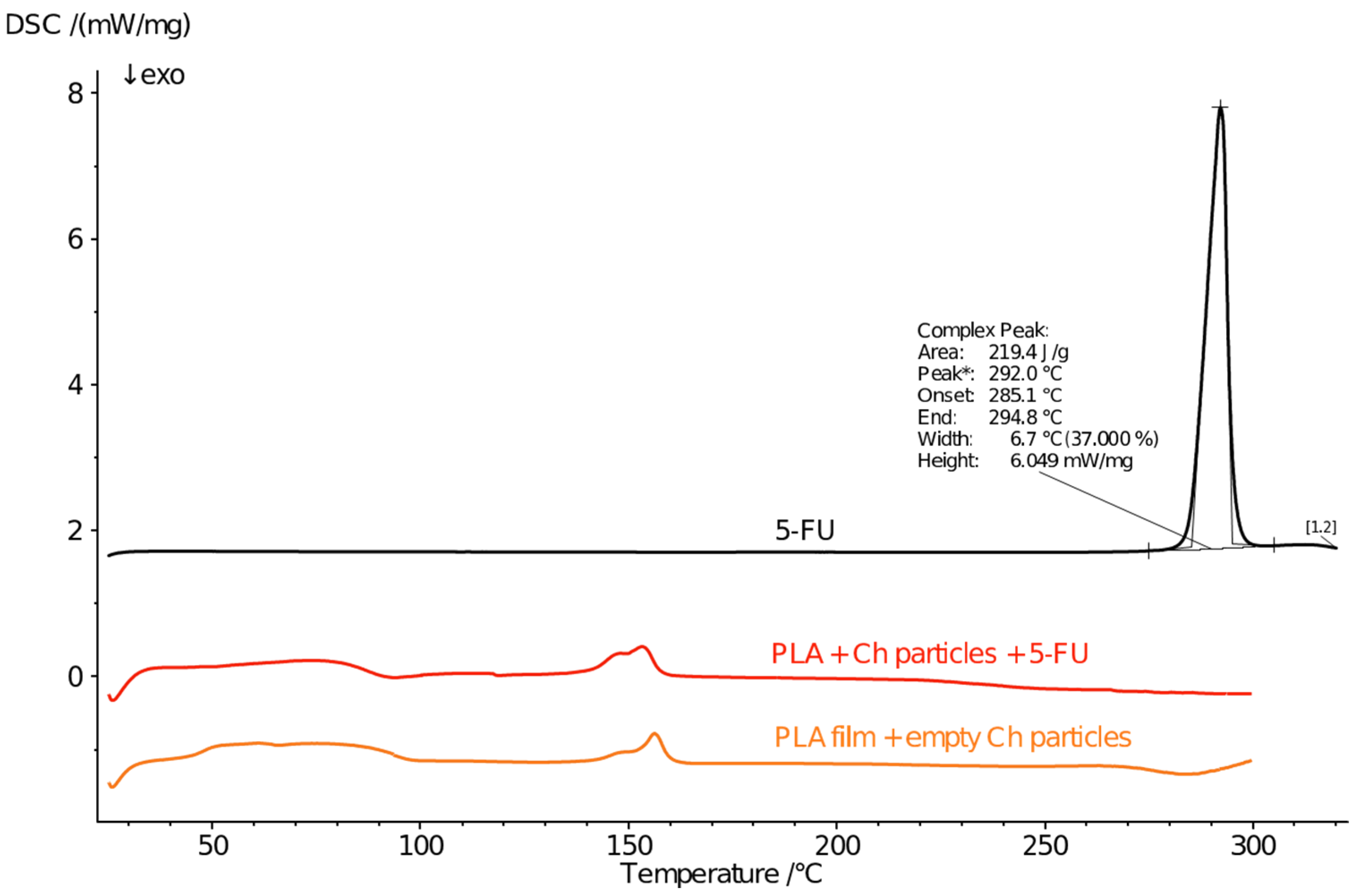
3.5. Phase State and Thermal Behaviour

The phase state of drugs and bioactive compounds affects the overall characteristics of the cargo system, such as mechanical properties, solubility, release rate, and kinetics [28,29]. In addition, it affects the bioavailability of the drug [30]. Hence, one of the many desirable outcomes of drug encapsulation in the delivery matrix is the potential phase transition from crystalline to a partly or fully amorphous phase state [31]. Therefore, the next step in the characterisation of the developed polymeric cargo systems was thermal and phase state studies. Figure 6 and Figure 7 present the resultant thermograms of these tests for all components used in the preparation process, namely, pure 5-FU, PLA film with and without 5-FU, and the same for chitosan particles.
The thermogram of native 5-FU is evident in its high-crystallinity phase state. The melting point of the drug was found to be around 285 °C, which is consistent with the findings of other studies [32]. Usually, the starting point and duration of the fusion process are affected by the purity of the drug and the occurrence of decomposition along with melting [33]. After incorporating the drug into the PLA film, it underwent a partial phase transition. The resultant crystallinity degree of 5-FU in this structure was around 14%. This means that most of the drug was able to get molecularly dispersed within the polymer matrix, but not completely. This is mainly a result of the strong hydrogen bonding among the 5-FU molecules, which enables them to form fibrillar structures under certain conditions, such as at higher concentrations and lower temperatures. Banerjee et al. pointed out that depending on the drug concentration and dissolution medium, it can form rod-like, spherical, flower-like, and helical crystalline structures [34]. Therefore, recrystallisation, in this case, is due to the high concentration of the drug and its propensity to self-assemble. After encapsulation of the drug in chitosan particles, the melting peak of 5-FU was no longer present, indicating that it was fully dispersed and unable to recrystallise. It can be seen that chitosan particles also influence the cold crystallisation of PLA, and this process is not as strongly pronounced as in the bare PLA film. Elsawy et al. had a similar discovery [19]. They studied the effects of adding chitosan micro- and nanoparticles to PLA films at different ratios. The cold crystallisation temperature shifted to lower values, and there was more than a 10% decrease in the enthalpy of the process. This is due to hindered nucleation and crystallisation, along with the reduction of the PLA chain length. In addition, the size and distribution of the particles influence the cold crystallisation process [35].

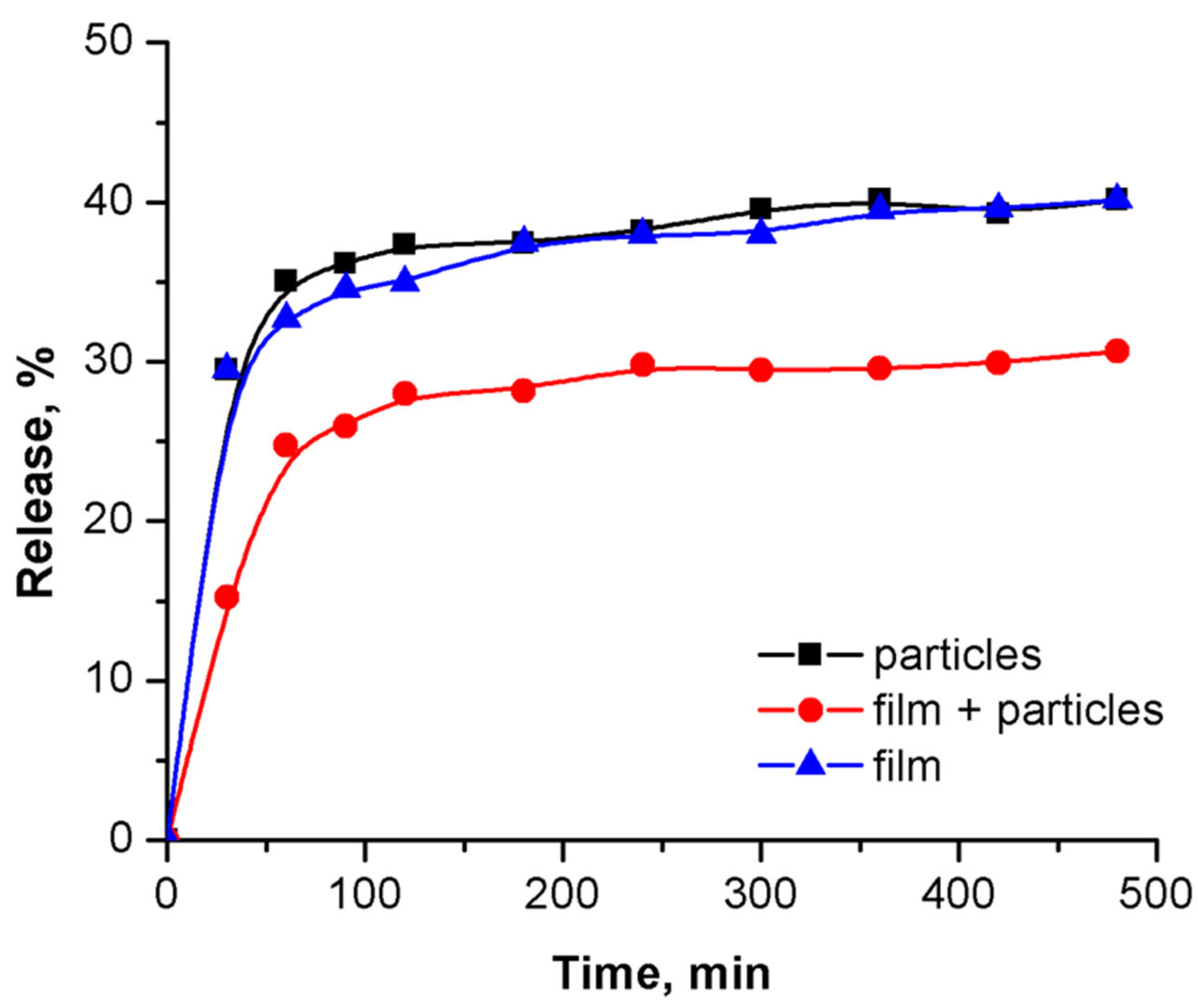
3.6. Drug Release Profiles and Optimal Model Fitting of the Process

To confirm the potential of a given structure as a cargo system, its ability to entrap and prolong the release of the active compound of choice should be examined and evaluated. To do this, the release profiles of each prepared sample were tested under conditions that simulated the administration route and spot as closely as possible. Figure 8 shows the 5-FU release profiles from chitosan particles, PLA film, and PLA film with embedded particles.
All samples exhibited biphasic behaviour. As previously reported by our research group, in the first hour, the highest amount of released drug is out of the particles due to their ability to swell and dissolve in slightly acidic conditions, as they are in the buffer media [16]. A slightly lower amount was released by the PLA film. Despite its hydrophobicity, which should slow down the migration of 5-FU, the release from this structure was comparable to that of hydrophilic chitosan. Jones et al. pointed out that the size of the pores is dependent on the drug concentration and crystal size and affects the release process [36]. It turns out that when the pores’ size is big enough, this facilitates drug migration into the pores and through the porous surface. As the pores are only on the surface and not intraparticular, this enables sustained release for the drug encapsulated more tightly into the volume of the PLA film after the initial dissolution of the surface-located crystals. Both particles and film structures, as separate systems, showed a similar trend and drug release amount for the tested 8-h period, namely around 40–45% of the total amount loaded into them. When 5-FU was loaded into chitosan particles and then embedded into the PLA film, the amount of released drug decreased by nearly 2-fold in the first hour, reducing the slope of the release curve. Hence, the initial fast release observed for the other two types of samples was not observed for this type of structure. These findings were confirmed by a mathematical model of the process. Based on R2 and the distribution of the residuals, the best-fitting model was the Korsmeyer–Peppas model [37]. The values of the model parameters after regression modelling of the experimental data are listed in Table 3.
As can be seen, the time-dependent constant, namely parameter K, had the lowest value for the sample PLA + CH + 5-FU. The steepest curve, and hence the largest K value, was observed for the chitosan particles. PLA + 5-FU had a slightly lower value for this parameter, indicating a slightly lower release rate compared to the particles. The parameter indicating the main release mechanism is n. For all developed structures, despite their shape and morphology, it was lower than 0.45; therefore, the release process obeyed Fickian diffusion. Such low values for n, as observed in this case, suggest that there is a significant hindrance to the diffusion process, which is also known as pseudo-Fickian diffusion [38]. Such a type of release is characterised by a release rate lower than ideal Fickian diffusion, which is typically a result of structural or morphological barriers within the polymer matrix that limit drug mobility, resulting in a slower achievement of equilibrium.

4. Conclusions

The present study shows the successful preparation of a PLA film loaded with 5-FU for the first time and a comparison between such a structure and a PLA film embedded with 5-FU-loaded chitosan microparticles. Testing of the mechanical and barrier properties revealed that the addition of 5-FU and chitosan particles altered the film flexibility and permeability, resulting in a lower strain at break and permeability. The water vapour rate increased from 189 g/m2·24 h to 241 g/m2·24 h after the addition of 5-FU due to the formation of voids on the film surface. Chitosan particles decreased this value to 174 g/m2·24 h and increased the surface free energy to 24 mJ/m2. Morphological analysis confirmed the uniformity of the films. The DSC study indicated a favourable transition of 5-FU to an amorphous state, particularly in the chitosan-encapsulated form. In the PLA + 5-FU film, recrystallisation of about 14% of the drug was observed. The drug release profiles demonstrated that pure chitosan particles and PLA films loaded with 5-FU, exhibited a burst release, resulting in the release of above 30% of the amount of the loaded drug. The most promising behaviour was observed in PLA films embedded with chitosan particles, which offered sustained drug delivery, minimised the initial burst, and maintained prolonged drug availability.

Author Contributions

Conceptualisation, S.M. and M.M.; methodology, S.M. and M.M.; software, S.M. and M.M.; validation, S.M. and M.M.; formal analysis, M.M.; investigation, S.M. and M.M.; resources, S.M.; data curation, S.M.; writing—original draft preparation, S.M.; writing—review and editing, M.M.; visualisation, S.M. and M.M.; supervision, M.M.; project administration, S.M.; funding acquisition, S.M. All authors have read and agreed to the published version of the manuscript.

Funding

This research received no external funding.

Data Availability Statement

Data are available upon request.

Acknowledgments

Sofia Milenkova acknowledges the financial help during this study from the National Science Program “Young Scientists and Post-Doctoral Students 2” of the Ministry of Education and Science, Republic of Bulgaria.

Conflicts of Interest

The authors declare no conflicts of interest.

Abbreviations

The following abbreviations are used in this manuscript:
5-FU5-fluorouracil
CHChitosan particles
PPermeability
PLAPoly(lactic acid); film based on poly(lactic acid)
PLA + 5-FUfilm based on poly(lactic acid) and loaded with 5-fluorouracil
PLA + CH + 5-FUPLA film containing drug-loaded chitosan particles
WVTRWater Vapour Transmission Rate
YMYoung’s Modulus

References

  1. Zhang, L.; Lv, J.; Yin, Y.; Ling, G.; Zhang, P. Rapidly Separable Microneedle Patch for the Controlled and Sustained Release of 5-Fluorouracil. Int. J. Pharm. 2023, 635, 122730. [Google Scholar] [CrossRef] [PubMed]
  2. Sahu, P.; Kashaw, S.K.; Sau, S.; Kushwah, V.; Jain, S.; Agrawal, R.K.; Iyer, A.K. PH Responsive 5-Fluorouracil Loaded Biocompatible Nanogels for Topical Chemotherapy of Aggressive Melanoma. Colloids Surf. B Biointerfaces 2019, 174, 232–245. [Google Scholar] [CrossRef] [PubMed]
  3. Park, J.; Kim, Y.-C. Topical Delivery of 5-Fluorouracil-Loaded Carboxymethyl Chitosan Nanoparticles Using Microneedles for Keloid Treatment. Drug Deliv. Transl. Res. 2020, 11, 205–213. [Google Scholar] [CrossRef] [PubMed]
  4. Zahra, F.T.; Adil, M.; Amin, S.S.; Mohtashim, M.; Bansal, R.; Khan, H.Q. Efficacy of Topical 5% 5-Fluorouracil with Needling versus 5% 5-Fluorouracil Alone in Stable Vitiligo: A Randomised Controlled Study. J. Cutan. Aesthetic Surg. 2020, 13, 197–203. [Google Scholar] [CrossRef]
  5. Raviraj, V.; Pham, B.T.T.; Kim, B.J.; Pham, N.T.H.; Kok, L.F.; Painter, N.; Delic, N.C.; Jones, S.K.; Hawkett, B.S.; Lyons, J.G. Non-Invasive Transdermal Delivery of Chemotherapeutic Molecules in Vivo Using Superparamagnetic Iron Oxide Nanoparticles. Cancer Nanotechnol. 2021, 12, 6. [Google Scholar] [CrossRef]
  6. Santos, L.F.; Correia, I.J.; Silva, A.S.; Mano, J.F. Biomaterials for Drug Delivery Patches. Eur. J. Pharm. Sci. 2018, 118, 49–66. [Google Scholar] [CrossRef]
  7. Tyler, B.; Gullotti, D.; Mangraviti, A.; Utsuki, T.; Brem, H. Polylactic Acid (PLA) Controlled Delivery Carriers for Biomedical Applications. Adv. Drug Deliv. Rev. 2016, 107, 163–175. [Google Scholar] [CrossRef]
  8. Hijazi, N.; Le Moigne, N.; Rodier, E.; Sauceau, M.; Vincent, T.; Benezet, J.-C.; Fages, J. Biocomposite Films Based on Poly (Lactic Acid) and Chitosan Nanoparticles: Elaboration, Microstructural and Thermal Characterisation. Polym. Eng. Sci. 2018, 59, E350–E360. [Google Scholar] [CrossRef]
  9. Garavand, F.; Rouhi, M.; Jafarzadeh, S.; Khodaei, D.; Cacciotti, I.; Zargar, M.; Razavi, S.H. Tuning the Physicochemical, Structural, and Antimicrobial Attributes of Whey-Based Poly (L-Lactic Acid) (PLLA) Films by Chitosan Nanoparticles. Front. Nutr. 2022, 9, 880520. [Google Scholar] [CrossRef]
  10. Tığlı Aydın, R.S.; Pulat, M. 5-Fluorouracil Encapsulated Chitosan Nanoparticles for PH-Stimulated Drug Delivery: Evaluation of Controlled Release Kinetics. J. Nanomater. 2012, 2012, 313961. [Google Scholar] [CrossRef]
  11. Khan, T.A.; Azad, A.K.; Fuloria, S.; Nawaz, A.; Subramaniyan, V.; Akhlaq, M.; Safdar, M.; Sathasivam, K.V.; Sekar, M.; Porwal, O.; et al. Chitosan-Coated 5-Fluorouracil Incorporated Emulsions as Transdermal Drug Delivery Matrices. Polymers 2021, 13, 3345. [Google Scholar] [CrossRef] [PubMed]
  12. Sabitha, M.; Sanoj Rejinold, N.; Nair, A.; Lakshmanan, V.-K.; Nair, S.V.; Jayakumar, R. Development and Evaluation of 5-Fluorouracil Loaded Chitin Nanogels for Treatment of Skin Cancer. Carbohydr. Polym. 2013, 91, 48–57. [Google Scholar] [CrossRef] [PubMed]
  13. Das, S.; Saha, M.; Mahata, L.C.; China, A.; Chatterjee, N. Krishna Das Saha Quercetin and 5-Fu Loaded Chitosan Nanoparticles Trigger Cell-Cycle Arrest and Induce Apoptosis in HCT116 Cells via Modulation of the P53/P21 Axis. ACS Omega 2023, 8, 36893–36905. [Google Scholar] [CrossRef] [PubMed]
  14. Sun, L.; Chen, Y.; Zhou, Y.; Guo, D.; Fan, Y.; Guo, F.; Zheng, Y.; Chen, W. Preparation of 5-Fluorouracil-Loaded Chitosan Nanoparticles and Study of the Sustained Release In Vitro and In Vivo. Asian J. Pharm. Sci. 2017, 12, 418–423. [Google Scholar] [CrossRef]
  15. Entezar-Almahdi, E.; Mohammadi-Samani, S.; Tayebi, L.; Farjadian, F. Recent Advances in Designing 5-Fluorouracil Delivery Systems: A Stepping Stone in the Safe Treatment of Colorectal Cancer. Int. J. Nanomed. 2020, 15, 5445–5458. [Google Scholar] [CrossRef]
  16. Milenkova, S.; Tashkov, S.; Zahariev, N.; Pilicheva, B.; Marudova, M. 5-Fluorouracil Encapsulated Chitosan Microspheres. J. Chem. Technol. Metall. 2024, 59, 887–896. [Google Scholar] [CrossRef]
  17. ASTM D882-91; Standard Test Methods for Tensile Properties of Thin Plastic Sheeting. American Society for Testing and Materials: Philadelphia, PA, USA, 2017. Available online: https://www.astm.org/d0882-00.html (accessed on 26 May 2025).
  18. Owens, D.K.; Wendt, R.C. Estimation of the Surface Free Energy of Polymers. J. Appl. Polym. Sci. 1969, 13, 1741–1747. [Google Scholar] [CrossRef]
  19. Elsawy, M.A.; Saad, G.R.; Sayed, A.M. Mechanical, Thermal, and Dielectric Properties of Poly (Lactic Acid)/Chitosan Nanocomposites. Polym. Eng. Sci. 2016, 56, 987–994. [Google Scholar] [CrossRef]
  20. Pünnel, L.C.; Lunter, D.J. Film-Forming Systems for Dermal Drug Delivery. Pharmaceutics 2021, 13, 932. [Google Scholar] [CrossRef]
  21. Chedik, L.; Baybekov, S.; Cosnier, F.; Marcou, G.; Varnek, A.; Champmartin, C. An Update of Skin Permeability Data Based on a Systematic Review of Recent Research. Sci. Data 2024, 11, 224. [Google Scholar] [CrossRef]
  22. Zhang, Y.; Liu, L.; Yue, L.; Huang, Y.; Wang, B.; Liu, P. Uncovering Key Mechanisms and Intervention Therapies in Aging Skin. Cytokine Growth Factor Rev. 2024, 79, 66–80. [Google Scholar] [CrossRef] [PubMed]
  23. Guivier, M.; Chevigny, C.; Domenek, S.; Casalinho, J.; Perré, P.; Almeida, G. Water Vapour Transport Properties of Bio-Based Multilayer Materials Determined by Original and Complementary Methods. Sci. Rep. 2024, 14, 50. [Google Scholar] [CrossRef] [PubMed]
  24. Soltani, Z.; Tavakolipour, H.; Tabari, M. The Influence of Chitosan and Titanium Dioxide Nanoparticles Incorporated with Polylactic Acid on Prolonging Rye Bread Shelf Life. J. Food Meas. Characterisation 2022, 17, 1806–1816. [Google Scholar] [CrossRef]
  25. Singh, P.P.; Sharma, B.R.; Sidhu, K.S. Thermodynamics of Chloroform and Methanol Mixtures. Can. J. Chem. 1979, 57, 387–393. [Google Scholar] [CrossRef]
  26. Tümer, E.H.; Erbil, H.Y.; Akdoǧan, N. Wetting of Superhydrophobic Polylactic Acid Micropillared Patterns. Langmuir 2022, 38, 10052–10064. [Google Scholar] [CrossRef]
  27. Capra, P.; Musitelli, G.; Perugini, P. Wetting and Adhesion Evaluation of Cosmetic Ingredients and Products: Correlation of In Vitro-In Vivo Contact Angle Measurements. Int. J. Cosmet. Sci. 2017, 39, 393–401. [Google Scholar] [CrossRef]
  28. Choi, M.-J.; Woo, M.R.; Choi, H.-G.; Jin, S.G. Effects of Polymers on the Drug Solubility and Dissolution Enhancement of Poorly Water-Soluble Rivaroxaban. Int. J. Mol. Sci. 2022, 23, 9491. [Google Scholar] [CrossRef]
  29. Han, A.S.; Kim, J.; Park, J.W.; Jin, S.G. Novel Acyclovir-Loaded Film-Forming Gel with Enhanced Mechanical Properties and Skin Permeability. J. Drug Deliv. Sci. Technol. 2022, 70, 103213. [Google Scholar] [CrossRef]
  30. Adepu, S.; Ramakrishna, S. Controlled Drug Delivery Systems: Current Status and Future Directions. Molecules 2021, 26, 5905. [Google Scholar] [CrossRef]
  31. Castro, K.C.; de Costa, J.M.; Campos, M.G.N. Drug-Loaded Polymeric Nanoparticles: A Review. Int. J. Polym. Mater. Polym. Biomater. 2020, 71, 1–13. [Google Scholar] [CrossRef]
  32. Gautam, M.K.; Besan, M.; Pandit, D.; Mandal, S.; Chadha, R. Cocrystal of 5-Fluorouracil: Characterization and Evaluation of Biopharmaceutical Parameters. AAPS PharmSciTech 2019, 20, 149. [Google Scholar] [CrossRef] [PubMed]
  33. Samy, M.; Abdallah, H.M.; Awad, H.M.; Ayoub, H. Preparation, Characterisation and in Vitro Biological Activity of 5-Fluorouracil Loaded onto Poly (D, L-Lactic-Co-Glycolic Acid) Nanoparticles. Polym. Bull. 2022, 80, 6197–6219. [Google Scholar] [CrossRef]
  34. Banerjee, P.; Mukherjee, D.; Maiti, T.K.; Sarkar, N. Unveiling the Self-Assembling Behaviour of 5-Fluorouracil and Its N, N′-Dimethyl Derivative: A Spectroscopic and Microscopic Approach. Langmuir 2017, 33, 10978–10988. [Google Scholar] [CrossRef] [PubMed]
  35. Flores-León, J.R.; Rodríguez-Félix, D.E.; Quiroz-Castillo, J.M.; Burrola-Núñez, H.; Castillo-Ortega, M.M.; Encinas-Encinas, J.C.; Alvarado-Ibarra, J.; Santacruz-Ortega, H.; Valenzuela-García, J.L.; Herrera-Franco, P.J. Effect of Degradation on the Physicochemical and Mechanical Properties of Extruded Films of Poly (Lactic Acid) and Chitosan. ACS Omega 2024, 9, 9526–9535. [Google Scholar] [CrossRef]
  36. Jones, E.C.L.; Bimbo, L.M. Crystallisation Behaviour of Pharmaceutical Compounds Confined within Mesoporous Silicon. Pharmaceutics 2020, 12, 214. [Google Scholar] [CrossRef]
  37. Kelle, D.; Speth, K.R.; Martínez-Negro, M.; Mailänder, V.; Landfester, K. Banu Iyisan Effect of Protein Corona on Drug Release Behavior of PLGA Nanoparticles. Eur. J. Pharm. Biopharm. 2024, 207, 114611. [Google Scholar] [CrossRef]
  38. Kim, K.-J.; Hwang, M.-J.; Yun, Y.-H.; Yoon, S.-D. Synthesis and Drug Release Behaviour of Functional Montelukast Imprinted Inulin-Based Biomaterials as Asthma Treatment. J. Ind. Eng. Chem. 2022, 109, 221–229. [Google Scholar] [CrossRef]
Figure 1. Stress-strain curves for PLA (yellow line), PLA + 5-FU (black line), and PLA + CH + 5-FU (grey line).
Processes 13 01961 g001
Figure 2. SEM (A) and optical microscope image in the illuminating mode (B) of PLA.
Processes 13 01961 g002
Figure 3. SEM (A) and optical microscope image in the illuminating mode (B) of PLA + 5-FU.
Processes 13 01961 g003
Figure 4. SEM (A) and optical microscope image in illuminating mode (B) of PLA + CH + 5-FU.
Processes 13 01961 g004
Figure 5. Water contact angles (blue bars) and surface free energies (orange bars) of the obtained structures.
Processes 13 01961 g005
Figure 6. Thermograms of 5-FU (black line), PLA film loaded with 5-FU (red line), and pure PLA film (yellow line).
Processes 13 01961 g006
Figure 7. Thermograms of 5-FU (black line), PLA film loaded with 5-FU-chitosan particles (red line), and PLA film loaded with empty chitosan particles (yellow line).
Processes 13 01961 g007
Figure 8. Drug release profiles for 8 h from chitosan particles (black line), PLA film (blue line), and PLA film loaded with chitosan particles (red line).
Processes 13 01961 g008
Table 1. Sample names, thicknesses, and main mechanical properties.
PLAPLA + 5-FUPLA + CH + 5-FU
Film thickness, µm62.9 ± 8.468.5 ± 5.862.6 ± 3.7
Strain at break, %393 ± 287 ± 114 ± 2
Stress at break, MPa30 ± 230 ± 624 ± 2
Young’s Modulus, MPa292 ± 31630 ± 100514 ± 98
Table 2. Barrier property values for the three samples. All presented values are the average of three repetitions with standard deviation.
PLAPLA + 5-FUPLA + CH + 5-FU
WVTR, g/m2·24 h189 ± 7241 ± 6174 ± 9
P × 10−13, g·mm/m2·24 h·kPa2.44 ± 0.091.56 ± 0.152.23 ± 0.12
Table 3. Korsmeyer-Peppas model parameters after modelling the 5-FU release process.
KnR2
CH + 5-FU23.4618 ± 1.44540.0897 ± 0.01150.9915
PLA + 5-FU21.2348 ± 0.62890.1045 ± 0.00550.9981
PLA + CH + 5-FU11.7193 ± 2.10620.1621 ± 0.03320.9468
Disclaimer/Publisher’s Note: The statements, opinions and data contained in all publications are solely those of the individual author(s) and contributor(s) and not of MDPI and/or the editor(s). MDPI and/or the editor(s) disclaim responsibility for any injury to people or property resulting from any ideas, methods, instructions or products referred to in the content.

