Article

Integrated In Silico and In Vitro Assessment of the Anticancer Potential of Origanum vulgare L. Essential Oil

by
Gabriel Mardale
1,2,†,
Florina Caruntu
3,†,
Alexandra Mioc
1,2,
Marius Mioc
2,4,
Alexandra Teodora Lukinich-Gruia
5,
Maria-Alexandra Pricop
5,6,
Calin Jianu
7,
Armand Gogulescu
8,*,
Tamara Maksimovic
1,2 and
Codruța Șoica
1,2
1
Department of Pharmacology-Pharmacotherapy, Faculty of Pharmacy, “Victor Babes” University of Medicine and Pharmacy, Eftimie Murgu Square, No. 2, 300041 Timișoara, Romania
2
Research Center for Experimental Pharmacology and Drug Design (X-Pharm Design), “Victor Babes” University of Medicine and Pharmacy, Eftimie Murgu Square, No. 2, 300041 Timișoara, Romania
3
Department of Medical Semiology II, Faculty of Medicine, “Victor Babes” University of Medicine and Pharmacy, 2 Eftimie Murgu, 300041 Timisoara, Romania
4
Department of Pharmaceutical Chemistry, Faculty of Pharmacy, “Victor Babes” University of Medicine and Pharmacy, Eftimie Murgu Square, No. 2, 300041 Timișoara, Romania
5
OncoGen Centre, Clinical County Hospital “Pius Branzeu”, Blvd. Liviu Rebreanu 156, 300723 Timisoara, Romania
6
Department of Applied Chemistry and Environmental Engineering and Inorganic Compounds, Faculty of industrial Chemistry, Biotechnology and Environmental Engineering, Polytechnic University of Timisoara, Vasile Pârvan 6, 300223 Timisoara, Romania
7
Faculty of Food Engineering, Banat’s University of Agricultural Sciences and Veterinary Medicine “King Michael I of Romania” from Timisoara, Calea Aradului 119, 300645 Timișoara, Romania
8
Department XVI, Balneology, Medical Rehabilitation and Rheumatology, “Victor Babes” University of Medicine and Pharmacy, 2 Eftimie Murgu, 300041 Timisoara, Romania
*
Author to whom correspondence should be addressed.
These authors contributed equally to this work.
Processes 2025, 13(6), 1695; https://doi.org/10.3390/pr13061695
Submission received: 23 April 2025 / Revised: 19 May 2025 / Accepted: 27 May 2025 / Published: 28 May 2025
(This article belongs to the Special Issue Extraction, Separation, and Medicinal Analysis of Natural Products)

Abstract

:
Oregano essential oil (OEO) has gained attention for its broad pharmacological activities, such as anti-inflammatory, antimicrobial, and anticancer properties. This study aimed to analyze the phytochemical composition and biological activity of OEO obtained from wild-growing Origanum vulgare L. in Romania. Gas chromatography–mass spectrometry (GC–MS) analysis identified p-cymene (43.98%), γ-terpinene (22.16%), and thymol (11.46%) as major constituents, with notable differences from previously reported chemotypes. Antioxidant activity was assessed using the DPPH, ABTS radical scavenging assay, and TPC. OEO has a moderate antioxidant activity, with IC50 values of 134.67 ± 1.32 µg/mL (DPPH) and 88.15 ± 0.045 Inh% (ABTS) and a TPC of 159.63 mg GAE/g extract. The cytotoxicity of the simple water dispersion of OEO, OEO solubilized with polyethylene glycol 400 (OEO-PEG), and that solubilized with Tween 20 (OEO-Tw) was evaluated on human melanoma (A375) and human colorectal adenocarcinoma (HT-29) cancer cell lines, as well as on the normal human immortalized keratinocytes (HaCaT) cell line. The results demonstrated a significant inhibition of cancer cell viability with no recorded cytotoxic effect on normal cells. The highest inhibition of cell viability was recorded for OEO-PEG 200 µg/mL (7.22% ± 6.51 in A375 cell line and 22.25% ± 10.08 in HT-29 cell line). In cancer cells, OEO and its formulations significantly reduced malondialdehyde (MDA) levels (up to 41.24% in A375 cells and up to 48.58% in HT-29 cells), suggesting potent antioxidant activity. Moreover, treatment with OEO increased caspase 3/7 activation two-fold in treated A375 cells, while high-resolution respirometry studies revealed that OEO induces mitochondrial dysfunction by acting as a potential uncoupling agent. Molecular docking analysis suggested that β-caryophyllene oxide (CPO), a minor constituent of OEO, may act as a potential inhibitor of 3-phosphoinositide-dependent protein kinase-1 (PDPK1), indicating a possible mechanism of anticancer activity. Our findings highlight the potential of OEO as a natural anticancer agent, emphasizing the need for further investigations to elucidate its exact molecular mechanisms and therapeutic applicability.

1. Introduction

Cancer is the leading cause of death worldwide, thus becoming a major challenge to prolonging life expectancy [1]. It is characterized by an uncontrolled growth and spread of abnormal cells; such cells are able to invade and damage surrounding tissues and organs and may lead to death if left untreated [2]. Traditional treatments for cancer include surgery, chemotherapy, and radiotherapy [3] and although these methods can be effective, they often come with significant side effects [4]. The search for alternative and complementary therapies, including those derived from natural sources, has therefore intensified [5]. Plants are a vast source of bioactive compounds, many of which have demonstrated promising anticancer properties [6]. These compounds exert their effects through various mechanisms [7], including antioxidant activity [8], antiproliferative effects [9,10,11], induction of apoptosis [12,13], anti-inflammatory effects [14], cell cycle arrest [15], inhibition of angiogenesis [16], and modulation of immune function [17]. Such biological activities were reported for OEO, which has incited growing interest among researchers in recent decades.
OEO, extracted from Origanum vulgare L. (Lamiaceae), is rich in bioactive compounds, particularly carvacrol and thymol, which are usually pointed out as responsible for its diverse biological activities [18,19], including being antimicrobial [20,21], anti-inflammatory [22], antioxidant [23], and antiproliferative [24]. Notably, several studies have reported the potent anticancer properties of both OEO and its pure components. One such study found that both OEO and its main ingredients, carvacrol and thymol, exhibited significant cytotoxic activity against HepG2 hepatocellular carcinoma cells [25]; Wang et al. demonstrated the efficacy of OEO against JTC-26 human cervical JTC-26, HepG2 hepatic HepG2, and A549 lung cancer cell lines [26]. Other studies reported the antitumor activity of OEO against non-small cell lung cancer (NSCLC), CT26 colon cancer, and PC3 prostate cancer [27]. However, despite these promising findings, the precise molecular mechanisms underlying OEO’s anticancer activity are not sufficiently characterized. Moreover, due to a considerable variability in essential oil (EO) composition, the reproducibility of results can be complicated. The wide differences in OEO composition are a result of numerous factors, such as the geographic origin, harvesting period, extracting method, and plant chemotype. Also taking into account the huge variety of molecular and morphological traits exhibited by cancer cells, investigating as many cell lines as possible, as well as the molecular mechanisms involved in the antitumor action of OEO, may unravel its potential use as an anticancer agent.
Furthermore, the therapeutic application of EO is often limited by poor water solubility, volatility, and chemical and physical instability with high degradability [28]. By using surfactants, such as PEG 400 and Tween 20, such limitations can be overcome; it has been reported that surfactants enhance the solubility, stability, bioavailability, and cellular uptake of EOs [29,30]. Despite the fact that the results highlight the benefits, the impact of such formulations on the anticancer efficacy of EOs and OEO still remains underexplored.
The objective of this study was to conduct a detailed phytochemical analysis of OEO using GC–MS, while also assessing its anticancer efficacy against HT-29 and A375 cancer cell lines compared to normal HaCaT cells. The in vitro study also aimed to assess whether PEG-400 and Tween 20 formulations enhance OEO’s anticancer efficacy and to further explore its possible underlying mechanism of action. Additionally, the research includes an in silico docking-based analysis to identify potential biological targets of its main active compounds. This integrated approach aims to deepen the understanding of the biological potential and the molecular mechanism of OEO’s action, in order to highlight its possible application as an anticancer agent.

2. Materials and Methods

2.1. OEO Extraction and GC–MS Analysis

The dried plant material (Voucher number VSNH.BUASTM-93/3) was provided by “King Michael I” University of Life Sciences (Timisoara Romania Herbarium). A Craveiro-type device was used to grind the dried plant before it was steam hydrodistilled for four hours at 100 °C [31]. After being separated, the oil was treated with anhydrous sodium sulfate to eliminate any remaining water, and it was then packed and kept at −18 °C for further examination. Oil weight/dried plant weight × 100 (wt%) was the formula used to determine the extraction yield.
GC–MS analysis was performed using a Hewlett Packard HP 6890 Series gas chromatograph coupled to a Hewlett Packard HP 5973 Mass Selective Detector. A 1 μL aliquot (diluted 1:100 in hexane) was injected under the following conditions: a DB-WAX capillary column (30 m × 0.25 mm × 0.25 μm), oven temperature programmed from 50 °C to 250 °C at 6 °C/min with a 4-min solvent delay. The MS source and quadrupole were set at 230 °C and 150 °C, respectively, with helium as the carrier gas at a 1 mL/min flow rate. Compounds were scanned in the 50–600 amu range and identified by comparing their spectra to the NIST 02 library. Retention indices were calculated using C9–C18 alkanes and compared with the literature data for verification.

2.2. Determination of OEO DPPH Scavenging Activity

A 2,2-diphenyl-1-picrylhydrazyl (DPPH) assay was employed to determine the samples’ antioxidant activity [32]. A DPPH stock solution was obtained after dissolving 5 mg DPPH in 5 mL of ethanol. Subsequently, a calibration curve with concentrations ranging from 10 µg/mL to 0.5 mg/mL was created through serial dilutions. Positive controls, butylated hydroxyanisole (BHA) and ascorbic acid (AA), were prepared at concentrations ranging from 0.06 µg/mL to 1.2 mg/mL for comparative analysis.
A 0.25 mM DPPH ethanolic solution was combined with a 1:10 ethanol-diluted plant extract in a 1:4 (v/v) ratio. For half an hour, the reaction mixture was incubated at 25 °C in the dark. The absorbance of all the tested samples was measured at 515 nm using a Tecan Infinite 200Pro spectrophotometer (Tecan Group Ltd., Männedorf, Switzerland) equipped with i-control software (version 1.10.4.0), which was used to measure absorbance at 515 nm.
The following formula was used to determine the DPPH radical inhibition percentage (Inh%):
Inh% = [(A0 − As)/A0] × 100
where A1 represents the absorbance of the sample and A0 represents the absorbance of the control (DPPH solution without the sample). The inhibition values were plotted against sample concentrations using the corresponding calibration curve equation for each sample and control to determine the IC50. The antioxidant capacity of the sample is quantitatively measured by the IC50 values, which are given in µg/mL. Higher antioxidant activity is indicated by lower values.

2.3. ABTS Radical Scavenging Assay

The ABTS [2,20-azino-bis (3-ethylbenzothiazoline-6-sulphonic acid)] radical scavenging activity was assessed using a modified method [33,34]. To generate the ABTS cation (ABTS+•), a solution of ABTS (7.29 mM) was mixed in a 1:1 (v/v) ratio with K2S2O8 (2.47 mM) in an amber-colored bottle and kept in the dark at 25 °C for 14 h. The resulting ABTS+• solution was then diluted with ethanol to achieve an absorbance of 0.745 ± 0.047 at 734 nm. Next, 400 µL of the ABTS+• solution was combined with 100 µL of a 1:10 diluted sample extract and incubated at room temperature in the dark for 30 min. Absorbance was then measured at 734 nm, with 1 mg/mL solutions of butylated hydroxyanisole (BHA) and ascorbic acid (AA) used as reference standards. The following equation was used:
%ABTS+• inhibition = (Acontrol − Asample) × 100/Acontrol,
where Acontrol measures the absorbance of ABTS+• solution mixture without adding the sample and Asample measures the absorbance of the sample with ABTS+• solution mixture.

2.4. Determination of the Total Phenolic Content

The total phenolic content in the sample was assessed using a modified Folin–Ciocalteu procedure from the literature [35]. To perform the assay, a ratio of 1:5, 0.1 mL of the sample, diluted 1:10 in ethanol, was mixed with a 1:10 dilution of Folin–Ciocalteu 2 N reagent in distilled water. The reaction solution was vortexed and kept in the dark for 5 min at room temperature (23 °C). Furthermore, a volume of 7.5% sodium carbonate (Na2CO3) was added to adjust the pH and enhance the development of the color reaction. The mixture was thoroughly mixed and incubated in the dark at room temperature (23 °C) for 1 h to complete the reaction. Absorbance was measured at 725 nm using a Tecan Infinite 200Pro spectrophotometer (i-control software, version 1.10.4.0), with samples pipetted in quadruplicate into clear Polystyrol 96-well flat-bottom plates (Corning, Männedorf, Switzerland).
The final results of TPC were expressed in gallic acid equivalents (mg GAE/g extract) calculated after a propyl gallate calibration curve with concentrations between 9 µg/mL and 0.3 mg/mL. The concentrations of samples were calculated by using the linear equation obtained from the standard curve (y = 0.2264x − 0.0208; R2 = 0.9972), with Microsoft Excel v. 130.0.2849.52 (Microsoft Corporation, Redmond, WA, USA).

2.5. Cell Culture

The HaCaT cell line (CLS Cell Lines Service GmbH in Eppelheim, Germany) and A375 and HT-29 cell lines (American Type Culture Collection—ATCC, Lomianki, Poland) were chosen as the study material for our research. The cell culture medium used for both HaCaT and A375 cell line proliferation was Dulbecco’s Modified Eagle Medium, while HT-29 was cultured in McCoy’s 5A (Modified) medium. Both media were supplemented with 10% fetal bovine serum and 1% penicillin/streptomycin, following the guidelines provided by the manufacturer. The cells were maintained in a humidified incubator at 37 °C and a concentration of 5% CO2.

2.6. Cell Viability Assessment

The Alamar Blue colorimetric assay was used to assess the 24 h cell viability of HaCaT, A375, and HT-29 cell lines, as previously described [36]. Two methods were used to test the efficacy of OEO. One of the methods involved the use of OEO 0.5% and 1%, dissolved in the culture medium specific to each cell line. The other method involved dispersing a concentrated stock solution containing 10 mg/mL OEO in either Tween 20 or PEG 400 to obtain final concentrations of 100 μg/mL and 200 μg/mL. These concentrations were subsequently used to assess the viability of the above cell lines. The final concentrations of PEG 400 (1%) and Tween 20 (0.01%) were selected based on previous studies demonstrating their effectiveness in enhancing the solubility of EOs without exerting significant cytotoxicity on normal cells [37,38]. The work protocol involved culturing in 96-well plates at a cell concentration of 1 × 10⁴/well, followed by an incubation period at 37 °C with 5% CO2 until 80–85% confluency was obtained. After reaching the desired confluence, the medium in the wells was aspirated using an aspiration station, after which cells were exposed to working concentrations of OEO together with fresh medium specific to each compound. After 24 h of treatment, cells were stained with 0.01% Alamar Blue, then further incubated for another 4h. The results were obtained using an XMarkTM microplate spectrophotometer from Bio-Rad (Hercules, CA, USA) at a wavelength of 570 nm. The entire experimental procedure was performed in three biological replicates (independent cell cultures) and three technical replicates (triplicate measurements per biological replicate).

2.7. Molecular Docking

Molecular docking investigations were carried out using a previously established method [32]. The protein targets retrieved from the RCSB Protein Data Bank [39] and used as docking targets are presented in Table 1. The protein structures used represent either the full-length protein or specific functional domains, such as kinase or BH3-binding domains, as defined by their crystallized residue ranges (Table 1). The docking process was focused on the biologically relevant active sites, defined based on the coordinates of co-crystallized ligands or reported functional domains. The residue ranges and sequence lengths of the protein regions used in the docking simulations are also provided in Table 1. The chemical structures of the EO components were retrieved from PubChem as SDF files [40] and were later converted into 3D docking-suitable files with PyRx’s Open Babel module [41]. The PyRx v0.8-driven docking protocol that was employed to obtain the docking parameters for each protein target is also presented in Table 1. Key catalytic and functional binding site residues for protein targets were identified based on the analysis of co-crystallized ligands in the retrieved PDB structures, combined with literature reports describing their active sites. The co-crystallized native ligands listed in Table 1 were also retrieved and redocked as positive docking controls, since these compounds are experimentally validated inhibitors of their respective protein targets.

2.8. In Silico ADMET Predictions for the OEO Components

The PreADMET online tool was used to predict the OEO components’ absorption, distribution, metabolism, excretion, and toxicity (ADMET) profiles [42]. To create the necessary input structures, each compound was drawn using the platform’s built-in chemical sketching tool. Human intestinal absorption (HIA), Caco-2 cell permeability, plasma protein binding, lipophilicity (SKlogP), and inhibition of cytochrome P450 enzymes (CYP2C9, CYP2C19, CYP2D6, and CYP3A4) were among the pharmacokinetic characteristics predicted by the server. Toxicity predictions were made for rodent carcinogenicity (mouse and rat models), mutagenicity (Ames test), and hERG channel blockage (cardiotoxicity risk).
Table 1. Structural and docking parameters of the protein targets used in the molecular docking of OEO components, including domain definitions, sequence ranges, and native co-crystallized ligands used as positive docking controls.
Protein
(PDB ID)
Grid Box Center CoordinatesGrid Box SizeChain/
Domain Type
Residue Range (Sequence Length)Native Ligand Name/Description (PDB ID)Ref.
PDPK1
(2PE1)
x = −7.0111
y = 44.0378
z = 43.8068
x = 14.4075
y = 14.4075
z = 14.4075
A/Kinase domain82–360 (279 aa)BX-517/Indolinone-based ATP-competitive inhibitor (517)[43]
mTOR
(4JSX)
x = 51.6285
y = −2.0404
z = −48.8188
x = 18.7676
y = 16.3518
z = 18.7676
A/Kinase domain (ΔN-mTOR)2015–2549 (535 aa)Torin2/ATP-site inhibitor (17G)[44]
MEK1
(3DV3)
x = 37.7734
y = −13.7113
z = −1.1927
x = 17.4625
y = 14.0291
z = 12.8563
A/Kinase domain59–364 (306 aa)PF-04622664/MEK inhibitor (MEK)[45]
AKT/PKB
(4GV1)
x = −20.8519
y = 3.6032
z = 10.7428
x = 15.2397
y = 11.6572
z = 14.4921
A/Kinase domain144–480 (337 aa)AZD5363/ATP-competitive inhibitor (0XZ)[46]
PI3Kα
(6GVF)
x = −16.9238
y = 148.1857
z = 29.3169
x = 14.5772
y = 21.3565
z = 12.8758
A/Catalytic subunit (p110α)107–1051 (945 aa)3-(2-Amino-benzooxazol-5-yl)-1-isopropyl-1H-pyrazolo[3,4-d]pyrimidin-4-ylamine (FE5)[47]
PI3Kγ
(4FA6)
x = 45.4506
y = 14.5595
z = 30.3998
x = 20.2093
y = 12.4970
z = 16.1645
A/Catalytic subunit144–1102 (959 aa)PI3Kalpha/mTOR-IN1/Pyrrolidinyl pyrido pyrimidinone derivative (0TA)[48]
BCL-2
(4LVT)
x = 6.3606
y = −2.7407
z = −7.7449
x = 18.7676
y = 27.4958
z = 15.6622
A/BH3-binding domain1–34, 92–207 (150 aa)Navitoclax/ABT-263, BH3-mimetic inhibitor (1XJ)[49]
BCL-XL
(2YXJ)
x = −7.9224
y = −16.8631
z = 10.5365
x = 17.4625
y = 28.5719
z = 12.8563
A/BH3-binding domain1–209 (209 aa)ABT-737/BH3-mimetic inhibitor (N3C)[50]
EGFR1 (1XKK)x = 18.7130
y = 33.5455
z = 36.7358
x = 19.8569
y = 19.8569
z = 19.8569
A/Kinase domain695–1022 (328 aa)Lapatinib/GW572016, dual EGFR/ErbB2 inhibitor (FMM)[51]
VEGFR2 (4ASD)x = −25.0080
y = −0.1288
z = −10.9439
x = 20.2093
y = 14.4098
z = 16.1645
A/Kinase domain787–1171 (385 aa)Sorafenib/BAY 43-9006, ATP-competitive inhibitor (BAX)[52]

2.9. Lipid Peroxidation Assay

The level of malondialdehyde (MDA), a well-known marker of lipid peroxidation, was assessed using the Lipid Peroxidation (MDA) Assay Kit (ab118970, Abcam, Cambridge, UK) [53]. Treated and control A375 and HT-29 cells (2 × 10⁶/mL) were washed with PBS, harvested, and homogenized on ice in 303 of μL lysis solution. The lysate was sonicated and then centrifuged for 10 min at 13,000× g. The supernatant was collected for further use. To each well containing 200 μL of standard and 200 μL of sample, 600 μL of developer reagent was added. The controls and samples were incubated at 95 °C for 60 min and then cooled in an ice bath for another 10 min. All samples were transferred into a 96-well microplate for analysis. The fluorescence was read at Ex/Em = 532/553 nm with a BioTek Synergy HTX multimode microplate reader (Agilent Technologies, Santa Clara, CA, USA). This assay was performed independently three times using separate biological samples to ensure reproducibility.

2.10. Caspase-3/7 Assay

OEO’s ability to induce apoptosis was assessed using the CellEvent™ Caspase-3/7 Green Detection Reagent Kit (Thermo Fisher Scientific, Waltham, MA, USA). Cells treated with OEO 1%, OEO-PEG, and OEO-Tw (200 μg/mL) were stained and analyzed live using the Countess™ 3 FL Automated Cell Counter (Thermo Fisher Scientific) according to the manufacturer’s instructions [54]. This detection method relies on a dye that remains inactive while conjugated to a caspase-3/7-specific DEVD peptide sequence. Upon activation of caspase-3 or caspase-7 during apoptosis, the DEVD peptide is cleaved, allowing the dye to bind to DNA and emit green fluorescence (excitation/emission: 502/530 nm). The assay was conducted three times with different biological replicates.

2.11. High-Resolution Respirometry

Mitochondrial respiration was assessed at 37 °C using the Oxygraph-2k high-resolution respirometer (Oroboros Instruments, Innsbruck, Austria) following a modified substrate–uncoupler–inhibitor titration (SUIT) protocol as described by Petruș et al. [55]. A375 and HT-29 cells were cultured, trypsinized, and resuspended at 1 × 10⁶ cells/mL in a specialized mitochondrial respiration buffer (EGTA 0.5 mM, 3mM KH2PO4, taurine 20 mM, K-lactobionate 60 mM, MgCl2 10 mM, D-sucrose 110 mM, HEPES 20 mM, and BSA 1 g/L, pH 7.1). After a 15-min stabilization period in the chamber, digitonin was added to permeabilize the cells. Sequential additions of glutamate and malate (complex I substrates), ADP, and succinate (complex II substrate) were used to measure State 2CI, OXPHOSCI, and OXPHOSCI+II respiration. Oligomycin was added to assess State 4CI+II (leak respiration), followed by FCCP titration to determine the maximal electron transport system (ETSCI+II) capacity. Antimycin A was subsequently added to inhibit complex III, allowing the correction of all respiratory values after the residual oxygen consumption (ROX). The high-respirometry studies were performed three times with different biological replicates.

2.12. Statistical Analysis

One-way ANOVA followed by the application of Bonferroni’s Multiple Comparison Test conditions was used for statistical analysis determination with the aid of GraphPad Prism version 6.0.0 (GraphPad Software in San Diego, CA, USA). The differences between the experimental groups were considered to be statistically significant if p < 0.05 (* p < 0.05, ** p < 0.01, and *** p < 0.001).

3. Results

3.1. Chemical Composition of OEO

The hydrodistillation of the ground and previously dried oregano leaves produced 2.1% (wt) of a light yellow oil that was subjected to GC–MS analysis, revealing the presence of three major components, namely p-cymene (43.98%), γ-terpinene (22.16%), and thymol (11.46%), along with several minor compounds; the chemical constituents of OEO are depicted in Table 2.

3.2. Determination of the Antioxidant Activity of OEO

The antioxidant activity of OEO was determined by a 2,2-diphenyl-1-picrylhydrazyl (DPPH) radical scavenging assay, the [2,20-azino-bis (3-ethylbenzothiazoline-6-sulphonic acid)] (ABTS) radical scavenging activity, and the total polyphenolic content (TPC). For the first two mentioned methods, butylated hydroxyanisole (BHA) and ascorbic acid (AA) were used as positive controls. The obtained results are provided in Table 3. OEO reduced the stable free radical DPPH, recording an IC50 value of 134.67 μg/mL. However, the two positive controls reduced DPPH more effectively, recording lower IC50 values than OEO. A 1:10 dilution of OEO, corresponding to 94 mg/recorded a similar ABTS radical scavenging activity, expressed as inhibitory percentages, with a 1mg/mL solution of AA and BHA. The total polyphenolic content expressed as mg GAE/g recorded for OEO was 159.63.

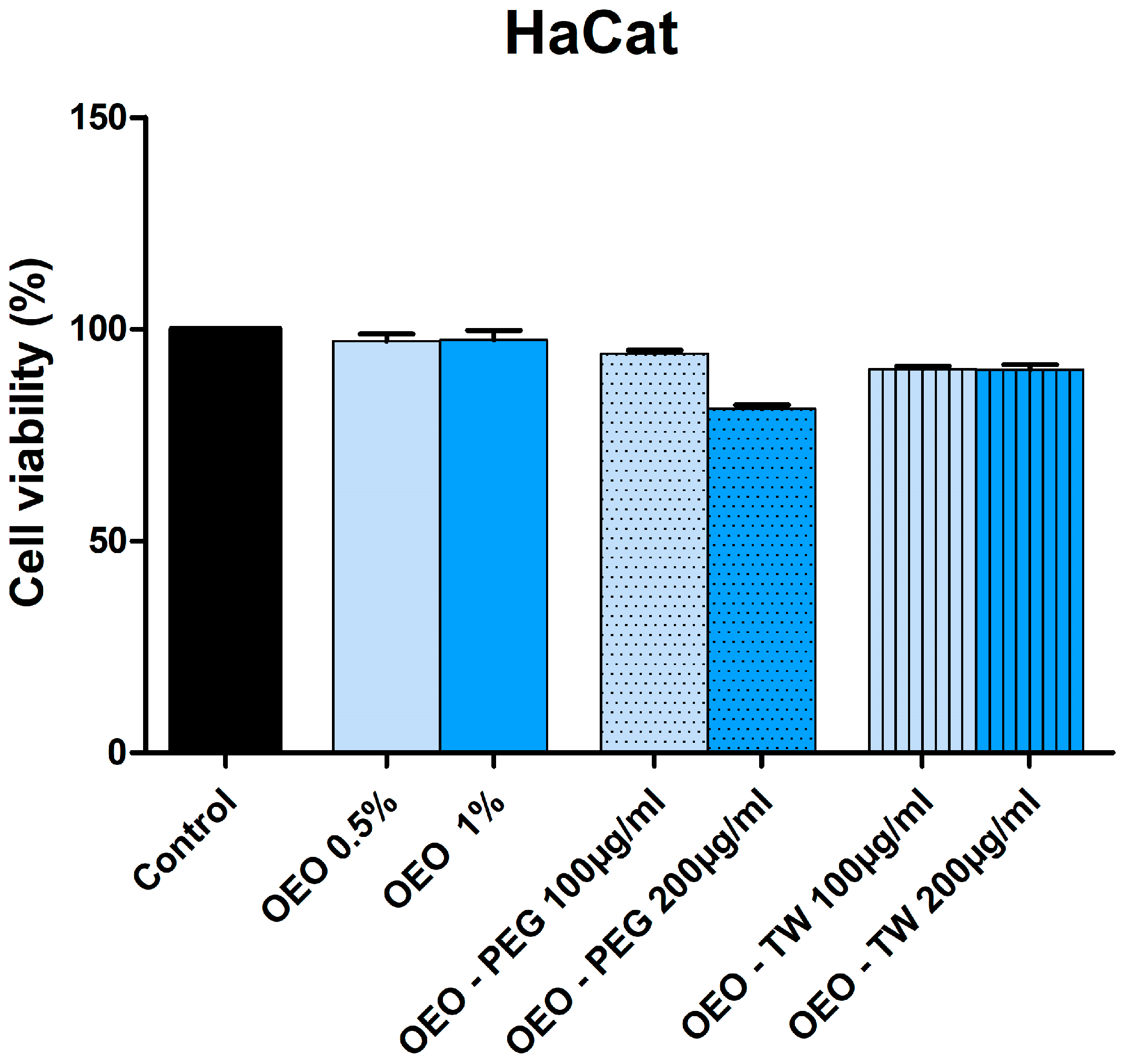
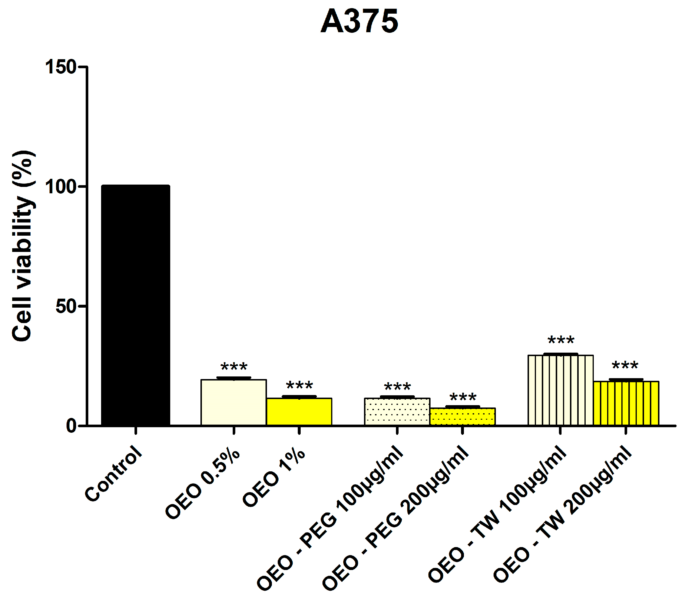
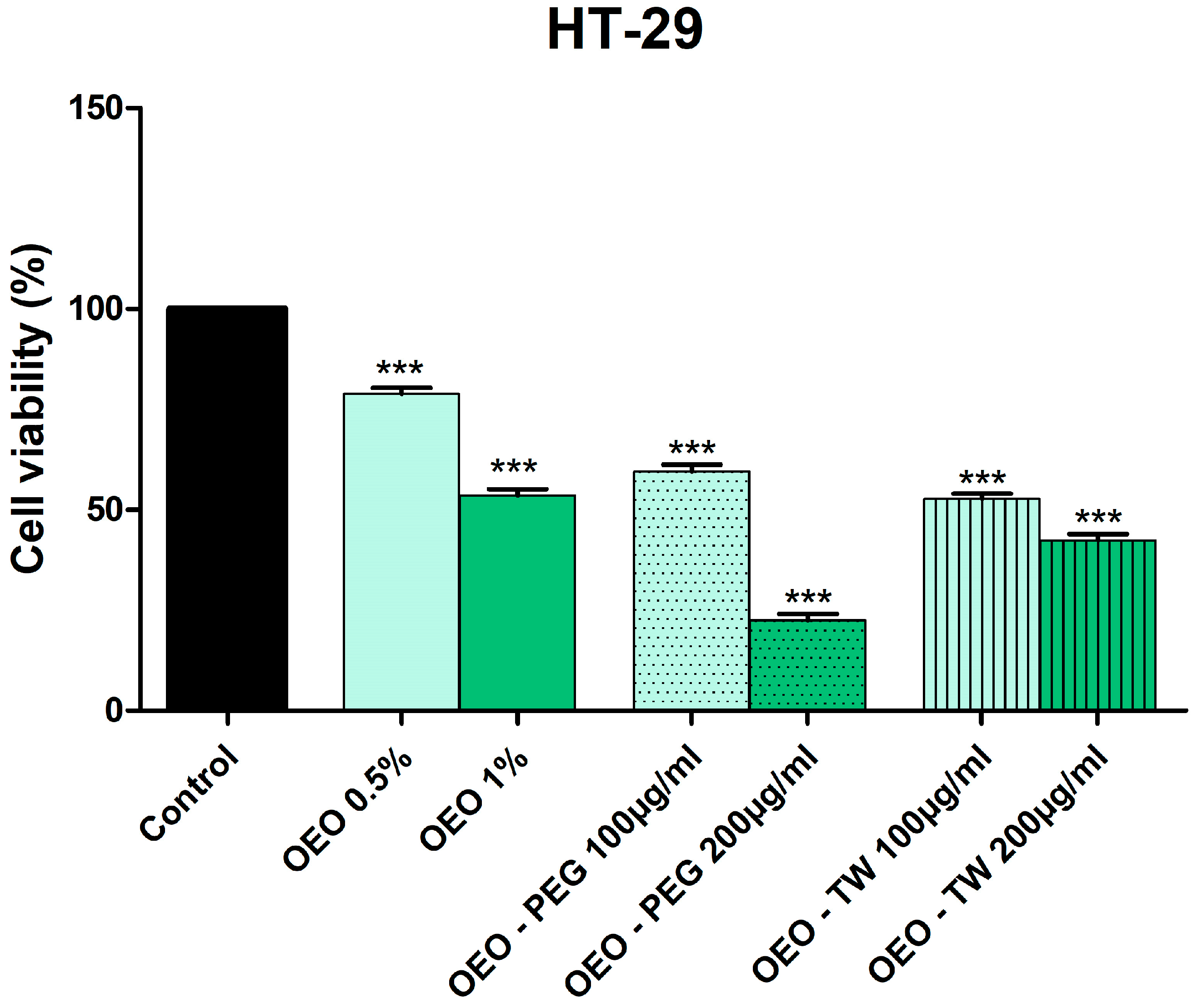
3.3. Evaluation of the Cytotoxic Effect of OEO

The viability of HaCaT, A375, and HT-29 cells after 24h treatment with different concentrations of OEO, 0.5% and 1%, as well as OEO-PEG and OEO-Tw (100 μg/mL and 200 μg/mL) was examined using the Alamar Blue assay.
The results showed no statistically significant reduction in HaCaT cell viability compared to the control group when treated with 0.5% or 1% OEO, as well as with 100 μg/mL and 200 μg/mL of OEO-PEG and OEO-Tw (Figure 1).
A significant reduction in A375 cell viability was observed following treatment with OEO, OEO-PEG, and OEO-Tw, compared to the control group. The exact variations in cell viability rates between the control group (100%) and tested cells were substantial, as follows: 19.75% ± 7.60 (OEO 0.5%), 11.56% ± 6.70 (OEO 1%), 11.79% ± 5.69 (OEO-PEG 100 μg/mL), 7.22% ± 6.51 (OEO-PEG 200 μg/mL), 29.49% ± 9.08 (OEO-Tw 100 μg/mL), and 18.39% ± 8.79 (OEO-Tw 200 μg/mL), as in Figure 2.
In HT-29 colorectal carcinoma cells, OEO reduced cell viability to 78.40% ± 6.49 (0.5%) and 0.5% and 53.66% ± 9.12 (1%). Dispersion in PEG 400 and Tween 20 further enhanced OEO’s effect, with OEO-PEG reducing viability to 59.28% ± 3.95 (100 μg/mL) and 22.25% ± 10.08 (200 μg/mL) and OEO-Tw to 52.67% ± 9.40 (100 μg/mL) and 42.00% ± 16.04 (200 μg/mL) (Figure 3).

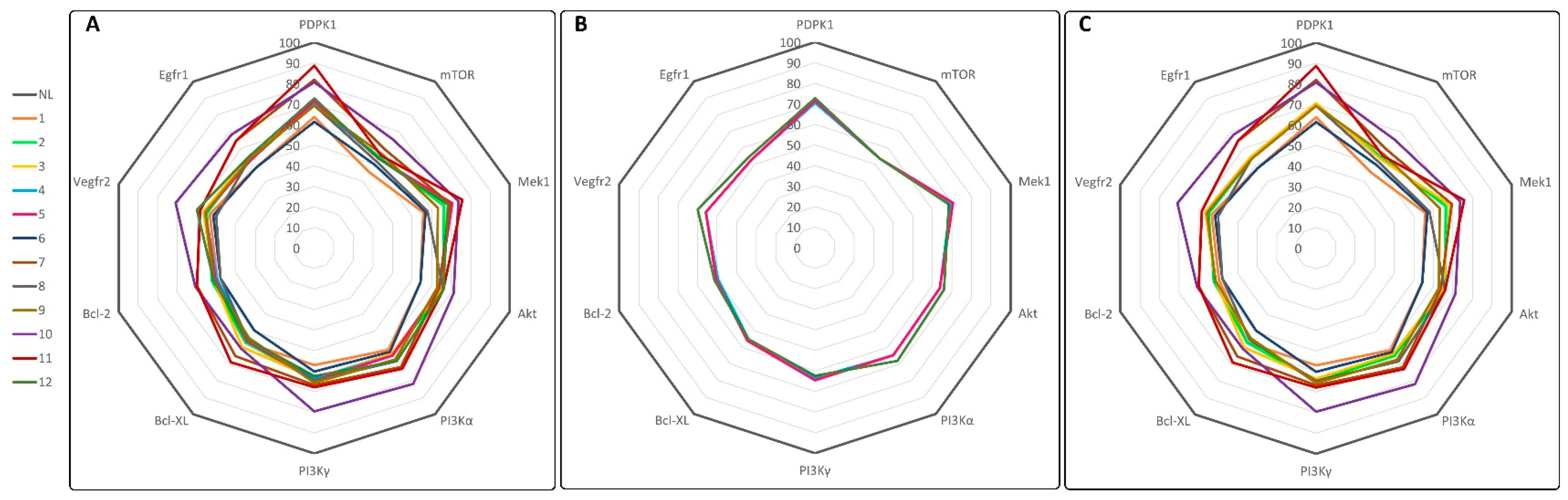
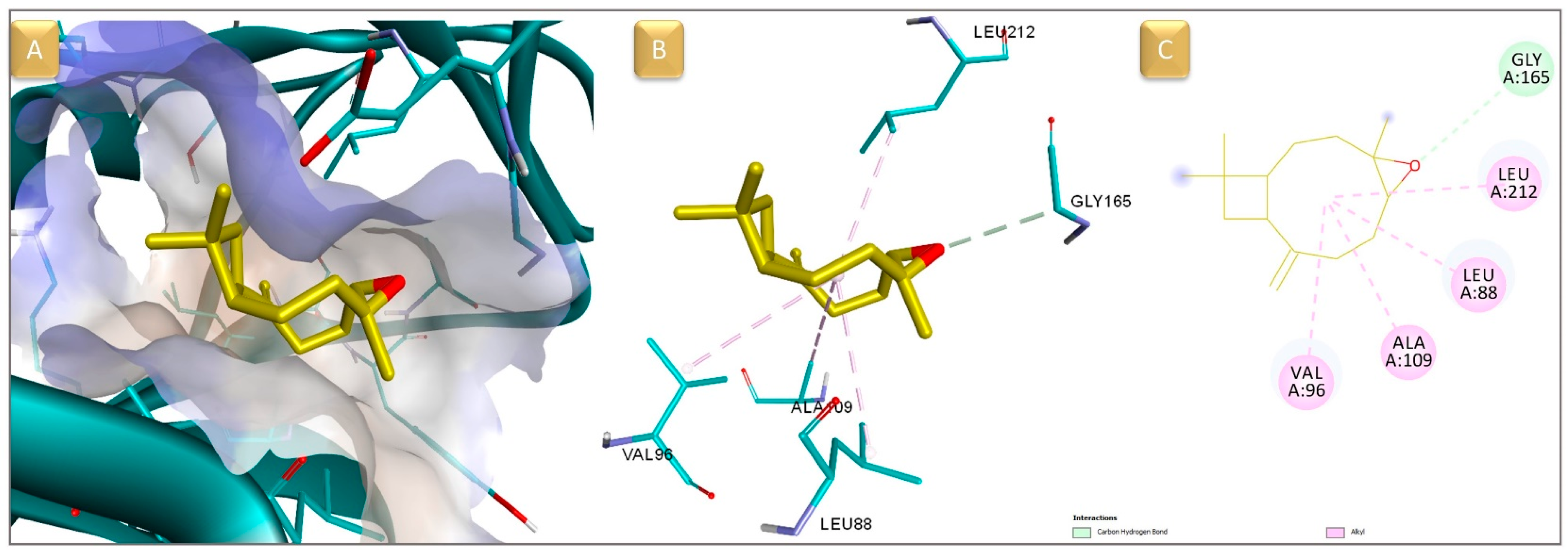
3.4. Molecular Docking and ADMET Prediction

In this study, a molecular docking in silico-based method was used to identify theoretical protein targets for the 12 structures contained in the OEO, providing a theoretical basis to support the observed in vitro cytotoxic effects against cancer cells. Next, we docked the previously discussed compounds into the binding site of anticancer protein targets, which are often used in anticancer drug design; the overexpression of these proteins is frequently correlated with cancer development, elevated cell proliferation, and survivability within multiple types of cancer. The protein structures chosen for the docking experiment were the epidermal growth factor receptor 1 (EGFR1), the vascular endothelial growth factor receptor 2 (VEGFR2), phosphoinositide-dependent kinase-1 (PDPK1), dual specificity mitogen-activated protein kinase kinase 1 (MEK1), phosphatidylinositol 4,5-bisphosphate 3-kinase catalytic subunit alpha isoform (PI3Kα), mammalian target of rapamycin (mTOR), protein kinase B (AKT/PKB), phosphatidylinositol 4,5-bisphosphate 3-kinase catalytic subunit gamma isoform (PI3K γ), apoptosis regulator Bcl-2 (Bcl-2), and apoptosis regulator Bcl-XL (Bcl-XL). The docking scores for compounds 112 and for the native ligands (NL) of each target, used as positive controls, are presented in Table 4. The docking scores of the native co-crystallized ligands, presented in Table 4, serve as reference benchmarks, allowing direct comparison with the binding affinities of the OEO compounds.
The 12 docked compounds were rated using Vina’s scoring function, which calculated the binding affinity value (kcal/mol) for each assessed structure. Binding affinity values represent a theoretical assessment of a molecule’s inhibitory activity. A structure can have a stronger inhibitory potential if its predicted binding affinity is lower. All docked compounds showed weaker binding affinities compared to the native ligands (NLs) used as reference inhibitors. However, to assess the cumulative inhibitory potential of the 12 OEO structures toward a specific target, the obtained generated docking results were converted into percentage values of their respective NL’s docking result and visualized as a radar plot, with each corner representing one of the 10 assessed targets (Figure 4). If there is an overall inhibitory propensity toward certain proteins, the graph should display lines (showing affinity percentage values) oriented closer to one or more of the graph’s corners (proteins).
In this case, the graph lines, which are generated based on docking score percentage values, are drawn closer to PDPK1 (Figure 4A). We divided the first graph into two subgraphs, representing the major (4, 5, and 12) and minor components (13 and 612), to see if the score trend held true in both cases. Figure 4B,C demonstrates a consistent preference for PDPK1 among OEO components. The major components 4 (γ-terpinene), 5 (p-cymene), and 12 (thymol) ranked poorly as PDPK1 inhibitors. CPO was the most effective theoretical inhibitor of PDPK1, except for the NL (90% of the NL score). This is clearly visible in the graphs, where the line generated from the docking scores of compound 11 is shown in dark red (Figure 4B,C).
Binding pattern analysis shows that CPO interacts with the binding site of PDPK1 through hydrophobic interactions with amino acid residues such as LEU88, VAL96, ALA109, and LEU212 and forms a C-hydrogen bond with GLY165 (Figure 5). While these amino acid residues are not part of the catalytic core, they are located within or adjacent to the ATP-binding site. Residues Lys111, Glu130, Phe149, Thr222, and Asp223 are key amino acids within the catalytic site and were identified based on the crystal structure of PDPK1 in a complex with its co-crystallized ligand, BX-517 [43]. The same analysis shows that the native ligand also extends toward these peripheral hydrophobic regions, interacting with similar residues like CPO. Among the interacting residues, Gly165 is part of the catalytic core hinge region, which is critical for anchoring ATP or similar inhibitors like BX-517, while Leu88, Val96, Ala109, and Leu212 are positioned adjacent to the catalytic core of the binding pocket, contributing to the shape and hydrophobic environment necessary for ligand accommodation. These findings support the hypothesis that CPO acts as a putative theoretical ATP competitive inhibitor of PDPK1, engaging both the core catalytic site and surrounding residues within the kinase domain.
In addition to molecular docking, the pharmacokinetic and toxicity profiles of the oregano essential oil components were assessed using in silico ADMET predictions performed with the PreADMET web tool [42]. This analysis complements the binding affinity results by providing insights into the drug-likeness and safety potential of the tested compounds.
The predicted ADME and toxicity profiles of the OEO components are summarized in Table 5 and Table 6. All compounds showed maximal predicted human intestinal absorption (100%) and high Caco-2 permeability, supporting good potential oral bioavailability. SKlogP values ranged from 2.7 to 5.6, indicating moderate to high lipophilicity, with β-bisabolene and β-caryophyllene being the most lipophilic. Plasma protein binding was predicted to be high for all compounds. This can lead to a potential reduction in the free circulating active plasma fraction. Several compounds, such as limonene, γ-terpinene, p-cymene, and thymol, were predicted to inhibit key cytochrome P450 enzymes (CYP2C9, CYP2C19, and CYP3A4), suggesting possible drug interaction risks. In this regard, δ-Carene and β-caryophyllene oxide were more advantageous since they displayed lower metabolic risks.
Regarding toxicity, all compounds were predicted as mutagenic in the Ames test. Carcinogenicity predictions indicated varying degrees of potential risk in rodents, either in mice, rats, or both. This prediction is based on a model derived from long-term in vivo data from the NTP and US FDA, as noted by the PreADMET server, although the dose-dependency of these outcomes remains unclear. Predicted hERG inhibition was mostly classified as medium risk, with linalool and thymol showing lower cardiotoxicity potential.
Overall, δ-carene and β-caryophyllene oxide appear as the most promising candidates, balancing favorable ADME profiles with fewer predicted metabolic and cardiotoxicity risks, though mutagenicity and carcinogenicity concerns require further validation.

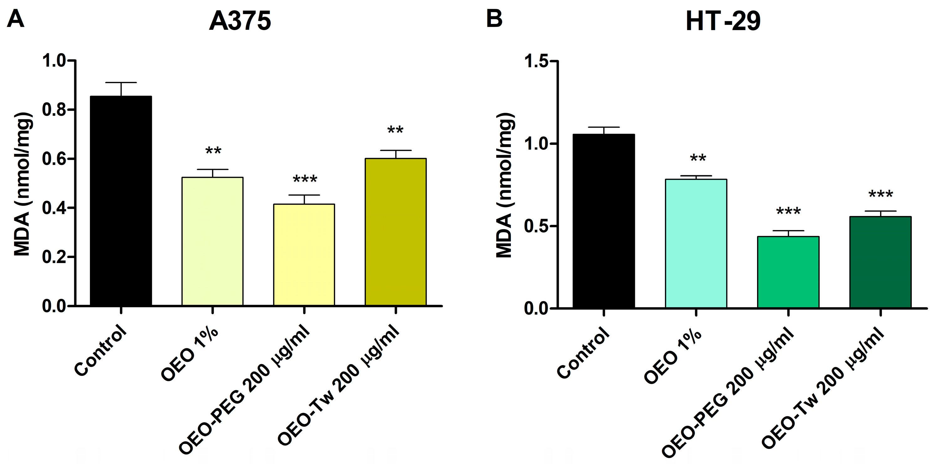
3.5. OEO Decreases Lipid Peroxidation

MDA levels, a marker of lipid peroxidation, were measured to evaluate oxidative stress in A375 and HT-29 cells treated with OEO, OEO-PEG, and OEO-Tw. The control group exhibited the highest MDA levels: 0.855 ± 0.09 in A375 cells and 1.057 ± 0.073 in HT-29 cells, reflecting the baseline lipid peroxidation. Treatment with 1% OEO significantly reduced MDA levels to 0.5253 ± 0.05 in A375 cells and to 0.783 ± 0.038 in HT-29 cells, suggesting a strong antioxidant effect. An even more pronounced reduction was observed following treatment with 200 mg/mL OEO-PEG, with MDA levels decreasing to 0.415 ± 0.065 in A375 cells and to 0.436 ± 0.064 in HT-29 cells. These results suggest an enhanced antioxidant activity, potentially due to improved solubility and bioavailability. Treatment with OEO-Tw (200 mg/mL) also reduced MDA levels to 0.601 ± 0.056 in A375 cells and to 0.556 ± 0.059 in HT-29 cells, although to a lesser extent than OEO-PEG. Overall, these results indicate that OEO and its formulations effectively mitigate lipid peroxidation, with OEO-PEG exerting the strongest protective effect (Figure 6).

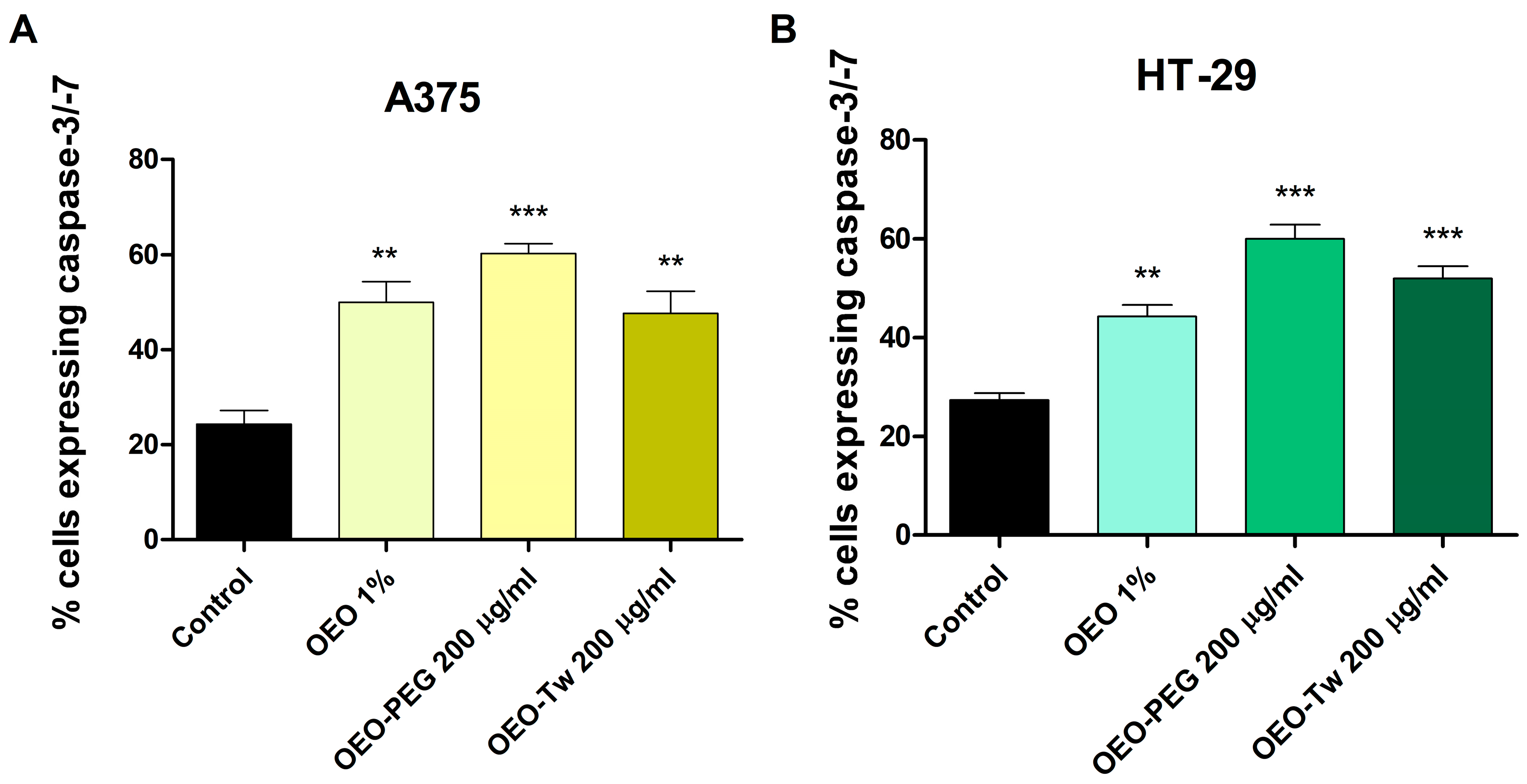
3.6. OEO Increases Caspase 3/7 Activation

Caspase-3/7 activity, a key indicator of apoptosis, was assessed in A375 and HT-29 cell lines after 24 h treatment with OEO 1%, OEO-PEG (200 μg/mL), and OEO-Tw (200 μg/mL). The results showed that in A375 cells, caspase-3/7 activity significantly increased from the control level of 20.12 ± 5.1 to 51.21 ± 7.5 after treatment with 1% OEO. OEO-PEG induced the highest caspase activation at 60.33 ± 3.1, while OEO-Tw increased the caspase activity to 47.67 ± 8.0, indicating induction of apoptosis. A similar trend was observed in HT-29 cells, where the control group showed a baseline activity of 25.33 ± 2.5. Treatment with 1% OEO increased caspase-3/7 activation to 44.33 ± 4.2, while OEO-PEG produced the highest caspase 3/7 activation at 63.19 ± 5.5, followed by OEO-Tw at 52.73 ± 4.39 (Figure 7). These findings indicate that OEO and its formulations can effectively trigger apoptotic pathways in both melanoma and colon cancer cells, with OEO-PEG demonstrating the strongest pro-apoptotic response in both cell lines.

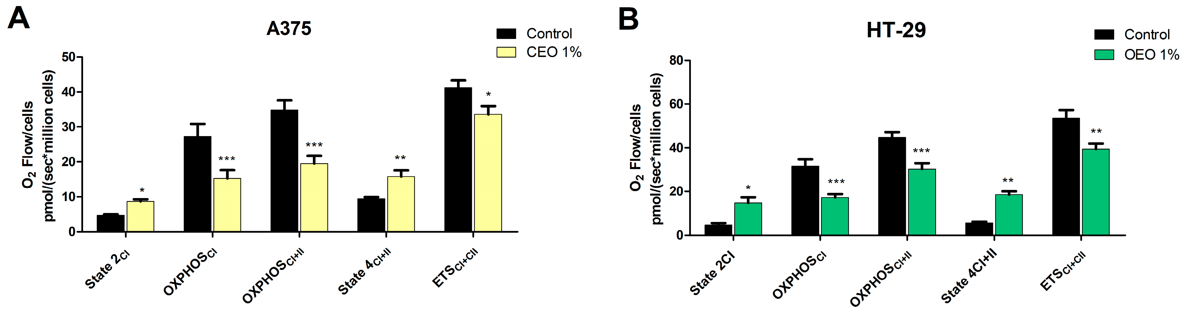
3.7. Evaluation of the Effect of OEO on Mitochondrial Function

High-resolution respirometry revealed significant alterations in mitochondrial function in both A375 and HT-29 cells following treatment with 1% OEO (Figure 8). In A375 cells, OEO markedly increased State 2CI from 4.77 ± 0.45 (control) to 8.67 ± 1.07, while also increasing proton leak respiration (State 4CI+II) from 9.45 ± 0.84 (control) to 15.80 ± 3.12 (Figure 8A). Moreover, OEO significantly suppressed CI ADP-stimulated respiration (OXPHOSCI, 15.32 ± 4.02) vs. control (27.26 ± 6.24) and also reduced OXPHOSCI+II from 34.86 ± 4.61 (control) to 19.48 ± 3.93 (Figure 8A). OEO treatment also decreased the maximal capacity of the electron transport system (ETSCI+II) from the control level of 41.23 ± 3.61 to 33.59 ± 4.00. A similar trend was observed in HT-29 cells; OEO treatment increased State 2CI respiration (14.77 ± 4.59 vs. 4.72 ± 1.38—control) and State 4CI+II respiration (18.55 ± 2.73 vs. 5.59 ± 0.92—control). Both OXPHOSCI and OXPHOSCI+II decreased after OEO treatment vs. control, as follows: 17.24 ± 2.65 vs. 31.64 ± 5.36 and 30.28 ± 4.54 vs. 44.79 ± 4.48. ETSCI+II also decreased after the treatment with 1% OEO from 53.68 ± 6.28 (control) to 39.42 ± 4.35. All these results indicate that OEO can impair mitochondrial respiratory function and can induce mitochondrial uncoupling in A375 and HT-29 cells.

4. Discussion

Recent studies have demonstrated the antiproliferative properties of EOs on different cancer cell lines, underscoring their complex biological potential [57]. OEO, traditionally known for its antimicrobial, anti-inflammatory, and digestive properties, has gained attention for its wide range of pharmacological activities that extend far beyond its traditional applications, including cancer [58,59,60].
The assessment of OEO chemical composition revealed the presence of high amounts of p-cymene, accompanied by two other compounds, γ-terpinene and thymol. Our results contrast with other publications that identified distinct chemical profiles for OEO. In the current study, the EO of Origanum vulgare L. originating from wild-growing plants from Romania proved to be devoid of the phenolic constituent carvacrol and contained only a small amount of thymol, usually cited as a main or marker compound for OEO. These two volatiles are also considered to be especially relevant for the favorable effects of Origanum vulgare L. oil on human health [61]. However, the absence of thymol and carvacrol in oregano oil has been pointed out by numerous other researchers. Lukas et al. [62] analyzed more than 500 plants from 17 countries and pointed out that different geographic regions of Europe are characterized by different Origanum vulgare L. chemotypes. Indeed, sabinene-rich EOs are typical for plants growing under a continental climate, in regions like the Balkan Peninsula, central Europe (Hungary and Slovakia), and Northern Europe (Baltic countries, Finland, and Norway). Other regional studies further confirm this chemotypic variability [63,64,65,66]. In Romania, Tamas et al. [67,68] demonstrated that wild-growing Origanum vulgare L. contains neither thymol nor carvacrol but is rich in bicyclic monoterpenes (sabinene and ∂-4-carene), sesquiterpenes (β-caryophylene and germacrene-D), and acyclic monoterpenes like trans-β-ocimene. Besides the geographical factor, additional factors, including misidentification [69] and commercial oregano adulteration with various other Mediterranean aromatic plants, including Origanum hirtum, Origanum majorana, and Thymus species [70], could also contribute to the reported chemical discrepancies.
The DPPH antioxidant activity data show that OEO has a moderate capacity to scavenge free radicals, with an IC50 value of 134.67 µg/mL. OEO showed similar ABTS radical inhibition to both controls at a concentration of 1 mg/mL. Although the ABTS inhibition values for OEO, AA, and BHA appear similar, it should be noted that OEO was tested at a higher concentration (94 mg/mL) compared to the 1 mg/mL solutions used for the reference antioxidants. Additionally, the high total polyphenolic content (159.63 mg GAE/g extract) is in line with the OEO phenol concentration (represented by thymol) determined through GC–MS.
The antiproliferative activity of OEO was investigated in A375 and HT-29 cancer cell lines, with PEG 400 and Tween 20 used as emulsifiers to improve the dispersion of the lipophilic oil in the aqueous-based culture medium [71]. Their stability, absence of toxicity, and high hydrosolubility were the key features that prompted their use in the current study [72]. An enhanced inhibitory effect was observed for both emulsified forms of OEO, with OEO-PEG showing the highest efficacy on both cancer cell lines, compared to the OEO water dispersion. This improvement can be attributed to the enhanced solubility, emulsion stability, and increased intracellular delivery of active volatiles, enhancing their antiproliferative action [30,72,73]. A similar increase in the cytotoxic effect after using emulsions of EO with Tween 20 and PEG 400 as surfactants was previously described in a recent study; the results showed that, similar to our case, clove EO, formulated with Tween-20 or PEG-400, enhanced the cytotoxic effect of the clove EO water dispersion in RPMI-7951 melanoma, A431 skin carcinoma, and NCI-H460 non-small lung cancer cells [32]. The inhibitory effects observed in our study demonstrated a dose-dependent profile and selectivity against cancer cells, as no antiproliferative effects in human HaCaT keratinocytes were detected.
In the A375 cell line, the strongest inhibitory activity was noticed for the PEG 400 emulsion, followed by the direct dispersion of OEO in aqueous medium, in a dose-dependent manner. However, Tween 20 emulsions were also effective in inhibiting A375 cell viability. To the best of our knowledge, the Origanum vulgare L. species investigated in the current study, without carvacrol, has rarely been the subject of cancer cell-based scientific investigations so far. Indeed, a similar chemical composition was reported for Origanum majorana, whose EO was found active in B16F10 melanoma cells through antioxidant and antiproliferative mechanisms [74].
In HT-29 cells, PEG 400 and Tween 20 emulsions inhibited cell viability more effectively than the water dispersion of OEO, presumably due to a better penetration of the active ingredients into the cells. Similar antiproliferative activity of Origanum vulgare EO was reported by Begnini et al. in HT-29 cells [75], while Spyridopoulou et al. [27] reported that in HT-29 and murine CT26 colon cancer cells, Origanum onitum EO has cytotoxic effects through apoptosis induction. The study of Kozics et al. [76], performed on HT-29 and HCT-116 colon carcinoma cell lines, revealed that among several EOs, such as peppermint, oregano, tea tree, lemon, lavender, frankincense, and two oil blends (Zengest and OnGuard), OEO had one of the highest cytotoxic effects; more precisely, the IC50 value for OEO in HT-29 cells was 0.056 µg/mL, while in the HCT-116 cell line, the IC50 value was 0.052 µg/mL. One study reporting on the biological effect of Origanum vulgare L. hydroalcoholic extract found a selective inhibitory activity against Caco2 cells through the activation of both initiator and effector caspases involved in apoptosis induction [77].
One main ingredient identified in the current study is p-cymene, a phytocompound that has been reported to exhibit anticancer effects against various cell lines, including A375 melanoma and HT-29 colorectal cancer cells [78,79]. This is consistent with the significant cytotoxicity observed in both cell types. One other major component, γ-terpinene, was shown to increase pro-apoptotic gene expression while suppressing A-2058 cell proliferation and anti-apoptotic gene expression [80]. Although thymol, the third main compound found in OEO, exhibited controversial antiproliferative effects in Caco2 cells [81] and other colon cancer cells, such as HCT-116 and HT-29 cells, thymol increased ROS production and induced apoptosis via the mitochondrial pathway [82,83]. Collectively, it is therefore safe to assume that the three compounds are at least in part responsible for the OEO’s cell inhibitory effect; however, as previously signaled by other authors, it is possible that the overall activity of OEO is caused by yet unidentified minor ingredients.
Molecular docking was employed to observe a potential mechanism of action for the OEO components related to cancer-active protein target inhibition. The results showed that CPO, a minor component, is a potential inhibitor for PDPK1. It was previously reported that CPO inhibits the constitutive activation of PI3K/AKT/mTOR/S6K1 signaling cascade [84]. PDPK1 is a crucial regulator of the PI3K/AKT pathway, and inhibiting this enzyme can further reduce AKT activity, which is essential for carcinogenesis [84]. This could mean that CPO might inhibit PI3K/AKT activation through PDPK1 inhibition as well. Unfortunately, there is no biological evidence yet that suggests the direct implication of CPO in PDPK1 inhibition.
MDA overproduction occurs as a result of increased production of free radicals and thus is considered an oxidative stress marker [85]. Due to its high reactivity, MDA is involved in the pathophysiology of various conditions, including cancer [86]. Our study revealed a significantly decreased MDA level in both types of cancer cells treated with OEO and an even stronger effect for OEO formulated with PEG 400 or Tween 20, presumably due to their increased aqueous solubility that facilitates a more intimate contact with cellular compounds. A similar decrease in the MDA level was reported in vivo for Origanum onites extract after oral administration and for Origanum vulgare EO both in vitro and in vivo [87,88]. Therefore, by reducing the amount of ROS as a result of its antioxidant activity, OEO is presumably able to act as a scavenger that hinders ROS-dependent signaling pathways involved in cancer progression.
The in vitro assay on caspase 3/7 activity indicated that OEO significantly increases the pro-apoptotic effect in both A375 melanoma and HT-29 colorectal cancer cells; apoptosis is an essential intracellular event consisting of programmed cell death in close relation with caspase enzyme activity [89]. This aligns with previous studies reporting similar caspase activation by Origanum majorana, which exhibited a similar chemical composition to that reported in the current study [90,91]. However, we were not able to identify any published data regarding the activity of OEO on caspase induction in melanoma or colon cancer cells; therefore, the current study provides useful information for future studies.
Considering the results obtained in the caspase 3/7 assay, this study also assessed the effect of OEO on the mitochondrial function of A375 and HT-29 cells. As far as we are aware, this study is the first to specifically evaluate the effects of 1% OEO on A375 and HT-29 cancer cell lines using high-resolution respirometry. Our results revealed that OEO significantly decreases the active respiration dependent on CI and CI+CII (OXPHOSCI and OXPHOSCI+II), indicating that OEO is able to inhibit oxidative phosphorylation, and thus, it can significantly impair mitochondrial function. Moreover, OEO increased both State2CI and proton leak respiration (State 4CI+II), suggesting that OEO induces mitochondrial dysfunction, potentially by uncoupling the oxidative phosphorylation. The uncoupling effect was first described by Terada H. [92]. Uncouplers are substances that can disrupt mitochondrial function by facilitating proton transfer through the inner mitochondrial membrane, bypassing complex V (ATP synthase), and hence they separate substrate oxidation from ADP phosphorylation [92]. In our study, the observed increase in State 4 respiration following treatment with OEO indicates their role as uncoupling agents. Another study revealed that p-Cymene, one of the major constituents of OEO, decreases OXPHOS, induces a proton leak, and uncouples oxidative phosphorylation [93]. In line with these findings, our results suggest that OEO can impair mitochondrial respiratory function and can induce mitochondrial uncoupling in A375 and HT-29 cells. However, a limitation of the current study is that, in order to confirm that OEO induces mitochondrial uncoupling, further mechanistic investigations employing specific assays targeting mitochondrial proton leak, ATP production, and uncoupling protein expression are necessary to validate this hypothesis.

5. Conclusions

This study offers strong evidence of the selective antiproliferative activity of OEO derived from Romanian Origanum vulgare L. against A375 melanoma and HT-29 colorectal carcinoma cell lines. GC–MS analysis revealed a unique chemical composition rich in p-cymene, γ-terpinene, and thymol, distinguishing it from other reported OEO chemotypes. OEO exhibited selective cytotoxicity against A375 and HT-29 cancer cells while sparing normal HaCaT cells. The enhanced cytotoxic effects of OEO when emulsified in PEG 400 and Tween 20 suggest that formulation strategies may play a crucial role in optimizing its bioactivity. OEO has a moderate antioxidant activity and proved to be better at scavenging ABTS radicals than DPPH, probably due to its thymol content. Molecular docking revealed that one of several anticancer mechanisms may be related through PDPK1 inhibition by minor components such as CPO. OEO and its formulations significantly reduced MDA levels, effectively mitigating lipid peroxidation, and increased caspase 3/7 activation, highlighting its ability to also trigger apoptosis in A375 and HT-29 cells. High-resolution respirometry studies demonstrated that OEO can inhibit mitochondrial respiration and that OEO might acts as a potential uncoupling agent in A375 and HT-29 cancer cells. These findings support the therapeutic potential of OEO as a promising candidate in cancer treatment strategies. Nevertheless, further research, including pharmacokinetic in vivo studies and mechanistic investigations, are required to validate these findings, to explore the therapeutic applications, and to advance its development as a therapeutic agent in cancer treatment.

Author Contributions

Conceptualization, G.M., F.C. and C.Ș.; methodology, A.G. and C.Ș.; software, M.M. and C.J.; validation, A.G. and C.Ș.; formal analysis, A.M., M.-A.P., A.T.L.-G., T.M. and C.J.; investigation, G.M., F.C., A.M., M.-A.P., A.T.L.-G., T.M. and M.M.; data curation, G.M., F.C. and C.J.; writing—original draft preparation, G.M. and F.C.; writing—review and editing, A.G. and C.Ș.; visualization, A.G. and C.Ș.; supervision, C.Ș. All authors have read and agreed to the published version of the manuscript.

Funding

We would like to acknowledge the “Victor Babes” University of Medicine and Pharmacy Timisoara for their support in covering the costs of publication for this research paper.

Data Availability Statement

The original contributions presented in the study are included in the article; further inquiries can be directed to the corresponding author.

Conflicts of Interest

The authors declare no conflicts of interest.

References

  1. Bray, F.; Laversanne, M.; Weiderpass, E.; Soerjomataram, I. The ever-increasing importance of cancer as a leading cause of premature death worldwide. Cancer 2021, 127, 3029–3030. [Google Scholar] [CrossRef]
  2. Anjum, S.; Hashim, M.; Malik, S.A.; Khan, M.; Lorenzo, J.M.; Abbasi, B.H.; Hano, C. Recent Advances in Zinc Oxide Nanoparticles (ZnO NPs) for Cancer Diagnosis, Target Drug Delivery, and Treatment. Cancers 2021, 13, 4570. [Google Scholar] [CrossRef] [PubMed]
  3. American Cancer Society. Global Cancer Facts & Figures, 5th ed.; American Cancer Society: Atlanta, GA, USA, 2024. [Google Scholar]
  4. Gogulescu, A.; Blidisel, A.; Soica, C.; Mioc, A.; Voicu, A.; Jojic, A.; Voicu, M.; Banciu, C. Neurological Side Effects of TNF-α Inhibitors Revisited: A Review of Case Reports. Medicina 2024, 60, 1409. [Google Scholar] [CrossRef]
  5. Chizenga, E.P.; Abrahamse, H. Biological Therapy with Complementary and Alternative Medicine in Innocuous Integrative Oncology: A Case of Cervical Cancer. Pharmaceutics 2021, 13, 626. [Google Scholar] [CrossRef]
  6. Jojic, A.A. Juniperi galbulus: Screening of phytochemicals and bioactivity investigation of the aqueous extract. Farmacia 2024, 72, 934–945. [Google Scholar] [CrossRef]
  7. Shaik, B.B.; Katari, N.K.; Jonnalagadda, S.B. Role of Natural Products in Developing Novel Anticancer Agents: A Perspective. Chem. Biodivers. 2022, 19, e202200535. [Google Scholar] [CrossRef] [PubMed]
  8. Asadi-Samani, M.; Kaffash Farkhad, N.; Reza Mahmoudian-Sani, M.; Shirzad, H. Antioxidants as a Double-Edged Sword in the Treatment of Cancer. In Antioxidants; IntechOpen: London, UK, 2019. [Google Scholar]
  9. Tietbohl, L.A.C.; Oliveira, A.P.; Esteves, R.S.; Albuquerque, R.D.D.G.; Folly, D.; Machado, F.P.; Correa, A.L.; Santos, M.G.; Ruiz, A.L.G.; Rocha, L. Antiproliferative activity in tumor cell lines, antioxidant capacity and total phenolic, flavonoid and tannin contents of Myrciaria floribunda. An. Acad. Bras. Cienc. 2017, 89, 1111–1120. [Google Scholar] [CrossRef] [PubMed]
  10. Meireles, D.R.; Fernandes, H.M.; Rolim, T.L.; Batista, T.M.; Mangueira, V.M.; de Sousa, T.K.; Pita, J.C.; Xavier, A.L.; Beltrão, D.M.; Tavares, J.F.; et al. Toxicity and antitumor efficacy of Croton polyandrus oil against Ehrlich ascites carcinoma cells. Rev. Bras. Farmacogn. 2016, 26, 751–758. [Google Scholar] [CrossRef]
  11. Mohamed Abdoul-Latif, F.; Ainane, A.; Houmed Aboubaker, I.; Mohamed, J.; Ainane, T. Exploring the Potent Anticancer Activity of Essential Oils and Their Bioactive Compounds: Mechanisms and Prospects for Future Cancer Therapy. Pharmaceuticals 2023, 16, 1086. [Google Scholar] [CrossRef]
  12. Pfeffer, C.; Singh, A. Apoptosis: A Target for Anticancer Therapy. Int. J. Mol. Sci. 2018, 19, 448. [Google Scholar] [CrossRef]
  13. Yu, J.; Zhong, B.; Xiao, Q.; Du, L.; Hou, Y.; Sun, H.-S.; Lu, J.-J.; Chen, X. Induction of programmed necrosis: A novel anti-cancer strategy for natural compounds. Pharmacol. Ther. 2020, 214, 107593. [Google Scholar] [CrossRef]
  14. Fu, W.; Chen, J.; Cai, Y.; Lei, Y.; Chen, L.; Pei, L.; Zhou, D.; Liang, X.; Ruan, J. Antioxidant, free radical scavenging, anti-inflammatory and hepatoprotective potential of the extract from Parathelypteris nipponica (Franch. et Sav.) Ching. J. Ethnopharmacol. 2010, 130, 521–528. [Google Scholar] [CrossRef]
  15. Luk, S.C.-W.; Siu, S.W.-F.; Lai, C.-K.; Wu, Y.-J.; Pang, S.-F. Cell Cycle Arrest by a Natural Product via G2/M Checkpoint. Int. J. Med. Sci. 2005, 2, 64–69. [Google Scholar] [CrossRef]
  16. Mohamed, F.; Essam, A. Regulation of Apoptosis, Invasion and Angiogenesis of Tumor Cells by Caffeic Acid Phenethyl Ester. In Carcinogenesis; InTech: Nappanee, IN, USA, 2013. [Google Scholar]
  17. Albonici, L.; Giganti, M.G.; Modesti, A.; Manzari, V.; Bei, R. Multifaceted Role of the Placental Growth Factor (PlGF) in the Antitumor Immune Response and Cancer Progression. Int. J. Mol. Sci. 2019, 20, 2970. [Google Scholar] [CrossRef] [PubMed]
  18. Morshedloo, M.R.; Craker, L.E.; Salami, A.; Nazeri, V.; Sang, H.; Maggi, F. Effect of prolonged water stress on essential oil content, compositions and gene expression patterns of mono- and sesquiterpene synthesis in two oregano (Origanum vulgare L.) subspecies. Plant Physiol. Biochem. 2017, 111, 119–128. [Google Scholar] [CrossRef]
  19. Guo, X.; Hao, Y.; Zhang, W.; Xia, F.; Bai, H.; Li, H.; Shi, L. Comparison of Origanum Essential Oil Chemical Compounds and Their Antibacterial Activity against Cronobacter sakazakii. Molecules 2022, 27, 6702. [Google Scholar] [CrossRef] [PubMed]
  20. Leyva-López, N.; Gutiérrez-Grijalva, E.; Vazquez-Olivo, G.; Heredia, J. Essential Oils of Oregano: Biological Activity beyond Their Antimicrobial Properties. Molecules 2017, 22, 989. [Google Scholar] [CrossRef]
  21. Lupea-Chilom, D.-S.; Solovan, C.S.; Farcas, S.S.; Gogulescu, A.; Andreescu, N.I. Latent Tuberculosis in Psoriasis Patients on Biologic Therapies: Real-World Data from a Care Center in Romania. Medicina 2023, 59, 1015. [Google Scholar] [CrossRef]
  22. Stojanović, N.M.; Mitić, K.V.; Nešić, M.; Stanković, M.; Petrović, V.; Baralić, M.; Randjelović, P.J.; Sokolović, D.; Radulović, N. Oregano (Origanum vulgare) Essential Oil and Its Constituents Prevent Rat Kidney Tissue Injury and Inflammation Induced by a High Dose of L-Arginine. Int. J. Mol. Sci. 2024, 25, 941. [Google Scholar] [CrossRef] [PubMed]
  23. Kosakowska, O.; Węglarz, Z.; Pióro-Jabrucka, E.; Przybył, J.L.; Kraśniewska, K.; Gniewosz, M.; Bączek, K. Antioxidant and Antibacterial Activity of Essential Oils and Hydroethanolic Extracts of Greek Oregano (O. vulgare L. subsp. hirtum (Link) Ietswaart) and Common Oregano (O. vulgare L. subsp. vulgare). Molecules 2021, 26, 988. [Google Scholar] [CrossRef]
  24. Erenler, R. Antiproliferative activity and cytotoxic effect of essential oil and water extract from origanum Vulgare L. Sigma J. Eng. Nat. Sci.–Sigma Mühendislik Ve Fen Bilim. Derg. 2023, 41, 202–206. [Google Scholar] [CrossRef]
  25. Elshafie, H.; Armentano, M.; Carmosino, M.; Bufo, S.; De Feo, V.; Camele, I. Cytotoxic Activity of Origanum Vulgare L. on Hepatocellular Carcinoma cell Line HepG2 and Evaluation of its Biological Activity. Molecules 2017, 22, 1435. [Google Scholar] [CrossRef] [PubMed]
  26. Wang, Y.; Wang, L.; Li, L.; Lu, J.X.; Ma, S.X. Anti-tumor activity of Origanum oil in vitro. Chin. J. Microecol. 2010, 22, 1101–1102. [Google Scholar]
  27. Spyridopoulou, K.; Fitsiou, E.; Bouloukosta, E.; Tiptiri-Kourpeti, A.; Vamvakias, M.; Oreopoulou, A.; Papavassilopoulou, E.; Pappa, A.; Chlichlia, K. Extraction, Chemical Composition, and Anticancer Potential of Origanum onites L. Essential Oil. Molecules 2019, 24, 2612. [Google Scholar] [CrossRef] [PubMed]
  28. Kebriti, I.; Solgi, M.; Velashjerdi, M. Improving quality of strawberry by novel essential oil nanoemulsions of Echinophora platyloba combined with Aloe vera gel and gum arabic. Sci. Rep. 2025, 15, 1731. [Google Scholar] [CrossRef]
  29. Campolo, O.; Giunti, G.; Laigle, M.; Michel, T.; Palmeri, V. Essential oil-based nano-emulsions: Effect of different surfactants, sonication and plant species on physicochemical characteristics. Ind. Crops Prod. 2020, 157, 112935. [Google Scholar] [CrossRef]
  30. Khairan, K.; Marfu’ah, M.; Idroes, R.; Sriwati, R.; Diah, M. An overview of essential oil-based nanoemulsion and their biological activities against some microbial pathogenic. IOP Conf. Ser. Earth Environ. Sci. 2024, 1297, 012083. [Google Scholar] [CrossRef]
  31. Craveiro, A.A. Oleos essenciais de plantas do Nordeste. In Coleção Ciência; Edição UFC: Fortaleza, Brazil, 1981; p. 209. [Google Scholar]
  32. Munteanu, A.; Gogulescu, A.; Șoica, C.; Mioc, A.; Mioc, M.; Milan, A.; Lukinich-Gruia, A.T.; Pricop, M.-A.; Jianu, C.; Banciu, C.; et al. In Vitro and In Silico Evaluation of Syzygium aromaticum Essential Oil: Effects on Mitochondrial Function and Cytotoxic Potential Against Cancer Cells. Plants 2024, 13, 3443. [Google Scholar] [CrossRef]
  33. Jianu, C.; Rusu, L.-C.; Muntean, I.; Cocan, I.; Lukinich-Gruia, A.T.; Goleț, I.; Horhat, D.; Mioc, M.; Mioc, A.; Șoica, C.; et al. In Vitro and In Silico Evaluation of the Antimicrobial and Antioxidant Potential of Thymus pulegioides Essential Oil. Antioxidants 2022, 11, 2472. [Google Scholar] [CrossRef]
  34. Pricop, M.-A.; Lukinich-Gruia, A.T.; Cristea, I.-M.; Păunescu, V.; Tatu, C.A. Aristolochia clematitis L. Ethanolic Extracts: In Vitro Evaluation of Antioxidant Activity and Cytotoxicity on Caco-2 Cell Line. Plants 2024, 13, 2987. [Google Scholar] [CrossRef]
  35. Jianu, C.; Goleț, I.; Stoin, D.; Cocan, I.; Lukinich-Gruia, A. Antioxidant Activity of Pastinaca sativa L. ssp. sylvestris [Mill.] Rouy and Camus Essential Oil. Molecules 2020, 25, 869. [Google Scholar] [CrossRef] [PubMed]
  36. Milan, A.; Mioc, M.; Mioc, A.; Gogulescu, A.; Mardale, G.; Avram, Ș.; Maksimović, T.; Mara, B.; Șoica, C. Cytotoxic Potential of Betulinic Acid Fatty Esters and Their Liposomal Formulations: Targeting Breast, Colon, and Lung Cancer Cell Lines. Molecules 2024, 29, 3399. [Google Scholar] [CrossRef] [PubMed]
  37. Liu, G.; Li, Y.; Yang, L.; Wei, Y.; Wang, X.; Wang, Z.; Tao, L. Cytotoxicity study of polyethylene glycol derivatives. RSC Adv. 2017, 7, 18252–18259. [Google Scholar] [CrossRef]
  38. Eskandani, M.; Hamishehkar, H.; Ezzati Nazhad Dolatabadi, J. Cyto/Genotoxicity study of polyoxyethylene (20) sorbitan monolaurate (tween 20). DNA Cell Biol. 2013, 32, 498–503. [Google Scholar] [CrossRef]
  39. Berman, H.M. The Protein Data Bank. Nucleic Acids Res. 2000, 28, 235–242. [Google Scholar] [CrossRef]
  40. Kim, S.; Chen, J.; Cheng, T.; Gindulyte, A.; He, J.; He, S.; Li, Q.; Shoemaker, B.A.; Thiessen, P.A.; Yu, B.; et al. PubChem 2023 update. Nucleic Acids Res. 2023, 51, D1373–D1380. [Google Scholar] [CrossRef]
  41. Dallakyan, S.; Olson, A.J. Small-Molecule Library Screening by Docking with PyRx. Methods Mol. Biol. 2015, 1263, 243–250. [Google Scholar]
  42. Toxicity Prediction—PreADMET. Available online: https://preadmet.webservice.bmdrc.org/toxicity-prediction (accessed on 16 May 2025).
  43. Islam, I.; Bryant, J.; Chou, Y.-L.; Kochanny, M.J.; Lee, W.; Phillips, G.B.; Yu, H.; Adler, M.; Whitlow, M.; Ho, E.; et al. Indolinone based phosphoinositide-dependent kinase-1 (PDK1) inhibitors. Part 1: Design, synthesis and biological activity. Bioorg. Med. Chem. Lett. 2007, 17, 3814–3818. [Google Scholar] [CrossRef]
  44. Yang, H.; Rudge, D.G.; Koos, J.D.; Vaidialingam, B.; Yang, H.J.; Pavletich, N.P. mTOR kinase structure, mechanism and regulation. Nature 2013, 497, 217–223. [Google Scholar] [CrossRef]
  45. Tecle, H.; Shao, J.; Li, Y.; Kothe, M.; Kazmirski, S.; Penzotti, J.; Ding, Y.-H.; Ohren, J.; Moshinsky, D.; Coli, R.; et al. Beyond the MEK-pocket: Can current MEK kinase inhibitors be utilized to synthesize novel type III NCKIs? Does the MEK-pocket exist in kinases other than MEK? Bioorg. Med. Chem. Lett. 2009, 19, 226–229. [Google Scholar] [CrossRef]
  46. Addie, M.; Ballard, P.; Buttar, D.; Crafter, C.; Currie, G.; Davies, B.R.; Debreczeni, J.; Dry, H.; Dudley, P.; Greenwood, R.; et al. Discovery of 4-Amino-N-[(1S)-1-(4-chlorophenyl)-3-hydroxypropyl]-1-(7H-pyrrolo[2,3-d]pyrimidin-4-yl)piperidine-4-carboxamide (AZD5363), an Orally Bioavailable, Potent Inhibitor of Akt Kinases. J. Med. Chem. 2013, 56, 2059–2073. [Google Scholar] [CrossRef] [PubMed]
  47. Ouvry, G.; Clary, L.; Tomas, L.; Aurelly, M.; Bonnary, L.; Borde, E.; Bouix-Peter, C.; Chantalat, L.; Defoin-Platel, C.; Deret, S.; et al. Impact of Minor Structural Modifications on Properties of a Series of mTOR Inhibitors. ACS Med. Chem. Lett. 2019, 10, 1561–1567. [Google Scholar] [CrossRef] [PubMed]
  48. Le, P.T.; Cheng, H.; Ninkovic, S.; Plewe, M.; Huang, X.; Wang, H.; Bagrodia, S.; Sun, S.; Knighton, D.R.; LaFleur Rogers, C.M.; et al. Design and synthesis of a novel pyrrolidinyl pyrido pyrimidinone derivative as a potent inhibitor of PI3Kα and mTOR. Bioorg. Med. Chem. Lett. 2012, 22, 5098–5103. [Google Scholar] [CrossRef]
  49. Souers, A.J.; Leverson, J.D.; Boghaert, E.R.; Ackler, S.L.; Catron, N.D.; Chen, J.; Dayton, B.D.; Ding, H.; Enschede, S.H.; Fairbrother, W.J.; et al. ABT-199, a potent and selective BCL-2 inhibitor, achieves antitumor activity while sparing platelets. Nat. Med. 2013, 19, 202–208. [Google Scholar] [CrossRef]
  50. Lee, E.F.; Czabotar, P.E.; Smith, B.J.; Deshayes, K.; Zobel, K.; Colman, P.M.; Fairlie, W.D. Crystal structure of ABT-737 complexed with Bcl-xL: Implications for selectivity of antagonists of the Bcl-2 family. Cell Death Differ. 2007, 14, 1711–1713. [Google Scholar] [CrossRef]
  51. Wood, E.R.; Truesdale, A.T.; McDonald, O.B.; Yuan, D.; Hassell, A.; Dickerson, S.H.; Ellis, B.; Pennisi, C.; Horne, E.; Lackey, K.; et al. A Unique Structure for Epidermal Growth Factor Receptor Bound to GW572016 (Lapatinib). Cancer Res. 2004, 64, 6652–6659. [Google Scholar] [CrossRef]
  52. McTigue, M.; Murray, B.W.; Chen, J.H.; Deng, Y.-L.; Solowiej, J.; Kania, R.S. Molecular conformations, interactions, and properties associated with drug efficiency and clinical performance among VEGFR TK inhibitors. Proc. Natl. Acad. Sci. USA 2012, 109, 18281–18289. [Google Scholar] [CrossRef]
  53. Abcam, C. (UK): Lipid Peroxidation (MDA) Assay Kit-ab118970. Available online: https://www.abcam.com/en-us/products/assay-kits/lipid-peroxidation-mda-assay-kit-colorimetric-fluorometric-ab118970?srsltid=AfmBOooSt5XYd52jEjk-dFQUE8Quw7ATT6_poQMSo-KMw40gzgHQJKne (accessed on 13 January 2025).
  54. Scientific, T. CellEventTM Caspase-3/7 Detection Reagents. Available online: https://www.thermofisher.com/order/catalog/product/C10423 (accessed on 27 January 2025).
  55. Petrus, A.; Ratiu, C.; Noveanu, L.; Lighezan, R.; Rosca, M.; Muntean, D.; Duicu, O. Assessment of Mitochondrial Respiration in Human Platelets. Rev. Chim. 2017, 68, 768–771. [Google Scholar] [CrossRef]
  56. Babushok, V.I.; Linstrom, P.J.; Zenkevich, I.G. Retention Indices for Frequently Reported Compounds of Plant Essential Oils. J. Phys. Chem. Ref. Data 2011, 40, 043101. [Google Scholar] [CrossRef]
  57. Bhalla, Y.; Gupta, V.K.; Jaitak, V. Anticancer activity of essential oils: A review. J. Sci. Food Agric. 2013, 93, 3643–3653. [Google Scholar] [CrossRef] [PubMed]
  58. Cinbilgel, I.; Kurt, Y. Oregano and/or marjoram: Traditional oil production and ethnomedical utilization of Origanum species in southern Turkey. J. Herb. Med. 2019, 16, 100257. [Google Scholar] [CrossRef]
  59. Lombrea, A.; Antal, D.; Ardelean, F.; Avram, S.; Pavel, I.Z.; Vlaia, L.; Mut, A.-M.; Diaconeasa, Z.; Dehelean, C.A.; Soica, C.; et al. A Recent Insight Regarding the Phytochemistry and Bioactivity of Origanum vulgare L. Essential Oil. Int. J. Mol. Sci. 2020, 21, 9653. [Google Scholar] [CrossRef]
  60. Bora, L.; Burkard, T.; Juan, M.H.S.; Radeke, H.H.; Muț, A.M.; Vlaia, L.L.; Magyari-Pavel, I.Z.; Diaconeasa, Z.; Socaci, S.; Borcan, F.; et al. Phytochemical Characterization and Biological Evaluation of Origanum vulgare L. Essential Oil Formulated as Polymeric Micelles Drug Delivery Systems. Pharmaceutics 2022, 14, 2413. [Google Scholar] [CrossRef]
  61. Bora, L.; Avram, S.; Pavel, I.; Muntean, D.; Liga, S.; Buda, V.; Gurgus, D.; Danciu, C. An up-to-date review regarding cutaneous benefits of Origanum vulgare L. essential oil. Antibiotics 2022, 11, 549. [Google Scholar] [CrossRef]
  62. Lukas, B.; Schmiderer, C.; Novak, J. Essential oil diversity of European Origanum vulgare L. (Lamiaceae). Phytochemistry 2015, 1, 32–40. [Google Scholar] [CrossRef] [PubMed]
  63. Mutu, A.; Budeanu, O.; Martea, R.; Clapco, S.; Gille, E. RAPD molecular marker study of the intraspecific variability of Origanum vulgare subsp. vulgare naturally occurring in Moldova. Rev. Bot. 2014, 1, 23–28. [Google Scholar]
  64. Nurzyńska-Wierdak, R.E.; Bogucka-Kocka, A.; Sowa, I.; Szymczak, G. The composition of essential oil from three ecotypes of Origanum vulgare L. ssp. vulgare cultivated in Poland. Farmacia 2012, 1, 571–577. [Google Scholar]
  65. Port, A.; Mutu, A.; Duca, M.; Gille, E. Analiza biochimică a plantelor de Origanum vulgare ssp. vulgare din flora spontană a Republicii Moldova. Bul. AŞM. Ştiinţele Vieţii. 2020, 340, 95–105. [Google Scholar]
  66. Gales, R.; Preotu, A.; Necula, R.; Gille, E.; Toma, C. Altitudinal variations of morphology, distribution and secretion of glandular hairs in Origanum vulgare L. leaves. Stud. Univ. Vasile Goldis. Ser. Stiint. Vietii. 2010, 1, 59–62. [Google Scholar]
  67. Tămaș, M.; Balica, G.; Ştefănescu, C. Research on some plant species contining essential oils performed at University of Medicine and Pharmacy „Iuliu Hațieganu” Cluj-Napoca. Contrib. Bot. 2020, 55, 141–151. [Google Scholar] [CrossRef]
  68. Tămaș, M.; Oprean, I. Precizări asupra compoziției chimice a uleiului volatil de Origanum vulgare L. Rev. Med. Chir. Soc. Med. Nat. 2003, 107, 31–34. [Google Scholar]
  69. Shafiee-Hajiabad, M.; Hardt, M.; Honermeier, B. Comparative investigation about the trichome morphology of Common oregano (Origanum vulgare L. subsp. vulgare) and Greek oregano (Origanum vulgare L. subsp. hirtum). J. Appl. Res. Med. Aromat. Plants 2014, 1, 50–58. [Google Scholar] [CrossRef]
  70. Raclariu-Manolică, A.C.; Anmarkrud, J.; Kierczak, M.; Rafati, N.; Thorbek, B.; Schrøder-Nielsen, A.; de Boer, H. DNA metabarcoding for quality control of basil, oregano, and paprika. Front. Plant Sci. 2021, 12, 665618. [Google Scholar] [CrossRef]
  71. Ke, W.-T.; Lin, S.-Y.; Ho, H.-O.; Sheu, M.-T. Physical characterizations of microemulsion systems using tocopheryl polyethylene glycol 1000 succinate (TPGS) as a surfactant for the oral delivery of protein drugs. J. Control. Release 2005, 102, 489–507. [Google Scholar] [CrossRef] [PubMed]
  72. Taher, S.S.; Al-Kinani, K.K.; Hammoudi, Z.M.; Ghareeb, M. mohammed Co-surfactant effect of polyethylene glycol 400 on microemulsion using BCS class II model drug. J. Adv. Pharm. Educ. Res. 2022, 12, 63–69. [Google Scholar] [CrossRef]
  73. Krishna, G.; Thrimothi, D. Nano Emulsions: A Novel Targeted Delivery of Cancer Therapeutics. In Design and Applications of Self-Assembly Aggregates—From Micelles to Nanoemulsions; IntechOpen: London, UK, 2024. [Google Scholar]
  74. da Silva, C.C.; Giordani, C.; Picoli, T.; Guterres, K.A.; Perera, S.C.; Capella, G.A.; Waller, S.B.; Corcini, C.D.; Junior, A.S.V.; Freitag, R.A.; et al. Potencial antiproliferativo do óleo essencial de Origanum majorana Linn. em células de melanoma e seus efeitos intracelulares em células neoplásicas e não-neoplásicas. Res. Soc. Dev. 2021, 10, e7110917788. [Google Scholar] [CrossRef]
  75. Begnini, K.R.; Nedel, F.; Lund, R.G.; Carvalho, P.H.d.A.; Rodrigues, M.R.A.; Beira, F.T.A.; Del-Pino, F.A.B. Composition and Antiproliferative Effect of Essential Oil of Origanum vulgare Against Tumor Cell Lines. J. Med. Food 2014, 17, 1129–1133. [Google Scholar] [CrossRef]
  76. Kozics, K.; Mesárošová, M.; Šramková, M.; Bučková, M.; Puškárová, A.; Galová, D.; Pangallo, D. Evaluation of Bioactivity of Essential Oils: Cytotoxic/Genotoxic Effects on Colorectal Cancer Cell Lines, Antibacterial Activity, and Survival of Lactic Acid Bacteria. Molecules 2025, 30, 890. [Google Scholar] [CrossRef]
  77. Savini, I.; Arnone, R.; Catani, M.V.; Avigliano, L. Origanum Vulgare Induces Apoptosis in Human Colon Cancer Caco 2 Cells. Nutr. Cancer 2009, 61, 381–389. [Google Scholar] [CrossRef]
  78. Wang, S.; Wang, X.; Wang, Y.; Leng, Q.; Sun, Y.; Hoffman, R.M.; Jin, H. The Anti-oxidant Monoterpene p -Cymene Reduced the Occurrence of Colorectal Cancer in a Hyperlipidemia Rat Model by Reducing Oxidative Stress and Expression of Inflammatory Cytokines. Anticancer Res. 2021, 41, 1213–1218. [Google Scholar] [CrossRef]
  79. Balahbib, A.; El Omari, N.; Hachlafi, N.E.; Lakhdar, F.; El Menyiy, N.; Salhi, N.; Mrabti, H.N.; Bakrim, S.; Zengin, G.; Bouyahya, A. Health beneficial and pharmacological properties of p-cymene. Food Chem. Toxicol. 2021, 153, 112259. [Google Scholar] [CrossRef] [PubMed]
  80. Acikgul, F.C.; Duran, N.; Kutlu, T.; Ay, E.; Tek, E.; Bayraktar, S. The therapeutic potential and molecular mechanism of Alpha-pinene, Gamma-terpinene, and P-cymene against melanoma cells. Heliyon 2024, 10, e36223. [Google Scholar] [CrossRef]
  81. Sampaio, L.A.; Pina, L.T.S.; Serafini, M.R.; Tavares, D.d.S.; Guimarães, A.G. Antitumor Effects of Carvacrol and Thymol: A Systematic Review. Front. Pharmacol. 2021, 12, 702487. [Google Scholar] [CrossRef] [PubMed]
  82. Chauhan, A.; Bahuguna, A.; Paul, S.; Chul, S. Thymol Elicits HCT-116 Colorectal Carcinoma Cell Death Through Induction of Oxidative Stress. Anticancer. Agents Med. Chem. 2017, 17, 1942–1950. [Google Scholar] [CrossRef]
  83. Blažíčková, M.; Blaško, J.; Kubinec, R.; Kozics, K. Newly Synthesized Thymol Derivative and Its Effect on Colorectal Cancer Cells. Molecules 2022, 27, 2622. [Google Scholar] [CrossRef] [PubMed]
  84. Park, K.-R.; Nam, D.; Yun, H.-M.; Lee, S.-G.; Jang, H.-J.; Sethi, G.; Cho, S.K.; Ahn, K.S. β-Caryophyllene oxide inhibits growth and induces apoptosis through the suppression of PI3K/AKT/mTOR/S6K1 pathways and ROS-mediated MAPKs activation. Cancer Lett. 2011, 312, 178–188. [Google Scholar] [CrossRef]
  85. Gaweł, S.; Wardas, M.; Niedworok, E.; Wardas, P. Malondialdehyde (MDA) as a lipid peroxidation marker. Wiad. Lek. 2004, 57, 453–455. [Google Scholar]
  86. Ayala, A.; Muñoz, M.F.; Argüelles, S. Lipid Peroxidation: Production, Metabolism, and Signaling Mechanisms of Malondialdehyde and 4-Hydroxy-2-Nonenal. Oxid. Med. Cell. Longev. 2014, 2014, 360438. [Google Scholar] [CrossRef]
  87. Kubiliene, A.; Munius, E.; Songailaite, G.; Kokyte, I.; Baranauskaite, J.; Liekis, A.; Sadauskiene, I. A Comparative Evaluation of Antioxidant Activity of Extract and Essential Oil of Origanum onites L. In Vivo. Molecules 2023, 28, 5302. [Google Scholar] [CrossRef]
  88. Nurzyńska-Wierdak, R.; Walasek-Janusz, M. Chemical Composition, Biological Activity, and Potential Uses of Oregano (Origanum vulgare L.) and Oregano Essential Oil. Pharmaceuticals 2025, 18, 267. [Google Scholar] [CrossRef]
  89. Shim, M.K.; Yoon, H.Y.; Lee, S.; Jo, M.K.; Park, J.; Kim, J.-H.; Jeong, S.Y.; Kwon, I.C.; Kim, K. Caspase-3/-7-Specific Metabolic Precursor for Bioorthogonal Tracking of Tumor Apoptosis. Sci. Rep. 2017, 7, 16635. [Google Scholar] [CrossRef] [PubMed]
  90. Gökhan, A. Evaluation of Cytotoxic, Membrane Damaging and Apoptotic Effects of Origanum majorana Essential Oil on Lung Cancer and Epidermoid Carcinoma Cells. Cyprus J. Med. Sci. 2022, 7, 201–206. [Google Scholar] [CrossRef]
  91. Arafat, K.; Sulaiman, S.; Al-Azawi, A.M.; Yasin, J.; Sugathan, S.; Nemmar, A.; Karam, S.; Attoub, S. Origanum majorana essential oil decreases lung tumor growth and metastasis in vitro and in vivo. Biomed. Pharmacother. 2022, 155, 113762. [Google Scholar] [CrossRef] [PubMed]
  92. Terada, H. The interaction of highly active uncouplers with mitochondria. Biochim. Biophys. Acta-Rev. Bioenerg. 1981, 639, 225–242. [Google Scholar] [CrossRef]
  93. Custodio, J.; Ribeiro, M.V.; Silva, F.; Rodrigues Machado, S.M.; Sousa, M. The essential oils component p-cymene induces proton leak through Fo-ATP synthase and uncoupling of mitochondrial respiration. J. Exp. Pharmacol. 2011, 3, 69–76. [Google Scholar] [CrossRef]
Figure 1. HaCaT cell viability after 24h treatment with OEO (0.5% and 1%), OEO-PEG (100 μg/mL and 200 μg/mL), and OEO-TW (100 μg/mL and 200 μg/mL). Results are presented as percentages of cell viability relative to the control group (set at 100%). Data are shown as mean ± SD from three independent experiments, each performed in triplicate.
Processes 13 01695 g001
Figure 2. A375 cell viability after 24h treatment with after 24h treatment with OEO (0.5% and 1%), OEO-PEG (100 μg/mL and 200 μg/mL), and OEO-TW (100 μg/mL and 200 μg/mL). Results are presented as percentages of cell viability relative to the control group (set at 100%). Data are shown as mean ± SD from three independent experiments, each performed in triplicate. Differences were considered statistically significant at p < 0.05 (*** p < 0.001).
Processes 13 01695 g002
Figure 3. HT-29 cell viability after 24h treatment with OEO (0.5% and 1%), OEO-PEG (100 μg/mL and 200 μg/mL), and OEO-TW (100 μg/mL and 200 μg/mL). Results are presented as percentages of cell viability relative to the control group (set at 100%). Data are shown as mean ± SD from three independent experiments, each performed in triplicate. Differences were considered statistically significant at p < 0.05 (*** p < 0.001).
Processes 13 01695 g003
Figure 4. Radar graphs generated by the docking scores of all docked components of OEO (A), the major constituents (B), and the minor components (C). The graph lines were generated based on a percentage of each compound’s docking score relative to the respective NL docking result (100%).
Processes 13 01695 g004
Figure 5. Compound 11 (β-caryophyllene oxide) docked into the binding site of the PDPK1 (PDB ID: 2PE1): hydrophobic surface representation (A) and 3D (B) and 2D representation of ligand–protein interactions (C); hydrophobic interactions are presented using purple dotted lines while green dotted lines represent hydrogen bonds.
Processes 13 01695 g005
Figure 6. The MDA level in A375 (A) and HT-29 (B) cells after 24 h treatment with OEO 1%, OEO-PEG, and OEO-Tw (200 μg/mL). Data are shown as mean ± SD from three independent experiments, each performed in triplicate. Differences were considered statistically significant at p < 0.05 (** p < 0.01 and *** p < 0.001).
Processes 13 01695 g006
Figure 7. The activation of caspase 3/7 in A375 (A) and HT-29 (B) cell lines after 24h treatment with OEO 1%, OEO-PEG, and OEO-Tw (200 μg/mL). Data are shown as mean ± SD from three independent experiments, each performed in triplicate. Differences were considered statistically significant at p < 0.05 (** p < 0.01 and *** p < 0.001).
Processes 13 01695 g007
Figure 8. Mitochondrial respiratory rates in permeabilized A375 (A) and HT-29 (B) cells after the treatment with OEO 1%. Data are presented as mean ± SD from three independent experiments (* p < 0.05, ** p < 0.01, and *** p < 0.001).
Processes 13 01695 g008
Table 2. Chemical composition of OEO as determined by GC–MS. The table presents the retention times (RT, min), calculated retention indices (RIcalc), and literature retention indices (RIlit) for the identified compounds; RIlit values from the literature, where available, were obtained under comparable chromatographic conditions using the same column type [56]. “—” indicates that no suitable literature RI value was found under comparable conditions.
IdCompound NameRTRIcalcRIlitArea
% Calc
1β-Pinene5.319599744.84
2δ-Carene5.639762.92
3Limonene5.9699310241.63
4γ-Terpinene6.841040100822.16
5p-Cymene7.31106543.98
6Linalool13.58139510953.11
7β-Caryophyllene13.84140914183.40
82-Isopropyl-4-methylanisole13.92141312322.08
9Anethole15.1914809132.23
10β-Bisabolene16.25153615051.10
11β-Caryophyllene oxide20.86178015821.09
12Thymol24.341963128911.46
Table 3. Antioxidant activity of OEO determined by DPPH and ABTS assays. DPPH results are expressed as IC50 (µg/mL). ABTS results are expressed as % inhibition determined for a 1:10 dilution of OEO, corresponding to 94 mg/mL, and for reference compounds (ascorbic acid (AA) and butylated hydroxyanisole (BHA)) at 1 mg/mL. TPC: Total Phenolic Content expressed as mg gallic acid equivalents (GAE) per gram of extract.
SampleDPPH IC50 (µg/mL)ABTS (Inh %)TPC (mg GAE/g Extract)
OEO134.67 ± 1.3288.15 ± 0.045159.63
AA10.53 ± 0.3388.84 ± 0.002 *-
BHA8.21 ± 0.4688.79 ± 0.002 *-
* ABTS inhibitory % determined for a control (AA and BHA) solution of 1 mg/mL.
Table 4. Docking scores of oregano essential oil (OEO) components compared to the native co-crystallized ligands of each protein target, used as reference benchmarks to validate docking performance.
CompoundProtein Targets
PDPK1mTOR Mek1AktPI3KαPI3KγBcl-XLBcl-2Vegfr2Egfr1
Binding Affinity (kcal/mol)
Native Ligand−8.8−11.2−9.5−9.4−8.8−9.3−10.9−11.5−12−11
1−5.6−5.1−5.3−5.1−5.4−5.3−6.2−5.7−6.4−5.3
2−6.1−6.1−6.3−6.2−5.7−6−6.2−6−6.6−5.9
3−6.2−6−6.5−6.1−5.6−5.9−6.5−5.9−6.8−6
4−6.2−6−6.6−6−5.7−5.9−6−5.7−6.7−5.8
5−6.3−6−6.7−6−5.7−6−6.1−5.8−6.7−5.8
6−5.4−5.6−5.4−5.1−5.5−5.6−5.4−5.5−6.2−5.3
7−7.2−6.6−6.6−6−6.3−6.2−7.1−6.9−7−7.1
8−6.1−5.8−5.5−6.2−6−6−6−5.5−6−5.9
9−6.1−6.3−6−5.9−5.9−6.1−5.9−5.9−6.7−5.9
10−7.1−7.3−7−6.7−7.2−7.4−6.6−7−8.5−7.5
11−7.9−6.2−7.2−6.2−6.4−6.3−7.5−6.9−7−7.1
12−6.4−6−6.5−6.2−6−5.8−6−5.9−7.2−6
Table 5. Predicted ADME (Absorption, Distribution, Metabolism, and Excretion) properties of OEO components based on PreADMET analysis. HIA: Human Intestinal Absorption (%, where 100 indicates high absorption), BBB: Predicted blood–brain barrier permeability (higher values suggest greater permeability), Caco-2: Predicted permeability through Caco-2 cell monolayers (nm/s), SKlogP: Predicted logarithm of the octanol–water partition coefficient (lipophilicity), Plasma protein binding: Predicted percentage of plasma protein binding, CYP 2C9, 2C19, 2D6, and 3A4: Predicted inhibition of major cytochrome P450 enzymes (Inhib: inhibitor, Non: non-inhibitor).
CompoundBBBCaco2CYP_2C19_InhibitionCYP_2C9_InhibitionCYP_2D6_InhibitionCYP_3A4_InhibitionHIAPlasma_Protein_BindingSKlogP_Value
β-Pinene5.75623.492NonInhibNonInhib1001002.952
δ-Carene5.53323.631NonInhibNonNon1001002.918
Limonene8.27823.631InhibInhibNonNon1001003.669
γ-Terpinene8.03723.640InhibInhibNonNon1001003.634
p-Cymene4.96923.433InhibInhibNonInhib1001003.559
Linalool6.12529.355InhibInhibNonNon1001002.749
β-Caryophyllene13.31923.631InhibInhibNonNon1001004.896
2-Isopropyl-4-methylanisole2.39357.964InhibInhibNonInhib1001003.544
Anethole1.47058.089InhibiInhibNonNon10089.242.938
β-Bisabolene15.06423.405InhibInhibNonNon1001005.613
β-Caryophyllene oxide3.75256.347NonInhibNonInhib10090.843.700
Thymol6.38838.012InhibInhibNonInhib1001003.405
Table 6. Predicted toxicity profiles of OEO components based on PreADMET analysis. Ames Test: Predicted mutagenicity (Mutagen/Non-mutagen) based on the bacterial reverse mutation assay using Salmonella typhimurium strains TA98, TA100, and TA1535. Carcinogenicity (Mouse/Rat): Predicted carcinogenic potential in rodent models (Positive/Negative). hERG Inhibition: Predicted risk of blocking the human ether-à-go-go-related gene (hERG) potassium channel (Low/Medium/High risk).
CompoundAmes_TestCarcino_MouseCarcino_RathERG_Inhibition
β-Pinenemutagennegativepositivemedium_risk
δ-Carenemutagennegativepositivemedium_risk
Limonenemutagennegativepositivemedium_risk
γ-Terpinenemutagenpositivepositivemedium_risk
p-Cymenemutagenpositivenegativemedium_risk
Linaloolmutagennegativenegativelow_risk
β-Caryophyllenemutagennegativepositivemedium_risk
2-Isopropyl-4-methylanisolemutagenpositivenegativemedium_risk
Anetholemutagenpositivenegativemedium_risk
β-Bisabolenemutagennegativepositivemedium_risk
β-Caryophyllene oxidemutagenpositivepositivemedium_risk
Thymolmutagennegativenegativelow_risk
Disclaimer/Publisher’s Note: The statements, opinions and data contained in all publications are solely those of the individual author(s) and contributor(s) and not of MDPI and/or the editor(s). MDPI and/or the editor(s) disclaim responsibility for any injury to people or property resulting from any ideas, methods, instructions or products referred to in the content.

