Article

Development of Chitosan–Hydroxyapatite Membranes from Bone of Armoured Catfish (Pterygoplichthys spp.) for Applications in Guided Bone Regeneration (GBR)

by
Ricardo de Jesús Figueroa López
1,
Carlos Roberto Luna-Domínguez
1,*,
Ana María Mendoza-Martínez
2,
Muradiye Şahin
3,
Bader Shafaqa Al-Anzi
4,
Ronaldo Câmara Cozza
5 and
Jorge Humberto Luna-Domínguez
1,*
1
Department of Periodontology, School of Dentistry, Autonomous University of Tamaulipas, Av. Universidad Esq. COm Blvd, Adolfo López Mateos, Tampico 89337, Tamaulipas, Mexico
2
Centro de Investigación en Petroquímica, Instituto Tecnológico de Ciudad Madero, Tecnológico Nacional de México, Altamira 89600, Tamaulipas, Mexico
3
Department of Chemistry, Kırşehir Ahi Evran University, 40100 Kırşehir, Turkey
4
Department of Environmental Sciences, Kuwait University, P.O. Box 5969, Safat 13060, Kuwait
5
CEETEPS—State Center of Technological Education “Paula Souza”, Faculty of Technology, Department of Mechanical Manufacturing, Campus Mauá, Av. Antônia Rosa Fioravante, Mauá 804-09390-120, SP, Brazil
*
Authors to whom correspondence should be addressed.
Processes 2025, 13(5), 1559; https://doi.org/10.3390/pr13051559
Submission received: 27 March 2025 / Revised: 8 May 2025 / Accepted: 12 May 2025 / Published: 18 May 2025
(This article belongs to the Section Materials Processes)

Abstract

:
Nowadays, there is an increasing interest in the development of novel bioresorbable membranes for Guided Bone Regeneration (GBR), and for this purpose, hydroxyapatite, from different sources, has been tested in combination with chitosan. This work details the production and the characterization of membranes of chitosan reinforced with hydroxyapatite derived from the bone of armoured catfish (Pterygoplichthys spp.), which is a widely available natural resource. The hydroxyapatite was characterized morphologically and chemically after the particles of hydroxyapatite were incorporated into a chitosan matrix. Then, the impact of adding hydroxyapatite particles into a matrix of chitosan on the roughness, mechanical properties, degradation, and cytotoxicity was evaluated. Subsequently, an in vivo test was carried out with the purpose of elucidating its guided bone regeneration activity, where the newly developed chitosan–hydroxyapatite membranes were implanted in rabbits with calvarial bone defects. The membranes of chitosan–hydroxyapatite presented a very rough surface morphology compared to the membranes of chitosan; moreover, the membranes of chitosan–hydroxyapatite showed superior mechanical tensile properties. The Masson’s trichrome staining analysis histologically demonstrated that the membranes of chitosan–hydroxyapatite enhanced the formation of a complete mineralized bone matrix in the calvarial bone defects. Finally, these findings confirm that the bone of armoured catfish (Pterygoplichthys spp.) is a viable, economic, and environmentally friendly source for isolating hydroxyapatite, which, combined with a matrix of chitosan, can be a suitable alternative to develop biocompatible GBR membranes.

1. Introduction

Tissue engineering has been adopted successfully in oral and maxillofacial surgical procedures in order to restore the density and volume augmentation of the alveolar bone [1,2,3,4]; for this purpose, Guided Bone Regeneration (GBR) is considered a promising technique.
In this technique of Guided Bone Regeneration (GBR), membranes are the fundamental components, being mainly used to cover the defective area because they prevent the connective-tissue cells’ migration—for example, fibroblasts—allowing the growth of bone cells [5,6,7,8]. Currently, according to their stability in the body, resorbable and non-resorbable membranes are utilized in the GBR technical therapy—in particular, biodegradable membranes available on the market, especially membranes developed with resorbable natural polymers, are more attractive for use in a wider spectrum of clinical situations [9], because they avoid a second surgical intervention [10,11,12,13,14].
Among the variety of bioresorbable polymers, chitosan has been reported to have a good affinity with the periodontal ligament cells. It also has a similar structure to glycosaminoglycans, resembling those of the bone tissue extracellular matrix, which facilitates the osteoblast attachment [15,16,17]. However, pristine chitosan membranes lack osteoinduction, and the mechanical properties may be a concern [18,19,20]. To further improve the osteoinductive properties (which are the bone regeneration properties) and the capacity to prevent the migration of epithelial cells to the socket (which is the barrier function of the membrane of chitosan), the incorporation of calcium phosphate into the chitosan matrix has been applied as a promising strategy [21,22,23,24], where hydroxyapatite can act as a source of calcium phosphate.
Hydroxyapatite is the main mineral component of hard tissues [2,14] like bones. It is a biomaterial with a plethora of human applications, being widely used as a bone graft substitute in orthopedic, dental, and maxillofacial applications due to its bioactivity, osteoconductivity, and osteoinductivity [25,26,27]. Hydroxyapatite can be produced from chemical synthesis or from natural sources, such as fish and chicken bones—the latter has better tissue engineering properties due to the presence of ions such as Na, K, Mg, Sr, Zn, and Si, which help to biomimetize the natural apatite of the human body. Generally, the bones used to extract the hydroxyapatite are obtained from animal residues, which, consequently, can generate a positive impact on the economy and the environment by adding high value to an organic resource [27,28,29,30,31,32]. In particular, hydroxyapatite obtained from the bones of armoured catfish can be a source of calcium phosphate for incorporation into the chitosan matrix.
Armoured catfish (Pterygoplichthys spp.) are found in countries located in the Gulf of Mexico [33], as well as on the Asian continent [34], and nowadays, this species is spread in the sea and the rivers [35]. In home aquariums, they are used to clean the bottom of the aquarium by controlling the algae and detritus build-up; however, their presence causes some damage to the marine fauna and to the anglers (fishermen), because while looking for food, they look for some plants, like the American Vallisneria, found in wetlands [10]. Their range of size is from 11 mm to 367 mm, and their reproduction is very easy, putting at risk the ecological (environmental) balance; they can survive in brackish environments, which can increase the probability of their dispersion [35].
It is well known that the fish bone is mainly composed of hydroxyapatite. However, nowadays, there are no dedicated efforts to extract hydroxyapatite from this species and direct it for human benefits. Thus, in the present work, the bone-armoured catfish (Pterygoplichthys spp.) is proposed as a source of hydroxyapatite for incorporation into a matrix of chitosan, with the purpose of developing a biomaterial (barrier membrane) with bioactivity and biocompatibility—particularly for repairing bone defects in the Guided Bone Regeneration (GBR) technique. The postulated research question is as follows: “Can membranes of chitosan–hydroxyapatite offer better medical performance than membranes of chitosan?

2. Materials and Methods

2.1. Preparation of the Hydroxyapatite Powder from Armoured Catfish (Pterygoplichthys spp.)

The hydroxyapatite powder has been isolated from the armoured catfish (Pterygoplichthys spp.) bone.
For this purpose, initially, specimens of armoured catfish (Pterygoplichthys spp.) were collected from the Champayán Lagoon, situated in Tampico, Tamaulipas, Mexico. Afterwards, the fish bones were washed with undistilled natural water for degreasing and dried at 80 °C for 6 h in a conventional oven (Magma, Renfert, São Caetano do Sul, Brazil). Then, the bones were cut into pieces to remove the bone marrow, followed by repeated washing and drying processes. The cortical bone pieces were pulverized using an agate mortar and followed by a heat treatment. During the heat treatment, the uncalcinated powder was subjected to 1000 °C for 1.5 h, obeying a heating rate of 6 °C/min in a catalytic burnout furnace under air atmosphere. The white powder obtained was ground and then sieved between #325 and #400 until reaching a particle size higher than 37 μm and lower than 44 μm for further characterization.

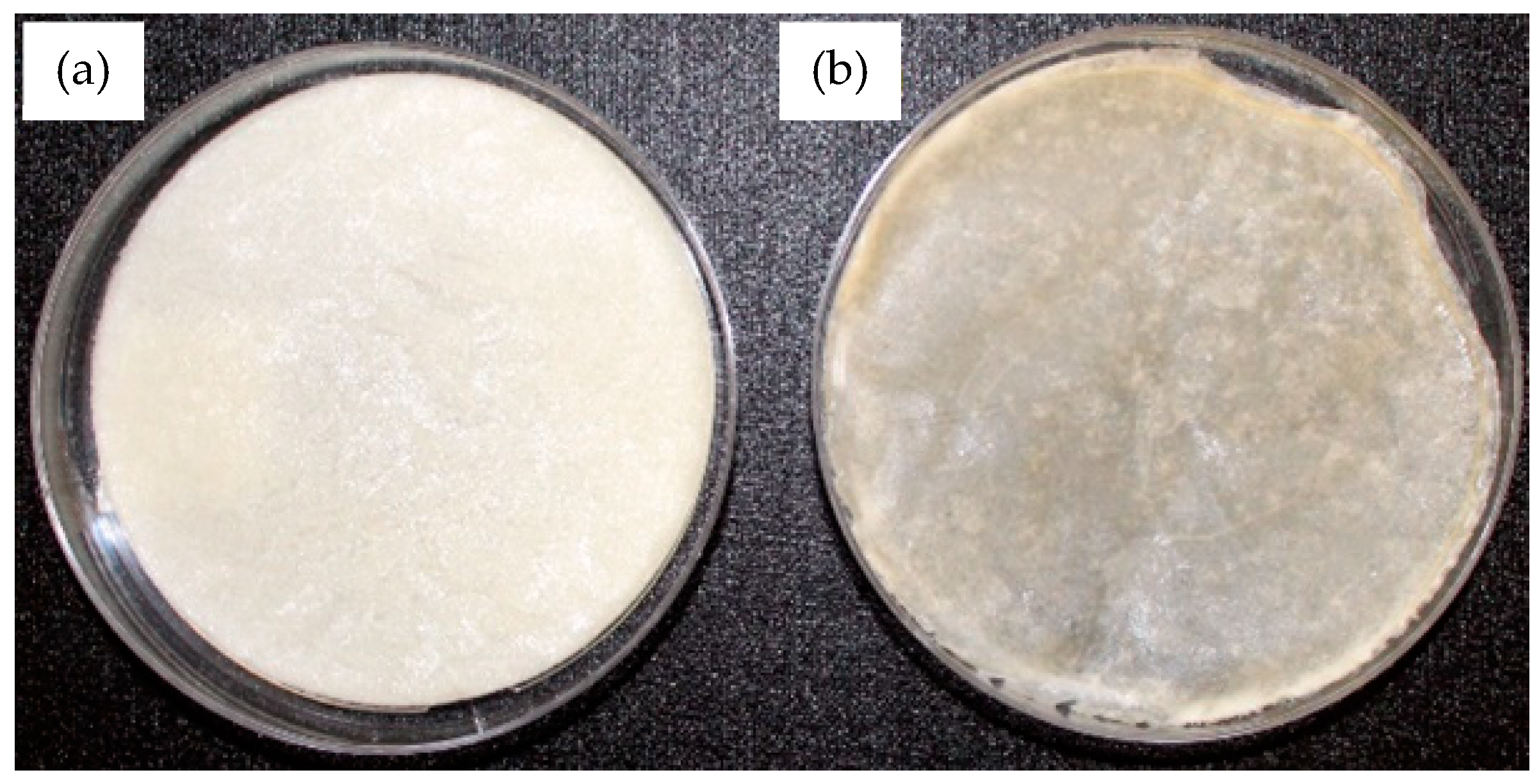
2.2. Fabrication of the Membranes of Chitosan and Chitosan–Hydroxyapatite

2.2.1. Preparation of the Chemical Solution for the Fabrication of the Membranes of Chitosan

Chitosan obtained from shrimp shells (deacetyl ≥ 75%) of 190,000–375,000 Da (Aldrich Chemical Company, Inc., Burlington, MA, USA) was used in the process of fabrication of the membranes of chitosan. Chitosan in a quantity of mc = 0.2 g was dissolved into 10 mL of 1 mol acetic acid solution, followed by the addition of 0.4 mL of edible glycerin under continuous stirring for 1 min, obtaining a gel-like chemical solution. Subsequently, the chemical solution obtained was deposited into glass Petri dishes and dried in a conventional heater (Magma, Renfert, São Caetano do Sul, Brazil) under 40 °C for a period of 24 h (Figure 1).
Finally, membranes of chitosan with diameter of 100 mm and thickness of 1 mm were produced.

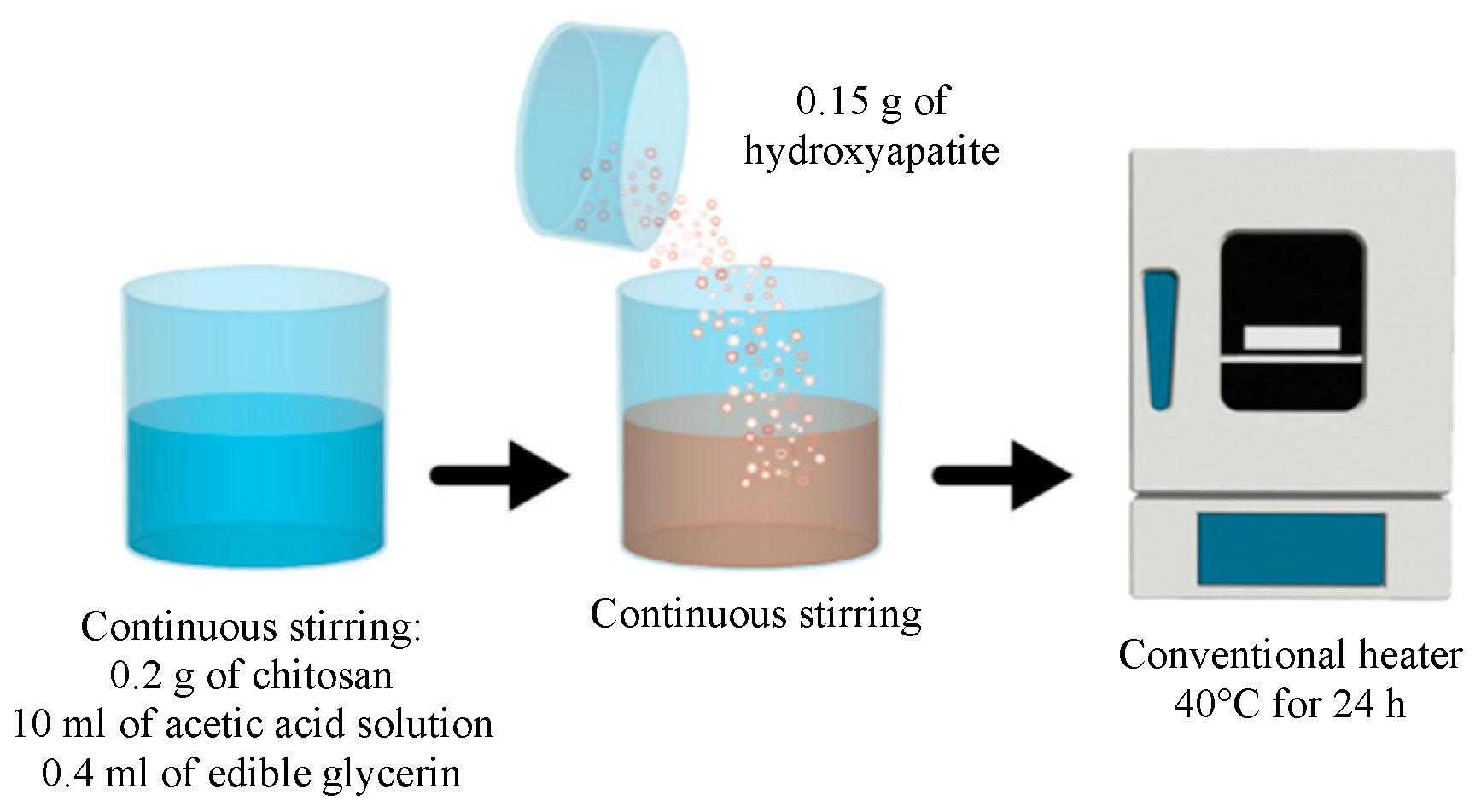
2.2.2. Preparation of the Chemical Solution for the Fabrication of the Membranes of Chitosan–Hydroxyapatite

Like the procedure carried out for the fabrication of the membranes of chitosan, for the fabrication of the membranes of chitosan–hydroxyapatite, mh = 0.15 g of hydroxyapatite powder was incorporated into the same chemical solution defined for the fabrication of the membranes of chitosan—mc = 0.2 g, 10 mL of 1 mol of acetic acid, and 0.4 mL of edible glycerin—and stirred for 1 min, obtaining a gel-like chemical solution. Subsequently, the chemical solution obtained was deposited into glass Petri dishes and dried in a conventional heater (Magma, Renfert, São Caetano do Sul, Brazil) under 40 °C for 24 h (Figure 2).
Finally, membranes of chitosan–hydroxyapatite with diameter of 100 mm and thickness of 1 mm were produced, where the ratio between the mass of hydroxyapatite, mh = 0.15 g, and the mass of chitosan, mc = 0.2 g, is defined by Equation (1):
m h m c = 0.15 g 0.2 g m h m c = 0.75

2.3. Characterization of the Powder of Hydroxyapatite, the Membranes of Chitosan, and the Membranes of Chitosan–Hydroxyapatite

The morphology of the powder of hydroxyapatite was analyzed by SEM (Scanning Electron Microscopy; JSM-7800F, JEOL Inc., Peabody, MA, USA) under different magnifications. Subsequently, EDX (Energy Dispersive X-Ray Spectroscopy; D2 Phaser, Bruker, Billerica, MA, USA) was carried out for characterizing its chemical composition.
The phase composition and the crystallinity of the hydroxyapatite were evaluated by X-Ray Diffraction (D2 Phaser, Bruker, Billerica, MA, USA) under CuKα radiation, from 2θ = 20° to 2θ = 60°, in steps of 0.030°. The functional groups of the powder of hydroxyapatite and of the membranes of chitosan and chitosan–hydroxyapatite were identified using a Fourier Transform Infrared Spectroscopy (FT-IR Spectrophotometer; FT 9700 Bench-top FT-NIR, PerkinElmer Inc., Waltham, MA, USA) using the Attenuated Total Reflectance (ATR) Method and scanned from 4000 cm−1 to 650 cm−1 under a resolution of 4 cm−1.

2.4. Mechanical and Superficial Evaluation of the Membranes of Chitosan and Chitosan–Hydroxyapatite

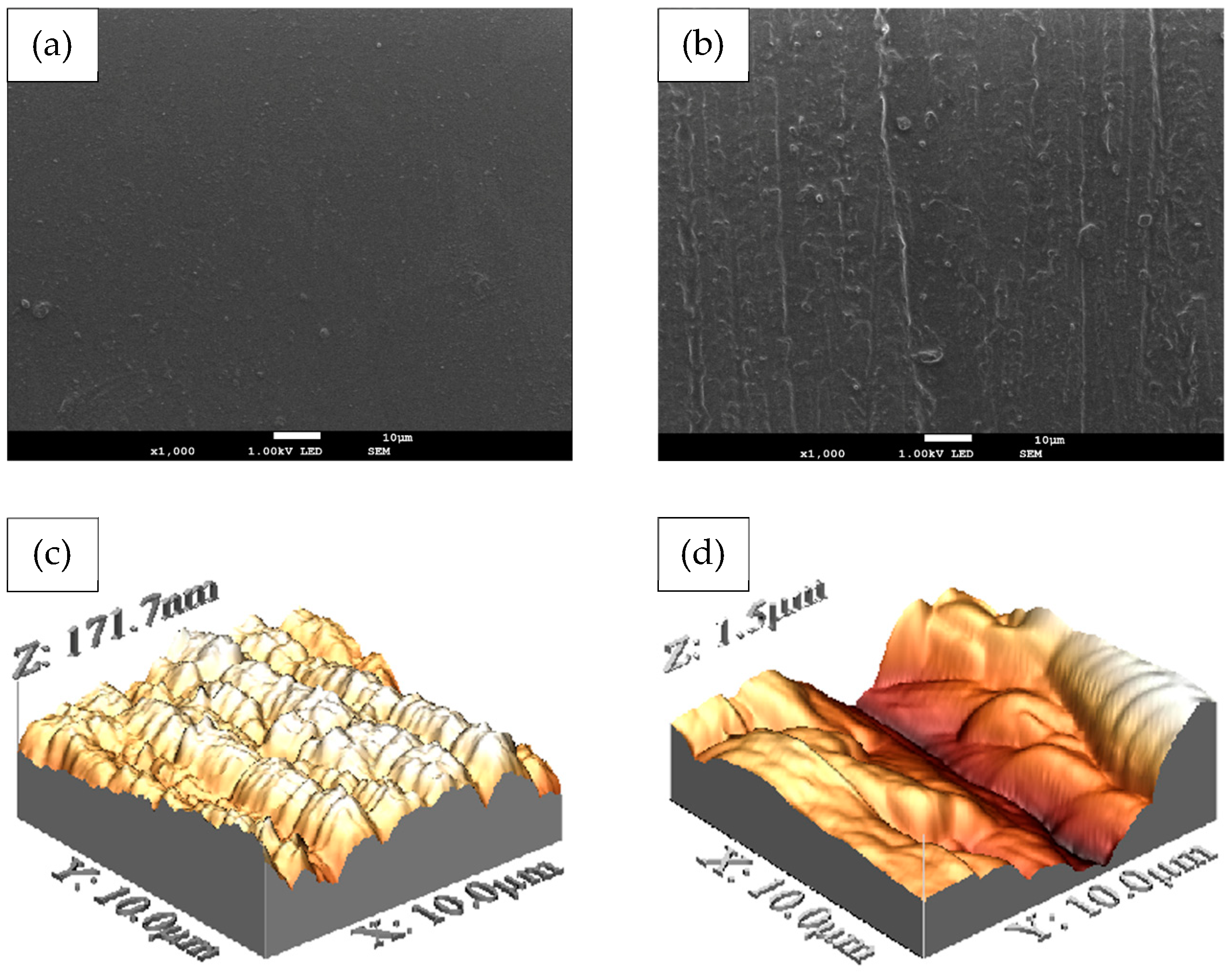
2.4.1. Evaluation of the Surface Roughness

The surface roughness of the membranes of chitosan and chitosan–hydroxyapatite was determined by a tapping mode using an Atomic Force Microscope (AFM; Alphacen 300 Flex, Nanosurf AG, Liestal, Switzerland).

2.4.2. Evaluation of the Tensile Mechanical Properties

The evaluation of tensile mechanical properties of the membranes of chitosan and chitosan–hydroxyapatite was performed according to Standard ASTM D 882-02 [36].
After the production of the membranes of chitosan and chitosan–hydroxyapatite, each membrane was cut into eight specimens with the dimensions of length, width, and thickness being 50 × 10 × 1 [mm], respectively. Subsequently, the sixteen specimens were immersed in distilled water at 37 °C in an incubator for 12 h.
The specimens were subjected to tensile tests parameterized in a Universal Testing Machine (Alliance RT/30, MTS Systems Corporation, Eden Prairie, MN, USA) equipped with a load cell of capacity 5 kN and under a velocity of 50 mm/min; all tensile tests were carried out at room temperature. The values of the Ultimate Tensile Strength (UTS) and elongation at break point (ε) were registered by a data acquisition system.

2.5. In Vitro Membrane Degradation

To characterize the in vitro degradation of the membranes of chitosan and chitosan–hydroxyapatite, initially, four specimens from membranes of chitosan and four specimens from membranes of chitosan–hydroxyapatite were cut in the dimensions of 20 × 20 × 1 [mm], and then, their initial masses (mi) were recorded using an electronic balance. Subsequently, the specimens were placed in containers filled with artificial saliva solution and, afterwards, placed in an incubator for eight weeks at a temperature of 37 °C. Once the interval time was over, the specimens were removed from the artificial saliva and passed by air-drying for 12 h, and then, the dry mass (md) of each membrane was recorded. Finally, the loss of mass (Δm) and the percentage loss of mass (Δm [%]) were calculated by Equations (2) and (3), respectively:
m = m i m d
m % = m d m i 100 %

2.6. Swelling Behaviour Analysis of the Membranes

The swelling behaviour analysis of the membranes of chitosan and chitosan–hydroxyapatite was determined by the gravimetric method in a controlled implantation environment in vitro [27]. Initially, eight membranes of chitosan and eight membranes of chitosan–hydroxyapatite—all membranes with dimensions of 20 × 20 × 1 [mm]—were subjected to air-drying for 12 h. Then, the masses of the eight dry membranes (md) of chitosan and the masses of the eight dry membranes (md) of chitosan–hydroxyapatite were measured.
Subsequently, the membranes were suspended in PBS (Phosphate-Buffered Saline) and incubated at 37 °C for 24 h. Afterwards, the membranes of chitosan and chitosan–hydroxyapatite were removed from incubator, and the excess PBS present on their surfaces was removed by placing each side of the membranes on a filter paper for 60 s.
Finally, the wet masses (mw) of the eight membranes of chitosan and of the eight membranes of chitosan–hydroxyapatite were measured, and the respective Swelling Ratio (SR) values were calculated using Equation (4):
S R % = m w + m d m d 100 %

2.7. Cell Culture

Human gingival fibroblasts (HGF) and osteoblastic cells were prepared from gingival tissues and mandibular bone fragments obtained by third molar surgical removal from a twenty-five-year old patient, who agreed to participate and signed an informed consent document.
The tissue samples were prepared in small fragments seeded into 10 cm culture plates and cultured in alpha-modified Eagle’s Minimum Essential Medium (α-MEM), supplemented with 20% heat-inactivated FBS (Fetal Bovine Serum; Thermo Fisher Scientific Inc., Waltham, MA, USA); to this solution were added 100 UI/mL of penicillin and 100 mg/mL of streptomycin (Thermo Fisher Scientific Inc., Waltham, MA, USA).
Then, the main culture was incubated at 37 °C in a humidified atmosphere with 5% CO2 until reaching a cell population of 80% of the area of the plate. Subsequently, the cells were harvested by treatment with 0.25% trypsin and 0.025% of ethylenediaminetetraacetic acid disodium salt in Phosphate-Buffered Saline (PBS). Finally, the cells were subcultured in α-MEM (Thermo Fisher Scientific Inc., Waltham, MA, USA) supplemented with 10% of Fetal Bovine Serum (FBS) and antibiotics in a humidified atmosphere with 5% CO2.

2.8. Cytotoxicity Assay

Cells, in a concentration of 2 × 10⁵ cells/mL, were inoculated into 96 micro-well plates and incubated for 48 h, with the purpose of achieving complete cell adhesion. Subsequently, the membranes were placed at the bottom of the culture plate and incubated for 24 h, 48 h, 72 h, and 96 h, followed by the determination of the number of viable cells using 0.2 mg/mL of Cell Proliferation Kit I—MTT (3-(4,5-Dimethylthiazol-2-yl)-2,5-Diphenyltetrazolium Bromide) (Sigma-Aldrich, Taufkirchen, Germany). Then, once the culture medium was replaced by MTT dissolved in DMEM (Dulbecco’s Modified Eagle’s Medium), the cells were incubated at 37 °C for 4 h. The formazan product was dissolved with DMSO (dimethyl sulfoxide) (Thermo Fisher Scientific Inc., Waltham, MA, USA), and the absorbance of the lysate was determined at 540 nm using a microplate reader.
Finally, the mean value of the 50% cytotoxic concentration (CC50) of the membranes of chitosan and of the membranes of chitosan–hydroxyapatite were calculated in triplicate from three independent experiments, and the asseveration was based on the interpretation of the ISO 10993-5: Biological Evaluation of Medical Devices [37].

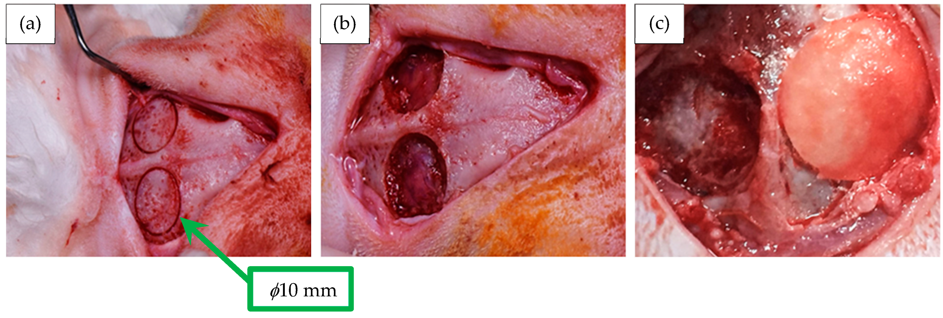
2.9. Membrane Implantation in the Animal Model

Twelve healthy adult male rabbits belonging to the New Zealand strain with an average mass of 4.1±0.31 kg were fed according to the Mexican Official Standard NOM 062 Z00-1999 [38]; this investigation was reviewed and approved by the Dentistry Faculty Animal Care and Use Committee of the Autonomous University of Tamaulipas, Mexico.
The surgery was followed as previously reported in references [28,30]. Two craniotomy defects were created in each one of the twelve rabbits to reach a total of twenty-four craniotomies. Thus, the defects were randomly selected and treated under the following conditions:
  • Eight craniotomy defects (n = 8) were treated with membranes of chitosan;
  • Eight craniotomy defects (n = 8) were treated with membranes of chitosan–hydroxyapatite;
  • Eight craniotomy defects (n = 8) were treated without membranes, which were considered as the negative control.
The surgeries were initiated with the administration of general anesthesia tiletamine/zolazepam 0.2 mL (Zoletil® 50, Virbac Corporation, Westlake, CA, USA), combined with acepromazine 0.1 mL (Calmivet®, Vetoquinol Unipessoal, Lda., Agualva-Cacém—POR); additionally, 2% of lidocaine with epinephrine under the ratio 1:100,000 was applied as a local anesthetic. Once the area of surgical intervention was shaved and disinfected with iodopovidone, an incision was made in the skin along the midline, with the muscular fascia being exposed and the periosteum from sagittal crest being removed. Subsequently, with a trephine contra-angle bur (Hu-Friedy® Trephine Burs, Pearson Dental Supply Co., Sylmar, CA, USA), two craniotomies were performed, under a diameter of 10 mm and a minimum depth that was sufficient to expose the dura mater. In sequence, these experimental defects were completely covered with the membranes—in each adult male rabbit, one experimental defect was covered with a membrane of chitosan and one experimental defect was covered with a membrane of chitosan–hydroxyapatite, according to the Guide Bone Regeneration (GBR) guidelines [39,40,41,42].
Finally, a 4-0 suture (Atramat®, Mexico City, Mexico) was applied, and the animals were medicated intramuscularly with enrofloxacin (1.03 mL/day) for five days, complemented by a single dose of 0.5 mg/kg of dexamethasone (Dexa-Jet NRV*, Mexico City, Mexico). During the postoperative course, the lesions were inspected four times a week for abnormal clinical signs. After four, six, and eight weeks of the surgical procedure, the rabbits were sacrificed with an overdose of sodium pentobarbital (PISABENTAL®, PiSA Salud Animal, Jalisco, Mexico) through the peritoneal route.

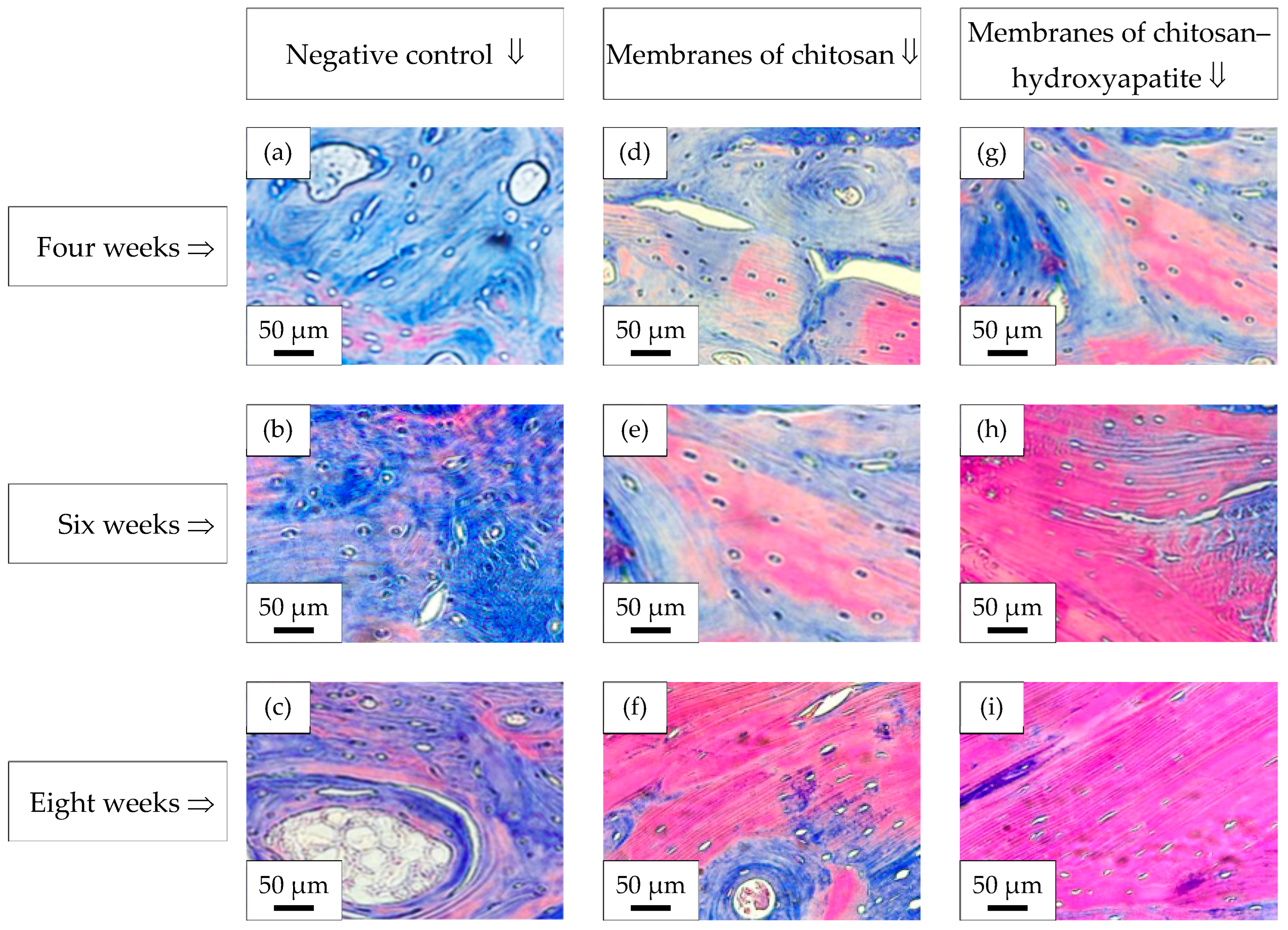
2.10. Histological Analysis—Masson’s Trichrome Stain

The specimens of bone tissue, obtained from the calvarial craniotomies, were dissected and fixed in 10% paraformaldehyde, followed by the application of 10% formic acid for 30 days for the decalcification process. Subsequently, the samples were gradually dehydrated in increasing concentrations of ethanol ⇒ 50%, 70%, 90%, and 100%. Then, the specimens were placed in xylene for 1 h, embedded in paraffin, and cut into sections of 6 mm using a rotary microtome (Model HM 340E, Thermo Fisher Scientific Microm International GmbH, Regensburg, Germany).
Finally, the sections obtained were mounted on a glass slide, and Masson’s trichrome staining was used to visualize the mineralized bone matrix and collagen with a digital microscope equipped with a digital camera (Model BX51, Olympus Co., Tokyo, Japan).

2.11. Statistical Analysis

Quantitative data were recorded descriptively as mean (σ) and standard deviation (SD) ⇒ σ±SD. After testing the normal distribution assumption with the Kolmogorov–Smirnov test, Student’s t-test was used to analyze significant differences between the membranes of chitosan and chitosan–hydroxyapatite under a statistical significance set at p < 0.05. IBM SPSS Statistics 23 software (IBM Co., New York, NY, USA) was used to perform the statistical analysis.

3. Results and Discussion

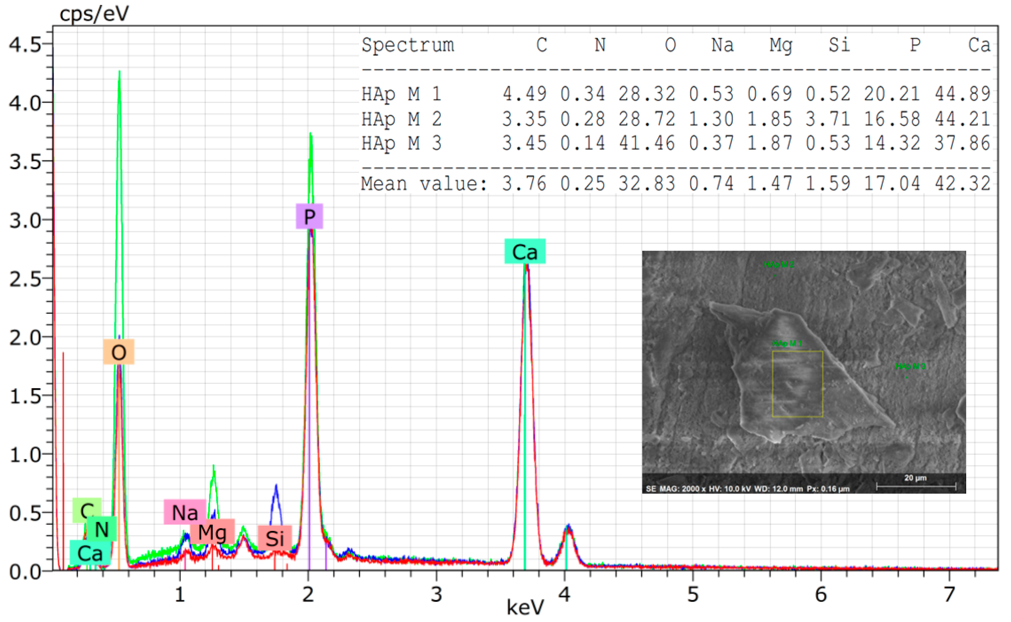
3.1. Characterization of the Hydroxyapatite Powder Isolated from Armoured Catfish

The morphology of the hydroxyapatite powder is shown in Figure 3a,b under different magnifications—it is possible to observe rod-shaped structures with a consistent diameter, ranging between 0.1 μm and 2.0 μm, and a length of approximately 6.0 μm or more; the structures also present small pores on their surface.
In other natural sources of hydroxyapatite under the same calcination temperature, similar particle sizes have been obtained, but rock-shaped, as reported in Reference [43]; similar results were obtained for synthesized nano-hydroxyapatite [44,45]. It is conjectured that the as-obtained rod-shaped morphology can improve the crosslinking during the formation of the membrane; additionally, the small pores on the surfaces of the particles of hydroxyapatite can ensure tissue growth as well as cell attachment.
Similar values for the particle sizes of hydroxyapatite produced from other types of natural sources have been obtained when the calcination temperature was similar to the ones developed in this work.
Through an analysis conducted by EDX (Energy Dispersive X-Ray Spectroscopy), it was possible to report traces of ions, such as Na, Mg, and Si, in the derived natural hydroxyapatite powder (Figure 4). The minor amounts of these chemical elements in the hydroxyapatite network increase the proliferation and differentiation of osteoblasts and, additionally, replace the inorganic component of human bones even more [42,43,45,46,47,48,49].
On the other hand, despite having calcined the powder at 1000 °C, the average Ca/P ratio was 1.90±0.03, and this result agrees with the data reported in the literature, where no relationship was observed between the Ca/P ratio and the temperature of calcination [43,50]. However, this behaviour differs from previous published results, where the authors mentioned that Ca/P ratios lower than 1.67 occur under temperatures of calcination above 800 °C [47,48,51,52]. Additionally, Ca/P ratios in the range from 0.90 to 1.11 were found in the jaw tissue of Wistar rats, suggesting that the hydroxyapatite obtained from armoured catfish can be used in engineering tissue [53,54].
It has been reported that calcination treatments carried out between 900 °C and 1100 °C lead to partial decomposition of the hydroxyapatite phase in tricalcium phosphate, a physico–chemical phenomenon that, in this study, was not observed in the X-Ray Diffraction patterns [27,51,55] of the hydroxyapatite powder, where the hydroxyapatite characteristic peaks correspond to the main intensities of the crystal planes 〈002〉, 〈120〉, 〈121〉, 〈112〉, 〈300〉, 〈202〉, 〈130〉, 〈222〉, 〈132〉, 〈123〉, 〈231〉, and 〈004〉 with hexagonal phase (Standard Hydroxyapatite, JCPDS Card # 96-901-2217). Additionally, the results obtained by X-ray diffractogram revealed narrow, high-intensity peaks that can be associated with larger crystallite sizes [44,45] and that quantitatively coincide with the values of crystallite sizes obtained for the synthesized nano-hydroxyapatite [44]. Complementing this analysis, the behaviour observed in this current work also was reported in calcined tuna bones under temperatures above 700 °C, indicating that the organic material has been eliminated [56].
Calcination at 1000 °C was selected because this temperature yields a highly crystalline hydroxyapatite with enhanced chemical, mechanical, and thermodynamic stability in biological fluids [57,58]. Such crystallinity and near-stoichiometric purity ensure that the hydroxyapatite maintains structural integrity when integrated into the membrane of chitosan for bone regeneration, providing a stable ceramic component within the polymeric scaffold [58,59].
Indeed, highly crystalline hydroxyapatite is a well-established osteoconductive material capable of serving as a scaffold for osteoblast adhesion, proliferation, and osteoid matrix deposition, as evidenced by the ceramic hydroxyapatite coatings on metallic implants that achieve direct bone bonding without adverse responses [60].
In contrast, lower calcination temperatures yield a carbonated hydroxyapatite containing bioactive trace elements but with increased solubility, while higher temperatures may induce the formation of biphasic calcium phosphate (e.g., hydroxyapatite/β-TCP), also characterized by elevated resorption rates [57,59]. Accordingly, the choice of 1000 °C maximizes the stability of the hydroxyapatite phase—approaching the ideal Ca/P ratio (≈1.67)—thus, avoiding excessive dissolution while preserving its osteoconductive behaviour [57].
Correlating the results obtained from X-Ray Diffraction (Figure 5a) with the analysis carried out by FT-IR (Fourier Transform Infrared Spectroscopy) (Figure 5b), it is possible to confirm that there was no partial decomposition of the hydroxyapatite phase in tricalcium phosphate, because additional absorption bands did not appear. In reality, the FT-IR spectrum presented in Figure 3b reveals absorption bands at 1088 cm−1, 1026 cm−1, and 962 cm−1, which are associated with the triple group of P O 4 3 ; consequently, this confirms a typical apatite structure, as reported in the scientific literature [45,56]. Additionally, according to the literature, positively charged chitosan amine groups, as well as their deprotonated portion, can lead to attraction by electrostatic forces with negatively charged P O 4 3 ions and cationic Ca ions present in the hydroxyapatite [27,28,54,61].
The likely explanation for this physico–chemical phenomenon is that the main inorganic component of the armoured catfish bones is a non-stoichiometric hydroxyapatite without calcium deficiency.
Under the treatment temperature of 1000 °C, the organic material has been completely removed without decomposing the hexagonal hydroxyapatite phase, i.e., pure hydroxyapatite has been obtained following the scientific methodology proposed in this current work.

3.2. Superficial, Chemical, and Mechanical Characterizations of the Membranes of Chitosan and Chitosan–Hydroxyapatite

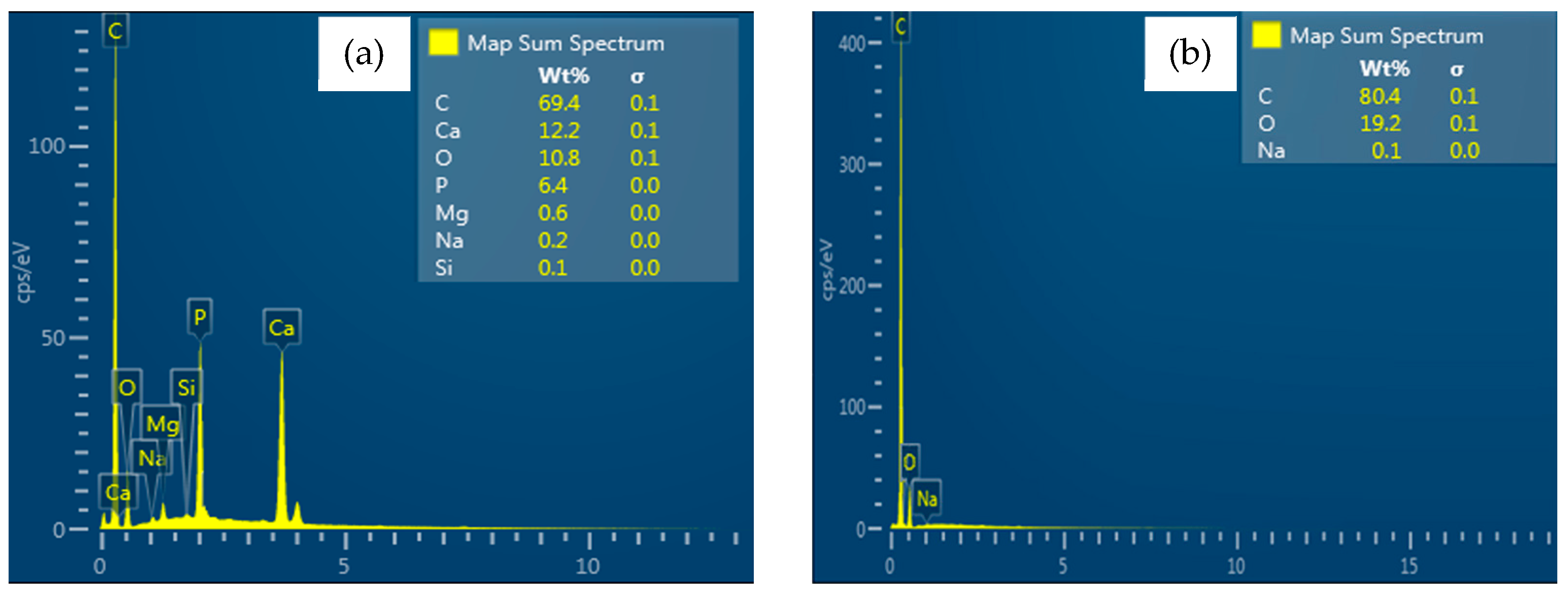
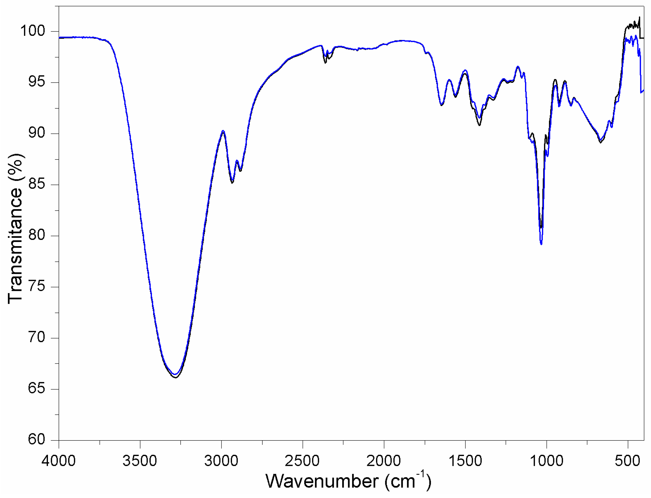
3.2.1. Chemical Characterization of the Membranes of Chitosan and Chitosan–Hydroxyapatite

The mapping analysis of the chemical elementary composition of each type of membrane produced in this work showed that the membranes of chitosan only presented C, O, and Na (Figure 6a), while the membranes of chitosan–hydroxyapatite presented, additionally, Ca, P, and trace chemical elements; the presence of Mg and Si was also reported (Figure 6b).
Furthermore, analyzing the results obtained by FT-IR (Fourier Transform Infrared Spectroscopy), it is possible to observe well-defined adsorption bands of chitosan in the membrane of chitosan (Figure 7—black spectrum), which confirms the chemical identity of the biopolymer [62].
Regarding the chitosan–hydroxyapatite membrane (Figure 7—blue spectrum), the phosphate and hydroxyl stretching vibration bands corresponding to the hydroxyapatite biomaterial overlap with the chitosan biomaterial stretching vibration bands of the C–O group under the wavenumbers ranging between λ = 1150 cm−1 and λ = 1040 cm−1; additionally, the stretching vibration bands of O–H and N–H are related to the wavenumbers of λ = 3420 cm−1 and λ = 3255 cm−1 [61], respectively. Similar results related to the overlapping of FT-IR (Fourier Transform Infrared Spectroscopy) were obtained in a recent study that developed and produced different scaffolds composed of chitin, chitosan, and hydroxyapatite [19,27].
Table 1 details the scientific meaning of the value of each wavenumber (λ) extracted from the FT-IR (Fourier Transform Infrared Spectroscopy) analysis of Figure 7 [63].
Therefore, according to the literature, due to its chemical composition, the type of chitosan–hydroxyapatite membrane developed and produced in this current work has the potential to provide qualitatively better mechanical integration with the bone tissue and better cell adhesion than the membrane of chitosan, because the chitosan–hydroxyapatite biomaterial favours the chelation of the chemical interactions of the cations [54,61].

3.2.2. Superficial Characterization of the Membranes of Chitosan and Chitosan–Hydroxyapatite

Figure 8 presents examples of the membranes produced in this work: a membrane of chitosan (Figure 8a) and a membrane of chitosan–hydroxyapatite (Figure 8b). Due to the dispersion of the powder of hydroxyapatite into the matrix of chitosan, the physical appearance of the membrane of chitosan–hydroxyapatite (Figure 8b) is slightly white when compared with the membrane of chitosan (Figure 8a).
Carrying out a visual analysis of the images obtained by Scanning Electron Microscopy (SEM; Figure 9), it is possible to observe that the two types of material membranes have a dense structure, i.e., they are visually absent of porosity, as shown in Figure 9a (membrane of chitosan) and Figure 9b (membrane of chitosan–hydroxyapatite).
Furthermore, while the membranes of chitosan have a smooth surface (Figure 9a), the membranes of chitosan–hydroxyapatite presented a rough surface morphology due to the rod shape of the hydroxyapatite particles that were incorporated into the matrix of chitosan; the particles of hydroxyapatite were organized in an aligned manner, uniformly distributed, and grouped under cylindrical formations (Figure 9b)—the scientific literature reports that the addition and incorporation of different calcium phosphate particles into a matrix of chitosan also provided qualitatively similar results [22,53,64] to the results obtained in this work. Additionally, surface topographical analysis conducted by Atomic Force Microscopy (AFM-3D) also supports the visual differences in the roughness of the two types of material membranes, as observed in Figure 9c (membranes of chitosan) and Figure 9d (membranes of chitosan–hydroxyapatite).
Moreover, through Atomic Force Microscopy (AFM) analysis, the numerical differences in the topography between the surfaces of the membranes of chitosan and the surfaces of the membranes of chitosan–hydroxyapatite could be quantified based on the values of average roughness (Ra), as displayed in Table 2.
A statistically significant difference in the values of average roughness (Ra) between the membranes of chitosan and the membranes of chitosan–hydroxyapatite has been reported. While the membranes of chitosan presented an average roughness value of Rach = 0.097±0.003 µm, an average roughness value of Rach-ha = 0.176±0.006 µm was reported for the membranes of chitosan–hydroxyapatite, where both statistical analyses were carried out under p < 0.001.
The average roughness value of the membranes of chitosan–hydroxyapatite is 1.81 times greater than the average roughness value of the membranes of chitosan (Equation (5)). The level of roughness of a biomedical membrane is an important physico-mechanical parameter in Guide Bone Regeneration (GBR) because, as a function of the level of roughness of a biomaterial, the chemical process of osteoblastic attachment can be favoured and accelerated, consequently decreasing the time of clinical recovery of patients. According to the literature, biomaterials that have a surface roughness greater (higher) than Ra = 0.2 µm cause the problem of accumulation of bacteria, which can potentialize inflammation and delay the bone healing of defects [53,54,55,58,64]. Fortunately, the scientific methodology proposed in this work for the development and production of the membranes of chitosan–hydroxyapatite allowed the maintenance of an adequate average roughness value, approximately 12% lower than the critical roughness value of Racrit = 0.2 µm, as can be observed in Table 2.
R a c h h a R a c h = 0.176 μ m 0.097 μ m R a c h h a R a c h = 1.81

3.3. Mechanical Evaluation of the Membranes of Chitosan and Chitosan–Hydroxyapatite

Table 3 presents the mechanical tensile properties of the membranes of chitosan and chitosan–hydroxyapatite, namely the values of Ultimate Tensile Strength (UTS) and Elongation at Break (Δl).
Analyzing the values of UTS and Δl available in Table 3, it can be said that significant improvements were achieved in the mechanical tensile properties of the membranes of chitosan–hydroxyapatite.
The value of the Ultimate Tensile Strength (UTS) of the membranes of chitosan–hydroxyapatite is 1.64 times higher than the value of the Ultimate Tensile Strength (UTS) of the membranes of chitosan, according to Equation (6), which represents higher mechanical strength of the membranes of chitosan–hydroxyapatite. Additionally, under a value of UTSch-ha = 5.40 MPa, the Ultimate Tensile Strength of the membranes of chitosan–hydroxyapatite developed and produced in this work has superior mechanical strength performance compared to that of commercial collagen membranes of natural origin, where UTS = 4.80 MPa [65], and to that of biodegradable PLGA (poly(L-lactide-co-glycolide)) membranes, which present a value of UTS = 4.57 MPa [66].
U T S c h h a U T S c h = 5.40   M P a 3.30   M P a U T S c h h a U T S c h = 1.64 U T S c h h a = 1.64 × U T S c h
Furthermore, while the Elongation at Break (Δl) of the membranes of chitosan was Δlch = 54.0%, the value of the Elongation at Break (Δl) reported for the membranes of chitosan–hydroxyapatite increased to approximately Δlch-ha = 92.4%, which represents higher ductility of the membranes of chitosan–hydroxyapatite.
These mechanical improvements observed in the “mechanical strength” and “ductility” performance of the membranes of chitosan–hydroxyapatite are related to the addition of the particles of hydroxyapatite into the chitosan matrix; this is because instead of just dispersing, the particles of hydroxyapatite can be mainly cross-linked to the polymeric matrix chains, where this effect can be associated with the rod shape of the particles of hydroxyapatite.

3.4. In Vitro Membrane Degradation and Swelling Analysis

Table 4 shows the percentage loss of mass (Δm [%])—which is indicative of the level of degradation of the membranes of chitosan and of the membranes of chitosan–hydroxyapatite—after eight weeks of exposure to artificial saliva solution at a temperature of 37 °C. Additionally, for both types of membranes, the values of Swelling Ratio (SR)—which indicates the capacity of liquid absorption—are also presented in Table 4, where the swelling behaviour of each type of membrane was analyzed for a period of 24 h at a temperature of 37 °C.
The membranes of chitosan presented an average percentage loss of mass of Δmch = 54.2±7.1%, while the membranes of chitosan–hydroxyapatite presented an average percentage loss of mass of Δmch-ha = 43.7±5.9%; for p < 0.001, Δmch and Δmch-ha are statistically different. Consequently, the level of degradation of the membranes of chitosan–hydroxyapatite is lower than the level of degradation of the membranes of chitosan, because Δmch-ha < Δmch.
Regarding the capacity of liquid absorption of each type of biomaterial, the values of Swelling Ratios (SR) demonstrated that the membranes of chitosan–hydroxyapatite presented an increase in the capacity of water (liquid) absorption when compared to the membranes of chitosan because SRch-ha = 62.5±3.0% > SRch = 54.4±3.3%, where, for p < 0.001, SRch and SRch-ha are statistically different.
The results obtained in this present research showed that the incorporation and the dispersion of the particles of hydroxyapatite into the matrix of chitosan made possible the decrease in the level of degradation and the increase in the capacity of liquid absorption of the biomaterial, because the particles of hydroxyapatite inhibited the entanglement of its chains and weakened its intermolecular hydrogen bonds. Furthermore, the increase in the capacity of liquid absorption is related to the hydrolysis of chitosan, where this chemical condition generated spaces that allowed the penetration of water and a greater exposure of free hydrophilic groups that conditioned the interaction of water molecules [27].

3.5. Cytotoxicity Assay

For both types of membranes, the results of cell viability in direct contact with “osteoblasts” and “human gingival fibroblasts” indicated a non-cytotoxic effect and presented a similar reduction—less than 30%—in the evaluated periods (Table 5).
Therefore, based on the interpretation of “ISO 10993-5: Biological Evaluation of Medical Devices. Part 5: In Vitro Cytotoxicity Tests” [37], the results of cell viability for each membrane were considered as non-cytotoxic.
For p < 0.001, no statistically significant difference was reported in the cell viability behaviour between the membranes of chitosan and the membranes of chitosan–hydroxyapatite. During in vitro cytotoxicity tests, the membranes of hydroxyapatite presented better performance under contact with “human gingival fibroblasts”.

3.6. Histological Analysis—Masson’s Trichrome Stain

Initially, the muscular fascia was exposed, and the periosteum from the sagittal crest was removed (Figure 10a). Subsequently, with a trephine contra-angle bur of diameter 10 mm, two bilateral craniotomies of diameter 10 mm were performed at a depth until the exposure of the dura mater (Figure 10b). Afterwards, the experimental defects were completely covered with the membranes of chitosan and with the membranes of chitosan–hydroxyapatite (Figure 10c), according to the GBR guidelines.
Figure 11 presents the postoperative histological sections of the rabbits’ calvarial defects, where, as a function of the time of recovery, clinical recovery was mapped under negative control—only defects (Figure 11a–c), under the implementation of the membranes of chitosan (Figure 11d–f), and under the implementation of the membranes of chitosan–hydroxyapatite.
Regarding the control group (negative control), with four weeks of postoperative clinical treatment, the presence of collagen was reported, characterized by the blue stain in Figure 11a. Subsequently, with six weeks of postoperative clinical treatment, the coverage of the bilateral craniotomies was initiated with woven bone tissue, highlighted by the pink stain in Figure 11b. Finally, after eight weeks of postoperative clinical treatment, the area of the bilateral craniotomies covered by natural woven bone tissue increased, as can be observed by the higher pink stain in Figure 11c; however, there is still a large area that needs to be covered and healed.
On analyzing the sequence of clinical recovery of the bilateral craniotomies treated with the membranes of chitosan, it is possible to observe that the mineralization has already started in four weeks of clinical treatment—as can be reported by the pink stains shown in Figure 11d. The level of mineralization provided by the membranes of chitosan increased over time, as can be noted by the pink stains in Figure 11e. Finally, after eight weeks of postoperative clinical treatment, the bilateral craniotomies clinically treated with the membranes of chitosan presented a significant improvement in the degree of mineralization reached, which is better than the degree of mineralization obtained with the control group (negative control), but still with the presence of collagen, characterized in Figure 11f by the blue stain.
The bilateral craniotomies clinically treated with the membranes of chitosan–hydroxyapatite provided the highest degrees of mineralization under four weeks, as can be observed by the pink stains reported in Figure 11g, when compared with the degrees of mineralization reached under negative control (Figure 11a) or under the adoption of the membranes of chitosan (Figure 11d). Upon increasing the duration of clinical treatment to six weeks, the mineralized region increased even more, almost completely healing the defects created, as can be reported in Figure 11h. Finally, after eight weeks of clinical treatment, the bilateral craniotomies treated with the membranes of chitosan–hydroxyapatite were the only ones completely mineralized by a bone matrix, and the full medical regeneration can be characterized by the pink area shown in Figure 11i.
Masson’s trichrome staining analysis confirmed successful bone tissue regeneration in calvarial defects treated under the Guided Bone Regeneration (GBR) technique. The histological results presented in Figure 11 support the clinical strategy to repopulate the bone defects exclusively with osteoprogenitor cells by preventing, with a barrier membrane, the entry of non-osteogenic tissue [5].
The particles of hydroxyapatite confer mechanical stability in physiological conditions [67], enhance the bone cell attachment, and allow the release of ions [2], mainly calcium, phosphates, and trace chemical elements, such as Si and Mg, which promote the differentiation of stem cells and osteoblasts [68,69,70,71]; moreover, the synergism between these two chemical elements biomimetizes the natural polymer-ceramic extracellular matrix of the bone tissue [18]. Likewise, the inclusion of non-stoichiometric hydroxyapatite particles isolated from armoured catfish (Pterygoplichthys spp.) into the matrix of chitosan resulted in a promising procedure to modify the composite-membrane properties in order to accelerate the new bone formation; this can be assumed owing to chitosan having possibly activated the immunocytes and inflammatory cells, accelerating the wound-healing process [72].
Consequently, due to the importance of hydroxyapatite medical material [73,74,75], hydroxyapatite obtained from armoured catfish (Pterygoplichthys spp.) bones added to a matrix of chitosan can be an alternative for the development of biodegradable membranes for use in the Guided Bone Regeneration (GBR) technique.

4. Conclusions

In this study, the bone of armoured catfish (Pterygoplichthys spp.) has been successfully used for the first time as a source of isolated calcium phosphate hydroxyapatite phase, where it was possible to make the following conclusions:
  • From the analysis conducted by X-Ray Diffraction, a diffractogram corresponding to a hexagonal phase was observed.
  • The FT-IR (Fourier Transform Infrared Spectroscopy) revealed the triple group P O 4 3 absorption, confirming an apatite structure.
  • The ratio Ca/P = 1.9±0.03 and the presence of trace chemical elements improve the biological performances of the powder of hydroxyapatite.
  • The incorporation of particles of hydroxyapatite into a matrix of chitosan enhances the barrier function properties and the bone-promoting capacity.
  • The surface of the membranes of chitosan–hydroxyapatite had sufficient roughness to increase the cell adhesion and the mechanical tissue integration.
  • The chemical interactions between the particles of hydroxyapatite and the matrix of chitosan chains increase the mechanical tensile properties of the membranes of chitosan–hydroxyapatite.
  • The membranes of chitosan–hydroxyapatite did not have a cytotoxic effect in direct contact with osteoblasts and human gingival fibroblasts.
  • The defects treated with the membranes of chitosan–hydroxyapatite induced the acceleration of new bone formation in comparison with the defects treated with the membranes of chitosan or control defects.
In fact, the findings reported in this research can impact the field of Guided Bone Regeneration (GBR) because the new membrane of chitosan–hydroxyapatite presented better chemical and mechanical performance compared to the membranes of chitosan. Furthermore, the membranes of chitosan–hydroxyapatite provided better performance of medical recovery.
Finally, the postulated research question of this work has been answered: “membranes of chitosan–hydroxyapatite can offer better medical performance than membranes of chitosan”.

Author Contributions

Methodology, R.d.J.F.L.; Software, R.d.J.F.L.; Validation, C.R.L.-D.; Formal analysis, A.M.M.-M.; Investigation, A.M.M.-M. and M.Ş.; Resources, M.Ş. and J.H.L.-D.; Data curation, B.S.A.-A.; Writing—original draft, J.H.L.-D.; Writing—review & editing, R.C.C.; Supervision, R.C.C. and J.H.L.-D.; Project administration, R.C.C. and J.H.L.-D. All authors have read and agreed to the published version of the manuscript.

Funding

The APC was funded by Prof. Jorge Humberto Luna-Domínguez.

Data Availability Statement

The original contributions presented in this study are included in the article. Further inquiries can be directed to the corresponding authors.

Acknowledgments

The authors would like to thank “Sistema Nacional de Investigadores—SNI”, Universidad Autónoma de Tamaulipas, Facultad de Odontología, and Instituto Politécnico Nacional—IPN, all from Mexico.

Conflicts of Interest

The authors declare no conflict of interest.

References

  1. Lambert, F.; Bacevic, M.; Layrolle, P.; Schüpbach, P.; Drion, P.; Rompen, E. Impact of biomaterial microtopography on bone regeneration: Comparison of three hydroxyapatites. Clin. Oral Implant. Res. 2016, 28, e201–e207. [Google Scholar] [CrossRef] [PubMed]
  2. Smolka, W.; Eggensperger, N.; Carollo, V.; Ozdoba, C.; Iizuka, T. Changes in the volume and density of calvarial split bone grafts after alveolar ridge augmentation. Clin. Oral Implant. Res. 2006, 17, 149–155. [Google Scholar] [CrossRef] [PubMed]
  3. Qasim, S.B.; Delaine-Smith, R.M.; Fey, T.; Rawlinson, A.; Rehman, I.U. Freeze gelated porous membranes for periodontal tissue regeneration. Acta Biomater. 2015, 23, 317–328. [Google Scholar] [CrossRef] [PubMed]
  4. Sheikh, Z.; Sima, C.; Glogauer, M. Bone Replacement Materials and Techniques Used for Achieving Vertical Alveolar Bone Augmentation. Materials 2015, 8, 2953–2993. [Google Scholar] [CrossRef]
  5. Elgali, I.; Omar, O.; Dahlin, C.; Thomsen, P. Guided bone regeneration: Materials and biological mechanisms revisited. Eur. J. Oral Sci. 2017, 125, 315–337. [Google Scholar] [CrossRef]
  6. Irinakis, T. Rationale for Socket Preservation after Extraction of a Single-Rooted Tooth when Planning for Future Implant Placement. J. Can. Dent. Assoc. 2006, 72, 917–922. [Google Scholar]
  7. Fenbo, M.; Xingyu, X.; Bin, T. Strontium chondroitin sulfate/silk fibroin blend membrane containing microporous structure modulates macrophage responses for guided bone regeneration. Carbohydr. Polym. 2019, 213, 266–275. [Google Scholar] [CrossRef]
  8. Rocchietta, I.; Simion, M.; Hoffmann, M.; Trisciuoglio, D.; Benigni, M.; Dahlin, C. Vertical Bone Augmentation with an Autogenous Block or Particles in Combination with Guided Bone Regeneration: A Clinical and Histological Preliminary Study in Humans. Clin. Implant Dent. Relat. Res. 2016, 18, 19–29. [Google Scholar] [CrossRef]
  9. Wang, J.; Wang, L.; Zhou, Z.; Lai, H.; Xu, P.; Liao, L.; Wei, J. Biodegradable Polymer Membranes Applied in Guided Bone/Tissue Regeneration: A Review. Polymers 2016, 8, 115. [Google Scholar] [CrossRef]
  10. Hoover, J.J.; Killgore, K.J.; Cofrancesco, A.F. Suckermouth Catfishes: Threats to Aquatic Ecosystems of the United States? ANSRP Bulletin, Vol-04-1; US Army Corp of Engineers Research and Development Center: Vicksburg, MS, USA, 2004. [Google Scholar]
  11. Atrian, M.; Kharaziha, M.; Emadi, R.; Alihosseini, F. Silk-Laponite® fibrous membranes for bone tissue engineering. Appl. Clay Sci. 2019, 174, 90–99. [Google Scholar] [CrossRef]
  12. Stankovic, D.; Labudovic-Borovic, M.; Radosavljevic, R.; Marinkovic, M.; Isenovic, E. Use of acellular collagen matrix for the closure of the open oral wound in bone regeneration. J. Stomatol. Oral Maxillofac. Surg. 2018, 119, 446–449. [Google Scholar] [CrossRef] [PubMed]
  13. Rad, M.M.; Khorasani, S.N.; Ghasemi-Mobarakeh, L.; Prabhakaran, M.P.; Foroughi, M.R.; Kharaziha, M.; Saadatkish, N.; Ramakrishna, S. Fabrication and characterization of two-layered nanofibrous membrane for guided bone and tissue regeneration application. Mater. Sci. Eng. C 2017, 80, 75–87. [Google Scholar] [CrossRef]
  14. Cheng, H.; Chabok, R.; Guan, X.; Chawla, A.; Li, Y.; Khademhosseini, A.; Jang, H.L. Synergistic interplay between the two major bone minerals, hydroxyapatite and whitlockite nanoparticles, for osteogenic differentiation of mesenchymal stem cells. Acta Biomater. 2018, 69, 342–351. [Google Scholar] [CrossRef] [PubMed]
  15. Arancibia, R.; Maturana, C.; Silva, D.; Tobar, N.; Tapia, C.; Salazar, J.C.; Martínez, J.; Smith, P.C. Effects of Chitosan Particles in Periodontal Pathogens and Gingival Fibroblasts. J. Dent. Res. 2013, 92, 740–745. [Google Scholar] [CrossRef]
  16. Venkatesan, J.; Kim, S.-K. Chitosan Composites for Bone Tissue Engineering—An Overview. Mar. Drugs 2010, 8, 2252–2266. [Google Scholar] [CrossRef]
  17. Acevedo, C.A.; Olguín, Y.; Briceño, M.; Forero, J.C.; Osses, N.; Díaz-Calderón, P.; Jaques, A.; Ortiz, R. Design of a biodegradable UV-irradiated gelatin-chitosan/nanocomposed membrane with osteogenic ability for application in bone regeneration. Mater. Sci. Eng. C 2019, 99, 875–886. [Google Scholar] [CrossRef]
  18. Munhoz, M.A.S.; Hirata, H.H.; Plepis, A.M.G.; Martins, V.C.A.; Cunha, M.R. Use of collagen/chitosan sponges mineralized with hydroxyapatite for the repair of cranial defects in rats. Injury 2018, 49, 2154–2160. [Google Scholar] [CrossRef]
  19. Pereira, I.C.; Duarte, A.S.; Neto, A.S.; Ferreira, J. Chitosan and polyethylene glycol based membranes with antibacterial properties for tissue regeneration. Mater. Sci. Eng. C 2019, 96, 606–615. [Google Scholar] [CrossRef]
  20. Cheng, Y.; Hu, Z.; Zhao, Y.; Zou, Z.; Lu, S.; Zhang, B.; Li, S. Sponges of Carboxymethyl Chitosan Grafted with Collagen Peptides for Wound Healing. Int. J. Mol. Sci. 2019, 20, 3890. [Google Scholar] [CrossRef]
  21. Chen, P.; Liu, L.; Pan, J.; Mei, J.; Li, C.; Zheng, Y. Biomimetic composite scaffold of hydroxyapatite/gelatin-chitosan core-shell nanofibers for bone tissue engineering. Mater. Sci. Eng. C 2019, 97, 325–335. [Google Scholar] [CrossRef]
  22. Chen, Y.-H.; Tai, H.-Y.; Fu, E.; Don, T.-M. Guided bone regeneration activity of different calcium phosphate/chitosan hybrid membranes. Int. J. Biol. Macromol. 2019, 126, 159–169. [Google Scholar] [CrossRef] [PubMed]
  23. Lee, J.S.; Baek, S.D.; Venkatesan, J.; Bhatnagar, I.; Chang, H.K.; Kim, H.T.; Kim, S.-K. In vivo study of chitosan-natural nano hydroxyapatite scaffolds for bone tissue regeneration. Int. J. Biol. Macromol. 2014, 67, 360–366. [Google Scholar] [CrossRef]
  24. Park, K.-W.; Yun, Y.-P.; Kim, S.E.; Song, H.-R. The Effect of Alendronate Loaded Biphasic Calcium Phosphate Scaffolds on Bone Regeneration in a Rat Tibial Defect Model. Int. J. Mol. Sci. 2015, 16, 26738–26753. [Google Scholar] [CrossRef] [PubMed]
  25. De Tullio, I.; Caputi, S.; Perfetti, G.; Mavriqi, L.; Wismeijer, D.; Traini, T. A Human Clinical and Histomorphometrical Study on Different Resorbable and Non-Resorbable Bone Substitutes Used in Post-Extractive Sites. Preliminary Results. Materials 2019, 12, 2408. [Google Scholar] [CrossRef] [PubMed]
  26. Šupová, M. Substituted hydroxyapatites for biomedical applications: A review. Ceram. Int. 2015, 41, 9203–9231. [Google Scholar] [CrossRef]
  27. Bee, S.-L.; Hamid, Z.A. Characterization of chicken bone waste-derived hydroxyapatite and its functionality on chitosan membrane for guided bone regeneration. Compos. Part B Eng. 2019, 163, 562–573. [Google Scholar] [CrossRef]
  28. Luna-Domínguez, J.H.; Téllez-Jiménez, H.; Hernández-Cocoletzi, H.; García-Hernández, M.; Melo-Banda, J.A.; Nygren, H. Development and in vivo response of hydroxyapatite/whitlockite from chicken bones as bone substitute using a chitosan membrane for guided bone regeneration. Ceram. Int. 2018, 44, 22583–22591. [Google Scholar] [CrossRef]
  29. Mondal, S.; Mondal, B.; Dey, A.; Mukhopadhyay, S.S. Studies on Processing and Characterization of Hydroxyapatite Biomaterials from Different Bio Wastes. J. Miner. Mater. Charact. Eng. 2012, 11, 55–67. [Google Scholar] [CrossRef]
  30. Saber-Samandari, S.; Saber-Samandari, S.; Ghonjizade-Samani, F.; Aghazadeh, J.; Sadeghi, A. Bioactivity evaluation of novel nanocomposite scaffolds for bone tissue engineering: The impact of hydroxyapatite. Ceram. Int. 2016, 42, 11055–11062. [Google Scholar] [CrossRef]
  31. Pon-On, W.; Suntornsaratoon, P.; Charoenphandhu, N.; Thongbunchoo, J.; Krishnamra, N.; Tang, I.M. Hydroxyapatite from fish scale for potential use as bone scaffold or regenerative material. Mater. Sci. Eng. C 2016, 62, 183–189. [Google Scholar] [CrossRef]
  32. Kamalanathan, P.; Ramesh, S.; Bang, L.; Niakan, A.; Tan, C.; Purbolaksono, J.; Chandran, H.; Teng, W. Synthesis and sintering of hydroxyapatite derived from eggshells as a calcium precursor. Ceram. Int. 2014, 40, 16349–16359. [Google Scholar] [CrossRef]
  33. Wakida-Kusunoki, A.T.; Ruiz-Carus, R.; Amador-Del-Angel, E. Amazon sailfin catfish, Pterygoplichthys pardalis (Castelnau, 1855) (Loricariidae), Another Exotic Species Established in Southeastern Mexico. Southwest. Nat. 2007, 52, 141–144. [Google Scholar] [CrossRef]
  34. Wu, L.-W.; Liu, C.-C.; Lin, S.-M. Identification of Exotic Sailfin Catfish Species (Pterygoplichthys, Loricariidae) in Taiwan Based on Morphology and mtDNA Sequences. Zool. Stud. 2011, 50, 235–246. [Google Scholar]
  35. Capps, K.A.; Nico, L.G.; Mendoza-Carranza, M.; Arévalo-Frías, W.; Ropicki, A.J.; Heilpern, S.A.; Rodiles-Hernández, R. Salinity tolerance of non-native suckermouth armoured catfish (Loricariidae: Pterygoplichthys) in south-eastern Mexico: Implications for invasion and dispersal. Aquat. Conserv. Mar. Freshw. Ecosyst. 2011, 21, 528–540. [Google Scholar] [CrossRef]
  36. ASTM D 882-02; Standard Test Method for Tensile Properties of Thin Plastic Sheeting. ASTM: West Conshohocken, PA, USA, 2002.
  37. ISO 10993-5: 2009; Biological Evaluation of Medical Devices—Part 5: Tests for In Vitro Cytotoxicity. ISO: Geneva, Switzerland, 2009.
  38. NOM-062-ZOO-1999; Especificaciones Técnicas para la Producción, Cuidado y Uso de los Animales de Laboratorio; Miércoles 22 de Agosto de 2001. Diario Oficial (Primera Sección): Washington, DC, USA, 2001.
  39. Laney, R.W. Glossary of Oral and Maxillofacial Implants; Quintessence Publishing Co, Ltd.: Berlin, Germany, 2007; ISBN 978-3-938947-00-5. [Google Scholar]
  40. Retzepi, M.; Donos, N. Guided Bone Regeneration: Biological principle and therapeutic applications. Clin. Oral Implant. Res. 2010, 21, 567–576. [Google Scholar] [CrossRef]
  41. Dimitriou, R.; Mataliotakis, G.I.; Calori, G.M.; Giannoudis, P.V. The role of barrier membranes for guided bone regeneration and restoration of large bone defects: Current experimental and clinical evidence. BMC Med. 2012, 10, 81. [Google Scholar] [CrossRef]
  42. Bornstein, M.M.; Halbritter, S.; Harnisch, H.; Weber, H.-P.; Buser, D. A Retrospective Analysis of Patients Referred for Implant Placement to a Specialty Clinic: Indications, Surgical Procedures, and Early Failures. Int. J. Oral Maxillofac. Implant. 2008, 23, 1109–1116. [Google Scholar]
  43. Mohd Pu’Ad, N.A.S.; Koshy, P.; Abdullah, H.Z.; Idris, M.I.; Lee, T.C. Syntheses of hydroxyapatite from natural sources. Heliyon 2019, 5, e01588. [Google Scholar] [CrossRef]
  44. Shi, P.; Liu, M.; Fan, F.; Yu, C.; Lu, W.; Du, M. Characterization of natural hydroxyapatite originated from fish bone and its biocompatibility with osteoblasts. Mater. Sci. Eng. C 2018, 90, 706–712. [Google Scholar] [CrossRef]
  45. Poinern, G.E.; Brundavanam, R.; Le, T.; Djordjevic, S.; Prokic, M.; Fawcett, D.; Poinern, G. Thermal and ultrasonic influence in the formation of nanometer scale hydroxyapatite bio-ceramic. Int. J. Nanomed. 2011, 6, 2083–2095. [Google Scholar] [CrossRef]
  46. Camargo, N.H.A.; de Lima, E.G.S.A. Synthesis and Characterization of Hydroxyapatite/TiO2n Nanocomposites for Bone Tissue Regeneration. Am. J. Biomed. Eng. 2012, 2, 41–47. [Google Scholar] [CrossRef]
  47. Bee, S.-L.; Mariatti, M.; Ahmad, N.; Yahaya, B.; Hamid, Z.A. Effect of the calcination temperature on the properties of natural hydroxyapatite derived from chicken bone wastes. Mater. Today Proc. 2019, 16, 1876–1885. [Google Scholar] [CrossRef]
  48. Sunil, B.R.; Jagannatham, M. Producing hydroxyapatite from fish bones by heat treatment. Mater. Lett. 2016, 185, 411–414. [Google Scholar] [CrossRef]
  49. Venkatesan, J.; Lowe, B.; Manivasagan, P.; Kang, K.-H.; Chalisserry, E.P.; Anil, S.; Kim, D.G.; Kim, S.-K. Isolation and Characterization of Nano-Hydroxyapatite from Salmon Fish Bone. Materials 2015, 8, 5426–5439. [Google Scholar] [CrossRef]
  50. Akram, M.; Ahmed, R.; Shakir, I.; Ibrahim, W.A.W.; Hussain, R. Extracting hydroxyapatite and its precursors from natural resources. J. Mater. Sci. 2014, 49, 1461–1475. [Google Scholar] [CrossRef]
  51. Zhu, Q.; Ablikim, Z.; Chen, T.; Cai, Q.; Xia, J.; Jiang, D.; Wang, S. The preparation and characterization of HA/β-TCP biphasic ceramics from fish bones. Ceram. Int. 2017, 43, 12213–12220. [Google Scholar] [CrossRef]
  52. Zainol, I.; Adenan, N.; Rahim, N.; Jaafar, C.A. Extraction of natural hydroxyapatite from tilapia fish scales using alkaline treatment. Mater. Today Proc. 2019, 16, 1942–1948. [Google Scholar] [CrossRef]
  53. Tai, H.-Y.; Fu, E.; Don, T.-M. Calcium phosphates synthesized by reverse emulsion method for the preparation of chitosan composite membranes. Carbohydr. Polym. 2012, 88, 904–911. [Google Scholar] [CrossRef]
  54. Matinfar, M.; Mesgar, A.S.; Mohammadi, Z. Evaluation of physicochemical, mechanical and biological properties of chitosan/carboxymethyl cellulose reinforced with multiphasic calcium phosphate whisker-like fibers for bone tissue engineering. Mater. Sci. Eng. C 2019, 100, 341–353. [Google Scholar] [CrossRef]
  55. Chu, C.; Deng, J.; Xiang, L.; Wu, Y.; Wei, X.; Qu, Y.; Man, Y. Evaluation of epigallocatechin-3-gallate (EGCG) cross-linked collagen membranes and concerns on osteoblasts. Mater. Sci. Eng. C 2016, 67, 386–394. [Google Scholar] [CrossRef]
  56. Venkatesan, J.; Kim, S.K. Effect of Temperature on Isolation and Characterization of Hydroxyapatite from Tuna (Thunnus obesus) Bone. Materials 2010, 3, 4761–4772. [Google Scholar] [CrossRef] [PubMed]
  57. Forero-Sossa, P.A.; Salazar-Martínez, J.D.; Giraldo-Betancur, A.L.; Segura-Giraldo, B.; Restrepo-Parra, E. Temperature effect in physicochemical and bioactive behavior of biogenic hydroxyapatite obtained from porcine bones. Sci. Rep. 2021, 11, 11069. [Google Scholar] [CrossRef] [PubMed]
  58. Figueiredo, M.; Fernando, A.; Martins, G.; Freitas, J.; Judas, F.; Figueiredo, H. Effect of the calcination temperature on the composition and microstructure of hydroxyapatite derived from human and animal bone. Ceram. Int. 2010, 36, 2383–2393. [Google Scholar] [CrossRef]
  59. Aarthy, S.; Thenmuhil, D.; Dharunya, G.; Manohar, P. Exploring the effect of sintering temperature on naturally derived hydroxyapatite for bio-medical applications. J. Mater. Sci. Mater. Med. 2019, 30, 21. [Google Scholar] [CrossRef]
  60. Lama-Odría, M.d.C.D.; del Valle, L.J.; Puiggalí, J. Lanthanides-Substituted Hydroxyapatite for Biomedical Applications. Int. J. Mol. Sci. 2023, 24, 3446. [Google Scholar] [CrossRef]
  61. Sathiyavimal, S.; Vasantharaj, S.; LewisOscar, F.; Pugazhendhi, A.; Subashkumar, R. Biosynthesis and characterization of hydroxyapatite and its composite (hydroxyapatite-gelatin-chitosan-fibrin-bone ash) for bone tissue engineering applications. Int. J. Biol. Macromol. 2019, 129, 844–852. [Google Scholar] [CrossRef]
  62. Fraga, A.F.; Filho, E.d.A.; Rigo, E.C.d.S.; Boschi, A.O. Synthesis of chitosan/hydroxyapatite membranes coated with hydroxycarbonate apatite for guided tissue regeneration purposes. Appl. Surf. Sci. 2011, 257, 3888–3892. [Google Scholar] [CrossRef]
  63. Muzzarelli, R.A.; Morganti, P.; Morganti, G.; Palombo, P.; Palombo, M.; Biagini, G.; Belmonte, M.M.; Giantomassi, F.; Orlandi, F.; Muzzarelli, C. Chitin nanofibrils/chitosan glycolate composites as wound medicaments. Carbohydr. Polym. 2007, 70, 274–284. [Google Scholar] [CrossRef]
  64. Banerjee, S.; Bagchi, B.; Bhandary, S.; Kool, A.; Hoque, N.A.; Biswas, P.; Pal, K.; Thakur, P.; Das, K.; Karmakar, P.; et al. Antimicrobial and biocompatible fluorescent hydroxyapatite-chitosan nanocomposite films for biomedical applications. Colloids Surf. B Biointerfaces 2018, 171, 300–307. [Google Scholar] [CrossRef]
  65. Ortolani, E.; Quadrini, F.; Bellisario, D.; Santo, L.; Polimeni, A.; Santarsiero, A. Mechanical qualification of collagen membranes used in dentistry. Ann. dell’Istituto Super. Sanita 2015, 51, 229–235. [Google Scholar] [CrossRef]
  66. Guo, M.; Chu, Z.; Yao, J.; Feng, W.; Wang, Y.; Wang, L.; Fan, Y. The effects of tensile stress on degradation of biodegradable PLGA membranes: A quantitative study. Polym. Degrad. Stab. 2016, 124, 95–100. [Google Scholar] [CrossRef]
  67. Kitayama, S.; Wong, L.O.; Ma, L.; Hao, J.; Kasugai, S.; Lang, N.P.; Mattheos, N. Regeneration of rabbit calvarial defects using biphasic calcium phosphate and a strontium hydroxyapatite-containing collagen membrane. Clin. Oral Implant. Res. 2016, 27, 206–214. [Google Scholar] [CrossRef]
  68. Gnaneshwar, P.V.; Sudakaran, S.V.; Abisegapriyan, S.; Sherine, J.; Ramakrishna, S.; Rahim, M.H.A.; Yusoff, M.M.; Jose, R.; Venugopal, J.R. Ramification of zinc oxide doped hydroxyapatite biocomposites for the mineralization of osteoblasts. Mater. Sci. Eng. C 2019, 96, 337–346. [Google Scholar] [CrossRef] [PubMed]
  69. Zhou, D.; Qi, C.; Chen, Y.-X.; Zhu, Y.-J.; Sun, T.-W.; Chen, F.; Zhang, C.-Q. Comparative study of porous hydroxyapatite/chitosan and whitlockite/chitosan scaffolds for bone regeneration in calvarial defects. Int. J. Nanomed. 2017, 12, 2673–2687. [Google Scholar] [CrossRef] [PubMed]
  70. Nygren, H.; Bigdeli, N.; Ilver, L.; Malmberg, P. Mg-corrosion, hydroxyapatite, and bone healing. Biointerphases 2017, 12, 02C407. [Google Scholar] [CrossRef]
  71. Liao, S.; Watari, F.; Zhu, Y.; Uo, M.; Akasaka, T.; Wang, W.; Xu, G.; Cui, F. The degradation of the three layered nano-carbonated hydroxyapatite/collagen/PLGA composite membrane in vitro. Dent. Mater. 2007, 23, 1120–1128. [Google Scholar] [CrossRef]
  72. Wu, H.-D.; Ji, D.-Y.; Chang, W.-J.; Yang, J.-C.; Lee, S.-Y. Chitosan-based polyelectrolyte complex scaffolds with antibacterial properties for treating dental bone defects. Mater. Sci. Eng. C 2012, 32, 207–214. [Google Scholar] [CrossRef]
  73. Pawinska, M.; Paszynska, E.; Amaechi, B.T.; Meyer, F.; Enax, J.; Limeback, H. Clinical evidence of caries prevention by hydroxyapatite: An updated systematic review and meta-analysis. J. Dent. 2024, 151, 105429. [Google Scholar] [CrossRef]
  74. Sun, Y.; Wang, Y.; Ji, C.; Ma, J.; He, B. The impact of hydroxyapatite crystal structures and protein interactions on bone’s mechanical properties. Sci. Rep. 2024, 14, 9786. [Google Scholar] [CrossRef]
  75. Damiri, F.; Fatimi, A.; Musuc, A.M.; Santos, A.C.P.; Paszkiewicz, S.; Idumah, C.I.; Singh, S.; Varma, R.S.; Berrada, M. Nano-hydroxyapatite (nHAp) scaffolds for bone regeneration: Preparation, characterization and biological applications. J. Drug Deliv. Sci. Technol. 2024, 95, 105601. [Google Scholar] [CrossRef]
Figure 1. Schematic representation of the sequence of preparation of the gel-like chemical mixture and production of the membranes of chitosan.
Processes 13 01559 g001
Figure 2. Schematic representation of the sequence of preparation of the gel-like chemical mixture and production of the membranes of chitosan–hydroxyapatite.
Processes 13 01559 g002
Figure 3. (a,b): Morphological characteristics of the hydroxyapatite powder isolated from armoured catfish; images obtained by Scanning Electron Microscopy (SEM) under different magnifications.
Processes 13 01559 g003
Figure 4. Analysis conducted by EDX (Energy Dispersive X-Ray Spectroscopy) of the hydroxyapatite powder isolated from armoured catfish.
Processes 13 01559 g004
Figure 5. Hydroxyapatite powder isolated from armoured catfish: (a) X-Ray Diffraction patterns, and (b) Fourier Transform Infrared Spectroscopy spectrum.
Processes 13 01559 g005
Figure 6. Analysis of the chemical elementary compositions of the membranes carried out by EDX (Energy Dispersive X-Ray Spectroscopy): (a) membranes of chitosan and (b) membranes of chitosan–hydroxyapatite.
Processes 13 01559 g006
Figure 7. FT-IR (Fourier Transform Infrared Spectroscopy) of the membranes of chitosan (black curve) and of the membranes of chitosan–hydroxyapatite (blue curve).
Processes 13 01559 g007
Figure 8. Macroscopic images of a (a) membrane of chitosan and a (b) membrane of chitosan–hydroxyapatite.
Processes 13 01559 g008
Figure 9. Images obtained by Scanning Electron Microscopy (SEM) from the surfaces of the (a) membranes of chitosan and the (b) membranes of chitosan–hydroxyapatite. Images obtained by Atomic Force Microscopy (AFM-3D) detailing the surface topographical features of the (c) membranes of chitosan and the (d) membranes of chitosan–hydroxyapatite.
Processes 13 01559 g009
Figure 10. Clinical images: (a) Bilateral craniotomies of diameter 10 mm, (b) exposure of the dura mater, and (c) chitosan–hydroxyapatite membrane in position to cover the defective area.
Processes 13 01559 g010
Figure 11. Histological section analysis of postoperative rabbit calvarial defects conducted under Masson’s trichrome stain. (ac) Negative control (defects only) under four weeks, six weeks, and eight weeks of treatment, respectively. (df) Group of defects treated with the placement of the membranes of chitosan under four weeks, six weeks, and eight weeks of treatment with Guided Bone Regeneration (GBR) technique, respectively. (gi) Group of defects treated with the placement of the membranes of chitosan–hydroxyapatite under four weeks, six weeks, and eight weeks of treatment with Guided Bone Regeneration (GBR) technique, respectively.
Processes 13 01559 g011
Table 1. Scientific meaning of the value of each wavenumber (λ) extracted from the FT-IR (Fourier Transform Infrared Spectroscopy) analysis of Figure 7 [63].
Wavenumber—λ [cm−1]
(from Figure 7)
Chemical Occurrence
3420Stretching vibration of O–H of alkenes
3255Stretching vibration of N–H of alkenes
2879Stretching vibration of C–H of alkenes
1654Stretching vibration of C=O—characteristic of Amide I
1625Stretching vibration of C=O—characteristic of Amide II
1570NH2 flexion—corresponds to the deformation in the CONH plane
1423Stretching vibration of secondary absorption of CH2 alkenes
1375Stretching vibration of C–CH amide
1315Stretching vibration of C–N
1157Stretching vibration of C–O–C bridge
1079Stretching vibration of symmetric C–O–C in the ring
1023Stretching vibration of C–O–C glycoside
890Stretching vibration of C–O–C glycoside
713Stretching vibration of C–O–C glycoside
Table 2. Roughness Ra of the membranes of chitosan and chitosan–hydroxyapatite quantified through analysis carried out by Atomic Force Microscopy (AFM).
MembraneRoughness—Ra [μm]Critical Value of Roughness—Ra [µm]
Chitosan0.097±0.003>0.2
Chitosan–hydroxyapatite0.176±0.006
Table 3. Mechanical tensile properties of the membranes of chitosan and chitosan–hydroxyapatite—values of Ultimate Tensile Strength (UTS) and Elongation at Break (Δl).
MembraneUltimate Tensile Strength—UTS [MPa]Elongation at Break—Δl [%]
Chitosan3.30±0.4654.0±6.1
Chitosan–hydroxyapatite5.40±0.6292.4±5.2
Table 4. Values of loss of mass (Δm [%]) after eight weeks of exposure to artificial saliva solution at a temperature of 37 °C and Swelling Ratio (SR [%])—swelling behaviour analyzed for a period of 24 h at a temperature of 37 °C.
MembraneLoss of Mass—Δm [%]
(Level of Degradation)
Swelling Ratio—SR [%]
(Capacity of Liquid Absorption)
Chitosan54.2±7.154.4±3.3
Chitosan–hydroxyapatite43.7±5.962.5±3.0
Table 5. Cytotoxicity of the membranes of chitosan and of the membranes of chitosan–hydroxyapatite in direct contact with “osteoblasts” and “human gingival fibroblasts” under different times of analysis.
Time of AnalysisContact with “Osteoblasts”Viability [%]—Membranes of ChitosanViability [%]—Membranes of Chitosan–Hydroxyapatite
24 h100±9.889±12.088±11.4
48 h100±8.385±7.784±8.1
72 h100±4.282±6.181±6.9
96 h100±1.381±4.278±4.7
Time of analysisContact with “human
gingival fibroblasts”
Viability [%]—Membranes of chitosanViability [%]—Membranes of chitosan–hydroxyapatite
24 h100±4.087±7.189±7.8
48 h100±3.183±4.987±6.4
72 h100±1.681±3.783±4.1
96 h100±0.879±2.881±2.2
Disclaimer/Publisher’s Note: The statements, opinions and data contained in all publications are solely those of the individual author(s) and contributor(s) and not of MDPI and/or the editor(s). MDPI and/or the editor(s) disclaim responsibility for any injury to people or property resulting from any ideas, methods, instructions or products referred to in the content.

