Article

Antibacterial Ability and Feature of Polyvinyl Alcohol/Chitosan/Montmorillonite/Copper Nanoparticle Composite Gel Beads

1
School of Animal Pharmacy, Jiangsu Agri-Animal Husbandry Vocational College, Taizhou 225300, China
2
School of Pharmacy & School of Biological and Food Engineering, Changzhou University, Changzhou 213164, China
3
College of Biology and the Environment, Nanjing Forestry University, Nanjing 210037, China
*
Author to whom correspondence should be addressed.
These authors contributed equally to this work.
Processes 2025, 13(11), 3518; https://doi.org/10.3390/pr13113518
Submission received: 15 October 2025 / Revised: 26 October 2025 / Accepted: 28 October 2025 / Published: 3 November 2025
(This article belongs to the Section Materials Processes)

Abstract

In the field of water treatment, the development of efficient and environmentally friendly antibacterial materials to combat pathogenic contamination is of great significance. This work aimed to synthesize copper nanoparticles (CuNPs) using Rosa roxburghii extract (RRT) and Trichoderma harzianum mycelia-free cell filtrate (MFCF) as reducing agents. It was found that RRT-CuNPs had higher antibacterial ability than MFCF-CuNPs. Therefore, RRT-CuNPs were selected for further study. Through a functionalization modification strategy, polyvinyl alcohol (PVA) and chitosan (CTS) served as carrier matrices, with RRT-CuNPs as the highly efficient antibacterial active component and montmorillonite (MMT) as a reinforcing filler. The CTS/PVA/MMT/RRT-CuNPs composite gel beads were successfully fabricated via a cross-linking and blending method. For RRT-CuNPs-based gel beads, Fourier transform infrared spectroscopy (FTIR) displays that the composite hydrogel particles contain characteristic peaks of PVA, CTS, and MMT. By comparison, it is confirmed that MMT acts as both a reinforcing agent and a molecular structure regulator through interfacial interactions. X-ray diffraction (XRD) shows that MMT and CuNPs are dispersed in the particles. The study illustrates that the optimal initial concentrations of MMT, CTS, and CuNPs added to RRT-CuNPs-based composite gel beads were 4, 30, and 0.5 g/L, respectively. The prepared composite gel beads exhibited significant inhibitory activity towards Gram–positive bacteria (S. aureus) and Gram–negative bacteria (P. aeruginosa and E. coli), acquiring inhibition zone diameters of nearly 21 mm. As the dose of gel beads was 0.3 g/L and the action time was four h, the inhibition rate reached 100% through the plate counting method analysis. In conclusion, RRT-CuNPs-based composite gel beads have excellent antimicrobial activity, showing high potential application in the fields of water treatment.

1. Introduction

Due to poor management and inadequate post-treatment measures [1], large amounts of wastewater discharge cause persistent water pollution [2,3,4]. Additionally, harmful bacteria in domestic and industrial wastewater can spread waterborne diseases, further increasing environmental and public health risks [5,6,7]. Accordingly, developing new materials and technologies to address serious environmental problems has become a key scientific challenge. Gel beads, as one type of environmentally functional material, have demonstrated a wide range of application prospects in wastewater treatment [8,9,10], offering a three-dimensional network structure to enhance the treatment capacity. With antimicrobial components, gel beads can effectively inhibit microbial growth and reproduction [11], offering an effective and sustainable approach for the treatment of wastewater containing pathogens [12].
Recent advances in antimicrobial research are exploring a variety of antimicrobial reagents and materials [9,13,14]. Nanometallic particles possess notable features such as broad-spectrum antimicrobial resistance and drug resistance [15,16,17,18]. Among them, copper nanoparticles (CuNPs) stand out because of their low cost, high antimicrobial efficiency, and good biocompatibility [19]. Currently, the methods for manufacturing metal nanoparticles include physical, chemical, and biological approaches [20]. In view of the plant extract-based green synthesis strategy, metallic nanoparticles can be rapidly prepared under mild conditions [21]. Rosa roxburghii, a deciduous shrub belonging to the Rosaceae family, is a dual-purpose medicinal and edible plant rich in bioactive components such as phenolics and organic acids [22,23]. Rosa roxburghii extract (RRT) exhibits potential in anti-atherosclerosis, immune regulation, and respiratory disease prevention [24]. Trichoderma harzianum is known as the main mycoparasite used against plant pathogens, and its mycelia-free cell filtrate (MFCF) can synthesize nanoparticles with great potential as antimicrobials [25]. Probably, RRT and MFCF serve as reducing agents, wherein their flavonoids, polyphenols, and other bioactive components not only efficiently reduce and stabilize the synthesized nanoparticles but also stabilize the formed nanoparticles through hydroxyl and carboxyl functional groups. This approach not only eliminates the need for harmful chemical reagents and simplifies the synthesis process but also enhances the dispersion and biosafety of the products [26]. Polyvinyl alcohol (PVA) is a non-toxic, water-soluble polymer that can serve as the gel backbone and provide gel beads with a stable shape and mechanical strength [27,28]. The hydroxyl groups on the PVA molecular chain can augment the dye adsorption capacity of gel beads through hydrogen bonding and electrostatic interaction [29,30]. Chitosan (CTS), which contains amino and hydroxyl groups in its molecular structure, has good biocompatibility, degradability, and antibacterial properties [31,32,33]. The amino groups of CTS and the hydroxyl groups of PVA can connect through a three-dimensional network structure, enhancing the performance of the gel beads. However, gel beads crosslinked solely by PVA and CTS have limitations in practical utilization [34,35], such as insufficient mechanical strength and limited adsorption capacity. The layered silicate clay mineral, with high porosity, surface area, and good adsorption capacity, can be introduced into the PVA/CTS composite system to enhance the mechanical properties of the gel beads through hydrogen bonding with the polymer, electrostatic interactions, and physical filling [36,37,38,39].
Considerable attention has been devoted to testing biobased chemicals such as phenols, acids, and amines in RRT and MFCF [25,40]. We synthesized CuNPs through green reduction using RRT and MFCF and embedded them into composite gel beads with PVA and CTS as carriers to confer antimicrobial properties (Figure 1). Montmorillonite (MMT) was incorporated into the gel beads, resulting in CTS/PVA/MMT/CuNPs beads. The optimal materials were obtained by comparing these two composite gel beads. The structure and morphology of the composites were characterized using Fourier transform infrared spectroscopy (FTIR) and X-ray diffraction (XRD). The broad-spectrum inhibitory effect against Gram–positive bacteria (S. aureus) and Gram–negative bacteria (P. aeruginosa and E. coli) was estimated via the filter paper sheet agar diffusion method. The optimal antibacterial conditions were constructed, realizing the efficient antibacterial performance in the water body. This study aims to address the limitation of the single function of biobased antibacterial materials, prepare CuNPs via a green reduction method, leverage the structural enhancement effect of montmorillonite (MMT), develop CTS/PVA/MMT/CuNPs composite hydrogel materials with broad-spectrum antibacterial performance, and provide an efficient and biocompatible practical scheme for bacterial removal in wastewater treatment.

2. Materials and Methods

2.1. Materials

Copper sulfate pentahydrate (CuSO4·5H2O) (>99.0%), polyvinyl alcohol (PVA ≥ 98.5%), chitosan (CTS) (deacetylation ≥ 95%; CAS 9012-76-4, molecular weight 161.16 (monomer)), acetic acid (glacial, ≥99.5%), montmorillonite (MMT), anhydrous ethanol, and sodium hydroxide (NaOH) were from Guoyao Reagent Co. (Shanghai, China). E. coli ATCC 25922, P. aeruginosa ATCC 9027, and S. aureus ATCC 6538 were obtained from the Shanghai Institute of Microbiology, Shanghai, China. Rosa roxburghii was collected in Taizhou, Jiangsu Province, China. Trichoderma harzianum CCZU2023 was stored in our laboratory.

2.2. Preparation of Extracts, CuNPs, and Gel Beads

Two different biological extracts were prepared as green reducing agents for the synthesis of CuNPs. The RRT and MFCF preparation processes were described as follows:
RRT preparation (Figure 2a): The collected Rosa roxburghii fruits were washed three times with deionized water (DI water) to remove surface debris. After removing the seeds, the fruit pulp was cut into small pieces. Subsequently, the treated Rosa roxburghii fruits were placed in an oven (forced air drying oven, Shanghai Yiheng Scientific Instruments Co., Ltd., Shanghai, China) at 40 °C for 48 h to obtain dried Rosa roxburghii. The obtained dried Rosa roxburghii was pulverized using a pulverizer (FW-100, Tianjin Taisite Instrument Co., Ltd., Tianjin, China), and the pulverized product was passed through a 60-mesh sieve to obtain a conforming powder for proper storage. For extraction, 2.0 g of the powder was dispersed in 80% (v/v) ethanol to prepare a 50 mL mixture. The mixture was subjected to 200 W ultrasonic-assisted extraction in a 60 °C constant-temperature water bath for 1 h. After completing the heat treatment, the extract was cooled naturally to room temperature, and RRT was obtained by vacuum filtration, transferred to a sterilized glass container, and stored in an airtight condition at 4 °C under the condition of light protection.
MFCF preparation (Figure 2b): The preparation of MFCF followed a procedure adapted from the previously reported protocol [41], with specific details as outlined below. Trichoderma harzianum was first cultivated in 500 mL Erlenmeyer flasks, each containing 200 mL of sterile liquid medium composed of glucose (10.0 g/L), (NH4)2SO4 (1.0 g/L), yeast extract (0.60 g/L), MgSO4·7H2O (0.1 g/L), K2HPO4 (2.0 g/L), and KH2PO4 (7.0 g/L). The flasks were incubated on a rotary shaker (ZHWY-2112B, Zhichu Instruments, Shanghai, China) at 160 rpm and 25 °C in the dark for 7 days to achieve maximum mycelial growth. Following the incubation period, the culture broth was subjected to a two-stage separation process to obtain a cell-free filtrate. First, the bulk of the mycelia was removed by vacuum filtration using a Büchner funnel and Whatman No. 1 filter paper. The resulting filtrate was then further clarified to remove any remaining spores or cellular debris by centrifugation at 8000 rpm for 15 min at 4 °C using a high-speed refrigerated centrifuge (H2050R, Xiangyi Instruments, Changsha, China). The final supernatant, which is the Mycelia Free Cell Filtrate (MFCF), was carefully collected and passed through a 0.22 μm sterile cellulose nitrate membrane filter (Xingya Purification Equipment, Shanghai, China) under sterile conditions. The clear, sterile MFCF was stored at 4 °C and used directly as the reducing and capping agent for the subsequent synthesis of CuNPs.
Once the reducing agent was ready, the synthesis of CuNPs was conducted as follows: 1 g of CuSO4·5H2O was dissolved in 99 mL of DI water to prepare a 1.0 wt% CuSO4·5H2O solution. The solution was mixed with RRT or MFCF at a ratio of 9:1 by volume, and then stirred magnetically under the condition of a constant temperature water bath at 80 °C for 60 min. A color change was observed, and reddish-brown precipitate was generated in the solution, indicating that CuNPs formed. The particle size distribution of CuNPs was measured using dynamic light scattering (DLS) technology on a Zetasizer Nano ZS90 particle size analyzer (Malvern Instruments, Malvern, UK). An appropriate amount of CuNPs dispersion was placed in a quartz cuvette and measured after equilibrating at 25 °C.
Finally, the newly synthesized CuNPs are incorporated into the polymer matrix to form composite gel beads (Figure 2c). 1.5 g of chitosan (CTS) powder was uniformly dispersed in 48.5 mL of 1.0 wt% acetic acid aqueous solution and continuously stirred for 30 min at room temperature using a magnetic stirrer until complete dissolution. We take 2 g of polyvinyl alcohol (PVA) and 48 mL of DI water, place them in a beaker, and stir for 15 min, and then this mixture was transferred to a 90 °C thermostatic water bath with stirring for 2 h to obtain a PVA (4.0 wt%) solution. The above CTS and PVA solutions were blended in equal volume proportions and stirred magnetically for 20 min at room temperature to prepare 100 mL of mixed solution. Subsequently, 2 mL of 0.5 g/L CuNPs dispersion was added, and the mixture continued magnetic stirring for 10 min to promote system mixing. Additionally, 0.4 g of MMT powder was supplemented to the mixed system, uniformly dispersed to form a homogeneous suspension. The final mixed dispersion has a total volume of 102 mL, which was used for bead formation. The concentrations of each component in the final mixture are approximately: PVA 19.6 g/L, CTS 14.7 g/L, MMT 3.9 g/L, CuNPs 0.1 g/L. Using a 0.6 mm syringe nozzle, the suspension was added dropwise into a 2.0 wt% NaOH aqueous solution to initiate the crosslinking reaction. After completion of the dropwise addition, the mixture was allowed to stand in NaOH solution for 2 h to complete the gel curing process and form gel beads. Subsequently, the gel beads were transferred to DI water and washed repeatedly until the surface pH was neutral. Finally, the residual water on the surface was dried to obtain the final gel bead product. The yield of the wet gel beads was determined to be 92.5 ± 1.5%, calculated based on the initial mass of the polymers used. The average diameter of the resulting wet gel beads was measured to be 3.2 ± 0.2 mm.

2.3. Antibacterial Testing

2.3.1. Agar Diffusion Method

The agar disk diffusion test was performed using the standard techniques recommended by CLSI [42]. Gram–positive (S. aureus) and Gram–negative (E. coli and P. aeruginosa) bacteria were used as representative strains to examine the optimal inhibitory concentration and duration of action of gel beads to inhibit microbial growth. 100 μL of bacterial suspension (1 × 10⁶ CFU/mL) was uniformly spread on the surface of sterile Luria–Bertani agar plates, agar wells were prepared using a hole punch (diameter 9 mm), 0.30 g of gel beads were added to the wells, the diameter of the antimicrobial zone was determined to the nearest 0.1 mm using a digital caliper after the petri dish was placed in a 37 °C constant temperature incubator for 18 h. All experiments were repeated three times to ensure the reliability of the data.

2.3.2. Colony Count Method in Aqueous Solution

The colony counting method was slightly modified with reference to previous reports [43]. Using the plate counting method, the strains were cultured to the logarithmic growth stage and diluted to 1 × 10⁶ CFU/mL with sterile water. The bacterial suspensions were mixed with certain mass concentrations of gel beads in sterile centrifuge tubes, and meanwhile, bacterial suspensions without gel beads served as a negative control, and sterile water served as a blank control. All the mixed solutions were placed in a thermostatic shaker (37 °C, 180 rpm) and incubated with shaking, and samples were taken for testing every 1 h for 6 h. After sampling, the bacterial suspensions were diluted with sterile saline to the appropriate concentration and then uniformly coated on the surface of Luria–Bertani agar plates. The petri dishes were incubated in an incubator at 37 °C for 18 h, and the inhibition rate was calculated based on the results of CFU counting. All reported dosages are based on the wet weight of the gel beads. All the experiments were repeated three times to ensure the reliability of the data.

2.4. Characterization of Gel Beads

To systematically analyze the microstructure of the composite gel beads and the interactions among their components, the following characterization methods were employed. FTIR Spectroscopy: The chemical bonding and changes in the chemical environment of the samples were investigated using a Nicolet iS50, Thermo Fisher Scientific, Waltham, MA, USA, Fourier-transform infrared spectrometer. The spectra were recorded in the wavenumber range of 4000–500 cm−1 in attenuated total reflectance (ATR) mode, with a resolution of 4 cm−1 and an accumulation of 32 scans.
XRD: The crystalline state of the components within the composite gel beads was analyzed by powder X-ray diffraction. The analysis was performed on a D8 Advance, Bruker, Billerica, MA, USA diffractometer equipped with a Cu Kα radiation source (λ = 1.5406 Å) and a Ni filter. Diffraction patterns were collected in the 2θ range of 10° to 80°.
The surface morphology and microstructure of CTS/PVA/MMT and CTS/PVA/MMT/CuNPs composite gel beads were characterized by scanning electron microscopy (SEM). The analysis was conducted on an SU8010 scanning electron microscope (Hitachi, Tokyo, Japan).
All experiments were repeated at least three times unless otherwise stated. SPSS 25.0 software was used for one-way analysis of variance (ANOVA). The significance of the difference is as follows: * indicates p < 0.05, ** indicates p < 0.01, *** indicates p < 0.001, **** indicates p < 0.0001, and ns indicates p > 0.05.

3. Results and Discussion

3.1. Comparing Antibacterial Activity of CuNPs by Using RRT or MFCF as a Natural Reducing Agent

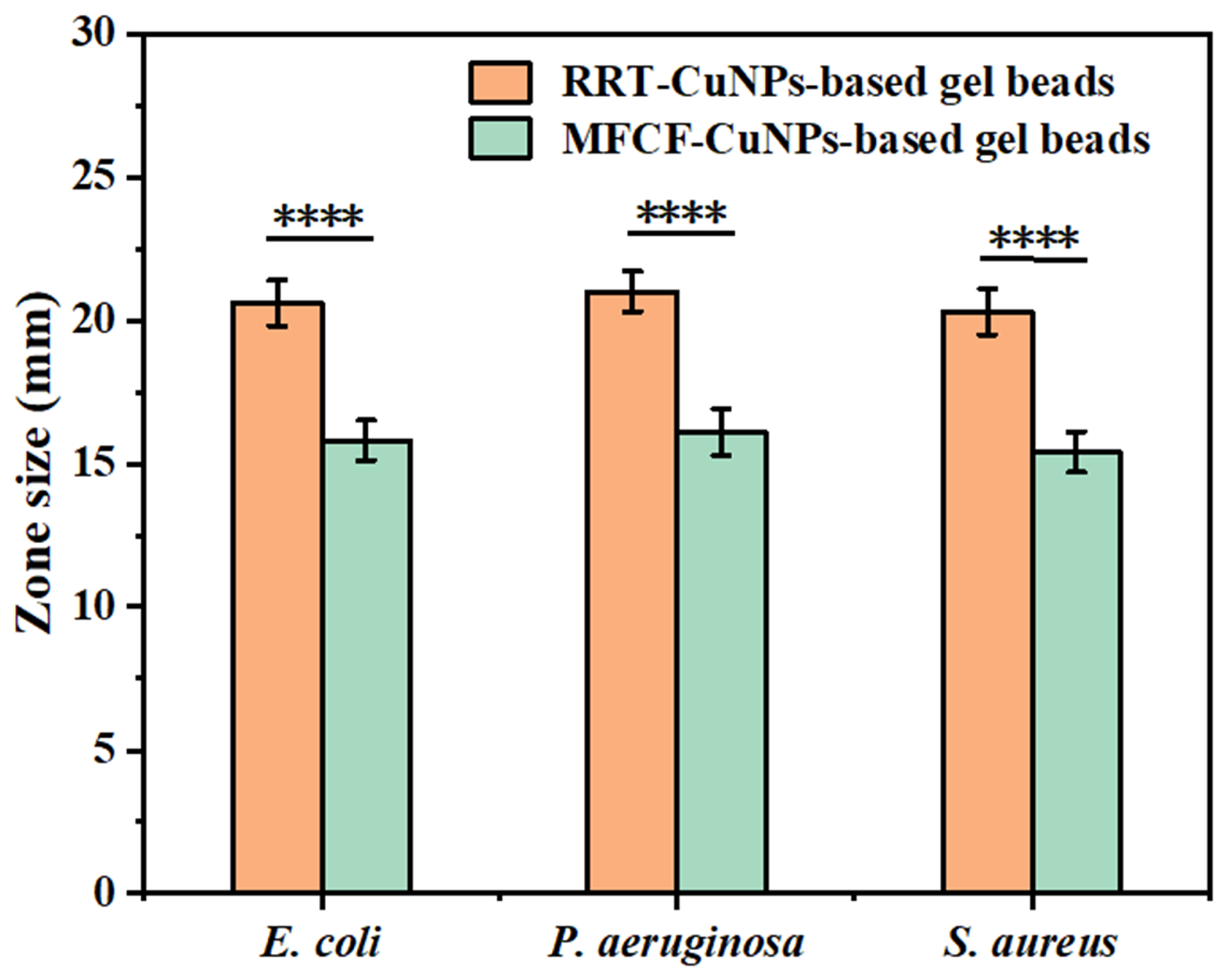
The antibacterial capacity of these two synthesized CuNPs was compared with the disk diffusion method. The antibacterial activity was evaluated against E. coli, P. aeruginosa, and S. aureus (Figure 3 and Figure S1). Under the tested concentration, RRT-CuNPs-based gel beads exhibited a higher antibacterial ability than MFCF-CuNPs-based gel beads. The former showed the best inhibitory effect, and the zone sizes of E. coli, P. aeruginosa, and S. aureus were in the range of 20–21 mm. While MFCF-CuNPs-based gel beads could inhibit E. coli, P. aeruginosa, and S. aureus with the zone sizes of 15.4–16.1 mm. This superior performance is likely attributed to the synergistic effect between the copper core and the bioactive polyphenols from the RRT extract that cap the nanoparticles, which are absent in the MFCF system. Therefore, based on these preliminary results, RRT-CuNPs-based gel beads were selected as the most promising antibacterial material for further investigation in this study. In future work, the chemical characterization of RRT and MFCF will be conducted, and the antibacterial ability and its components will be further explored.

3.2. Particle Size Distribution of RRT-CuNPs

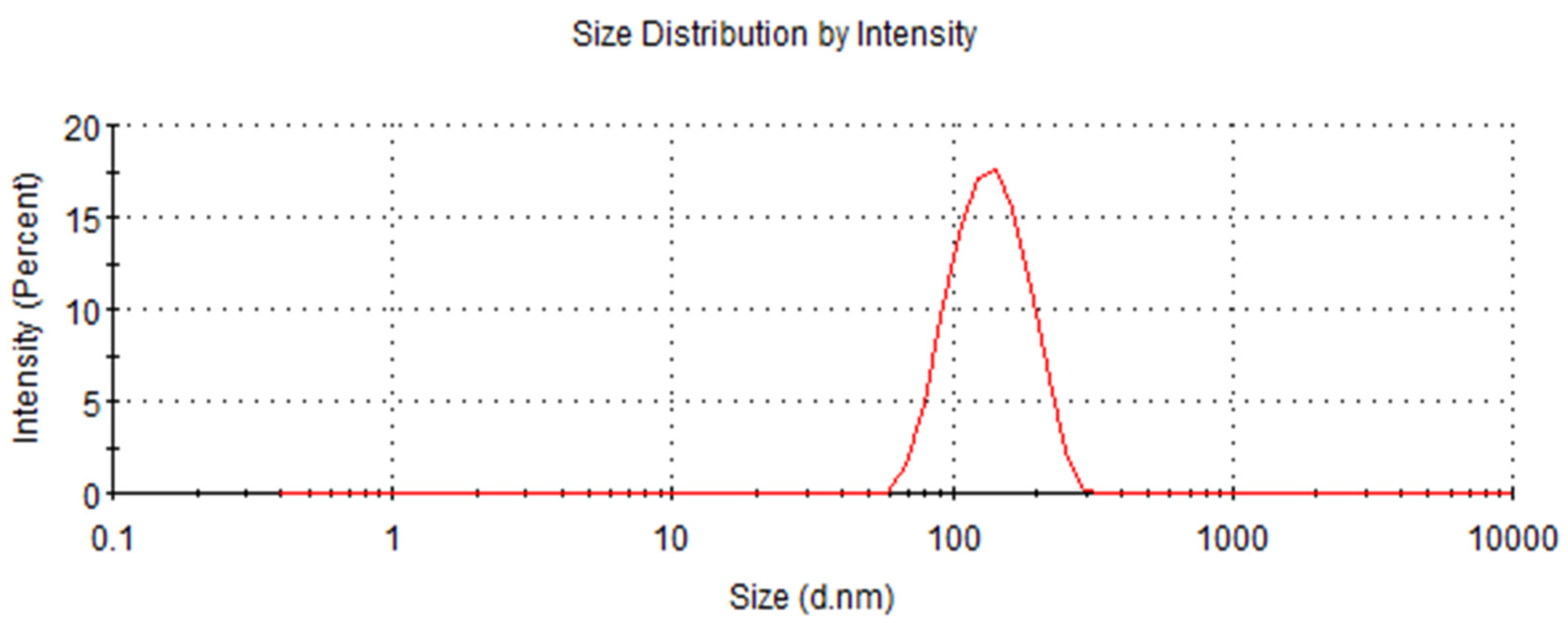
Based on the Brownian motion of particles, DLS technology was used for particle size measurement. Figure 4 shows that the particle size of CuNPs obtained by RRT green reduction was mainly around 134.9 ± 6.9 nm (n = 3), with a PDI of 0.26 ± 0.05. PDI measured the uniformity of NPs, and the PDI value below 0.3 indicates that the CuNPs had a limited size distribution and good homogeneity [44].

3.3. FTIR of CTS/PVA/MMT and CTS/PVA/MMT/RRT-CuNPs

The chemical composition of CTS/PVA/MMT and CTS/PVA/MMT/RRT-CuNPs composite gel beads was systematically characterized using FTIR spectroscopy, as evidenced in Figure 5. A prominent absorption peak near 3470 cm−1 was observed, mainly attributed to the superposition of hydroxyl groups (-OH) from PVA and CTS [45,46]. The peak around 2850 cm−1 corresponds to the methylene stretching absorption peak [47]. The absorption peak at about 2920 cm−1 is attributed to C–H asymmetric stretching vibration [48,49] in the CTS/PVA/MMT and CTS/PVA/MMT/RRT-CuNPs composite gel beads. The absorption peak at about 1467 cm−1 was mainly attributed to N-H bending vibrations of the amino group in CTS and the deformation vibration of -CH2. The peak near 1072 cm−1 in all the composite gel beads was primarily due to Si-O-Si vibrations, characteristic of MMT [50,51]. The peak around 850 cm−1 was mainly caused by C–H bending vibrations of the β-1,4-glycosidic bond in CTS, reflecting the vibrational properties of its sugar ring structure [49].

3.4. XRD of CTS/PVA/MMT and CTS/PVA/MMT/RRT-CuNPs

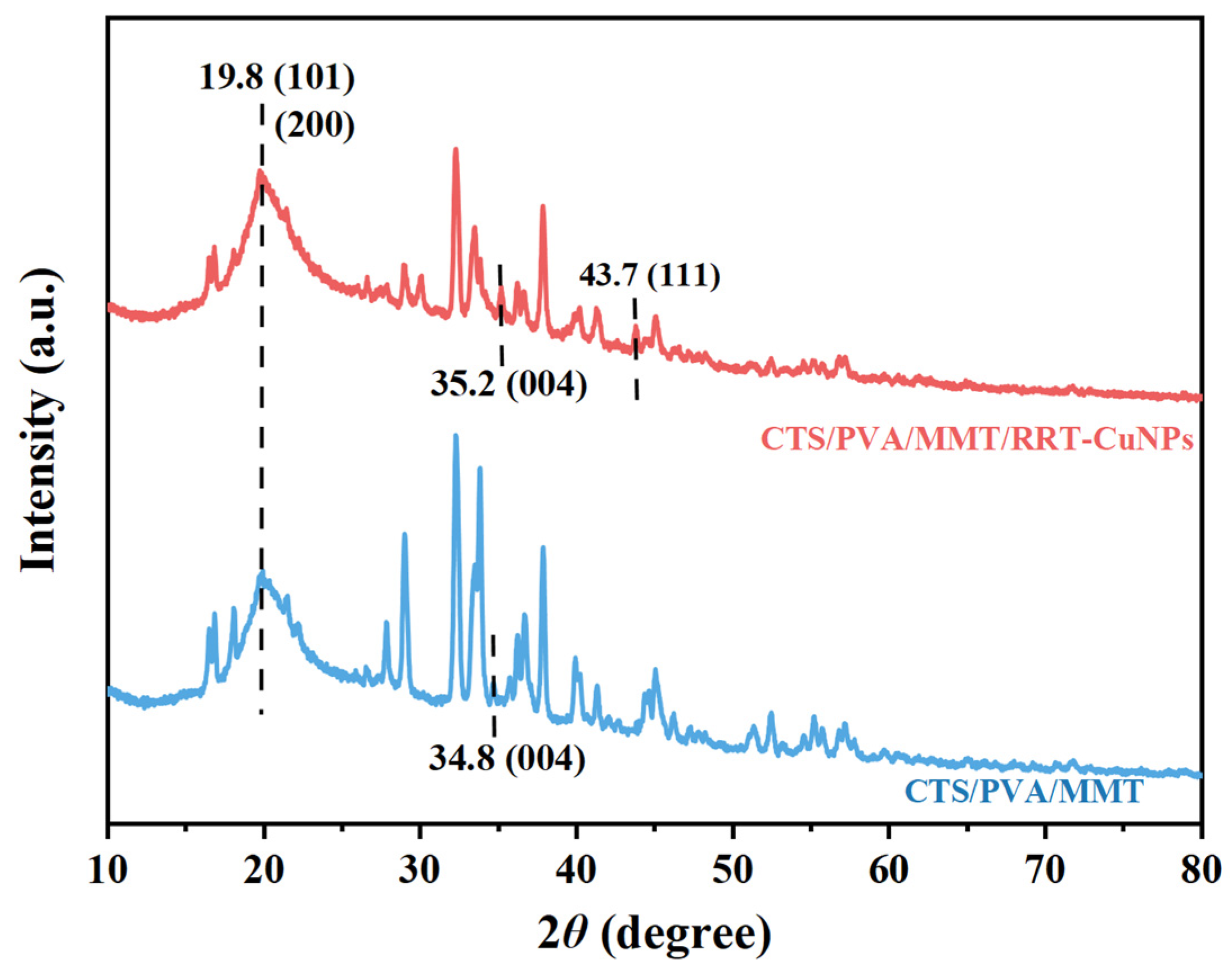
The crystallinity of the composite gel beads was estimated by XRD, and the results are shown in Figure 6. Diffraction peaks existed at 19.8° for both CTS/PVA/MMT and CTS/PVA/MMT/RRT-CuNPs composite gel beads, which mainly originated from the superposition of diffraction of the (101) crystallographic plane of PVA and the (200) crystallographic plane of CTS [52]. By comparison, it could be seen that the composite gel beads with the addition of MMT showed the diffraction peak at 35.2°, which mainly originated from the (004) crystal plane characteristic diffraction of MMT. In addition, the diffraction peaks of the composite gel beads around 43.7° correspond to the (111) crystallographic facet characteristic peaks of CuNPs, indicating that the CuNPs were uniformly dispersed in the gel beads in a crystalline state [53].

3.5. SEM of CTS/PVA/MMT and CTS/PVA/MMT/CuNPs

SEM was used to observe and analyze the composite gel beads. The SEM characterization images of CTS/PVA/MMT and CTS/PVA/MMT/CuNPs composite gel beads are shown in Figure 7a,b. The image was obtained using SEM at an accelerating voltage of 5 kV. Figure 7a showed that the surface of CTS/PVA/MMT composite gel beads was relatively dense. With the addition of CuNPs, the morphology of the composite gel beads changes, forming more porous structures, indicating that the addition of CuNPs effectively alters the pore characteristics of the gel beads. The increase in porosity may be due to the introduction of CuNPs during the crosslinking process, which creates void spaces in the polymer matrix and hinders the close packing of polymer chains, thereby forming the observed porous structure [54]. The picture of CTS/PVA/MMT/RRT-CuNPs composite gel beads is shown in Figure 7c.

3.6. Effect of CTS, MMT, and CuNPs Loading on the Antibacterial Efficacy of Gel Beads

The effect of CTS concentration in the initial solution on the antibacterial activity of composite gel beads was systematically evaluated by the agar diffusion method, as indicated in Figure 8. The results displayed that the occurrence of CTS showcased a clear correlation with the antibacterial effect of gel beads. With the increase in CTS concentration, the antibacterial killing size of composite gel beads against E. coli, P. aeruginosa, and S. aureus showed a trend of increasing first and then decreasing. When the CTS concentration was 30 g/L, the composite hydrogel beads exhibited a significant inhibitory effect (p < 0.05), with inhibition zone diameters of around 21 mm for E. coli, P. aeruginosa, and S. aureus, which were significantly better than at other tested concentrations. This phenomenon might be attributed to the fact that CTS itself has a certain inhibitory effect, and the amino group (-NH3+) was protonated under acidic conditions, which could electrostatically bind to the bacterial cell membrane and destroy the membrane integrity [55,56,57]. Excessive CTS might weaken the porosity of gel beads, and CuNPs were confined inside the high-density cross-linking network, which reduced their effective contact rate with bacteria. Accordingly, the optimization of CTS addition was crucial for balancing the gel network structure and the effectiveness of antimicrobial components. The experimental data confirmed that a 30 g/L CTS concentration could be used to form a uniform three-dimensional network structure, to ensure high loading and homogeneous dispersion of CuNPs and avoid nanoparticle agglomeration, thus maximizing the exposure efficiency of the antibacterial active sites. In the comprehensive evaluation, when the CTS solution concentration was 30 g/L, realizing the optimal balance between the antimicrobial performance and the gel structure.
The antibacterial effect of gel beads with different contents was tested by the agar diffusion method, and the results are shown in Figure 9. Supplementing MMT had a certain effect on the antibacterial efficacy of the composite gel beads. Upon raising MMT content, the size of the antibacterial zones of the composite gel beads firstly an increase in slowly, and the antibacterial activity of the composite gel beads reached the maximum when the MMT concentration in the initial solution system is 4 g/L, and the inhibitory zone sizes of the composite gel beads against E. coli, P. aeruginosa, and S. aureus were near 21 mm, respectively, and the difference is statistically significant (p < 0.05). This might be due to the high specific surface area and cation exchange capacity of MMT, which could adsorb and stabilize the CuNPs particles, preventing them from agglomeration and improving the contact rate of CuNPs with bacteria [58]. It was worth noting that the bacteriostatic effect of the composite gel beads did not increase linearly with an increase in MMT content, and its bacteriostatic activity began to decrease when the MMT content exceeded 4 g/L. This might be because the high content of MMT was prone to agglomeration, which destroyed the gel bead structure and made the internal pore distribution uneven, or blocked the pores, and affected the distribution and release of CuNPs [59]. Consequently, among the concentrations tested, an MMT loading of 4.0 g/L yielded the highest antibacterial activity and was thus identified as the optimal concentration within this experimental framework.
The antibacterial effect of composite hydrogel beads with different initial concentrations of CuNPs suspension was evaluated using the agar diffusion method, and the results are illustrated in Figure 10. The experimental results indicated that the composite gel beads (CuNPs at 0.5 g/L) had the best bacteriostatic effect against E. coli, P. aeruginosa, and S. aureus, with inhibitory zone sizes of nearly 21 mm (p < 0.05). With the increase in CuNPs content, the bacteriostatic effect of gel beads showed a trend of first increase and then decrease. The decrease in the bacteriostatic effect might be due to the fact that the excess CuNPs exceeded the loading capacity of MMT and CTS, agglomeration occurred, the specific surface area declined, and the antibacterial efficiency decreased [60]. Accordingly, CuNPs with an initial concentration of 0.5 g/L were added to the composite hydrogel beads for subsequent experiments. Kazeminava et al. [61] incorporated silver nanoparticles into PEG-based hydrogels, achieving inhibition zones of 17 mm and 16 mm against S. aureus and E. coli, respectively; Song et al. [62] prepared a porous gallium-doped TiO2 composite aerogel, which manifested the maximum inhibition zones of 18 mm and 16 mm against E. coli and S. aureus, respectively. These findings revealed that the CTS/PVA/MMT/CuNPs hydrogel beads exhibited favorable antimicrobial efficacy.

3.7. The Effect of the Dose and Action Duration of Gel Beads on the Bacteriostatic Efficacy

The plate-counting method was used to assess the effects of the dose and action duration of the CTS/PVA/MMT/CuNPs gel beads on the bacteriostatic activity of E. coli, P. aeruginosa, and S. aureus (Figure 11). For E. coli, the inhibition rate of the CTS/PVA/MMT/CuNPs gel beads (0.2 g/L) could reach more than 90% in four h. Increasing the loading of gel beads to 0.3 g/L, the inhibition ability reached 100% in three h (Figure 11a and Figure S2a). For P. aeruginosa, the antimicrobial capacity of CTS/PVA/MMT/CuNPs gel beads (0.2 g/L) could reach more than 90% in three h. As the dose of gel beads was 0.3 g/L and the action time was 4 h, the inhibition rate reached 100% (Figure 11b and Figure S2b). For S. aureus, the inhibition rate of the CTS/PVA/MMT/CuNPs gel beads (0.2 g/L) could reach more than 90% in 3 h, and the antimicrobial rate reached 100% in four h (Figure 11c and Figure S2b). Ma et al. [63] prepared a quaternary ammonium-oxidized sodium alginate injectable hydrogel, which exhibited inhibition rates of 91.3% against E. coli and 97.9% against S. aureus. Zhu et al. [64] developed a multifunctional medical flexible material exhibiting a 91% inhibition rate against S. aureus. Consequently, CTS/PVA/MMT/CuNPs gel beads demonstrated superior antimicrobial efficacy.

4. Conclusions

In this research, CuNPs were prepared through green reduction using RRT and MFCF. Higher antibacterial activity is achieved by using RRT as a natural reducing agent. Furthermore, the composite gel bead with bacteriostatic activity was successfully prepared by the green reduction method using RRT as a natural reducing agent and highly biocompatible CuNPs, with PVA and CTS as the composite carrier matrix and MMT as the structural filler. To study the optimal ratio of the composite gel, the effects of different initial CTS concentrations, MMT concentrations, and CuNPs concentrations added to the composite gel beads on antibacterial activity were explored. When the initial concentrations of CTS, MMT, and CuNPs were 30.0, 4.0, and 0.5 g/L, respectively, the composite gel beads exhibited optimal inhibitory activity, and the diameters of the inhibitory zone of CTS/PVA/MMT/CuNPs composite beads on E. coli, P. aeruginosa, and S. aureus were around 21 mm. The rate of bacterial inhibition of the composite gel beads could reach 100% after four h at a dosage of 0.30 g/L. To sum up, the composite gel beads of CTS/PVA/MMT/CuNPs function as highly effective antibacterial materials. From a process engineering perspective, technology demonstrates significant potential for scalability. The synthesis method, involving the mixing of aqueous solutions and ionic cross-linking, is straightforward, low-energy, and amenable to continuous production lines. While the extraction of active reducing agents from raw materials represents a key step, established industrial extraction technologies can be readily adapted for this purpose. The green nature, high efficacy, and use of biocompatible materials position this technology as a strong candidate for upscaling, offering a sustainable solution for the remediation of pathogen-laden wastewater. However, this study still has some key limitations. The primary constraint is that the antibacterial tests were conducted in a controlled laboratory environment using pure cultures, and their performance in complex real wastewater matrices may differ. In addition, we did not adequately assess the stability of CuNPs in the gel beads during long-term use or potential leaching, both of which are crucial for environmental safety. Finally, this study focused solely on antibacterial applications and did not explore the potential of this material for removing other water pollutants, such as dyes or heavy metals. Therefore, future work should prioritize testing in real wastewater, conduct long-term leaching studies, and expand its application scope to address a broader range of pollutants, thereby providing more comprehensive water treatment solutions.

Supplementary Materials

The following supporting information can be downloaded at: https://www.mdpi.com/article/10.3390/pr13113518/s1.

Author Contributions

Conceptualization, methodology, and writing—original draft, M.H.; data curation, software, resources, and writing—original draft, T.Z.; methodology, and software, W.H.; supervision, review, and revising manuscript, Y.H. All authors have read and agreed to the published version of the manuscript.

Funding

The research is kindly funded by the Research Project of Jiangsu Agri-animal Husbandry Vocational College (NSF2024ZR12) and the Scientific and Technological Innovation Team for Chinese Herbal Medicine Planting Technology, Development & Application of Jiangsu Agri-animal Husbandry Vocational College (NSF2024TC02).

Institutional Review Board Statement

Not applicable.

Informed Consent Statement

Not applicable.

Data Availability Statement

Data are contained within the article.

Acknowledgments

The authors thank the Analysis and Testing Center (Changzhou University) for the analysis of biomass samples.

Conflicts of Interest

The authors declare no conflicts of interest.

References

  1. Goswami, R.K.; Mehariya, S.; Verma, P.; Lavecchia, R.; Zuorro, A. Microalgae-based biorefineries for sustainable resource recovery from wastewater. J. Water Process Eng. 2021, 40, 101747. [Google Scholar] [CrossRef]
  2. Salim Dantas, M.; Rodrigues Barroso, G.; Corrêa Oliveira, S. Performance of sewage treatment plants and impact of effluent discharge on receiving water quality within an urbanized area. Environ. Monit. Assess. 2021, 193, 289. [Google Scholar] [CrossRef] [PubMed]
  3. Al Aukidy, M.; Verlicchi, P. Contributions of combined sewer overflows and treated effluents to the bacterial load released into a coastal area. Sci. Total. Environ. 2017, 607–608, 483–496. [Google Scholar] [CrossRef]
  4. Pistocchi, A.; Alygizakis, N.A.; Brack, W.; Boxall, A.; Cousins, I.T.; Drewes, J.E.; Finckh, S.; Gallé, T.; Launay, M.A.; McLachlan, M.S.; et al. European scale assessment of the potential of ozonation and activated carbon treatment to reduce micropollutant emissions with wastewater. Sci. Total. Environ. 2022, 848, 157124. [Google Scholar] [CrossRef]
  5. Wang, R.; Liu, Y.; Guan, J.; Zuo, C.; Dai, C.; Liu, X.; Liang, Z.; Zhao, G. Environmental justice of texas recreational water quality—The disproportionate E. coli levels and trends. J. Environ. Manag. 2024, 370, 122969. [Google Scholar] [CrossRef]
  6. Deepnarain, N.; Nasr, M.; Amoah, I.D.; Enitan-Folami, A.M.; Reddy, P.; Stenström, T.A.; Kumari, S.; Bux, F. Impact of sludge bulking on receiving environment using quantitative microbial risk assessment (QMRA)-based management for full-scale wastewater treatment plants. J. Environ. Manag. 2020, 267, 110660. [Google Scholar] [CrossRef] [PubMed]
  7. Jia, L.; Liu, W.; Cao, J.; Wu, Z.; Yang, C. Foam fractionation for effective removal of Pseudomonas aeruginosa from water body: Strengthening foam drainage by artificially inducing foam evolution. J. Environ. Manag. 2021, 291, 112628. [Google Scholar] [CrossRef] [PubMed]
  8. Pattnaik, A.; Ghosh, P.; Poonia, A.K. An overview on advancements in hydrogels for effective wastewater treatment. J. Mol. Liq. 2025, 424, 127120. [Google Scholar] [CrossRef]
  9. Xie, L.; Zhang, Z.; He, Y. Antibacterial effect of polyvinyl alcohol/biochar–nano silver/sodium alginate gel beads. Processes 2023, 11, 2330. [Google Scholar] [CrossRef]
  10. Singh, S.; Khurana, P.; Kaur, G.; Prasad, S.; Thatai, S. Antibacterial activity of biogenic and chemically synthesized silver nanoparticles and its application in wastewater treatment. Inorg. Chem. Commun. 2024, 170, 113205. [Google Scholar] [CrossRef]
  11. Atkinson, S.; Thomas, S.F.; Goddard, P.; Bransgrove, R.M.; Mason, P.T.; Oak, A.; Bansode, A.; Patankar, R.; Gleason, Z.D.; Sim, M.K.; et al. Swirl Flow Bioreactor coupled with Cu-alginate beads: A system for the eradication of Coliform and Escherichia coli from biological effluents. Sci. Rep. 2015, 5, 9461. [Google Scholar] [CrossRef]
  12. Mukherjee, A.; Das, S.; Ahn, Y.-H. Highly recyclable and stable CdS-alginate hydrogel beads towards prevention of oxidative release of nanoparticles in complete photocatalytic disinfection of multidrug-resistant Escherichia coli. Environ. Sci. Water Res. Technol. 2023, 9, 2954–2964. [Google Scholar] [CrossRef]
  13. Li, J.L.; Jiang, X.; Liu, X.; He, C.; Di, Y.; Lu, S.; Huang, H.; Lin, B.; Wang, D.; Fan, B. Antibacterial anthraquinone dimers from marine derived fungus Aspergillus sp. Fitoterapia 2019, 133, 1–4. [Google Scholar] [CrossRef] [PubMed]
  14. Shen, X.-Y.; Song, Y.-Z. Synthesis of uniform zinc peroxide nanoparticles for antibacterial application. Indian J. Pharm. Sci. 2024, 86, 219–225. [Google Scholar]
  15. Chen, H.; Li, Y.; Chen, D.; Fang, Y.; Gong, X.; Wang, K.; Ma, C. Photothermally enhanced antibacterial wound healing using albumin-loaded tanshinone IIA and IR780 nanoparticles. Front. Bioeng. Biotechnol. 2024, 12, 1487660. [Google Scholar] [CrossRef]
  16. Zhao, Q.-M.; Sun, Y.-Y.; Wu, C.-S.; Yang, J.; Bao, G.-F.; Cui, Z.-M. Enhanced osteogenic activity and antibacterial ability of manganese–titanium dioxide microporous coating on titanium surfaces. Nanotoxicology 2019, 14, 289–309. [Google Scholar] [CrossRef] [PubMed]
  17. Sedighi, O.; Johnsen, T.; Badireddy, A.R.; Wargo, M.J.; Doiron, A.L. Synergistic effects of iron oxide nanoparticles and hydrogen peroxide in inhibiting Pseudomonas aeruginosa growth to combat bacterial contamination in water recovery systems. Environ. Sci. Nano 2025, 12, 2449–2461. [Google Scholar] [CrossRef]
  18. Zhang, W.; Hu, E.; Wang, Y.; Miao, S.; Liu, Y.; Hu, Y.; Liu, J.; Xu, B.; Chen, D.; Shen, Y. Emerging antibacterial strategies with application of targeting drug delivery system and combined treatment. Int. J. Nanomed. 2021, 16, 6141–6156. [Google Scholar] [CrossRef]
  19. Ortega-Nieto, C.; Losada-Garcia, N.; Pessela, B.C.; Domingo-Calap, P.; Palomo, J.M. Design and synthesis of copper nanobiomaterials with antimicrobial properties. ACS Bio Med Chem Au 2023, 3, 349–358. [Google Scholar] [CrossRef]
  20. Vodyashkin, A.; Stoinova, A.; Kezimana, P. Promising biomedical systems based on copper nanoparticles: Synthesis, characterization, and applications. Colloids Surf. B Biointerfaces 2024, 237, 113861. [Google Scholar] [CrossRef]
  21. Siddiqi, K.S.; Husen, A. Current status of plant metabolite-based fabrication of copper/copper oxide nanoparticles and their applications: A review. Biomater. Res. 2020, 24, 11. [Google Scholar] [CrossRef]
  22. Liu, W.; Li, N.; Hou, J.; Cao, R.; Jia, L.; Guo, Y.; Xu, J. Structure and antitumor activity of a polysaccharide from Rosa roxburghii. Int. J. Biol. Macromol. 2024, 273, 132807. [Google Scholar] [CrossRef]
  23. Wang, L.; Wei, T.; Zheng, L.; Jiang, F.; Ma, W.; Lu, M.; Wu, X.; An, H. Recent advances on main active ingredients, pharmacological activities of Rosa roxbughii and its development and utilization. Foods 2023, 12, 1051. [Google Scholar] [CrossRef]
  24. Tang, Z.; Chen, Y.; Cai, C.; Ma, S.; Tan, Z. CO2-responsive ionic liquid for the sustainable extraction and separation of Rosa roxburghii polysaccharides. Int. J. Biol. Macromol. 2025, 308, 142708. [Google Scholar] [CrossRef]
  25. Mishra, D.N.; Prasad, L.; Suyal, U. Synthesis of zinc oxide nanoparticles using Trichoderma harzianum and its bio-efficacy on Alternaria brassicae. Front. Microbiol. 2025, 16, 1506695. [Google Scholar] [CrossRef]
  26. Chand Mali, S.; Dhaka, A.; Sharma, S.; Trivedi, R. Review on biogenic synthesis of copper nanoparticles and its potential applications. Inorg. Chem. Commun. 2023, 149, 110448. [Google Scholar] [CrossRef]
  27. Han, M.; Zhang, Y.; Zhang, Y.; Ye, Q.; Sillanpää, M.; Zhu, X.; Yang, W. A mini-review on polyvinyl alcohol/lignin (nano)composites: Preparation, applications and perspectives. Sustain. Chem. Pharm. 2024, 42, 101861. [Google Scholar] [CrossRef]
  28. Wang, M.; Bai, J.; Shao, K.; Tang, W.; Zhao, X.; Lin, D.; Huang, S.; Chen, C.; Ding, Z.; Ye, J. Poly(vinyl alcohol) hydrogels: The Old and New Functional Materials. Int. J. Polym. Sci. 2021, 2021, 2225426. [Google Scholar] [CrossRef]
  29. Khadivi, H.; Sirousazar, M.; Abbasi-Chianeh, V.; Jalilnejad, E. Egg white/polyvinyl alcohol/clay bionanocomposite hydrogel adsorbents for dye removal. J. Polym. Environ. 2022, 30, 3186–3202. [Google Scholar] [CrossRef]
  30. Zhang, P.; Zou, K.; Yuan, L.; Liu, J.; Liu, B.; Qing, T.-P.; Feng, B. A biomass resource strategy for alginate-polyvinyl alcohol double network hydrogels and their adsorption to heavy metals. Sep. Purif. Technol. 2022, 301, 122050. [Google Scholar] [CrossRef]
  31. Enoch, K.; C․S, R.; Somasundaram, A.A. Thixotropic chitosan hydrogels for biomedical applications: Unravelling the effect of chitosan concentration on the mechanical behaviour. Surf. Interfaces 2024, 50, 104475. [Google Scholar] [CrossRef]
  32. Wang, J.; Yuan, Y.; Liu, Y.; Li, X.; Wu, S. Application of chitosan in fruit preservation: A review. Food Chem. X 2024, 23, 101589. [Google Scholar] [CrossRef]
  33. Wu, K.; Yan, Z.; Wu, Z.; Li, J.; Zhong, W.; Ding, L.; Zhong, T.; Jiang, T. Recent advances in the preparation, antibacterial mechanisms, and applications of chitosan. J. Funct. Biomater. 2024, 15, 318. [Google Scholar] [CrossRef] [PubMed]
  34. Xia, C.; Qian, J. Recent advances in montmorillonite-based composites for biomedical applications: A review. J. Drug Deliv. Sci. Technol. 2025, 109, 107012. [Google Scholar] [CrossRef]
  35. Sarkar, A.; Mushahary, N.; Basumatary, F.; Das, B.; Basumatary, S.F.; Venkatesan, K.; Selvaraj, M.; Rokhum, S.L.; Basumatary, S. Efficiency of montmorillonite-based materials as adsorbents in dye removal for wastewater treatment. J. Environ. Chem. Eng. 2024, 12, 112519. [Google Scholar] [CrossRef]
  36. Tang, Z.; Guo, H.; Xu, J.; Li, Z.; Sun, G. Cationic poly(diallyldimethylammonium chloride) based hydrogel for effective anionic dyes adsorption from aqueous solution. React. Funct. Polym. 2022, 174, 105239. [Google Scholar] [CrossRef]
  37. Phu, N.A.M.M.; Wi, E.; Jeong, G.; Kim, H.; Singha, N.R.; Chang, M. Highly efficient dye adsorption by hierarchical porous SA/PVA/ZIF-8 composite microgels prepared via microfluidics. Carbohydr. Polym. 2024, 350, 123016. [Google Scholar] [CrossRef] [PubMed]
  38. Njuguna, D.G.; Schönherr, H. Smart and regeneratable Xanthan gum hydrogel adsorbents for selective removal of cationic dyes. J. Environ. Chem. Eng. 2022, 10, 107620. [Google Scholar] [CrossRef]
  39. Duru Kamacı, U.; Kamacı, M. Hydrogel beads based on sodium alginate and quince seed nanoparticles for the adsorption of methylene blue. Inorg. Chem. Commun. 2023, 160, 111919. [Google Scholar] [CrossRef]
  40. Zhou, X.; Qin, Y.; Wang, Y.; Wang, Y.; Qin, Z. Phytochemical profile and antioxidant characteristics of bound and free phenolics from Rosa roxburghii tratt. Food Biosci. 2024, 57, 103576. [Google Scholar] [CrossRef]
  41. Ahluwalia, V.; Kumar, J.; Sisodia, R.; Shakil, N.A.; Walia, S. Green synthesis of silver nanoparticles by Trichoderma harzianum and their bio-efficacy evaluation against Staphylococcus aureus and Klebsiella pneumonia. Ind. Crops Prod. 2014, 55, 202–206. [Google Scholar] [CrossRef]
  42. Chiu, C.-T.; Lai, C.-H.; Huang, Y.-H.; Yang, C.-H.; Lin, J.-N. Comparative analysis of gradient diffusion and disk diffusion with agar dilution for susceptibility testing of Elizabethkingia anopheles. Antibiotics 2021, 10, 450. [Google Scholar] [CrossRef] [PubMed]
  43. Tang, M.; Hao, X.; Kang, Y.; He, X.; Zhao, H. Preparation and antibacterial properties of polyelectrolyte complexed nanoparticles aggregated from PHMG and sodium caffeate. ACS Appl. Bio Mater. 2024, 7, 6467–6476. [Google Scholar] [CrossRef] [PubMed]
  44. Ranaware, A.S.; Kushwaha, S.B.; Kunchge, N.; Prakash, G.; Lele, S.S. Effect of different size silver nanoparticles synthesized at varying pH of plant extract for germination improvement of tetraploid watermelon (Citrullus lanatus). J. Clust. Sci. 2025, 36, 101. [Google Scholar] [CrossRef]
  45. Sacourbaravi, R.; Ansari-Asl, Z.; Hoveizi, E.; Darabpour, E. Poly(vinyl alcohol)/chitosan hydrogel containing gallic acid-modified Fe, Cu, and Zn metal–organic frameworks (MOFs): Preparation, characterization, and biological applications. ACS Appl. Mater. Interfaces 2024, 16, 61609–61620. [Google Scholar] [CrossRef]
  46. Roufegari-Nejhad, E.; Sirousazar, M.; Abbasi-Chiyaneh, V.; Kheiri, F. Removal of methylene blue from aqueous solutions using poly(vinyl alcohol)/montmorillonite nanocomposite hydrogels: Taguchi optimization. J. Polym. Environ. 2019, 27, 2239–2249. [Google Scholar] [CrossRef]
  47. Zhou, C.; Qi, S.; Zhu, P.; Zhao, Y.; Xu, Y.; Dong, X.; Wang, D. The methylene infrared vibration and dielectric behavior monitored by amide group arrangement for long chain polyamides. Polymer 2020, 190, 122231. [Google Scholar] [CrossRef]
  48. Sanjay, V.; Rajashekara, K.M.; Pattar, V.; Murugendrappa, M.V. Effect on electrical and dielectric properties of Te nanoparticle-doped PVA composite. J. Mater. Sci. Mater. Electron. 2022, 33, 17382–17394. [Google Scholar] [CrossRef]
  49. Liu, J.; Song, Z.; Li, B.; Ren, J.; Chen, F.; Xiao, M. Experimental study on the microstructure of coal with different particle sizes. Energies 2022, 15, 4043. [Google Scholar] [CrossRef]
  50. Wu, Y.; Guan, Y.; Gao, H.; Zhou, L.; Peng, F. Novel high-strength montmorillonite/polyvinyl alcohol composite film enhanced by chitin nanowhiskers. J. Appl. Polym. Sci. 2020, 138, 50344. [Google Scholar] [CrossRef]
  51. Moja, T.N.; Phiri, Z.; Nure, J.F.; Nkambule, T.T.; de Kock, L. Insights into preparation of polycaprolactone–chitosan–montmorillonite composite for Pb(II) and Cd(II) removal. ChemistrySelect 2025, 10, e202500798. [Google Scholar] [CrossRef]
  52. Radoor, S.; Karayil, J.; Jayakumar, A.; Siengchin, S.; Parameswaranpillai, J. A low cost and eco-friendly membrane from polyvinyl alcohol, chitosan and honey: Synthesis, characterization and antibacterial property. J. Polym. Res. 2021, 28, 82. [Google Scholar] [CrossRef]
  53. Suneetha, G.; Ayodhya, D.; Srikanth, K.; Manjari, P.S. Fabrication of CuNPs using schiff base ligand and their catalytic reduction of pharmaceutical drugs, fluorescence selective detection of Cd2+, antimicrobial, and antioxidant activities. J. Fluoresc. 2023, 34, 1307–1318. [Google Scholar] [CrossRef] [PubMed]
  54. Kumari, S.; Singh, B.N.; Srivastava, P. Effect of copper nanoparticles on physico-chemical properties of chitosan and gelatin-based scaffold developed for skin tissue engineering application. 3 Biotech 2019, 9, 102. [Google Scholar] [CrossRef]
  55. Varma, R.; Vasudevan, S. Extraction, characterization, and antimicrobial activity of chitosan from horse mussel modiolus modiolus. ACS Omega 2020, 5, 20224–20230. [Google Scholar] [CrossRef] [PubMed]
  56. Laanoja, J.; Sihtmäe, M.; Vihodceva, S.; Iesalnieks, M.; Otsus, M.; Kurvet, I.; Kahru, A.; Kasemets, K. Synthesis and synergistic antibacterial efficiency of chitosan-copper oxide nanocomposites. Heliyon 2024, 10, e35588. [Google Scholar] [CrossRef] [PubMed]
  57. Sánchez-Machado, D.I.; López-Cervantes, J.; Vega-Cázarez, C.A.; Hernández-Ruiz, K.L.; Campas-Baypoli, O.N.; Soto-Cota, A.; Madera-Santana, T.J. Functional and antibacterial characterization of electrospun nanofiber membranes made of chitosan and polyvinyl alcohol. Results Chem. 2024, 7, 101314. [Google Scholar] [CrossRef]
  58. Liang, X.; Wang, W.; Tang, M.; Kang, Y.; Cui, M.; Zhao, H. Preparation, characterization and antibacterial activities of bio-based schiff base copper complex intercalated montmorillonite. Appl. Clay Sci. 2025, 271, 107809. [Google Scholar] [CrossRef]
  59. Huang, Y.; Wu, L.; Chang, L.; Peng, W.; Wang, W.; Cao, Y. Sodium alginate-crosslinked montmorillonite nanosheets hydrogel for efficient gallium recovery. Int. J. Biol. Macromol. 2025, 295, 139474. [Google Scholar] [CrossRef]
  60. Noppradit, B.; Uthaipan, N.; Klinnawee, L.; Kongtragoul, P.; Phengdaam, A. Antifungal copper nanocomposite-rubber compound for tree wound dressings. Ind. Crops Prod. 2024, 222, 119798. [Google Scholar] [CrossRef]
  61. Kazeminava, F.; Arsalani, N.; Ahmadi, R.; Kafil, H.S.; Geckeler, K.E. A facile approach to incorporate silver nanoparticles into solvent-free synthesized PEG-based hydrogels for antibacterial and catalytical applications. Polym. Test. 2021, 101, 106909. [Google Scholar] [CrossRef]
  62. Song, Z.; Huang, L.; Cui, S. Preparation of porous Ga-doped TiO2 composite aerogel and its bactericidal activity against Escherichia coli and Staphylococcus aureus. Adv. Eng. Mater. 2024, 26, 2302042. [Google Scholar] [CrossRef]
  63. Ma, S.; Chen, K.; Ding, Q.; Zhang, S.; Lu, Y.; Yu, T.; Ding, C.; Liu, W.; Liu, S. Quaternized oxidized sodium alginate injectable hydrogel with high antimicrobial and hemostatic efficacy promotes diabetic wound healing. Int. J. Pharm. 2024, 661, 124421. [Google Scholar] [CrossRef] [PubMed]
  64. Zhu, X.; Du, C.; Gao, B.; Xiang, G.; Huang, Y.; He, B. Dual enzymatic preparation of flexible biocomplex with full amino-acid composition. Polymer 2024, 294, 126713. [Google Scholar] [CrossRef]
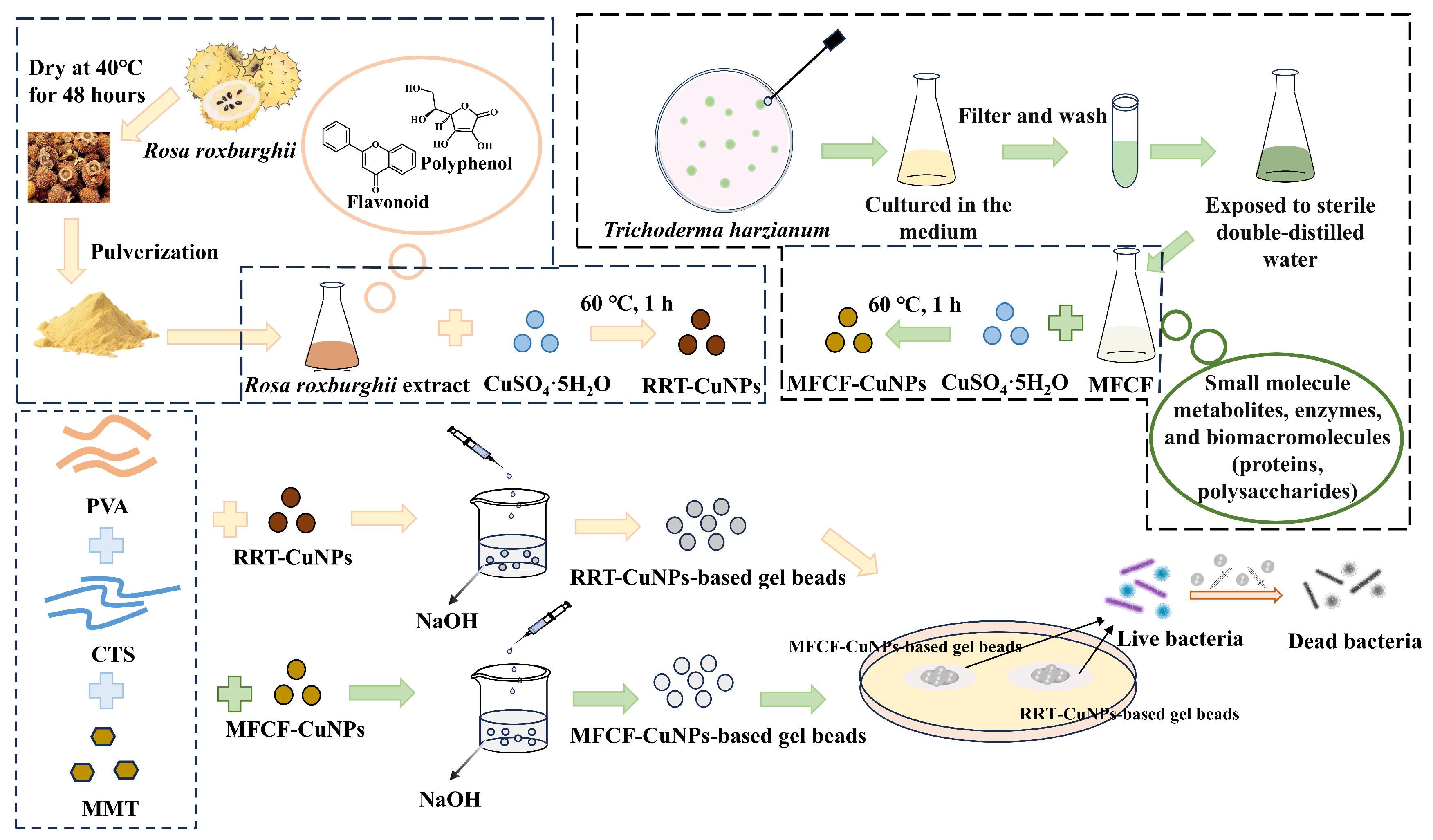
Figure 1. Schematic of the experimental workflow. The process involves the green synthesis of copper nanoparticles (CuNPs) using two different biological agents (RRT and MFCF), the fabrication of composite gel beads by embedding the CuNPs in a PVA/CTS/MMT matrix, and the antibacterial testing of the beads against bacteria.
Processes 13 03518 g001
Figure 2. Preparation of RRT and RRT-CuNPs (a), preparation of MFCF and MFCF-CuNPs (b), preparation of RRT-CuNPs-based gel beads and MFCF-CuNPs-based gel beads (c).
Processes 13 03518 g002
Figure 3. Antibacterial activity of RRT-CuNPs-based gel beads and MFCF-CuNPs-based gel beads (The concentration of CuNPs: 0.5 g/L; initial bacterial concentration: 108 CFU/mL).
Processes 13 03518 g003
Figure 4. Particle size of RRT-CuNPs.
Processes 13 03518 g004
Figure 5. FTIR spectra of CTS/PVA/MMT and CTS/PVA/MMT/RRT-CuNPs composite gel beads: 4000 to 2250 cm−1 (a); 2250 to 500 cm−1 regions (b).
Processes 13 03518 g005
Figure 6. X-ray diffraction spectra of CTS/PVA/MMT and CTS/PVA/MMT/RRT-CuNPs composite gel beads.
Processes 13 03518 g006
Figure 7. SEM images of CTS/PVA/MMT (a) and CTS/PVA/MMT/RRT-CuNPs (b) at magnifications of ×1000; Picture of CTS/PVA/MMT/RRT-CuNPs (c).
Processes 13 03518 g007
Figure 8. The zone size of CTS/PVA/MMT/CuNPs composite gel beads prepared from solutions with different CTS concentrations in the range of 25–40 g/L against E. coli, S. aureus, and P. aeruginosa (The concentrations of the other components in the precursor solution were held constant at 4 g/L for MMT and 0.5 g/L for CuNPs).
Processes 13 03518 g008
Figure 9. The zone size of CTS/PVA/MMT/CuNPs composite gel beads prepared from solutions with different MMT contentrations (0, 2, 3, 4, 5, and 6 g/L) against E. coli, S. aureus, and P. aeruginosa (The concentrations of the other components in the precursor solution were held constant at 30 g/L for CTS and 0.5 g/L for CuNPs).
Processes 13 03518 g009
Figure 10. The zone size of CTS/PVA/MMT/CuNPs composite gel beads prepared from solutions with different CuNPs contentrations (0.2, 0.4, 0.5, 0.6, 0.7, and 1 g/L) against E. coli, S. aureus, and P. aeruginosa (The concentrations of the other components in the precursor solution were held constant at 30 g/L for CTS and 4 g/L for MMT).
Processes 13 03518 g010
Figure 11. Inhibition of E. coli (a), P. aeruginosa (b), and S. aureus (c) by CTS/PVA/MMT/CuNPs composite gel beads at different use doses (0.1, 0.2, 0.3, 0.4, and 0.5 g/L) and different action duration (1, 2, 3, 4, 5, and 6 h) (The lines are guide to the eye).
Processes 13 03518 g011
Disclaimer/Publisher’s Note: The statements, opinions and data contained in all publications are solely those of the individual author(s) and contributor(s) and not of MDPI and/or the editor(s). MDPI and/or the editor(s) disclaim responsibility for any injury to people or property resulting from any ideas, methods, instructions or products referred to in the content.